Winner of the ESTS Young Investigators Award 2011
Signi
ficance of a new fluorodeoxyglucose-positive lesion on restaging
positron emission tomography/computed tomography after induction
therapy for non-small-cell lung cancer
†
Stéphane Collaud
a,*
‡, Didier Lardinois
a,‡, Verena Tischler
b, Hans C. Steinert
c, Rolf Stahel
dand Walter Weder
aa Division of Thoracic Surgery, University Hospital Zurich, Zurich, Switzerland b
Division of Pathology, University Hospital Zurich, Zurich, Switzerland
c Division of Nuclear Medicine, University Hospital Zurich, Zurich, Switzerland d Division of Oncology, University Hospital Zurich, Zurich, Switzerland
* Corresponding author. Division of Thoracic Surgery, Zurich University Hospital, Rämistrasse 100, CH-8091 Zurich, Switzerland. Tel: +41-44-2558802; fax: +41-44-2558805; e-mail: [email protected] (S. Collaud).
Received 17 June 2011; received in revised form 12 August 2011; accepted 17 August 2011
Abstract
OBJECTIVES: Restaging of patients with locally advanced non-small-cell lung cancer (NSCLC) is of paramount importance, since only
patients with down-staging after induction therapy will benefit from surgery. In this study, we assessed the aetiology of new18
fluoro-2-deoxy-D-glucose (FDG)-positive focal abnormalities on restaging positron emission tomography/computed tomography (PET/CT) in
patients with a good response after induction chemotherapy in the primary tumour and lymph nodes.
METHODS: Between 2004 and 2008, 31 patients with histological proven stage III NSCLC had a PET/CT prior and after induction chemotherapy. Their medical charts were retrospectively reviewed.
RESULTS: Restaging PET/CT revealed a new FDG-positive lesion in 6 of 31 (20%) patients. The initial clinical stage of the disease was
IIIA N2 in four and IIIB T4 in two patients. The maximal standard uptake value in the primary tumour (P = 0.043) and in the initially
involved mediastinal nodes (P = 0.068) decreased after induction treatment in all patients. The new PET/CT findings were located in an
ipsilateral cervical lymph node in two patients, a contralateral mediastinal in one patient and an ipsilateral mammary internal lymph
node in one patient. Two other patients had a lesion on the contralateral lung. Malignant lymph node infiltrations were excluded
fol-lowingfine-needle puncture, intraoperative biopsy or follow-up PET/CT. Contralateral pulmonary lesions were diagnosed as benign
fol-lowing mini thoracotomy and pulmonary wedge resection.
CONCLUSIONS: New solitary FDG-positive lesions on restaging PET/CT after induction chemotherapy for NSCLC are not rare in good responders to chemotherapy. In our experience, all these lesions were not associated with malignancy.
Keywords:Lung• Cancer • Positron emission tomography • Imaging • Adjuvant/neoadjuvant therapy
INTRODUCTION
Patients with clinical stage III-N2 or -T4 non-small-cell lung cancer (NSCLC) have a poor prognosis when treated with surgery alone. In order to improve outcome, the concept of preoperative induction therapy was introduced. It has been shown that induction chemotherapy (or radiochemotherapy) combined with surgery is effective in selected subgroups of patients such as
patients with IIIA-N2 disease [1,2]. Recent studies suggested that
mediastinal down-staging and complete resection after induction are significant factors for better outcome in cases of stage IIIA or
IIIB disease. But several reports have also described an increased perioperative morbidity and mortality for surgical resection following induction therapy compared with resection without
induction treatment [3]. Therefore, restaging after induction
therapy plays a central role in selecting candidates for resection.
18
Fluoro-2-deoxy-D-glucose positron-emission tomography
with integrated computed tomography (FDG-PET/CT) has become widely adopted as a major tool for the staging of NSCLC and has been increasingly incorporated into the routine work-up for restaging after induction therapy. However, due to
poor sensitivity of 50–60%, PET scan for mediastinal restaging is
not as accurate as prior to induction [4–8].
In this study, we assessed the significance of new solitary
FDG-positive lesions on restaging PET/CT located in lymph †Presented at the 19th European Conference on General Thoracic Surgery,
Marseille, France, 5–8 June 2011
‡Both authors contributed equally to this work.
nodes or in the contralateral lung in patients who showed a good radiological response in the primary tumour and in the mediastinal nodes after induction therapy.
MATERIALS AND METHODS
Between January 2004 and March 2008, an integrated whole-body PET/CT was carried out on a consecutive series of 603 patients with potentially operable NSCLC. Conventional staging
by means of a history, physical findings, blood test,
bronchos-copy and contrast medium-enhanced CT scan of the chest and upper abdomen was carried out in all patients. A hundred and
forty-five (24%) of these patients with clinical stage III disease
underwent induction chemotherapy or radiochemotherapy. Restaging was carried out by use of CT scan. Patients without evidence of disease progression after restaging underwent surgery. Anatomical resection of the primary tumour combined with a mediastinal lymph node dissection was carried out when-ever possible according to the lung function test after induction
and to the intraoperativefindings.
In 31 patients, additional restaging PET/CT was carried out 4 weeks after induction therapy. Stage IIIA was observed in 26 patients and stage IIIB in 5 patients. Stage IIIA included T1N2, T2N2, T3N1 and T3N2 in 2, 16, 1 and 7 patients, respectively, whereas stage IIIB included T4N0, T4N1 and T4N2 in 1, 2 and 2 patients. In patients with stage IIIA N2, suspected tumour in-volvement of the mediastinal lymph nodes on PET/CT had been
confirmed histologically by use of videomediastinoscopy before
induction treatment. In patients with clinical stage IIIB T4, PET/ CT demonstrated several positive satellite nodes in the same
lobe in three patients and suspicion of infiltration of the superior
vena cava in two patients. All patients underwent induction chemotherapy alone, consisting of a combination of platinum
(100 mg/m2) and gemcitabine (1000 mg/m2) in 13 patients and
of platinum and taxotere (85 mg/m2) in 18 patients. Stable
disease, partial and total remissions occurred in 9 (29%), 21 (68%) and 1 (3%) patients. The data of the pre- and post-induction PET/CT examinations were reviewed in these 31 patients. Patient informed consent was obtained prior to surgery for performing this analysis.
RESULTS
Restaging PET/CT revealed a new solitary focal abnormality in 6 of 31 (20%) patients after induction therapy. Characteristics of these six patients, histology, clinical stage of the disease and
location of the primary are shown in Table 1. Induction
treatment could be completed in all patients. Transient neutro-penia and gastroenteritis were observed in one patient.
In all patients, restaging PET/CT showed an important decrease of the FDG uptake in the primary tumour as well as in the mediastinal lymph nodes which were strongly PET positive before induction treatment. Despite the small number of patients, the decrease of maximal standard uptake value
(SUVmax) was significant for the primary tumour
(non-parametric Wilcoxon test). Table2gives the values of SUVmax in
the primary and in the lymph nodes before and after induction therapy. This radiological response to chemotherapy could be
confirmed histopathologically by the presence of necrosis in the
operative specimen. Median necrosis values for the primary tumour and for the lymph nodes were 45 and 20%, respectively.
The six new focal abnormalities revealed at restaging PET/CT after induction chemotherapy were located in an ipsilateral cer-vical lymph node in two patients, in the contralateral upper lobe in two patients and in a contralateral paratracheal lymph node and an ipsilateral mammary internal lymph node in one patient,
respectively (Table3). These new PET-positive lesions showed a
high SUVmax, with a mean value of 5.8 ± 2.2. Diagnostic
proce-dures were carried out in five of six patients. Two patients
Table 2: Values of SUVmax before and after induction
treatment in the primary lung tumour and in the mediastinal lymph nodes
Patient Primary tumour, SUVmax Mediastinal lymph nodes, SUVmax
Pre-induction Post-induction Pre-induction Post-induction
1 7.3 2.7 6.6 0.1 2 10.7 4.4 8.4 3.8 3 7.9 4.8 4.3 2.8 4 16.8 6.4 16.7 3.2 5 10.4 1.2 6 9.6 5.7 P = 0.043a P = 0.068a
*aNon-parametric Wilcoxon test.
Table 3: Characteristics of the new focal abnormalities
revealed on restaging PET/CT after induction
chemotherapy
Patient New focal post-induction abnormality (SUVmax)
Diagnostic procedure
Diagnosis
1 Ipsilateral cervical LN (5.4) FNP Reactive LN
2 Ipsilateral cervical LN (7.2) FNP Reactive LN
3 Contralateral LN ATS 2L (5.7)
Follow-up PET/ CT
Reactive LN 4 Contralateral LUL (3.8) Wedge resection
LUL Pneumonia 5 Ipsilateral mammary internal LN (3.1) Intraoperative resection Reactive LN 6 Contralateral LUL (9.6) Wedge resection
LUL
Aspergilloma
LN, lymph node; LUL, left upper lobe; FNP, fine-needle puncture.
Table 1: Patient and tumour characteristics
Patient Gender Age (years)
Histology cStage Location of the primary
1 w 44 Adeno T1N2 Left upper lobe
2 m 61 Large cell T2N2 Left upper lobe
3 m 58 Squamous cell T2N2 Middle lobe
4 w 51 Adeno T2N2 Right upper lobe
5 m 62 Large cell T4N0 Left upper lobe
6 m 60 Squamous cell T4N1 Right lower lobe
THORA
C
underwent a mini thoracotomy followed by a wedge resection of the contralateral left upper lobe without postoperative com-plication. Histopathological diagnosis revealed aspergilloma in
the first (Fig. 1) and pneumonia in the second patient. Both
patients could undergo complete lung resection consisting of right pneumonectomy and right upper lobectomy.
Two patients had preoperative fine-needle punctures of
cervical lymph node (Fig.2) and one had intraoperative resection
of a mammary internal lymph node. Histological/cytological examination revealed benign reactive lymph nodes in all patients. In the last patient, follow-up PET/CT at 1 month showed no focal abnormality anymore and could exclude therefore tumour involvement of the contralateral mediastinal lymph node.
DISCUSSION
In the 1990s, induction therapy, including preoperative radio-chemotherapy or radio-chemotherapy alone, has been increasingly used for locally advanced stage III NSCLC in order to downstage
tumours and render them completely resectable [9,10]. Several
studies have shown a strong survival benefit in patients with
stage IIIA-N2 disease who have been down-staged by induction
therapy in comparison with patients with residual N2 disease
[1,2]. It has also been shown that surgery can be carried out in a
curative intent in highly selected patients with stage IIIB-T4 disease within a multimodality therapy concept. The most im-portant prognostic factors are complete resection and the absence of mediastinal lymph node involvement. As a conse-quence, accurate restaging after induction therapy is of para-mount importance in these subgroups of patients. The difficulty is to assess the pathological response after induction treatment. In many centres restaging CT alone is carried out, despite its low accuracy in restaging the mediastinum. Its sensitivity varied from
41 to 59% and its specificity from 75 to 62% with an accuracy of
58 and 60% [11,12]. More invasive techniques such as
endoeso-phageal ultrasound-guided fine-needle aspiration (EUS-FNA),
endobronchial ultrasound-guided transbronchial needle aspir-ation (EBUS-TBNA) and mediastinoscopy offer the advantage of providing cytological/histological evidence of response after in-duction treatment. Until recently, these endoscopic techniques have been studied fully in restaging N2 patients. In a pioneer study, Annema and collaborators assessed the accuracy of EUS-FNA for restaging the mediastinum after induction chemo-therapy in 19 patients with proven N2 disease. The positive
pre-dictive value, negative prepre-dictive value, sensitivity, specificity and
Figure 1:A 60-year-old man with a central squamous cell carcinoma coming from the right lower lobe. (a) Axial PET/CT scan showed the high FDG primary tumour before induction therapy. (b) Restaging axial PET/CT scan demonstrated the partial response of the primary tumour after induction therapy. (c) Restaging axial PET/CT scan revealed the new solitary PET-positive lesion in the contralateral upper lobe. (d) Histopathology of the lung parenchyma FDG-positive lesion revealed an aspergilloma.
diagnostic accuracy of EUS-FNA in this small group of patients
were 100, 67, 75, 100 and 83%, respectively [13]. More recently,
Herth et al. published the results of a large trial evaluating
EBUS-TBNA in restaging the mediastinum after induction chemotherapy in 124 patients with NSCLC. Overall sensitivity,
specificity, positive predictive value, negative predictive value
and diagnostic accuracy were 76, 100, 100, 20 and 77%,
respect-ively. Therefore, the authors recommended the need to confirm
EBUS-FNA tumour-negative mediastinal nodes by surgical
staging before thoracotomy [14]. Repeated mediastinoscopy,
al-though technically more difficult than the first procedure due to
adhesions and mediastinal fibrosis, is technically feasible in
experienced hands [15]. In different series, its sensitivity after
in-duction therapy for mediastinoscopy proven N2 disease was reported from 70 to 78% except in one prospective study with a
reported sensitivity to detect mediastinal disease of 29% [11,12,
16–18]. This low sensitivity was largely explained by the fact that
biopsy of the subcarinal nodes was not adequately carried out in two of three of patients.
In our study, we used whole-body integrated PET/CT for restaging in 31 patients. Accuracy of PET/CT was assessed in dif-ferent settings related to induction protocol (chemotherapy or chemoradiotherapy), timing of imaging (from 1 to 10 weeks post-induction) and interpretation of imaging (visual or standardized
uptake value). Its sensitivity and specificity in three different
studies were 77 and 92%, 73 and 89% and 62 and 88%,
respect-ively [12,19,20]. Although accuracy of PET/CT in restaging is lower
than for staging untreated patients, it enables the direct correl-ation of FDG-accumulating lesions with morphologic structures throughout the body. It has also been shown that the comparison of SUVmax values before and after induction treatment allowed prediction of histopathological response in the primary tumour
and in the mediastinal lymph nodes, therefore carrying an
important prognostic value [19,21]. In the present study, restaging
PET/CT showed a marked response in primary tumour
(P = 0.043) and in mediastinal nodes (P = 0.068) after induction
chemotherapy for all patients, indicating a favourable outcome. Surprisingly, restaging PET/CT revealed new solitary high FDG-positive lesions in cervical or contralateral mediastinal lymph nodes as well as in the contralateral lung. There was a dis-crepancy since FDG uptake had strongly decreased in the primary
and in the involved lymph nodes. The clinical significance of
these newfindings was unclear. Was it a metastasis or a second
tumour resistant to the induction treatment? Further manage-ment of the patient would strongly differ according to the
neoplastic or inflammatory nature for the lesions. Cytological/
histopathological diagnosis was then mandatory. We carried out
preoperativefine-needle punctures for cervical lymph nodes as
well as an intraoperative resection of the mammary interna
lymph node. These procedures revealed inflammatory reactive
lymph nodes. The same diagnosis could be deducted in a medi-astinal contralateral lymph node from the disappearance of the
lesion’s high FDG-uptake on follow-up PET/CT. Regarding
FDG-positive lesions in the contralateral lung, histopathological examination revealed an aspergilloma and pneumonia.
New solitary high FDG accumulation in lymph node or contra-lateral parenchymal lung on restaging PET/CT after induction chemotherapy in good responders with locally advanced NSCLC were not rare (6 of 31 or 20%). In our experience, these FDG-positive lesions did not imply progression of the disease since they were diagnosed as benign lesions in all six patients. Therefore, extensive diagnostic procedures for these lesions
could be avoided in the future if larger studies confirm our
findings.
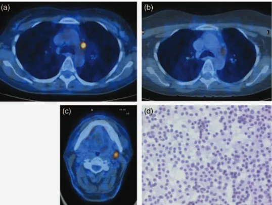
Figure 2:A 61-year-old man with a large-cell carcinoma of the left upper lobe and ipsilateral mediastinal lymph node invasion. (a) Axial PET/CT scan showed the high FDG activity within the ipsilateral mediastinal lymph node. (b) Restaging axial PET/CT scan demonstrated the partial response of the mediastinal lymph node after induction therapy. (c) Restaging axial PET/CT scan revealed the new solitary PET-positive lesion in the cervical lymph node. (d) Cytology afterfine-needle puncture revealed a reactive lymph node.
THORA
C
REFERENCES
[1] Bueno R, Richards WG, Swanson SJ, Jaklitsch MT, Lukanich JM, Mentzer SJet al. Nodal stage after induction therapy for stage IIIA lung cancer determines patient survival. Ann Thorac Surg 2000;70:1826–31. [2] Betticher DC, Hsu Schmitz SF, Totsch M, Hansen E, Joss C, von Briel C
et al. Mediastinal lymph node clearance after docetaxel-cisplatin neoad-juvant chemotherapy is prognostic of survival in patients with stage IIIA pN2 non-small-cell lung cancer: a multicenter phase II trial. J Clin Oncol 2003;21:1752–9.
[3] Stamatis G. Risks of neoadjuvant chemotherapy and radiation therapy. Thorac Surg Clin 2008;18:71–80.
[4] Akhurst T, Downey RJ, Ginsberg MS, Gonen M, Bains M, Korst Ret al. An initial experience with FDG-PET in the imaging of residual disease after induction therapy for lung cancer. Ann Thorac Surg 2002;73:259–64; discussion 264–266.
[5] Ryu JS, Choi NC, Fischman AJ, Lynch TJ, Mathisen DJ. FDG-PET in staging and restaging non-small cell lung cancer after neoadjuvant che-moradiotherapy: correlation with histopathology. Lung Cancer 2002;35: 179–87.
[6] Port JL, Lee PC, Korst RJ, Liss Y, Meherally D, Christos Pet al. Positron emission tomographic scanning predicts survival after induction chemo-therapy for esophageal carcinoma. Ann Thorac Surg 2007;84:393–400; discussion 400.
[7] Cerfolio RJ, Ojha B, Mukherjee S, Pask AH, Bass CS, Katholi CR. Positron emission tomography scanning with 2-fluoro-2-deoxy-d-glucose as a predictor of response of neoadjuvant treatment for non-small cell car-cinoma. J Thorac Cardiovasc Surg 2003;125:938–44.
[8] Hellwig D, Graeter TP, Ukena D, Georg T, Kirsch CM, Schafers HJ. Value of F-18-fluorodeoxyglucose positron emission tomography after induc-tion therapy of locally advanced bronchogenic carcinoma. J Thorac Cardiovasc Surg 2004;128:892–9.
[9] Rosell R, Gomez-Codina J, Camps C, Maestre J, Padille J, Canto Aet al. A randomized trial comparing preoperative chemotherapy plus surgery with surgery alone in patients with non-small-cell lung cancer. N Engl J Med 1994;330:153–8.
[10] Roth JA, Fossella F, Komaki R, Ryan MB, Putnam JB Jr, Lee JSet al. A ran-domized trial comparing perioperative chemotherapy and surgery with surgery alone in resectable stage IIIA non-small-cell lung cancer. J Natl Cancer Inst 1994;86:673–80.
Gonzalez-Pont G. Remediastinoscopy after induction chemotherapy in non-small cell lung cancer. Ann Thorac Surg 2000;70:391–5.
[12] De Leyn P, Stroobants S, De Wever W, Lerut T, Coosemans W, Decker G et al. Prospective comparative study of integrated positron emission tomography-computed tomography scan compared with remediastino-scopy in the assessment of residual mediastinal lymph node disease after induction chemotherapy for mediastinoscopy-proven stage IIIA-N2 non-small-cell lung cancer: a Leuven Lung Cancer Group Study. J Clin Oncol 2006;24:3333–9.
[13] Annema JT, Veselic M, Versteegh MI, Willems LN, Rabe KF. Mediastinal restaging: EUS-FNA offers a new perspective. Lung Cancer 2003;42: 311–8.
[14] Herth FJ, Annema JT, Eberhardt R, Yasufuku K, Ernst A, Krasnik M et al. Endobronchial ultrasound with transbronchial needle aspiration for restaging the mediastinum in lung cancer. J Clin Oncol 2008;26: 3346–50.
[15] Lardinois D, Schallberger A, Betticher D, Ris HB. Postinduction video-mediastinoscopy is as accurate and safe as video-video-mediastinoscopy in patients without pretreatment for potentially operable non-small cell lung cancer. Ann Thorac Surg 2003;75:1102–6.
[16] Van Schil P, van der Schoot J, Poniewierski J, Pauwels M, Carp L, Germonpre P et al. Remediastinoscopy after neoadjuvant therapy for non-small cell lung cancer. Lung Cancer 2002;37:281–5.
[17] Rami-Porta R, Mateu-Navarro M, Serra-Mitjans M, Hernandez-Rodriguez H. Remediastinoscopy: comments and updated results. Lung Cancer 2003;42:363–4.
[18] Stamatis G, Fechner S, Hillejan L, Hinterthaner M, Krbek T. Repeat mediastinoscopy as a restaging procedure. Pneumologie 2005;59: 862–6.
[19] Pottgen C, Levegrun S, Theegarten D, Marnitz S, Grehl S, Pink Ret al. Value of 18F-fluoro-2-deoxy-D-glucose-positron emission tomography/ computed tomography in non-small-cell lung cancer for prediction of pathologic response and times to relapse after neoadjuvant chemora-diotherapy. Clin Cancer Res 2006;12:97–106.
[20] Cerfolio RJ, Bryant AS, Ojha B. Restaging patients with N2 (stage IIIa) non-small cell lung cancer after neoadjuvant chemoradiotherapy: a pro-spective study. J Thorac Cardiovasc Surg 2006;131:1229–35.
[21] Dooms C, Verbeken E, Stroobants S, Nackaerts K, De Leyn P, Vansteenkiste J. Prognostic stratification of stage IIIA-N2 non-small-cell lung cancer after induction chemotherapy: a model based on the combination of morphometric-pathologic response in mediastinal nodes and primary tumor response on serial 18-fluoro-2-deoxy-glucose positron emission tomography. J Clin Oncol 2008;26:1128–34.