Functional analysis of the Bacillus subtilis cysK and cysJI genes
Jan R. van der Ploeg *, Mario Barone, Thomas Leisinger
Institute of Microbiology, Swiss Federal Institute of Technology, ETH Zentrum, CH-8092 Zu«rich, SwitzerlandReceived 30 April 2001; accepted 7 May 2001 First published online 7 June 2001
Abstract
The function of the Bacillus subtilis cysK and cysJI (previously designated yvgQR) genes, expected to be involved in the assimilatory sulfate reduction pathway, was investigated. A B. subtilis mutant with a deletion in the cysJI genes was unable to use sulfate or sulfite as sulfur source, which confirmed that these genes encode sulfite reductase. A mutant with a transposon insertion in the cysK gene, whose deduced protein sequence showed similarity to cysteine synthases, grew poorly on sulfate and butanesulfonate. A strain in which cysK and yrhA, a cysK paralog, were inactivated was unable to grow with sulfate. Whereas expression of the cysJI genes was induced by sulfate, expression of cysK was repressed both by sulfate and by cysteine. ß 2001 Federation of European Microbiological Societies. Published by Elsevier Science B.V. All rights reserved.
Keywords: Sulfate; Cysteine biosynthesis; Sul¢te reductase; Cysteine synthase; Bacillus subtilis
1. Introduction
The pathway by which Bacillus subtilis utilizes inorganic sulfate as source of sulfur appears to be very similar to the one in Escherichia coli (Fig. 1). Sulfate is transported by a sulfate permease [1], rather than by an ABC type transport system like in E. coli [2]. The gene encoding sulfate per-mease, cysP, resides in an operon together with the cysH gene, which encodes 3P-phospho-adenosine-5P-phosphosul-fate (PAPS) reductase [3]. Two other genes in this operon, ylnB and ylnC, are expected to encode enzymes responsi-ble for activation of sulfate to PAPS [1]. In addition, the 6.0-kb cysH operon also contains the genes ylnD, ylnE and ylnF. ylnD and ylnF are required for biosynthesis of the siroheme cofactor of sul¢te reductase [4], and the function of ylnE is not known. Until now, little is known about the genes and enzymes involved in the last two steps of cys-teine biosynthesis in B. subtilis, the reduction of sul¢te to sul¢de and incorporation of sul¢de into O-acetylserine (OAS).
The expression of the cysH operon is thought to be
regulated at the level of transcription initiation [5]. OAS appears to be an important signalling molecule for sulfur limitation, since addition of this compound to the medium resulted in elevated expression of a cysHP^lacZ fusion [5]. OAS is synthesized from serine and acetyl-CoA by the product of the cysE gene, which is located in one operon together with gltX, encoding glutamyl-tRNA synthetase, and cysS, which encodes cysteinyl-tRNA synthetase [6]. The expression of cysE and cysS is regulated by cys-teinyl-tRNA-mediated anti-termination of transcription [6]. In the absence of sulfate, B. subtilis can utilize aliphatic sulfonates as a source of sulfur. This is enabled by the genes of the ssuBACDygaN operon [7], and we have car-ried out studies to identify e¡ectors and regulators in-volved in expression of the ssu genes [8]. Within this framework, we have investigated the function of the cysK gene, encoding cysteine synthase, and that of the cysJI genes, which encode sul¢te reductase.
2. Materials and methods 2.1. Chemicals
All chemicals used as sulfur source were of the highest quality available and were obtained from Fluka, Acros, Aldrich or Sigma. OAS was from Sigma.
* Corresponding author. Present address: Institute of oral Microbiol-ogy and general ImmunolMicrobiol-ogy Center for dental and oral Medicine and Maxillofacial Surgery University of Zu«rich, Plattenstrasse 11, CH-8028 Zu«rich, Switzerland. Tel.: +41-1-6343329; Fax: +41-1-6344310; E-mail: [email protected]
2.2. Plasmids, primers and bacterial strains
The strains, plasmids and primers used in this study are listed in Table 1. B. subtilis and E. coli strains were grown at 30 or 37³C in LB medium or in sulfur free M63 medium [9]. Sulfur sources were added to a ¢nal concentration of 250 WM. If required, amino acids were added at the fol-lowing concentrations:L-histidine at 100 Wg ml31,L -threo-nine at 100 Wg ml31 andL-tryptophan at 40 Wg ml31. For B. subtilis, antibiotics were added at the following concen-trations when necessary: chloramphenicol, 5 Wg ml31; neomycin, 5 Wg ml31; erythromycin, 0.5 Wg ml31;
specti-nomycin, 100 Wg ml31. For E. coli, antibiotics were used at the following concentrations: kanamycin (50 Wg ml31), ampicillin (100 Wg ml31) and chloramphenicol (35 Wg ml31), spectinomycin (100 Wg ml31). Solid media con-tained 1.5% agar (Serva). The sequences of the primers used for ampli¢cation of B. subtilis genes were based on the genome sequence [10].
2.3. DNA and RNA manipulation
For plasmid isolation, restriction enzyme digestion, li-gation and transformation of E. coli, standard procedures
Table 1
Strains, plasmids and used in this study
Strain Genotype or relevant properties Ref. or source
E. coli
DH5K supE44 vlacU169(P80 lacZvM15) hsdR17 recA1 endA1 gyrA96 thi-1 relA1 Life Technologies XL1-Blue recA1 endA1 gyrA96 thi-1 hsdR17 supE44 relA1 lac [FP proAB laclqZvM15 Tn10 (Tetr)] Stratagene NK3 vtrpE5 leu6 thi cysK cysM rbsrm
k M. Hryniewicz
B. subtilis
1A1 trpC2 BGSC
BFA2063 trpC2 yrhA-lacZ vyrhA W. Schumann
MS-11 hisA1 trpC2 thr-5 ssuD: :lacZ (Tn917-lac) [7]
MS11-6 hisA1 trpC2 thr-5 ssuD: :lacZ (Tn917-lac) cysK: :Tn10 this study
SB11 trpC2 cysK: :Tn10 this study
SB12 trpC2 vyvgQr: :Nmr this study
SB36 trpC2 amyE: :pME4834 this study
SB27 trpC2 amyE: :pME4881 this study
SB28 trpC2 cysK: :Tn10 amyE: :pME4881 this study
SB29 trpC2 amyE: :pME4886 this study
SB30 trpC2 cysK: :Tn10 amyE: :pME4886 this study
SB49 trpC2 vytkP: :Nmr this study
SB50 trpC2 yrhA-lacZ vyrhA cysK: :Tn10 this study
SB51 trpC2 yrhA-lacZ vyrhA vytkP: :Nmr this study
SB52 trpC2 vytkP: :Nmr, cysK: :Tn10 this study
SB53 trpC2 yrhA-lacZ vyrhA cysK: :Tn10 ytkP: :Nmr this study
Plasmid
pBluescript II KS E. coli cloning vector, Apr Stratagene
pUC19 E. coli cloning vector, Apr [20]
pDH32M Apr CmramyE front amyE back lacZ [16]
pMLK83 Apr NmramyE front amyE back gus [17]
pRB374 E. coli/B. subtilis shuttle vector, Nmr [21]
pRSM40 pT7T3, cysK from S. typhimurium N. Kredich
pBEST501 Apr, Nmrcassette [15]
pIC333 pBR322 ori, repTs, SprEryr E. Bremer
pME4823 pUC19, cysJP-kan-PcysI this study
pME4833 pBluescript containing cysJI promoter region this study
pME4834 pDH32M containing cysJI promoter region this study
pME4857 cysKSTin pRB374 this study
pME4864 cysKBSin pRB374 this study
pME4881 cysKP^gusA in pMLK83 this study
pME4886 cysKP^gusA (truncated) in pMLK83 this study
pME4899 pBluescript II KS, ytkP: :kan this study
Primer Sequence (5P^3P) Used for
BcysK2 GCGAACCTGCAGTTTTGGC cloning of B. subtilis cysK
BscysK3 AATAAGCTTTACAAATAGTCGG cloning of B. subtilis cysK
cysKf3 GCTTTGCATGCAGTTAAGGACAG cloning of S. typhimurium cysK
cysKr TTAGGATCCTGGCATCACTG cloning of S. typhimurium cysK
were used [11]. B. subtilis was transformed as described [12]. Chromosomal DNA from B. subtilis was isolated us-ing the CTAB method [11]. Total RNA was isolated from strain 1A1 grown in minimal medium with sulfate or glu-tathione as sulfur source to an optical density at 600 nm of approximately 0.5 as described [13]. Primer extension anal-ysis was according to Babst et al. [14].
2.4. Construction of deletion insertions in cysJI and in ytkP For the construction of a cysJI knock-out, a neomycin resistance cassette from pBEST501 [15] was cloned in be-tween two fragments located within the cysJI genes which had been generated by PCR. The resulting plasmid, pME4823, was transformed into the wild-type B. subtilis strain 1A1 by selecting for neomycin resistance. PCR anal-ysis of one of the resulting colonies (designated SB12) showed that a correct replacement had occurred.
For the construction of strains containing a deletion/ insertion of ytkP, a similar strategy was used. Two frag-ments, encompassing the 5P end of ytkP and the 3P end of ytkP, were ampli¢ed by PCR and cloned into pBluescript II KS. A neomycin resistance cassette was then cloned in between the two fragments to give plasmid pME4899. Transformation of linearized pME4899 into B. subtilis and selection for neomycin resistance yielded ytkP mu-tants.
2.5. Transposon mutagenesis
Transposon mutagenesis was carried out using pIC333, which contains a temperature sensitive origin of replica-tion and harbors a derivative of Tn10 (Steinmetz and Richter, unpublished). Strain MS11 (ssuDP^lacZ), which does not produce su¤cient L-galactosidase to form blue colonies on LB plates containing X-gal (40 Wg ml31), was transformed with plasmid pIC333 and grown at 30³C on LB containing spectinomycin. Transformants were grown overnight in LB containing spectinomycin at 30³C, diluted 1:100 in the same medium and grown for 3 h at 30³C. The temperature was then shifted to 37³C and incubation was continued for another 4 h. A total of about 15 000 mutants were screened for the formation of blue colonies on LB plates containing X-gal. Chromosomal DNA from mu-tants was isolated and digested with EcoRI and HindIII. The DNA was ligated under conditions that favor circu-larization of fragments and transformed to E. coli by se-lection for spectinomycin resistance. Plasmids were iso-lated from the resulting transformants, and the sequence £anking the transposon insertion site was determined. 2.6. Construction of cysJP^lacZ and cysKP^gusA fusions
For the construction of a transcriptional cysJP^lacZ fu-sion, a 311-bp fragment encompassing 222 bp of the re-gion upstream of the start of cysJ was ampli¢ed by PCR.
The product was cloned in pBluescript KS and in pDH32M [16] to give pME4833 and pME4834 respec-tively. A transcriptional cysKP^gusA fusion was con-structed by cloning a 440-bp fragment, encompassing 317 bp of the region upstream of the cysK start codon, into pMLK83 [17] to give pME4881. A truncated cysKP^ gusA fusion, which contained 84 bp of the region up-stream of the cysK start codon, was constructed on plas-mid pME4886. The plasplas-mids containing the fusions were introduced in B. subtilis strain 1A1 by transformation [18] by selection for chloramphenicol or neomycin resistance. Double crossover events were con¢rmed by testing for an amyE negative phenotype. L-Galactosidase and L-glucu-ronidase activities were measured according to Miller [19]. 2.7. Cloning of B. subtilis and Salmonella typhimurium
cysK genes and complementation experiments
The cysK gene from B. subtilis was cloned by PCR ampli¢cation using the forward primer BcysK2 and the reverse primer BscysK3 (Table 1) with chromosomal DNA from strain BD99 as template. The PCR fragment was digested with HindIII and PstI and cloned in pRB374 to give pME4864.
The S. typhimurium cysK gene was ampli¢ed using PCR with the forward primer cysKf3 and the reverse primer cysKr using plasmid pRSM40 as template. A PaeI^BamHI fragment was cloned in pRB374 to give pME4857. E. coli strain NK3 (cysKcysM) and B. subtilis strain SB11 (cysK: :Tn10) were used as recipient in complementation studies to verify the functionality of cysKBS and cysKST. 3. Results
3.1. Functional analysis of the B. subtilis cysJI genes In E. coli, reduction of sul¢te to sul¢de is catalyzed by sul¢te reductase, encoded by the cysI and cysJ genes [2]. The cysJ gene product accepts electrons from NADPH, which are subsequently transferred to the hemoprotein CysI, and from there to sul¢te to give sul¢de. In B. sub-tilis, open reading frames with sequence similarity to the E. coli cysI and cysJ genes are encoded by yvgQ (51% identity over 567 amino acid residues) and yvgR (42% identity over 600 amino acid residues) respectively, which are positioned at 293 min on the chromosome. Sequence analysis showed that YvgQ contains a siroheme motif, which is conserved in sul¢te reductases. The YvgR protein contains putative FAD and NADH binding sites.
A mutant with a deletion in yvgRQ and a simultaneous insertion of a neomycin resistance cassette was constructed as described in Section 2. The resulting mutant, strain SB12, was unable to utilize sulfate, sul¢te or butanesulfo-nate as a source of sulfur and grew poorly with sul¢de, but it could still grow with thiosulfate, cysteine or methionine.
These data suggested that yvgRQ indeed encodes sul¢te reductase. We have therefore renamed yvgR and yvgQ into cysJ and cysI respectively. The results also indicate that, unlike in E. coli [2], a direct pathway from methio-nine to cysteine exists since growth with methiomethio-nine was not a¡ected.
3.2. Regulation of expression of cysJI
To study whether expression of the B. subtilis cysJI genes is regulated by the sulfur source used for growth, a strain containing a transcriptional cysJP^lacZ fusion in-tegrated at amyE was constructed (strain SB36) and L-ga-lactosidase was measured in cells grown with di¡erent sul-fur sources (Table 2). The levels of L-galactosidase were low compared to those found with ssuDP^lacZ fusions [7] or cysHP^lacZ fusions [5]. L-Galactosidase levels were higher in sulfate or butanesulfonate grown cells than in cells grown with other sulfur sources.
The transcriptional start of cysJI was mapped by primer extension analysis using RNA isolated from strain 1A1 grown with sulfate as sulfur source. It was located 46 bp
upstream of the translation initiation codon of cysJ (re-sults not shown). The band was weak, which con¢rmed the low levels of L-galactosidase obtained with the cysJP^ lacZ fusion.
3.3. Isolation and characterization of a cysK mutant In an attempt to identify proteins involved in regulation of the ssu operon, transposon mutagenesis with a Tn10 derivative was used to isolate mutants that exhibited con-stitutive expression of L-galactosidase from a transcrip-tional ssuDP^lacZ fusion. Although regulatory mutants were not obtained, a mutant containing a transposon in-sertion at bp 341 of the cysK gene exhibited constitutive expression of L-galactosidase [8]. The cysK gene from B. subtilis has not been previously characterized, but it may be assumed that it encodes cysteine synthase (O-ace-tylserine (thiol)-lyase), since it shows 43% amino acid se-quence identity to the cysK gene from E. coli. The cysK mutant, strain MS11-6, could still grow with sulfate, bu-tanesulfonate and sul¢te, but its growth rates were lower than those of the wild-type strain (not shown). Thus, cysK might be involved in the biosynthesis of cysteine from sulfate, but B. subtilis contains probably more than one gene encoding this enzyme activity. There are still two more cysK paralogs present on the B. subtilis chromo-some, yrhA and ytkP. The function of these paralogs is not known, but either both or one of them apparently can only partly take over the function of cysK, since the cysK mutant grew slowly with sulfate and butanesulfonate. We constructed several mutants that contained knock-outs in yrhA, ytkP and cysK and tested their growth properties. All strains containing single mutations were able to grow with sulfate as sulfur source. The yrhA cysK double mu-tant SB50 as well as the yrhA cysK ytkP triple mumu-tant SB53 could not grow anymore with sulfate or with
thio-Table 2
Expression L-galactosidase from a transcriptional cysJP^lacZ fusion Sulfur source L-Galactosidase activity (Miller units)
Sulfate 17.4 þ 5.2 Cystine 1.2 þ 1.4 Sulfate+cystine 8.2 þ 1.4 Methionine 5.8 þ 0.6 Methionine+sulfate 6.6 þ 0.9 Butanesulfonate 12.7 þ 2.3 Glutathione 7.5 þ 2.1
An overnight culture of B. subtilis strain SB36, containing a chromoso-mal cysJP^lacZ fusion, was 100-fold diluted in fresh medium containing the indicated sulfur source and L-galactosidase was measured in the mid-exponential phase of growth.
Fig. 2. Determination of the cysK transcription start site. Approximately equal amounts of RNA were isolated from cells grown with butanesul-fonate (lane 1), sulfate (lane 2), cystine (lane 3) or glutathione (lane 4) and were reverse-transcribed using the primer indicated in Fig. 3. The sequencing ladder was obtained using the same primer. The transcrip-tion start is indicated by an arrow.
Fig. 1. Cysteine biosynthesis from sulfate and aliphatic sulfonates in B. subtilis.
sulfate. This indicates that yrhA encodes cysteine synthase activity as well.
We used complementation analysis to con¢rm the func-tion of B. subtilis cysK. The gene was cloned in plasmid pRB374 under the control of the vegII promoter. The resulting plasmid, pME4864, could complement growth with sulfate as sulfur source of E. coli strain NK3, a cysK cysM mutant lacking O-acetylserine (thiol)-lyase ac-tivity. This suggests that B. subtilis cysK indeed encodes this enzyme. Using the same plasmid, the cysK mutation in B. subtilis strains MS11-6 or SB11 could not be com-plemented however. Plasmid pME4857, which contains the S. typhimurium cysK gene, could also complement E. coli NK3, but not B. subtilis MS11-6 or SB11 (results not shown). It is unlikely that the transposon insertion has a polar e¡ect on expression of genes downstream of cysK, since there is a transcription terminator present immedi-ately after the cysK stop codon. It is possible that the S. typhimurium and B. subtilis cysK genes were somehow not well expressed from the plasmid in B. subtilis. 3.4. Regulation of expression of cysK
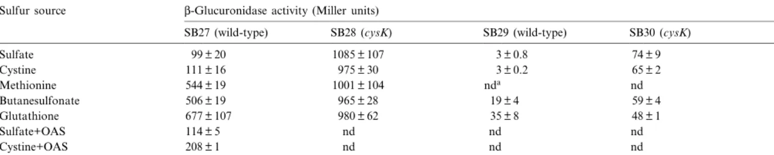
We used strains SB27 and SB29, which contain chromo-somally located transcriptional cysKP^gusA fusions in a
wild-type background (Fig. 3), to investigate the condi-tions under which the cysK gene was expressed. In sulfate or cystine grown SB27, L-glucuronidase activities were about 5-fold lower than in cells grown with methionine, butanesulfonate or glutathione (Table 3). Strain SB29, which harbors a fusion containing just 9 bp from the re-gion upstream of the 335 rere-gion, produced lower levels of L-glucuronidase, but repression by sulfate and cystine was retained. This indicates that this fusion lacked a region necessary for full expression, but we were unable to ¢nd a sequence which could function as enhancer.
Addition of OAS to the medium resulted in a 2-fold increase in activity in cystine grown cells, but had only a marginal e¡ect on expression of the cysKP^gusA fusion in sulfate grown cells.
When the cysKP^gusA fusions were introduced in the cysK mutant, the resulting strains SB28 and SB30 pro-duced L-glucuronidase constitutively. These results indi-cate that either CysK has also a regulatory function, or that intermediates that accumulate when CysK is inactive give rise to higher expression levels.
Using primer extension analysis, the transcriptional start of cysK was determined (Fig. 2). One band was ob-served, which was strongest when RNA from cells grown with glutathione or butanesulfonate was used. The
inten-Table 3
Expression of L-glucuronidase from transcriptional cysKP^gusA fusions in a wild-type strain and in a cysK mutant Sulfur source L-Glucuronidase activity (Miller units)
SB27 (wild-type) SB28 (cysK) SB29 (wild-type) SB30 (cysK)
Sulfate 99 þ 20 1085 þ 107 3 þ 0.8 74 þ 9 Cystine 111 þ 16 975 þ 30 3 þ 0.2 65 þ 2 Methionine 544 þ 19 1001 þ 104 nda nd Butanesulfonate 506 þ 19 965 þ 28 19 þ 4 59 þ 4 Glutathione 677 þ 107 980 þ 62 35 þ 8 48 þ 1 Sulfate+OAS 114 þ 5 nd nd nd Cystine+OAS 208 þ 1 nd nd nd
Overnight cultures of B. subtilis strains containing chromosomal cysKP^gusA fusions were diluted 100-fold in fresh medium containing the indicated sul-fur source and grown to the mid-exponential phase. When indicated, OAS was added to a ¢nal concentration of 0.5 mM 2 h before measurement of L-glucuronidase activities.
aNot done.
Fig. 3. Promoter region of the cysK gene. The 335 and 310 regions of the cysK promoter are boxed and the transcription start site is indicated. The stop codon of the yacD gene is underlined. The 5P ends of the promoter regions contained in strains harboring cysKP^gusA fusions are indicated by ar-rows. The 3P end of the cysk portion of these fusions coincides with the last nucleotide shown. The primer used for primer extension analysis (Fig. 2) is indicated by a stippled arrow.
sity of the bands was in agreement with the results ob-tained from measurements of L-glucuronidase activities from transcriptional cysKP^gusA fusions. The 335 region of the cysK promoter is located immediately downstream of the stop codon of the yacD gene (Fig. 3), whose func-tion is unknown.
4. Discussion
Until now, four operons and one gene involved in the biosynthesis of cysteine from sulfate and from sulfonates have been identi¢ed. The cysE gene encodes serine trans-acetylase, the enzyme responsible for synthesis of OAS, and resides in one operon with gltX and cysS [6]. The cysH operon is required for uptake and reduction of sul-fate to sul¢te [5]. The ssu operon encodes transport and liberation of sul¢te from aliphatic sulfonates [7]. In this study we have investigated the function and regulation of the cysJI operon, which is required for reduction of sul¢te to sul¢de, and that of the cysK gene, which encodes the ¢nal step in cysteine biosynthesis. The function of cysK can be taken over by its paralog yrhA. This is reminiscent to the situation in E. coli, which contains two proteins with O-acetylserine (thiol)-lyase activity, encoded by cysK and cysM [2]. The E. coli CysK protein is more active with sul¢de, while the CysM protein has a higher a¤nity for thiosulfate [2]. Further analysis of the proteins encoded by cysK and yrhA should give insight in their substrate speci¢city.
In E. coli, expression of the cys genes and the ssu genes is under coordinate control of the transcriptional regulator CysB [2]. Whether the B. subtilis cys and ssu genes are also controlled by one regulatory system is as yet unknown, but we have been unable to ¢nd sequence similarity be-tween the promoter regions of the cysH, the cysJI, the cysK and the ssu genes.
Similar as with the cysH and ssu operons, expression of cysK was repressed by sulfate and cysteine. The mecha-nism by which repression by these compounds is mediated remains unclear, but OAS or a closely related molecule plays an important role. In E. coli, OAS functions as co-inducer of the transcriptional regulatory protein CysB [2]. The level of OAS is a measure of sulfur availability for the cell, since cysteine exerts feedback inhibition on serine transacetylase. Sulfur limitation results in increased syn-thesis of OAS, which induces transcription of the cys genes. Although it is not known whether in B. subtilis serine transacetylase is inhibited by OAS, the expression of the cysE gene is regulated by transcription attenuation through binding of uncharged cysteinyl-tRNA to the lead-er RNA, thlead-ereby promoting formation of an anti-tlead-ermina- anti-termina-tor, which results in transcription readthrough [6]. Cys-teine could thus indirectly regulate the amount of OAS, which in turn could act as e¡ector in regulation of the cys and ssu genes.
Acknowledgements
We thank Erhard Bremer, Reinhard Bru«ckner, Monika Hryniewicz, Nicholas Kredich, Patrick Piggot, and Wolf-gang Schumann for gifts of strains or plasmids.
References
[1] Mansilla, M.C. and de Mendoza, D. (2000) The Bacillus subtilis cysP gene encodes a novel sulphate permease related to the inorganic phosphate transporter (Pit) family. Microbiology 146, 815^821. [2] Kredich, N.M. (1996) Biosynthesis of cysteine. In: Escherichia coli
and Salmonella, 2nd edn. (Neidhardt, F.C., Curtiss, R., Ingraham, J.L., Lin, E.C.C., Low, K.B., Magasanik, B., Rezniko¡, W.S., Riley, M., Schaechter, M. and Umbarger, H.E., Eds.), pp. 514^527. ASM Press, Washington, DC.
[3] Mansilla, M.C. and de Mendoza, D. (1997)L-cysteine biosynthesis in Bacillus subtilis: identi¢cation, sequencing, and functional character-ization of the gene coding for phosphoadenylylsulfate sulfotransfer-ase. J. Bacteriol. 179, 976^981.
[4] Johansson, P. and Hederstedt, L. (1999) Organization of genes for tetrapyrrole biosynthesis in Gram-positive bacteria. Microbiology 145, 529^538.
[5] Mansilla, M.C., Albanesi, D. and de Mendoza, D. (2000) Transcrip-tional control of the sulfur-regulated cysH operon, containing genes involved inL-cysteine biosynthesis in Bacillus subtilis. J. Bacteriol. 182, 5885^5892.
[6] Gagnon, Y., Breton, R., Putzer, H., Pelchat, M., Grunberg Manago, M. and Lapointe, J. (1994) Clustering and co-transcription of the Bacillus subtilis genes encoding the aminoacyl-tRNA synthetases spe-ci¢c for glutamate and for cysteine and the ¢rst enzyme for cysteine biosynthesis. J. Biol. Chem. 269, 7473^7482.
[7] Van der Ploeg, J.R., Cummings, N.J., Leisinger, T. and Connerton, I.F. (1998) Bacillus subtilis genes for the utilization of sulfur from aliphatic sulfonates. Microbiology 144, 2555^2561.
[8] Van der Ploeg, J.R., Barone, M. and Leisinger, T. (2001) Expression of the Bacillus subtilis sulphonate-sulphur utilization genes is regu-lated at the levels of transcription initiation and termination. Mol. Microbiol. 39, 1356^1365.
[9] Van der Ploeg, J.R., Weiss, M.A., Saller, E., Nashimoto, H., Saito, N., Kertesz, M.A. and Leisinger, T. (1996) Identi¢cation of sulfate starvation-regulated genes in Escherichia coli: a gene cluster involved in the utilization of taurine as a sulfur source. J. Bacteriol. 178, 5438^ 5446.
[10] Kunst, F., Ogasawara, N., Moszer, I., Albertini, A.M., Alloni, G., Azevedo, V., Bertero, M.G., Bessieres, P., Bolotin, A., Borchert, S., Borriss, R., Boursier, L., Brans, A., Braun, M., Brignell, S.C., Bron, S., Brouillet, S., Bruschi, C.V., Caldwell, B., Capuano, V., Carter, N.M., Choi, S.K., Codani, J.J., Connerton, I.F., Cummings, N.J., Daniel, R.A., Denizot, F., Devine, K.M., Du«sterho«ft, A., Ehrlich, S.D., Emmerson, P.T., Entian, K.D., Errington, J., Fabret, C., Fer-rari, E., Foulger, D., Fritz, C., Fujita, M., Fujita, Y., Fuma, S., Galizzi, A., Galleron, N., Ghim, S.-Y., Glaser, P., Go¡eau, A., Go-lightly, E.J., Grandi, G., Guiseppi, G., Guy, B.J., Haga, K., Haiech, J., Harwood, C.R., He¨naut, A., Hilbert, H., Holsappel, S., Hosono, S., Hullo, M.-F., Itaya, M., Jones, L., Joris, B., Karamata, D., Ka-sahara, Y., Klaerr-Blanchard, M., Klein, C., Kobayashi, Y., Koetter, P., Koningstein, G., Krogh, S., Kumano, M., Kurita, K., Lapidus, A., Lardinois, S., Lauber, J., Lazarevic, V., Lee, S.-M., Levine, A., Liu, H., Masuda, S., Maue«l, C., Me¨digue, C., Medina, N., Mellado, R.P., Mizuno, M., Moestl, D., Nakai, S., Noback, M., Noone, D., O'Reilly, M., Ogawa, K., Ogiwara, A., Oudega, B., Park, S.-H., Parro, V., Pohl, T.M., Portetelle, D., Porwollik, S., Prescott, A.M., Presecan, E., Pujic, P., Purnelle, B., Rapoport, G., Rey, M.,
Rey-nolds, S., Rieger, M., Rivolta, C., Rocha, E., Roche, B., Rose, M., Sadaie, Y., Sato, T., Scanlan, E., Schleich, S., Schroeter, R., Scof-fone, F., Sekiguchi, J., Sekowska, A., Seror, S.J., Serror, P., Shin, B.-S., Soldo, B., Sorokin, A., Tacconi, E., Takagi, T., Takahashi, H., Takemaru, K., Takeuchi, M., Tamakoshi, A., Tanaka, T., Terpstra, P., Tognoni, A., Tosato, V., Uchiyama, S., Vandenbol, M., Vannier, F., Vassarotti, A., Viari, A., Wambutt, R., Wedler, E., Wedler, H., Weitzenegger, T., Winters, P., Wipat, A., Yamamoto, H., Yamane, K., Yasumoto, K., Yata, K., Yoshida, K., Yoshikawa, H.-F., Zum-stein, E., Yoshikawa, H. and Danchin, A. (1997) The complete ge-nome sequence of the Gram-positive bacterium Bacillus subtilis. Na-ture 390, 249^256.
[11] Ausubel, F.M., Brent, R., Kingston, R.E., Moore, D.E., Seidman, J.G., Smith, J.A. and Struhl, K. (1987) Current Protocols in Molec-ular Biology. Greene Publishing Associates/Wiley Interscience, New York.
[12] Cutting, S.M. and Youngman, P. (1994) Gene transfer in Gram-pos-itive bacteria. In: Methods for General and Molecular Bacteriology (Gerhardt, P., Murray, G.E., Wood, W.A. and Krieg, N.R., Eds.), pp. 348^364. ASM Press, Washington, DC.
[13] Vo«lker, U., Engelmann, S., Maul, B., Riethdorf, S., Vo«lker, A., Schmid, R., Mach, H. and Hecker, M. (1994) Analysis of the induc-tion of general stress proteins of Bacillus subtilis. Microbiology 140, 741^752.
[14] Babst, M., Hennecke, H. and Fischer, H.M. (1996) Two di¡erent mechanisms are involved in the heat shock regulation of chaperonin gene expression in Bradyrhizobium japonicum. Mol. Microbiol. 19, 827^839.
[15] Itaya, M., Kondo, K. and Tanaka, T. (1989) A neomycin resistance gene cassette selectable in a single copy state in the Bacillus subtilis chromosome. Nucleic Acids Res. 17, 4410.
[16] Kraus, A., Hueck, C., Gartner, D. and Hillen, W. (1994) Catabolite repression of the Bacillus subtilis xyl operon involves a cis element functional in the context of an unrelated sequence, and glucose exerts additional XylR-dependent repression. J. Bacteriol. 176, 1738^1745. [17] Karow, M.L. and Piggot, P.J. (1995) Construction of gusA
transcrip-tional fusion vectors for Bacillus subtilis and their utilization for studies of spore formation. Gene 163, 69^74.
[18] Anagnostopoulos, C. and Spizizen, J. (1961) Requirements for trans-formation in Bacillus subtilis. J. Bacteriol. 81, 741^746.
[19] Miller, J.H. (1992) Cold Spring Harbor Laboratory Press, Cold Spring Harbor, NY.
[20] Yanisch-Perron, C., Vieira, J. and Messing, J. (1985) Improved M13 phage cloning vectors and host strains: nucleotide sequences of the M13mp18 and pUC19 vectors. Gene 33, 103^119.
[21] Bru«ckner, R. (1992) A series of shuttle vectors for Bacillus subtilis and Escherichia coli. Gene 122, 187^192.