The functions of RMR proteins in the
Physcomitrella patens secretory pathway
A dissertation submitted to the University of Neuchâtel For the degree of Doctor in Sciences by
Noémie FAHR
Institute of Biology - Laboratory of Cell and Molecular Biology
Accepted on the recommendation of
Prof. Jean-Marc NEUHAUS, thesis director, University of Neuchâtel, CH
Dr. Sébastien THOMINE, CNRS – Gif-sur-Yvette, FR
Dr. Shanmugabalaji VENKATASALAM, University of Neuchâtel, CH
Dr. Didier SCHAEFER, University of Neuchâtel, CH
5
Keywords: Vacuole, RMR, Vacuolar Sorting Determinant, Physcomitrella patens, E3 ligase,
Confocal microscopy
Mots-clés: Vacuole, RMR, Déterminant d’adressage vacuolaire, Physcomitrella patens, E3
ligase, Microscopie confocale
7
Table of Contents
Abstract ... 13
Résumé ... 15
List of abbreviations ... 17
Chapter 1 General Introduction ... 21
A. Generalities about the plant secretory pathway and the endomembrane system ... 21
A.1 Early secretory pathway ... 22
A.2 Late secretory pathway ... 30
A.3 Vesicular traffic ... 32
A.4 Endocytosis ... 35
A.5. Protein secretion ... 38
B. Vacuole types and protein targeting to vacuole ... 39
B.1 Vacuole types ... 39
B.2.Vacuolar sorting determinants... 40
B.3 Vacuolar sorting receptors in plant cells ... 41
The VSR family ... 41
The RMR family ... 43
Protease-Associated domain ... 45
C. C-terminal VSD used in this work ... 46
C.1 Ct-VSD of tobacco Chitinase A ... 46
C.2 Ct-VSD of barley lectin (BL) ... 46
C.3 Ct-VSD of Phaseolin ... 47
C.4. Ct-VSD and PSI of Cardosins ... 47
Generalities about Aspartic proteinases (APs) ... 47
Plant APs ... 48
Plant AP domain structure ... 48
Cardosin A... 48
Cardosin B ... 49
C.5 Ct-VSD and PSI of moss Aspartic proteinase ... 49
8
D.1 GRAIL (mammals) ... 51
D.2 Goliath and Godzilla ... 52
D.3 RING Finger protein 13... 52
D.4 Other PA-TM-RING proteins ... 53
E. RMR proteins and ubiquitylation activity: a key role for the RING domain? ... 54
E.1 Ubiquitylation mechanism ... 54
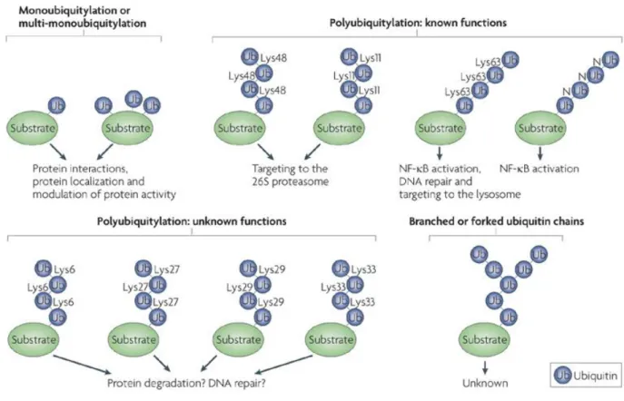
E.2 Different ubiquitin tags for diverse purposes ... 56
E.3 RING domain and ubiquitylation: the E3 ligases family ... 57
RING E3 ligases ... 57
HECT E3 ligases ... 58
U-box E3 ligases ... 59
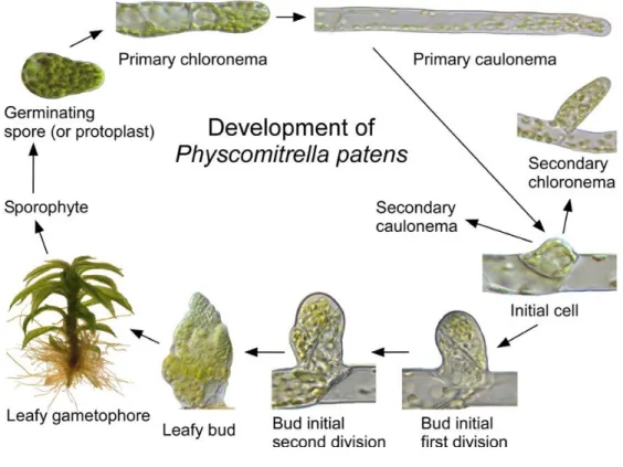
F. The moss Physcomitrella patens ... 60
F.1 P. patens as an experimental model ... 60
F.1.a Life cycle ... 60
F.1.b Genome ... 62
F.1.c Homologous recombination and gene targeting in P. patens ... 63
F.1.d RNAi in moss ... 64
F.1.e Abiotic stress tolerance in moss ... 65
F.2 The moss vacuole ... 65
F.2.a Different kind of vacuoles ... 65
F.2.b Vacuole structure and dynamics ... 66
F.3 Quintuple RMR Knock-Out lines ... 67
G. Aim of this thesis ... 68
Chapter 2
Study of the secretory system in moss: fluorescent reporters library ... 69
A. General secretory system markers ... 70
A.1 Reporter for the endoplasmic reticulum ... 70
A.2 Reporter for the acidic vacuole ... 72
A.3 Secreted reporter ... 74
A.3.a Citrine with just a signal peptide ... 74
A.3.b Secreted chitinase from P. patens ... 76
B. Reporters for the neutral vacuole (ctVSD) ... 78
9
B.2 CtVSD of cardosin A ... 78
B.3 CtVSD of moss aspartic proteinase 1 ... 81
C. Plant Specific Insert reporters ... 83
C.1 PSI domain from Cardosin A ... 83
C.2 PSI domain of the cardosin B... 83
D. Discussion ... 86
Chapter 3
Characterization of RMR Knock-out mutants ... 89
A. Phenotypic characterization of RMR mutants with fluorescent reporters ... 90
A.1 CtVSD reporters ... 90
A.1.a CtVSD of tobacco chitinase I ... 90
A.1.b CtVSD of cardosin A ... 91
A.1.c CtVSD of PpAP1 ... 95
A.2 PSI reporters ... 96
A.2.a PSI from cardosin A ... 96
A.2.b PSI domain from cardosin B ... 96
A.3 Further vacuolar markers ... 99
A.3.a Secreted Citrine (SP-Citrine) ... 99
A.3.b Acidic vacuole reporter ... 99
A.3.c Secreted moss chitinase ... 100
A.3.d ER-retained reporter ... 100
A.4 Discussion ... 102
B. Vacuolar routes studies: secretory pathway inhibitors effects ... 104
B.1 Results ... 105
B.2 Discussion ... 110
C. Localization of PpRMR2 ... 111
C.1 PpRMR2 localization in Physcomitrella patens ... 113
C.1.a Preliminary observations ... 113
C.1.b Control of transgene insertion and expression ... 114
C.2 Proteasome inhibitors ... 116
C.3 Discussion ... 118
D. Phenotypic study of RMR mutants ... 119
10
D.2 Discussion ... 122
Chapter 4
Identification of RMR2 partners: clues to the role of RMRs in moss? ... 123
1. Pull-down assay using a GST fusion protein ... 123
2. Results ... 125
3. Discussion ... 127
Chapter 5 General discussion and perspectives ... 129
1. The moss secretory pathway ... 129
2. Are RMRs vacuolar receptors? ... 131
3. Are RMRs E3 ubiquitin ligases? ... 133
Chapter 6 Material and methods ... 137
A. Material ... 137
A.1 Plant material ... 137
A.2 Bacterial strains ... 138
B. Methods related to bacteria ... 138
B.1 E. Coli Transformation by heat-shock ... 138
B.2 Preparation of XL-1 competent cells ... 138
B.3 Plasmid DNA extraction: miniprep ... 139
C. Methods related to plants ... 140
C.1 Protoplasts isolation and transformation ... 140
Protoplasts isolation ... 140
Protoplasts transformation by PEG ... 140
C.2 Heat-shock on protonema for protein expression ... 141
C.3 Secretory pathway inhibitors treatment ... 141
C.4 Proteasome II inhibitor treatment ... 141
D. Molecular biology ... 142
D.1 PCR ... 142
D.2 DNA digestion ... 142
D.3 DNA precipitation... 142
11
D.5 DNA electrophoresis ... 143
D.6 DNA extraction from agarose gel ... 143
D.7 DNA fragment ligation in a plasmid ... 143
D.8 Extraction of total RNA from P. patens ... 144
D.9 Reverse transcription: synthesis of cDNA ... 144
E. Methods related to proteins ... 144
E.1 Moss total proteins extraction in SB buffer ... 144
E.2 SDS-PAGE ... 144
E.3 Western blot ... 145
E.4 Colloidal Coomassie blue staining ... 146
E.5 Production of GST-RMR recombinant proteins in BL-21 strain ... 146
E.6 Pull-down assay ... 147
F. Microscopy ... 147
Confocal microscopy ... 147
Acknowledgements ... 149
Annexes ... 151
1. Primers... 151
2. Vectors and constructs ... 153
2.a Cloning vectors ... 153
2.b Cloning strategies ... 153
3. Vector maps ... 154
13
Abstract
Plant vacuoles play a wide range of functions within the cell, from the maintenance of turgor pressure and rigidity, to the storage or degradation of various molecules. Two types of vacuoles with distinct pH can coexist in the same single cell. Acidic vacuoles can be considered as homologues to animal lysosomes while neutral vacuoles are involved in proteins and secondary metabolites storage. Targeting of proteins to the lytic vacuole has been extensively studied and the vacuolar receptors involved, the VSRs, are now well characterized in higher plants. However, less is known about the traffic of proteins to the neutral/storage vacuole. RMR proteins are thought to be vacuolar receptors for the neutral/storage vacuole. However, the complete deletion of the five RMR genes in
Physcomitrella patens did not lead to any developmental phenotype. This work aimed to investigate
the role of RMR proteins in the moss.
In the first chapter, we review the plant secretory pathway system and how proteins are targeted to vacuoles.
In the second chapter, we studied the moss secretory pathway by developing a fluorescent reporter library. Several mechanisms seem to be conserved between the moss and the flowering plants.
In the third chapter, we focused on the characterization of the single and quintuple knock-out RMR mutants. We finally obtained a trafficking phenotype: the fluorescent reporter Citrine-Card was mistargeted in the single, triple and quintuple KO mutants. Fluorescent signal was detected in endoplasmic reticulum in the mutants, while it was observed in the central vacuole in WT. Trafficking to vacuole of a protein carrying this ctVSD was RMR-dependent.
In the last part of this thesis, we identified some putative binding partners of the cytosolic part of PpRMR2 by GST pull-down assay and mass spectrometry analysis.
15
Résumé
Chez les plantes, les vacuoles occupent un grand nombre de fonctions, allant du maintien de la pression de turgescence et de la rigidité cellulaire en passant par le stockage ou la dégradation de diverses molécules. Deux types de vacuoles ayant un pH distinct peuvent coexister au sein d’une même cellule. Les vacuoles acides sont considérées comme étant des homologues aux lysosomes présents dans les cellules animales, tandis que les vacuoles neutres sont impliquées dans le stockage de protéines et de métabolites secondaires. L’adressage des protéines à la vacuole lytique a été largement étudié et les récepteurs vacuolaires impliqués, les VSRs, sont des protéines bien caractérisées. À l’opposé, les connaissances sur l’adressage des protéines à la vacuole neutre ou de stockage sont moindres. Les protéines RMR sont très probablement les récepteurs vacuolaires impliqués, bien que la délétion des cinq gènes RMR chez Physcomitrella patens n’ait conduit à aucun phénotype visible. Ce travail a donc pour objectif l’élucidation du rôle des protéines RMR chez la mousse.
Dans le premier chapitre de cette thèse nous avons regroupé les principales données concernant le système sécrétoire et endomembranaire chez les plantes, et nous avons également documenté comment les protéines sont adressées aux vacuoles.
Dans le second chapitre, nous avons étudié le système sécrétoire de la mousse en développant une bibliothèque de marqueurs fluorescents. Différents mécanismes cellulaires semblent conservés entre les mousses et les plantes à fleurs.
Dans le troisième chapitre, nous nous sommes intéressés à la caractérisation des simples et quintuple knock-out mutants RMR. Nous avons finalement obtenu un phénotype de tri vacuolaire: un défaut d’adressage est observé avec le marqueur fluorescent Citrine-Card dans les simples, triple et quintuple KO mutants. Le signal fluorescent a été détecté dans le réticulum endoplasmique chez les mutants, tandis que la fluorescence est observée dans la vacuole centrale chez le WT. Cela montre que l’adressage à la vacuole d’une protéine comportant ce ctVSD est dépendant des RMRs.
Dans la dernière partie de ce travail, nous avons identifié des partenaires interagissant très probablement avec la partie cytosolique de PpRMR2 par des analyses de GST pull-down et de spectrométrie de masse.
17
List of abbreviations
% (v/v) mL / 100mL % (w/v) g / 100 mL µg microgram µl microliterAPs Aspartic proteinases
ARF1 ADP-Ribosylation factor 1
ATP Adenosine triphosphate
ATPase Adenosine triphosphate hydrolase
A. thaliana Arabidopsis thaliana
BFA Brefeldin A
BiFC Bimolecular fluorescence complementation
BiP Binding immunoglobulin protein
BP-80 Binding protein of 80 kDa
CCV Clathrin-coated vesicle
cDNA Complementary DNA
CME Clathrin-mediated endocytosis
COPI Coat protein I
COPII Coat protein II
CTPP Carboxyl-terminal propeptide
ctVSD C-terminal vacuolar sorting determinant
C-ter C-terminal
DIP Dark-induced protein
DMSO Dimethyl sulfoxide
DNA Deoxyribonucleic acid
DV Dense vesicle
E. coli Escherichia coli
EE Early endosome
EDTA Ethylenediaminetetraacetic acid
ER Endoplasmic reticulum
ERAD ER-associated protein degradation
ERES ER-export sites
ESCRT Endosomal sorting complex required for transport
GDP Guanine diphosphate
GEF Guanine exchange factor
GFP Green fluorescent protein
Gly Glycine
GRAIL Gene related to anergy in lymphocytes GREUL Goliath Related E3 Ubiquitin Ligase 1
GST Glutathione-S-transferase
GTP Guanosine triphosphate
HECT Homologous to the E6-AP carboxyl terminus
HR Homologous recombination
HSP70 70 kilodalton Heat-shock protein
Ile Isoleucine
IPTG Isopropyl β-D-1-thiogalactopyranoside IRT1 Iron regulated transporter 1
18 Kb Kilobase KDa Kilodalton KO Knock-out LE Late endosome Leu Leucine
LSP Leaderless secretory proteins
LV Lytic vacuole
M Molar
Mbp Mega base pairs
mg milligram
ml milliliter
mM milliMolar
mRNA Messenger RNA
miRNA Micro RNA
MVB Multivesicular bodies
N. benthamiana Nicotiana benthamiana N. tabacum Nicotiana tabacum
nm Nanometer
nM NanoMolar
NR Neutral red
NV Neutral vacuole
OD Optical density
PA domain Protease-associated domain
PAC Precursor Accumulating Vesicles
PBS Phosphate buffer saline
PCR Polymerase chain reaction
PEG Polyethylene glycol
PI3K Phosphatidylinositol 3-kinases PI4K Phosphatidylinositol 4-kinases
PM Plasma membrane
P. patens Physcomitrella patens
PSI Plant specific insert
PSV Protein storage vacuole
psVSD Physical structure vacuolar sorting determinant
PVC Prevacuolar compartment
Rab Ras-related in brain
RFP Red fluorescent protein
RING Really interesting new gene
RMR Receptor-like membrane RING-H2
RNA Ribonucleic acid
RNAse Ribonuclease
siRNA Small interfering RNA RFN13 RING finger protein 13
SCAMP1 Secretory carrier membrane protein 1
SDS Sodium dodecyl sulfate
SDS-PAGE SDS-Polyacrylamide gel electrophoresis
SNARE Soluble N-ethylmaleimide sensitive factor adaptor protein receptor
SP Signal peptide
ssVSD Sequence-specific vacuolar sorting determinant
SRP Signal recognition particle
UPR Unfolded protein response
19 TGN Trans-Golgi network
TIP Tonoplast intrinsic protein
TM Transmembrane domain
UV Ultraviolet
VAMP Vesicle associated membrane protein
VSD Vacuolar sorting determinant
VSR Vacuolar sorting receptor
WT Wild type
21
Chapter 1
General Introduction
A. Generalities about the plant secretory pathway and the endomembrane
system
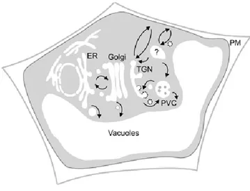
The plant endomembrane system consists of different organelles: the nuclear envelope, the endoplasmic reticulum (ER), the Golgi apparatus, the trans-Golgi Network (TGN), the pre-vacuolar compartment, the endosomes, the vacuoles and the plasma membrane. Some are directly connected by a maturation process while others are connected by trafficking vesicles. These different components collaborate for cargo molecule processing, packaging, transport and delivery to their residence site within the cell. Proteins are transported from the ER through the Golgi apparatus and then are directed to their final destination (plasma membrane, vacuole…) (Figure 1.1). The maintenance of this cellular compartmentation is primordial for the function of the plant cell. The secretory pathway plays a major role for the cell, because it is involved in cell interaction, differentiation and division, but also in various biotic and abiotic stress responses.
Figure 1.1 Simplified representation of the secretory pathway in plants (Foresti and Denecke, 2008)
Three routes of proteins trafficking to the vacuole have been described. First, via the classical route, proteins traffic through the ER, Golgi and TGN before reaching prevacuolar compartment. In the second route, cargoes bypass the Golgi and traffic to vacuole directly from the ER. The last route is a post-Golgi clathrin-mediated route from the TGN through the PVC.
22
A.1 Early secretory pathway
Endoplasmic Reticulum
Role of the ER
The endoplasmic reticulum (ER) is the first organelle in the secretory pathway and plays a central role in the cell. It is a multifunctional compartment, involved in protein synthesis and post-translational modifications, addition of N-linked oligosaccharides, formation of disulfide bonds, lipid biosynthesis but also in calcium storage (Hammond & Helenius, 1995). The traditional model of ER structure describes two main parts, both of which are present in all eukaryote cells: the rough ER (RER) and the smooth ER (SER). The RER membrane is associated with ribosomes and is partially continuous with the nuclear envelope whereas the smooth ER is composed of tubular structures without ribosomes. Recent studies showed the ER as a highly dynamic organelle, composed by a network of interconnected tubules. This structure can adapt in response to specific stimuli by morphological modifications and ER movements are driven by the cytoskeleton (Sparkes et al., 2011; Sparkes 2013). In plants, ER forms a continuous compartment between adjacent cells through plasmodesmata (Sparkes 2013) and it is closely associated with the actin cytoskeleton (Sparkes et al., 2009).
Translocon and Signal peptide
The entry of secretory proteins into the ER is usually linked with the presence of a signal peptide (SP) at the N-terminus of the protein. Import of soluble and membrane resident proteins into the ER occurs with the association of ribosomes and a protein pore called translocon. The SP is recognized and bound by a signal recognition particle (SRP), which interacts with the rough ER membrane, allowing the translocation of the polypeptide across the membrane through the translocon (Lütcke 1995; Hamman et al., 1998). Translocons form structurally and functionally dynamic aqueous pores in the ER membrane, and play a major role in the maintenance of the membrane permeability. The co-translational translocation of a nascent protein starts when the latest is still bound to the ribosome. The SRP binds at the same time the SP of the nascent protein and the 60S ribosome unit, causing a pause of the translocation process. This complex interacts then with the SRP receptor localized on the ER membrane, triggering the transfer of the peptide to the translocon. During the further translation and transfer to the ER lumen, the growing polypeptide is protected from cytosol exposition through the ribosome’s alignment with the translocon pore (Lodish et al., 2008). The pore is open only when a peptide is being translocated, preventing ionic leakage between the cytosol and the ER lumen.
23 Removal of the SP occurs while the protein is translocated through the membrane and it is a major step contributing to correct folding of the protein (Nielsen et al, 1997; Vitale and Denecke 1999). The signal peptide is cleaved by a signal peptidase, a transmembrane protein associated with the translocon. Cleavage of the SP allows the protein release from the membrane and then its proper folding in the ER lumen (reviewed by Auclair et al., 2011).
ER quality control
Proteins are folded in the lumen of ER where glycosylation can also occur. At this step, the ER plays an important role in quality control whereby misfolded proteins are recognized and retained within the ER or directed to a degradation pathway (Hammond and Helenius 1995, Vitale and Denecke 1999). This quality control is primordial for the cell because it is involved in various stress responses and thus in plant adaptation to the environment (reviewed by Liu and Howell, 2010).
A misfolded protein can have three fates. Firstly, it may accumulate or aggregate in the lumen; secondly it may be targeted to the vacuole or the lysosome (in animals), where protein degradation occurs and thirdly it may be degraded in the cytosol by the proteasome machinery after re-exportation to the cytosolic face of the ER (Brodsky and McCracken 1999; Vashist and Ng 2004). How the cell determines which process to use and how misfolded protein are recognized is still unclear. In fact, a large percentage of ER-synthesized proteins are misfolded or unfolded, justifying the major role of the ER quality control: to avoid the dispatching of non-properly matured proteins to their final cellular compartments. Misfolded proteins are detected by various sensors, including the molecular chaperones. The main chaperone families of the ER are heat shock proteins (Hsp) including BiP (HSP70), the peptidyl-prolyl isomerases, the thiol-disulfide oxidoreductases, calnexin and calreticulin (reviewed by Ellgaard and Helenius, 2003).
ER Associated Degradation system (ERAD)
ERAD is a quality control system of the ER which clears the misfolded proteins from the ER through ubiquitylation. This cellular pathway plays an important role in cell homeostasis (reviewed by Ruggiano 2014). To date, most knowledge of this mechanism came from studies in yeast and mammals. Less is known about the plant ERAD system. This system involves an ubiquitin-proteasome system, where the substrate is ubiquitylated before degradation in the cytoplasm (Müller et al., 2005; reviewed by Liu and Li, 2014). Brandizzi et al. (2003) visualized a putative ERAD pathway using a GFP fusion protein recognized as misfolded by the cell, which was degraded after a retrograde translocation back to the cytosol. The ERAD system has also been shown to play a major role during salt stress in
24
Arabidopsis mutant line with a defective ERAD system (hrd3-a mutant) is more sensitive to salt stress
than the wild-type line: seedlings are smaller and weaker (Liu et al., 2011).
Unfolded Protein Response
The Unfolded Protein Response (UPR) is an adaptive response from the cell to environmental and internal stresses. This intracellular signaling pathway is an essential part of the ER quality control and helps to reduce the misfolded and unfolded proteins accumulation (Liu and Kaufman 2003). UPR allows the re-establishment of cell homeostasis in order to decrease the ER stress, caused by various factors such as luminal calcium luminal depletion, hypoxia, pathogen infection or genetic mutation. This pathway involves different mechanisms that will cooperate to deal with the stress such as the induction of UPR genes, the decrease of global protein synthesis and the activation of the ERAD system (Fanata et al., 2013). Extended activation of the UPR pathway can lead to apoptotic cell death.
From the ER to the Golgi apparatus
Two classes of coat proteins, COPI and COPII, are involved in the transfer of proteins between the ER and the Golgi apparatus. This vesiculation is highly conserved within the eukaryotic domain. Proteins destined to the Golgi apparatus are exported from the ER by COPII-coated vesicles. The COPII vesicles bud off from specific sites of the ER membrane, the ER-exit sites (ERES; Barlowe et al., 1994; reviewed by Viotti, 2014). In the reverse direction, COPI-coated vesicles mediate a retrograde transport that selectively recycles mistargeted proteins (e.g. KDEL targeted proteins) from the cis-Golgi complex back to the ER (reviewed by Duden 2003).
COP vesicles
COPII: anterograde traffic
COPII vesicles mediate the transport of cargo from the ER to the cis-Golgi (Robinson et al., 2007). Their formation requires ER export sites (ERES) where the packaging of cargo into vesicles occurs. COPII complex formation occurs in several steps with protein interactions (Figure 1.2). First, the ER-membrane-resident GEF (guanine nucleotide exchange factor) protein Sec12 catalyzes the activation of the small GTPase Sar1, helping it to release GDP and to bind GTP. Once Sar1 is activated, it anchors into the ER membrane by insertion of its amphipathic N-terminal tail. Sar1 GTP drives the assembly of the cytosolic COPII subunits. The coat is formed by Sec23/Sec24 dimers forming an internal layer and completed with Sec13/Sec31 for the external scaffold layer. When the coat is achieved, the vesicle buds and is released from the ER donor membrane. Finally, the hydrolysis of Sar1-GTP is
25 triggered by Sec23, which leads to the disassembly of the COPII coat (Jürgen 2004; Hanton et al., 2009; Hwang and Robinson 2009; reviewed by Tang et al., 2005; Hugues and Stephens 2008).
Figure 1.2 Mechanism of COPII coat assembly (adapted from Yorimitsu et al., 2014).
Vesicle formation starts upon the recruitment of Sar1 to the ER membrane. Then ER integral membrane protein Sec12 exchanges GDP for GTP bound to Sar1 through its GEF activity. Membrane-associated GTP-bound Sar1 recruits the inner coat Sec23/24 complex and then assembles along with cargo protein into the pre-budding complex. Outer coat Sec13/31 complexes are recruited to the pre-budding complexes and self-assembled. The polymerization of Sec13/31 by self-assembly drives membrane curvature to form a spherically shaped vesicle.
Mutations in gene coding for COPII components leads to developmental deficiencies in plants (reviewed by Chung et al., 2016) and interruption of the COPII-mediated transport has severe effects on protein traffic which can lead to cell death (Hanton et al., 2009).
COPI: retrograde traffic
COPI vesicles mediates recycling of proteins from Golgi to ER, in order to capture escaped ER resident proteins, and retrograde transport within the Golgi (from trans to cis cisternae; Pimpl et al., 2000). First studied in mammalian cells, they have been described in yeast and plant cells (reviewed by Hanton, 2005). They require a seven-subunit coat (named coatomer) and the GTPase ARF1 (Jürgens 2004). Assembly of the coat begins with the activation of ARF1 by the released of GDP and the binding of GTP catalyzed by an ARF-GEF (Figure 1.3). This activation leads to a direct interaction of ARF1 with the Golgi membrane. Activated and membrane-anchored ARF1 recruits the seven coatomer subunits from the cytosol. These subunits can be separated in two layers: the inner layer involved in cargo binding and membrane attachment, including ɣ, δ, ξ and βCOP; and the outer scaffold including , β’ and εCOP. The formation of the coat contributes to the capture of the cargo proteins and causes the curvature of the membrane. When the coat is totally formed, the vesicle buds. The uncoating of COPI
26
vesicle starts with GTP hydrolysis which triggers the dissociation of ARF1 from the vesicle membrane (Kirchhausen 2000; Beck 2009 and Barlowe 2013).
Figure 1.3 Mechanism of COPI coat assembly (adapted from Yorimitsu et al., 2014).
COPI vesicle formation is also initiated by GTP-GDP exchange on ARF1 through the action of the GEF Gea protein (Gea1 or Gea2), which is peripherally located on the Golgi membrane. GTP-bound ARF1 stably binds to the membrane by a myristoylated amphipathic helix. The heptamer complex of the COPI coat is recruited en bloc and associates with cargo as well as two ARF1 molecules though the inner layer coat complex (β/γ/δ/ζ-COP). As in COPII, vesicles are formed upon polymerization of the outercoat (α/β’/ε-COP). The amphipathic helix of ARF1 has some role in the scission of budded vesicles.
ER exit sites (ERES)
ERES are the sites of export for proteins to the Golgi, where COPII vesicles are forming. Their physical structure is still unclear but they have been described to be a sort of membrane microdomains. Little is known about ERES in plant because most studies come from animal cells and the features of ERES can be different depending on the cell type and between kingdoms (DaSilva et al., 2004; review by Tang et al., 2005). It was proposed to define them as sites where nascent COPII membrane and COPII vesicles in transit can be found (DaSilva et al., 2004). In tobacco cells, ERES and Golgi bodies were observed moving together and are probably closely associated. Even if the reason needs to be elucidated, ERES and cisternal Golgi stacks are physically close (DaSilva et al., 2004; Budnik and Stephens, 2009). It has been shown that Golgi stacks do not associate with only one ERES but can be bound to several ERES at the same time (Yang et al., 2005).
ER retention signal
ER resident proteins carry a retention signal. For soluble proteins most of the time this signal is a tetrapeptide KDEL, HDEL or RDEL found in C-terminal position. A substitution of one amino acid in
27 this tetrapeptide is enough to disrupt the retention of the protein (Denecke et al., 1992). Different studies have shown that secreted proteins fused with KDEL sequence in C-terminal position were then retained in the ER (reviewed by Pagny et al., 1999). This mechanism of retention is highly specific and mostly conserved between mammals and plants (Denecke et al., 1992; reviewed by Pagny et al., 1999).
Traffic from ER to Golgi
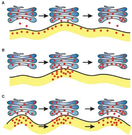
As described previously, ER proteins are packaged in vesicles and exported to the Golgi at ERES sites. Because ER and Golgi bodies are highly dynamic structures, different models of protein trafficking from ER to Golgi in plants have been proposed (Figure 1.4; Hanton et al., 2005). The first model called “vacuum cleaner model” is based on the Golgi bodies’ movements along the ER. Golgi stacks collect vesicles at ERES in order to obtain their cargoes. The second model is called “stop and go”. Here the Golgi bodies stop at ERES, obtain cargoes, move along the actin filaments, then stop at another ERES and collect more cargoes. This model suggests the involvement of a specific signal on ERES causing a transitory association between Golgi and ERES. Then last model is the “ERES mobile model” where Golgi and ERES together are highly mobile along the ER. This model describes a continuous transport of cargoes between these two compartments. However, the connections between Golgi and ER are still unclear and the mechanisms of interaction need to be elucidated (Hanton et al., 2005).
28
Figure 1.4 Models for proteins transport from ER to Golgi (Hanton et al., 2005).
A: The vacuum cleaner model. Golgi bodies move along the ER surface, picking up cargo. The entire ER is capable of exporting proteins. B: The stop-and-go model. Golgi bodies move along the ER and stop at fixed ERES, where protein transport takes place. After transfer of cargo from ER to Golgi, the Golgi body moves to the next site and collect more cargo. C: The mobile ERES model. Golgi bodies and ERES move together, allowing continual protein transport between the two organelles.
ER exit bypassing Golgi
Multiple export pathways from the ER have been described in plants. The classical export pathway from ER to the Golgi is sensitive to Brefeldin A (BFA), a fungal metabolite which can reversibly block the retrograde traffic by inhibiting the formation of COPI vesicles and consequently also block the anterograde traffic. Other BFA-insensitive pathways have been described but it is possible that most of them exist only in seeds (Vitale and Denecke 1999). It has been shown that some storage proteins may bypass the Golgi and traffic to the vacuole via precursor-accumulating vesicles (PAC) e.g. in maturing pumpkin seeds. Aggregates of storage proteins were visible within the ER by immunocytochemical staining (Hara-Nishimura et al., 1998). Toyooka et al. (2000) highlighted the transport of a vacuolar cysteine proteinase (SH-EP) from the ER to the protein storage vacuole (PSV)
29 bypassing the Golgi. This SH-EP has a C-terminal KDEL ER-retention signal. The proform of the enzyme has been observed accumulating at the edges of ER, in vesicles budding from it and in large vesicles distinct from PSV but not in the Golgi. Others studies support the hypothesis of a direct pathway from ER to lytic vacuoles. Originally discovered in yeast, a route from the cytosol to the lytic vacuole involving autophagy was also proposed in plants. Structures similar to autophagosomes containing vacuolar proteins like seed storage proteins were detected by electron microscopy (Michaeli et al., 2014). In 2013, Viotti et al., proposed a Golgi-bypassing route in Arabidopsis, where the ER membrane contributes to lytic vacuole biogenesis. Indeed, the tonoplast intrinsic protein α-TIP was able to reach the vacuole even after treatment with Brefeldin A in mesophyll protoplasts of tobacco plants (Gomez and Chrispeels 1993).
Golgi apparatus
The Golgi apparatus is a major organelle consisting of different stacked polarized cisternae, which can be divided in three main compartments: cis, medial and trans-Golgi which are functionally distinct (Staehelin and Moore 1995). The Golgi apparatus processes proteins arriving by vesicles from the ER at the cis-Golgi face and sends them from the trans-Golgi face to various locations in the cell (lysosomes, plasma membrane…) or out of the cell (secretory vesicle). Unlike mammalian cells which have one large Golgi apparatus next to the nucleus, numerous (up to one hundred) mobile Golgi stacks can be found in the cytoplasm of plant cells (DaSilva et al., 2004). Different major functions of the Golgi in the plant cell have been described. It can be considered as the glycan factory of the cell because it synthetizes and exports complex polysaccharides for the cell wall but also glycolipids for the plasma membrane and storage glycoproteins (Staehelin and Moore 1995). Golgi resident enzymes are single pass transmembrane proteins which are found in distinct cisternae depending on their function. The two main families of enzymes are the glycosidases and the glycosyltransferases, which are involved in the production of glycans and the modifications of N-or O-linked carbohydrates, attached to proteins and lipids (Saint-Jore-Dupas et al., 2004). After these modifications and maturations (glyco-) proteins are packaged in transport vesicles in the trans-Golgi and sent to their specific destinations in the cell (Driouich et al., 1993; Dupree and Sherrier 1998).
Plant Golgi bodies are highly motile because they are physically and mechanically bound to the actin cytoskeleton. In tobacco epidermal cells expressing a GFP fused to the actin-binding domain of talin, Golgi bodies were observed moving along the ER in an actin-dependent way but they did not interact with the microtubules (Brandizzi et al., 2002). Previous studies also showed a close link between Golgi stacks moving along actin cables and the ER, by expressing a Golgi protein fused to GFP (Boevink et al., 1998).
30
Two models of protein transport across the Golgi stacks have been proposed. The first one is based on the protein transport between the Golgi cisternae by vesicles budding from one cisterna and fusing with the next, in both anterograde and retrograde directions. The Golgi is here considered as a static organelle with small vesicles moving between compartments (Orci et al., 1998).
The second model proposes that proteins and lipids are transported in the cis-to-trans direction through cisternal maturation; while retrograde transport of Golgi-resident proteins is mediated by COPI vesicles (Glick et al., 1997; Allan and Balch 1999; Alberts et al., 2008). This model implies a dynamic organization of Golgi apparatus with the trans-cisternae being consumed while a new cis-cisternae forms. It is now the more generally accepted model. Transport of Golgi-resident proteins by COPI vesicles was also demonstrated (Orci et al., 1997).
A.2 Late secretory pathway
Trans-Golgi Network (TGN)
Initially thought to be part of the Golgi apparatus as in animal cells, the TGN was shown in plants to be an independent and separate organelle (Hawes and Satiat-Jeunemaitre 2005). Uemura et al. (2004) characterized in Arabidopsis protoplasts fluorescent plant TGN markers that localized in structures distinct from Golgi bodies. Foresti and Denecke (2008) also showed in tobacco leaf epidermis cells that fluorescent markers of plant TGN do not colocalize with Golgi markers.
The TGN has been described as a dynamic organelle, with a role in the exocytosis and endocytosis pathways (Foresti and Denecke 2008; Kang et al., 2011). However the definition of plant TGN is still unclear, it has been suggested to function as an early endosome while in animal cells these are two separate compartments (Lam et al., 2007; Viotti et al., 2010; reviewed by Uemura and Nakano 2013). One of the first studies used a rice secretory carrier membrane protein 1 (SCAMP1), known to be a post-Golgi proteins involved in endocytosis in animals, which was fused to YFP and expressed in tobacco BY-2 cells. The localization was observed in plasma membrane (PM) and mobile cytosolic organelles. These highly motile organelles are BFA-sensitive and form aggregates after treatment. Confocal immunofluorescence with SCAMP1 antibodies confirm that these organelles are distinct from Golgi and multivesicular bodies (MVB) and act as early endosomes (Lam et al., 2007). Viotti et al. (2010) observed in Arabidopsis that secretory proteins passed through the TGN before reaching their final destination, by studying the route of a brassinosteroid receptor BRI1 and a boron exporter BOR1, both localized in the PM. Using a secreted version of GFP, a YFP fusion of these membrane proteins and the endocytic tracer FM4-64, their work also allowed to distinguish the TGN and MVB as two distinct
31 compartments. The TGN was described as a highly mobile organelle which temporarily associates with the Golgi.
The size of the TGN may vary according to the cell function or specialization, but also depending on the number and type of vesicles that bud from it (Marty 1999; reviewed by Gendre et al., 2015). Three type of vesicles associated with the TGN have been described: the clathrin-coated vesicles (CCV), the COPI vesicles and the secretory vesicles (reviewed by Gendre et al., 2015). Proteins trafficking through the TGN can be directed to the plasma membrane or to the vacuole or lysosome in animal cells. At this point, targeting to the vacuole require a vacuolar sorting determinant, while targeting to the plasma membrane can be considered as the default pathway (Sanderfoot and Raikhel 1999).
Endosomes
Plants endosomes are highly dynamic membrane structures, considered as an intermediate compartment, which take part in endocytosis and biosynthesis pathways by receiving vesicles from the TGN or the plasma membrane. Endosomes are part of the late secretory system and are involved in the recycling or degradation of plasma membrane proteins and in the traffic of secreted proteins targeted to the vacuole or lysosomes (Geldner 2004; Otegui and Spitzer 2008). Endosomes also play a role in diverse cellular mechanisms such as polar tip growth, cell polarity, auxin mediated cell-cell communication and gravitropism (Geldner 2004; Samaj et al., 2005). With these numerous implications endosomes are important players in trafficking pathways signaling and regulation (Reyes
et al., 2011). In yeast and mammals, different types of endosomes have been described. The early
endosomes (EE) receive endocytic cargo vesicles from the plasma membrane. At this point, material can also be return to the PM via recycling endosomes. EE are considered as an important sorting station for vesicles in the endocytic and secretory pathway. Late endosomes (LE) are derived by maturation form the EE; they are also called multivesicular bodies (MVB) because they are formed by the invagination of the endosome membrane itself. They are involved in the degradation of membrane proteins by fusion with lysosomes or act in their recycling to the TGN (Otegui and Spitzer, 2008). The endosome system in plants differs from the other eukaryotes. Two main compartments have been described in literature: the TGN, which acts as early or recycling endosomes, and the MVB, considered as a late endosome (Otegui and Reyes, 2010; Contento and Bassham, 2012). These two compartments have been highlighted using the fluorescent dye FM4-64, which labels first the PM. This dye allows to study the endocytic pathway as it labels also very quickly the early endosomes and later the vacuole (Contento and Bassham, 2012; Bolte et al., 2004).
32
Early endosomes were not observed in plants while MVB were described to be active transport carriers for vacuolar proteins. Indeed, mutation in Rab5-like GTPase, a MVB localized protein, leads to vacuolar transport perturbation (reviewed by Otegui and Spitzer, 2008). Plant endosomes play a major role in endocytosis and more precisely in protein recycling, protein degradation and receptor-mediated signaling. Recycling of vacuolar cargo receptors via endosomes to the TGN needs a complex of proteins called retromer. This complex is conserved through eukaryote kingdom, localized in EE and it is involved in the recruitment of the cargoes (Seaman 2012). Some proteins were observed to be continually cycling between the PM and the EE, like the auxin carrier PIN1 (Otegui and Reyes 2010). Endosomes are also involved in degradation of membrane proteins via lysosomes or vacuoles. Membrane proteins that will be degraded are marked by an ubiquitin tag which causes their internalization into the MVB. Different protein complexes, called Endosomal Sorting Complex Required for Transport (ESCRT-0-I-II and III), are involved in this mechanism. ESCRT complexes differ in different organisms. The ESCRT-0 does not exist in plants while it is essential in animals, another unrelated complex replaces it. Endosomes play also a role in receptor-mediated signaling. In this process, activated receptors at the PM can be internalized by endocytosis into endosomes and then interacting with other downstream factors, in the endosomes membrane (Otegui and Spitzer 2008; Otegui and Reyes, 2010).
Vacuoles
Vacuoles are an endpoint of the secretory system in plants. They are dynamic organelles and important compartments for plant cell metabolism and life (Wink 1993). In general, a plant cell contains one large vacuole, which can occupy more than 90% of the total cell volume. Vacuole function depends of the cell type. They participate in homeostasis, cell turgor, growth and storage of metabolic products. They also have a hydrolytic role and can sequester toxic compounds. The size, number and function of vacuoles within a single cell depend on the kind of tissue and the cell type (reviewed by Marty 1999). These different functions can occur in the same compartment but a cell can also contain two different types of vacuoles (Paris et al., 1996).
A.3 Vesicular traffic
In the plant secretory pathway, vesicles mediate the transport of cargo proteins from an organelle to the next, until they reach their final destination. Vesicles move through the cytosol by interaction with cytoskeleton elements. The basis of this essential mechanism consists of the vesicle budding from a donor compartment, which contains cargo proteins, to the fusion of this vesicle to an
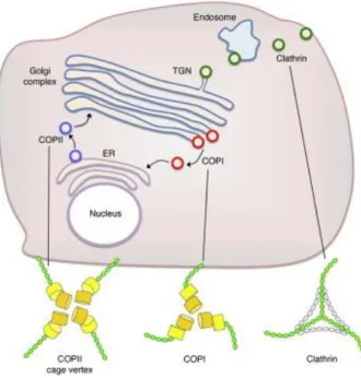
33 acceptor compartment where the cargo is released (Bonifacino and Glick 2004). This active process requires the recruitment of coat proteins and the intervention of small GTPases (Jürgens 2004). First studied in mammalian cells and yeast, the three major types of coated vesicles COPI, COPII and CCV are also found in plants (Figure 1.5). This classical model for vesicle transport requires different steps, from the formation of coated bud in the donor compartment, the vesicle formation and transport, to the coat protein release and fusion of the vesicle with the target membrane in order to discharge its content (Jürgens and Geldner 2002; Hwang and Robinson 2009).
Figure 1.5 The three type of coated vesicles and their localization in the cell (from Hughson 2010)
In the early secretory pathway, newly synthetized proteins are transported by COPII and COPI coated vesicles between the ER and the Golgi. From the TGN, cargo proteins traffic to the plasma membrane or recycling endosomes by clathrin coated vesicles.
Clathrin-coated vesicles
Clathrin-coated vesicles (CCV) consist of a two-layered coat: the outer layer of clathrin structural proteins and the inner layer of adaptor protein (AP) complexes. The clathrin molecule is composed of heavy and light polypeptide chains. Three heavy chains and three light chains assemble to form a structural unit called a triskelion, which assembles to form the clathrin lattice. AP complexes play a major role in vesicle formation. They mediate the clathrin lattice recognition and assembly of the cargo proteins (Schmid 1997). Different AP complexes have been identified (AP1 to AP4) in
34
eukaryotic cells, which are involved in different pathways (reviewed by McMahon and Gills 2004). The coat assembly is also controlled by the ARF1-GTPase. Protein sorting at the TGN and endosome is mediated by AP1 complex which recognize the luminal domain of the cargo protein, while AP2 mediates CCV formation at the PM. Other adaptor proteins have been identified in animal cells but have not been found in plants. CCV have been localized at the TGN and the plasma membrane in animal cells as well as in plant cells (Sanderfoot and Raikhel 1999; Jürgens and Geldner 2002).
Mechanism of membrane fusion
Vesicle and target membrane interact first via tethering proteins recruited by Rab proteins. Rab proteins are a large family of GTPases proteins, involved in the regulation of membrane trafficking and vesicles formation. They are key regulatory factors in docking of vesicles on target membranes (Hutagalung and Novick, 2011). The fusion of the transport vesicle with the acceptor compartment involves the action of SNAREs (soluble NSF attachment protein receptors): a v-SNARE in the vesicle membrane and t-SNAREs in the target membrane. This primes the interaction of the v-SNARE protein on the vesicle surface with the cytosolic domain of t-SNAREs localized on the target membrane. Then a SNARE complex is formed by four SNAREs, forming a bundle which brings the membranes close enough for fusion, where the vesicle releases the cargo to the acceptor compartment (Sanderfoot and Raikhel 1999). The dissociation of the SNARE complex is an ATP-dependent reaction, mediating by α-SNAP (soluble NSF attachment protein) and NSF proteins. (Hutagalung and Novick 2011) (Figure 1.6).
Figure 1.6 The SNARE mechanism of vesicle fusion (from Sanderfoot and Raikhel, 1999).
On the vesicular membrane, the v-SNARE and the Rab-type GTPase recognize on the target membrane the t-SNARE and Sec1 p-Homolog. After this docking event, Sec1 p-Homolog is removed and the formation of the SNARE complex allows the vesicle fusion with the target membrane. Finally, α-SNAP and NSF mediate the dissociation of the SNARE complex, allowing the retrograde recycling of the v-SNARE.
35
A.4 Endocytosis
Endocytosis general mechanisms
Endocytosis is a mechanism of internalization of material and protein cargoes into plasma membrane transport vesicles. This process plays a crucial part in numerous cellular mechanisms as turnover of proteins or hormone transport, but also in various responses to environmental signals like responses to pathogens or cell signaling (Otegui and Spitzer 2008; Reyes et al., 2011; Contento and Bassham 2012). It is the principal way for membrane and extracellular proteins to enter the cell (reviewed by Fan et al., 2015) (figure 1.7). In animals, three types of endocytosis have been described: phagocytosis, pinocytosis and receptor-mediated endocytosis (Alberts et al., 2008). Phagocytosis is the ingestion of large particles via vesicles called “phagosomes”. This process takes place in the immune response and in clearance of dead cells. Pinocytosis represents the internalization of fluid and solutes (proteins and polysaccharides) via small vesicles, formed from coated pits of the plasma membrane. This mechanism is involved in cellular metabolism and signaling. Receptor-mediated endocytosis, also described in plants, occurs for specific molecules transport (proteins and lipids) and implicates clathrin-coated vesicles. It plays a role in various cellular functions, from the turn-over of membrane proteins, the uptake of extracellular molecules, to the activation of signaling pathways (McMahon and Boucrot 2011).
36
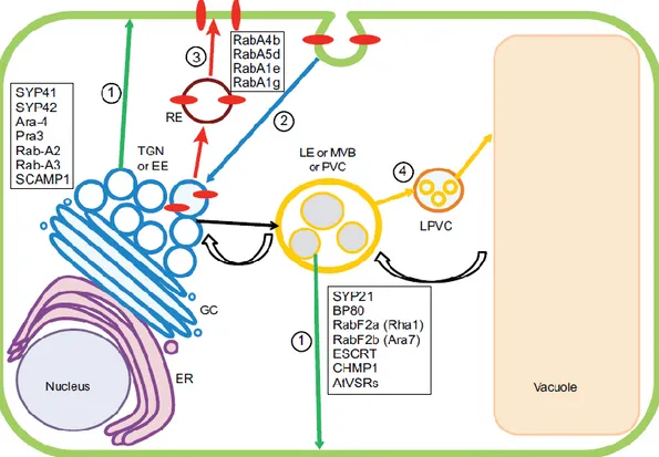
Figure 1.7 The endocytic pathway in plants (Contento and Bassham 2012)
Components of the endocytic pathway are involved in biosynthetic, degradative and recycling transport. The trans-Golgi network and early endosomes act as the point of organization for these three pathways. This diagram displays the organelles involved in endomembrane trafficking: nucleus, endoplasmic reticulum (ER), Golgi complex (GC), trans-Golgi network (TGN) / early endosome (EE), late endosome (LE) or multivesicular body (MVB) or prevacuolar compartment (PVC), late prevacuolar compartment (LPVC), vacuole surrounded by the tonoplast, recycling endosome (RE). The colored arrows designate potential traffic between organelles. The green arrows indicate pathway 1, the biosynthetic transport route to the plasma membrane that passes through the TGN and sometimes the MVB. The blue arrow shows a pathway (2) that is taken by plasma membrane components (red ovals) as they are internalized into endocytic vesicles and move through the TGN. Proteins associated with the TGN are listed. The thick, red arrows represent a recycling pathway (3), by which plasma membrane components might be returned to the plasma membrane through a specialized RE. Proteins associated with the RE are shown. The orange arrows designate the transport pathway (4) for newly synthesized vacuolar components, as well as cellular materials destined for degradation in the vacuole. Proteins associated with MVBs are shown. Transport from the TGN to MVBs is designated by a black arrow. Retrograde transport from the VAC to the MVB is represented by curved black arrows.
37 Clathrin mediated endocytosis in plants (CME)
In plants, the endocytic pathway is involved in various essential functions, as cell growth, nutrients uptake, hormonal signaling, detection of environmental signals and defense against pathogens. The main endocytic process is dependent on clathrin coated vesicles that pinch off from PM, where the cargoes are recognized, then packaged and incorporated into CCV. After the scission, newly formed CCV can fuse with the early endosomes, from where cargoes can continue to their final destination (Chen et al., 2011). CCV are also a main way for membrane protein retrieval and membrane receptor down regulation. Two major kinds of proteins are involved in plant CME. The first group includes the sub-units of the clathrin coat, the adaptor (AP) complexes. The second group is formed by various cytosolic proteins, playing a role in budding and fission events (Holstein 2002). Cargo selection requires adaptor complexes that recognize different motifs or post-translational modifications such as phosphorylation or ubiquitylation. The recognition of specific motifs is a mechanisms well studied in animals but still unclear in plants. The role of CME in plant development was highlighted by studies on the PIN proteins. These proteins were the first identified cargoes of CME, allowing to establish a link between endocytosis and auxin-mediated development (reviewed by Chen et al., 2011). CME was also described to be involved in the regulation of plant defense response against pathogens, specifically against bacterial and fungal elicitors. Pathogen attack could trigger plant immune response by regulating some membrane receptors via endocytosis. It is the case of the Arabidopsis FLS2 receptor, which recognizes the bacterial flagellin and is taken up from the PM into CCV (reviewed by Fan et al., 2015). CME plays also a role in uptake of nutrients such as metallic ions. The receptors IRT1 and BOR1, for iron and boron respectively, have been described to cycle from PM to endosomes. When needed, these proteins are sorted into vacuoles for downregulation in order to avoid metallic ion toxicity (Chen
et al., 2011).
Clathrin-independent endocytic routes in plants
Alternatives endocytic pathways have been described in plants. One of these involves membrane microdomains also called lipids rafts. Microdomains are well studied in yeast and mammals but less is known in plants. These dynamic platforms, insoluble in some detergents, are implicated in the concentration of some receptors and in the assembly of certain signal transduction machineries. Membrane microdomains play a role in the amplification or the attenuation of various cellular signaling cascades (Puri et al., 2004). In Arabidopsis, flotillin1 (flot1) was the first membrane protein identified which form microdomains, acting in an independent clathrin-endocytic pathway. This protein involved in seedling development was localized in plasma membrane and in intra-cellular vesicles. Flot1 was also observed to partially colocalize with the endocytic marker FM4-64 (Li et al., 2012; reviewed by Fan
38
Ubiquitylation mediated plant endocytosis
One of the well-known roles of ubiquitin tag is the marking of proteins for degradation by the 26S proteasome. However, ubiquitin is also involved in protein trafficking to the vacuole / lysosome by endocytosis. This pathway was first observed in yeast and mammals but today evidence is accumulating in plants. In Arabidopsis, it was observed in the study of the iron transporter ITR1. This transporter was described in the TGN/ EE of root hair cells and it is responsible for the iron uptake from the soil. IRT1 needs to be monoubiquitylated to enter the endocytic route for vacuolar degradation. Mutations in the two key lysines, sites of ubiquitination, increases its stability. IRT1 has been described to cycle between the PM and the early endosomes, depending on its monoubiquitylation (reviewed by Tian and Xie, 2013).
A.5. Protein secretion
Unconventional protein secretion
Proteins trafficking through the “classical” protein secretion pathway are transiting from the ER via the Golgi/TGN to their final destination. By default, soluble proteins are secreted at the plasma membrane. Cell secretion plays a role in cell to cell communication and pathogen defense, which can be constitutive of induced by internal or external stimuli (Krause et al., 2013). Uptake in the ER is determined by the presence of a Signal Peptide at the N-terminal end of the newly synthesized polypeptide. There are also some secreted proteins lacking this SP. These Leaderless Secretory Proteins (LSP) do not follow the ER/Golgi classic route but are trafficking through an “unconventional secretion pathway” and are directly secreted from the cytosol into the extracellular matrix (Agrawal et al., 2010; Ding et al., 2012; Krause et al., 2013). First identified in mammalian and yeast cells, data for unconventional secretion studies in plants are now accumulating (Drakakaki and Dandekar 2013).
Plant secretome studies
The term secretome was firstly defined by Tjalsma et al. (2000) after their work on protein transport in Bacillus subtilis. It was including the protein secretion machinery and the secreted proteins. Nowadays, the term secretome has evolved and has be defined for plants by Agrawal et al. (2010) as “the global group of secreted proteins into the extracellular space by a cell, tissue, organ or organism at any given time and conditions through known and unknown secretory mechanisms involving constitutive and regulated secretory organelles” (reviewed by Krause et al., 2013). Previous studies showed that LSPs can represent more than 50% of the identified secretome and suggested that LSP may play a role in stress responses and defense against pathogen (Agrawal et al., 2010). Early work
39 on the plant secretome was performed with cultured cells but it has been shown that these do not reflect natural conditions. Recent studies propose alternatives to cell cultures, allowing in planta work, such as gravity extraction from leaves or vacuum infiltration methods (Alexandersson et al., 2013; Krause et al., 2013).
B. Vacuole types and protein targeting to vacuole
B.1 Vacuole types
Plant cells can contain different vacuole types with different functions, depending of the cell type and the developmental stage. The distinction between Lytic Vacuole (LV) and Protein Storage Vacuole (PSV) and their presence within a single cell were demonstrated by different studies (Hoh et al., 1995; Paris et al., 1996; Vitale and Raikhel 1999). Epimashko et al. (2004) demonstrated that mesophyll cells of Mesembryanthemum crystallinum L. under salt stress conditions have two functionally different vacuoles, an acidic and a non-acidic. This allows the plant to compartmentalize salt storing and malate accumulation/mobilization.
The three kinds of vacuoles described in literature are the following:
Lytic Vacuole: it is an acidic compartment which can be compared to the lysosome in animal cells. This lytic vacuole contains hydrolytic enzymes and plays an important role to maintain cell turgor. This type of vacuole develops in vegetative organs in plants. Acidic vacuoles were shown to accumulate the pH-sensitive dye Neutral Red (NR) (Di Sansebastiano et al., 2001).
Neutral vacuole: it has been described in vegetative tissues and did not accumulate NR in contrast to the lytic vacuole (Di Sansebastiano et al., 2001).
Protein Storage Vacuole: involved in the accumulation of proteins, this type of vacuole is found in storage tissues of seeds and developing seeds.
According to the classical model, after trafficking through the ER and Golgi, vacuolar proteins are targeted to their final destination. Protein targeting to vacuoles is dependent on a signal, which can be found at different locations in the protein structure: at the N- or C-terminus or internal to the protein. The first model for the biogenesis of distinct vacuolar type was based on the localization of aquaporin Tonoplast Intrinsic Protein (TIP) isoforms and proposed an organelle-specific localization. Based on immunolocalization, these first studies showed that -TIP are present in Protein Storage Vacuole (PSV) whereas γ-TIP are found in Lytic vacuole (LV) (Paris et al., 1996; Neuhaus and Roger,
40
1998; Jauh et al., 1998) and δ-TIP in vacuole accumulating pigments and vegetative storage proteins (Jauh et al., 1999). More recent studies on A. thaliana demonstrated that TIP isoforms distribution and expression are regulated during development (Hunter et al., 2007; Gattolin et al., 2010). TIP isoforms distribution is tissue specific and depends on the developmental stage: γ and δ-TIP are found in vegetative tissues (but not in root tip) whereas -TIP predominates during seed maturation but decreases after germination (Hunter et al., 2007). In Soybean, the reversible functional switch from vegetative storage to lytic vacuole in paraveinal mesophyll is accompanied by a replacement of δ-TIP by γ-TIP (Murphy 2005).
B.2.Vacuolar sorting determinants
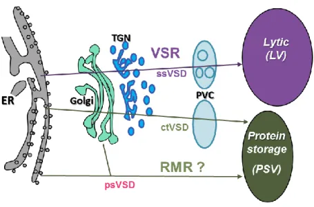
Soluble vacuolar proteins are sorted to the vacuoles by cargo receptors, which bind a specific component: a vacuolar sorting determinant (VSD). This sorting signal is found in the vacuolar protein precursor and it is mostly removed during maturation after vacuolar sorting. In plant cells, three vacuolar sorting determinants have been distinguished: sequence specific VSDs (ssVSD), C-terminal VSDs (ctVSD) and protein-structure-dependent VSDs (psVSD). Each determinant is structurally and functionally distinct (Neuhaus and Roger 1998) (Figure 1.8).
- ssVSDs: they address proteins to the lytic vacuole. SsVSDs were first characterized in sweet
potato prosporamin and in barley proaleurain (Matsuoka and Nakamura 1991; Koide et al., 1997; Holwerda et al., 1992). In mutants lacking the propeptide, sporamin was secreted, proving the presence of vacuolar sorting determinant in the propeptide (Matsuoka and Nakamura 1991). An ssVSD can be located in an N-terminal or C-terminal propeptide or in an internal propeptide, e.g. in castor bean ricin (Frigerio et al., 2001). In the case of sporamin and aleurain, the N-terminal propeptide contains a NPIR motif. Mutational analysis indicates that an Ile or Leu is critical for the function of the VSD, as well as less conserved amino acids around it (Matsuoka and Nakamura 1999).
- ctVSDs: they were first characterized at the C-terminal region of barley lectin (Bednarek et al.,
1990; Bednarek and Raikhel 1991), tobacco chitinase A (Neuhaus et al., 1991) and phaseolin (Frigerio et al., 1998). All three proteins are missorted if they are synthesized without their carboxyl-terminal propeptide (CTPP). The function could also be blocked by replacement of the terminal amino acid by a Gly (Neuhaus et al., 1991) or by the addition of two Gly or a
C-41 terminal N-glycan (Bednarek et al, 1990), indicating C-terminal binding by the putative receptor. There is no other conserved motif.
- psVSD: this determinant is a feature of some seed storage proteins, which aggregate in the ER
or in the Golgi. Vacuolar sorting could be caused by this aggregation. Castelli and Vitale showed that phaseolin forms membrane associated aggregates which are sorted to the vacuole while mutated phaseolin did not aggregate and was secreted (Castelli and Vitale 2005; Frigerio et al., 1998).
Figure 1.8 Proteins targeting to vacuole, current model. Different routes have been described, involving two cargo receptors. The first route involves a sequence specific VSD which target the protein to the lytic vacuole. The second route involves a C-terminal VSD which target the protein to the PSV. The third route involves a protein structure VSD forms aggregates in the ER or in the Golgi and go to the PSV.
B.3 Vacuolar sorting receptors in plant cells
Two families of vacuolar receptor are thought to be involved in protein trafficking to vacuoles: the VSR (Vacuolar Sorting Receptor) and the RMR (Receptor Membrane Ring-H2) families.
The VSR family
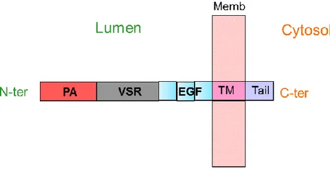
The VSR receptor was first identified as the BP-80 protein and isolated from pea clathrin-coated vesicles (Kirsch et al., 1994; Paris et al, 1997; Paris and Neuhaus, 2002). VSR are type I membrane proteins, constituted of an N-terminal luminal domain which includes a
Protease-42
Associated domain (PA), a large VSR specific domain, three Cys-Rich EGF Repeats (Epidermal Growth Factor) a single transmembrane domain, and a short cytosolic tail (Figure 1.9).
Figure 1.9 Schematic VSR structure (not to scale). N-ter: N-terminus; PA: Protease-associated domain; VSR: VSR domain; EGF: Epidermal Growth Factor domain (3 repeat); MP: plasma membrane; TM: Trans-membrane domain; C-ter: C-terminus
Kirsch et al. (1994) demonstrated that BP-80 binds in vitro the ssVSD of barley proaleurain, but showed no affinity for the pea prolectin and the barley prolectin, the latter carrying an already characterized ctVSD signal. Several publications have shown the involvement of VSR in the transport of vacuolar soluble proteins to the LV (Jiang and Rogers 1998; DaSilva et al., 2006; Zouhar et al., 2010). BP-80 was described by immunocytochemistry to localize in the trans-cisternae of the Golgi, in the TGN, and in the prevacuolar compartment (Paris et al., 1997). The current model suggests that BP-80 is a pH-dependent ligand binder. Cargo proteins are bound in the Golgi. Recycling of BP-80, from the prevacuolar compartment to the Golgi after cargo release, is a strongly supported hypothesis (DaSilva
et al., 2006). However, an alternative model implying ER to TGN traffic has been proposed (Niemes
2010; Robinson 2013). Reviewed by Robinson and Neuhaus (2016), this second model proposes that VSRs interact with their ligands earlier in the secretory pathway, in ER or in cis-Golgi. It also brings evidence that the dissociation of the ligand-receptor complex cannot occur in a pH-dependent way, because of the similarity of pH value between the two compartments.
VSR proteins are encoded by seven genes in A. thaliana (AtVSR1 to AtVSR7). Homologous receptors have been identified in all land plants including lycophytes and mosses, where eight VSR genes have been found.
43
The RMR family
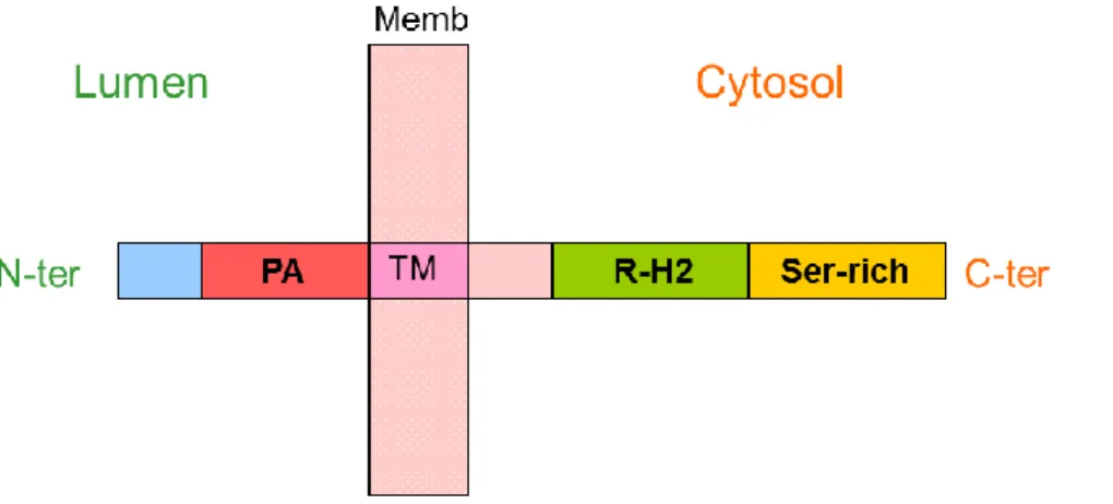
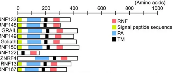
RMR proteins were identified by their homology to the PA domain of VSR proteins (Cao et al., 2000; reviewed by Wang et al., 2010). For this reason, RMRs were thought to be involved in ligand binding as vacuolar receptors. RMR structure consists of an N-terminal luminal domain, the Protease-associated domain, a single transmembrane domain and a cytosolic tail with a RING-H2 domain (Really Interesting New Gene, with two Histidines) (Figure 1.10). RING domains are cysteine-rich, zinc-binding domains and are known as protein-interaction domains. They consist of a pattern of conserved cysteine and histidine residues binding two zinc ions (Borden 2000; Kosarev et al., 2002). RING domains are involved in various cellular mechanisms in eukaryotes, such as signal transduction, transcription and protein-protein interactions (Borden and Freemont 1996). RMRs belong to the PA-TM-RING protein family, which combine PA, transmembrane domain and RING domains, and has been identified in many organisms: in plants, mammals (GRAIL; Seroogy et al., 2004), Xenopus (GREUL1; Borchers et al., 2002) and Drosophila (Goliath; Bouchard and Cote 1993) but not in yeast (Bocock et al., 2009). In plants, many RMRs include a C-terminal serine rich region (Jiang et al., 2000).
Figure 1.10 Schematic RMR structure (not to scale). N-ter : N-terminus ; PA : Protease-associated domain ; P : plasma membrane ; TM : Trans-membrane domain ; RING-H2 : RING H2 domain, Serine rich : Serine rich cytosolic tail; C-ter: C-terminus. Furthermore, in Arabidopsis thaliana RMR 1, 3 and 4 include a C-terminal serine rich region (Jiang et al., 2000).
In tobacco and tomato cells, RMR proteins were localized in organelles also labelled with anti-DIP antibodies: the PSV crystalloid (Jiang et al., 2000). anti-DIP (dark-induced tonoplast intrinsic protein) is an isoform of TIP, found in the crystalloid bodies, precursors of the PSV during seed development. Jiang
et al. (2000) proposed that DIP-positive organelle might function as PVC and fuse to form the PSV. Park et al. (2005) described localization of AtRMR1 in the prevacuolar compartment of PSV and in the Golgi.
44
showed an interaction of the luminal domain of AtRMR1 with the ctVSD of phaseolin. These results strongly suggest that AtRMR1 may act as a cargo receptor for proteins with ctVSD targeting to the PSV. However, in both tobacco leaves and Arabidopsis protoplasts, Occhialini (2011) found AtRMR2 mostly localized in the TGN while AtRMR1 localized in ER structures. Occhialini also described by BiFC (Bimolecular Fluorescence Complementation) that AtRMR2 can form homodimers and can also form heterodimers with AtRMR1, both localizing in the TGN. In rice, OsRMR1 was localized by immunogold labelling in Golgi apparatus, in TGN and in an electron-dense organelle similar but distinct from the MVB (Shen et al., 2011). This organelle was proposed to act as a PVC for the PSV pathway in rice. Moreover, Scabone et al. (2011) showed that transmembrane (TM) and C-terminal domain (CT) of
AtRMR1 fused to a RFP construct was localized in the lumen of central vacuole, in leaves, roots and
embryos of A. thaliana transgenic plants. These results highlight the involvement of the TM-CT of
AtRMR1 in protein targeting to vacuole.
RMR proteins are encoded by six genes in Arabidopsis (AtRMR1 to 6) and by five genes in
Physcomitrella patens. RMR proteins form two distinct subfamilies in Angiosperms and Gymnosperms.
Single and double mutants in AtRMR genes had no detectable phenotypes (Stigliano, PhD thesis; E. Rojo, personal communication). AtRMR1 belongs to the subfamily 1 while the five other AtRMRs belong to the subfamily 2. Moss RMRs form a clearly separate clade from Angiosperms and are represented by two subfamilies, the first one including RMR1 and 2, and the second one including RMR3, 4 and 5 (figure 1.11).