DOI 10.1007/s00018-013-1275-7
Cellular and Molecular Life Sciences
REVIEW
Deciphering microRNA code in pain and inflammation:
lessons from bladder pain syndrome
Ali Hashemi Gheinani · Fiona C. Burkhard ·
Katia Monastyrskaya
Received: 21 November 2012 / Revised: 22 January 2013 / Accepted: 28 January 2013 / Published online: 5 March 2013 © Springer Basel 2013
Introduction
MicroRNAs (miRNAs) are quickly gaining recognition for
their role in many biological processes and disease states
[
1
,
2
]. MiRNAs are endogenous non-coding single-stranded
RNAs of approximately 22 nucleotides that regulate gene
expression by post-transcriptional mechanisms upon
sequence-specific binding to the 3
′ untranslated regions
(3
′ UTRs) [
1
,
3
] or, occasionally, the 5
′UTRs [
4
,
5
] or
cod-ing regions [
6
–
8
] of their mRNA targets. MiRNAs exhibit
imperfect complementarity with mRNAs, allowing them
to regulate multiple genes and thus complicating efforts
to predict and functionally validate their targets [
9
]. Since
their discovery [
10
], miRNAs have been acknowledged as
important modulators of gene expression, and deficiency in
their synthesis or function contributes to many human
dis-eases [
11
,
12
]. MiRNA expression profiles characteristic of
a particular disorder may serve as a useful diagnostic tool,
but more importantly, it is the first step towards unraveling
miRNA functions. Follow-up studies in experimental
mod-els are carried out to identify the protein targets of
differen-tially expressed miRNAs and delineate the signaling
path-ways activated in a diseased state.
Although a wealth of information has been gathered on
miRNA expression in bladder cancer (BCa), which in this
respect remains the best-studied disorder, only a few
stud-ies have been carried out in bladder dysfunction. The first
miRNA profiling in bladder pain syndrome/interstitial
cysti-tis (BPS), which was performed in our laboratory, has
identi-fied several miRNAs, regulating the expression of signaling
and adhesion molecules [
13
,
14
]. Pain and inflammation are
characteristic of BPS, which has been suggested to share the
pathogenetic mechanisms with other inflammatory diseases
such as asthma, inflammatory bowel disease, and
autoim-mune diseases [
15
]. These are common co-morbidities
Abstract MicroRNAs (miRNAs), a novel class of
mol-ecules regulating gene expression, have been hailed as
modulators of many biological processes and disease states.
Recent studies demonstrated an important role of miRNAs
in the processes of inflammation and cancer, however, there
are little data implicating miRNAs in peripheral pain.
Blad-der pain syndrome/interstitial cystitis (BPS/IC) is a clinical
syndrome of pelvic pain and urinary urgency/frequency in
the absence of a specific cause. BPS is a chronic
inflam-matory condition that might share some of the pathogenetic
mechanisms with its common co-morbidities inflammatory
bowel disease (IBD), asthma and autoimmune diseases.
Using miRNA profiling in BPS and the information about
validated miRNA targets, we delineated the signaling
path-ways activated in this and other inflammatory pain disorders.
This review projects the miRNA profiling and functional
data originating from the research in bladder cancer and
immune-mediated diseases on the BPS-specific miRNAs
with the aim to gain new insight into the pathogenesis of this
enigmatic disorder, and highlighting the common regulatory
mechanisms of pain and inflammation.
Keywords MicroRNA · Bladder · Pain · Inflammation ·
Gene expression · Bladder cancer · Inflammatory bowel
disease
A. H. Gheinani · K. Monastyrskaya (*)
Department of Clinical Research, Urology Research Laboratory, University of Bern, Murtenstrasse 35, 3010 Bern, Switzerland e-mail: [email protected]
F. C. Burkhard
Department of Urology, University Hospital, 3010 Bern, Switzerland
of BPS/IC, and miRNA expression and function in these
immune-mediated diseases has been addressed in a
num-ber of studies. This review projects the miRNA profiling
and functional data originating from the research in bladder
cancer and immune-mediated diseases on the BPS-specific
miRNAs aiming to gain new insight into the pathogenesis of
this enigmatic disorder, and highlighting the common
regu-latory mechanisms of pain and inflammation.
MicroRNAs—general information
The first miRNAs lin-4 and let-7 were discovered as
impor-tant regulators of the normal temporal control of diverse
postembryonic developmental events in C. elegans [
10
,
16
].
Most miRNA reduce protein synthesis, whereas some, such
as miR369-3 and miR-373, enhance translation [
17
,
18
].
The first link between miRNAs and cancer was made after
uncovering the deletion of miR-15 and miR-16 in leukemias
[
19
], and the potential for the miRNA profiling in cancer
diagnosis became apparent [
20
]. This was followed by
stud-ies of the role of miRNAs in cardiovascular [
21
],
neuro-degenerative [
22
–
24
], and autoimmune disease [
25
,
26
].
Therapeutic use of miRNAs was pioneered by
administra-tion of miRNAs [
27
], or their antagonists called antagomirs
[
28
], to the affected cells restoring the normal levels of their
protein targets. The locked nucleic acid (LNA)-antagomirs
have been successfully introduced into the clinical practice:
LNA-anti-miR-122 suppresses hepatitis C virus (HCV)
rep-lication in chronically infected animals [
29
].
The details of miRNA synthesis are well characterized
and extensively reviewed [
30
–
32
]. Most canonical
mam-malian miRNA genes have been identified in introns of the
protein-coding or noncoding RNAs and around one-third
of them are located in the introns of target genes [
33
]. The
miRNA gene transcripts are the primary precursor RNAs
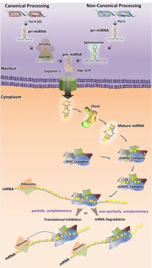
(pri-miRNA), which mature in two steps (Fig.
1
).
Pri-miRNAs are processed in the nucleus by Drosha–DGCR8
complex into a ~70-nucleotide RNA molecules [
34
]. These
precursor hairpin miRNAs (pre-miRNAs) are then exported
to the cytoplasm by Exportin-5 (Exp5) [
35
] and cleaved
by Dicer-TRBP complex into a ~20-bp miRNA/miRNA*
duplex. The guide strand of the miRNA/miRNA* duplex
is usually incorporated into miRNA-induced silencing
complex (miRISC) and the passenger strand or (miRNA*)
is released and degraded [
36
]. The final product is
RISC-loaded mature miRNA, which is guided to its target mRNA
by means of base pairing (Fig.
1
).
Some microRNA, such as miR-877 and miR-1226,
derive from short intronic hairpins named “mirtrons” [
37
,
38
]. Mirtrons are spliced from the host gene by the
spli-ceosome, become linearized by debranching enzyme and
then fold into hairpins, which enter the miRNA-processing
pathway without Drosha-mediated cleavage [
37
,
39
].
Recently, a third pathway of microRNA biogenesis has been
described: some microRNAs (miR-1225 and miR-1228) do
not need DGCR8, Dicer, Exportin-5, or Argonaute 2 for
their biogenesis, which nevertheless involves Drosha. This
class of microRNAs has been named “simtrons” [
40
]. The
miRNA registry in humans includes 18,226 entries for
hair-pin precursor miRNAs, expressing 21,643 mature miRNA
products [
41
,
42
].
Role of miRNAs in urinary bladder function
and pathology
Pathogenesis of bladder pain syndrome (BPS)
Bladder pain syndrome (BPS) is a clinical syndrome of
pel-vic pain and urinary urgency-frequency in the absence of a
specific cause. According to the European Society for the
Study of Interstitial Cystitis (ESSIC) proposal and
Euro-pean Association of Urology (EAU) guidelines [
43
], BPS
is diagnosed on the basis of symptoms of pain associated
with the urinary bladder, accompanied by at least one other
symptom, such as day-time and/or night-time urinary
fre-quency after exclusion of confounding symptoms. Overall,
this disease, which has a significant impact on social and
psychological well-being, affects approximately 1 million
patients in the USA alone [
44
], with at least 230 confirmed
cases per 100,000 females [
45
]. The etiology of BPS is
unknown, and its treatment largely empiric. A multitude
of pathogenetic mechanisms have been postulated ranging
from neuroinflammatory to autoimmune or possibly
infec-tious or toxic agents, but an inflammatory component is
commonly thought to be involved. Epithelial damage has
often been invoked: the mucinous layer of the healthy
blad-der is often compromised in patients with BPS/IC, as well
as in some animal models [
46
,
47
].
The integrity of the urothelium is indispensable for the
healthy bladder [
48
]. Mechanical or chemical damage, as
well as a bacterial infection can lead to a compromised
urothelium allowing urinary solutes to penetrate into the
interstitium [
49
]. The loss of epithelial integrity is a
pre-dominant histopathologic finding in biopsies from BPS
patients [
50
]. In human BPS patients, the molecular
mark-ers for bladder permeability and proteoglycan core proteins
are often down-regulated [
51
,
52
]. Recently, we have shown
that the mRNA levels of the tight junction (TJ) proteins
ZO-1, JAM-1, occludin, and tight claudins, normally
pre-sent in water impermeable epithelia and abundant in normal
urothelium, were significantly down-regulated in bladder
biopsies from BPS patients, indicating a compromised tight
junction structure and possibly increased permeability of the
urothelium [
13
]. Therefore it seems possible that urothelial
damage is a preceding feature of this disorder and might be
a causative factor in the pathogenesis of BPS. A decrease
of E-cadherin mRNA levels, which we observed at mRNA
level in BPS (Monastyrskaya, unpublished), was confirmed
by recent findings of lower E-cadherin and ZO-1
expres-sion in IC/BPS, but not in overactive (OAB) bladders, and
Fig. 1 Biogenesis of miRNAs. Mature microRNAs (miRNAs) are approximately 22 nucleo-tides long and generated by a two-step process. The first step takes place in the nucleus, where primary miRNAs (pri-miRNA) are converted into precursor miRNAs (pre-miRNAs) by the microprocessor complex containing Drosha and DGCR8 (in canonical process) or spliceosome, which excises the pri-microRNA transcribed from introns (in non-canonical process). Exportin-5 transports pre-miRNAs to the cytoplasm. In the second step, the hairpin structures of the pre-miRNAs are re-cleaved by the RNase III Dicer to generate mature miRNA. The functional strand of the mature miRNA gets incorporated into the RNA-induced Silencing Complex RISC (miRISC), where it guides miRISC to silence target mRNAs through translational repression or mRNA cleavage
suggesting the urothelial barrier function was compromised
in BPS but not affected in the OAB bladder [
53
].
BPS is characterized by several other gene expression
changes: tachykinin receptors NK1R and NK2R were
sig-nificantly down-regulated and bradykinin B1 receptor,
can-nabinoid receptor CB1, and muscarinic receptors M3–M5
were up-regulated in BPS patients’ biopsies [
13
]. In
addi-tion, expression of acid-sensing channels, important for
nociceptive pain, was altered in BPS: we detected an
up-regulation of ASIC2a and ASIC3 mRNA, whereas ASIC1a
remained unchanged [
54
]. These changes, in conjunction
with deficiencies of urothelial barrier, might account for
increased nociception in BPS patients.
MiRNAs in BPS and OAB
New information is emerging on the miRNA-mediated
regulation of epithelial permeability, bladder contractility,
and neurogenic inflammation in bladder dysfunction. While
studying the gene expression changes characteristic of BPS,
we observed a perplexing down-regulation of tachykinin
receptors in the biopsies of BPS patients which prompted
us to study its mechanisms [
13
]. Using cell-based models,
we showed that prolonged exposure of NK1R to Substance
P (SP) caused a decrease of NK1R mRNA levels and a
con-comitant increase of regulatory miRNAs miR-449b and
miR-500. In the biopsies of BPS patients, the same miRNAs
were significantly increased, suggesting that BPS promoted
an attenuation of NK1R synthesis via activation of specific
miRNAs. We confirmed this hypothesis by identifying 31
differentially expressed miRNAs in BPS patients and
dem-onstrated a direct correlation between miR-449b, miR-500,
miR-328, and miR-320 and a down-regulation of NK1R
mRNA and/or protein levels. The results of the first miRNA
profiling in biopsies of BPS patients are shown in Table
1
.
Recently, using a mouse model with an induced
dele-tion of Dicer, two groups examined the role of miRNAs in
the regulation of bladder contractility. An induced
smooth-muscle-specific Dicer knock-out resulted in significantly
reduced levels of miRNAs, including miR-145, miR-143,
miR-22, miR-125b-5p, and miR-27a, from detrusor
prepa-rations without mucosa. Deletion of Dicer resulted in a
disturbed micturition pattern in vivo and reduced
depolari-zation-induced pressure development in an isolated detrusor
due to decreased levels of L-type Ca
2+channels [
55
]. In
a similar study, the loss of Dicer exacerbated
cyclophos-phamide-induced bladder overactivity in mice, possibly
because the miRNAs capable of targeting P2X mRNAs
were impaired, leading to enhanced P2X receptor
expres-sion [
56
]. The authors describe an up-regulation of
miR-34a and miR-25 in the mouse model of OAB. Interestingly,
miR-25 is also elevated in BPS [
13
] (Table
1
), pointing to
the similarities of function of this miRNA in both disorders.
Our follow-up study of the miRNA function in BPS
concerned the role of these molecules in the regulation of
urothelial permeability [
14
]. We identified microRNA
miR-199a-5p, which was increased in BPS patients’ biopsies,
as an important regulator of intercellular junctions. Upon
overexpression in urothelial cells it impaired correct tight
junction formation and caused a permeability increase.
MiR-199a-5p directly targeted mRNAs encoding LIN7C,
ARHGAP12, PALS1, RND1, and PVRL1 and attenuated
their expression levels to a similar extent. Laser
micro-dissection revealed that miR-199a-5p was predominantly
expressed in the bladder smooth muscle, but also detected
in the mature bladder urothelium and primary urothelial
cultures. In the urothelium, its expression can be
up-reg-ulated following activation of cAMP signaling pathways.
Our results point to a possible link between miR-199a-5p
expression and the control of urothelial permeability in
bladder pain syndrome. Up-regulation of miR-199a-5p and
concomitant down-regulation of its multiple targets might
be detrimental for the establishment of a tight urothelial
bar-rier, leading to chronic pain.
miRNA profiling in bladder cancer (BCa)
Except the studies discussed above, most of the
informa-tion on the role of miRNAs in the bladder comes from the
work done in BCa. Here we summarize the published data
in order to identify the organ-specific miRNA expression
patterns and draw comparisons between BCa and bladder
dysfunction.
BCa is one of the most common cancers in the world
[
57
], the fifth most commonly diagnosed non-cutaneous
solid malignancy, and, after prostate cancer, the second
most frequently diagnosed genitourinary tumor [
58
]. In
2008, 386,300 new cases of bladder cancers were diagnosed
globally [
59
]. The early miRNA screening attempts were
aimed at defining the cancer-specific miRNAs, and
differen-tiating between the tumor stages by means of miRNA
profil-ing. The first study in human bladder compared the miRNA
expression profiles of bladder and kidney cancers [
60
]. Both
types of cancers were found to share many miRNAs. The
most significant up-regulated miRNAs in BCa versus
nor-mal bladder (1.2-fold change cutoff; p < 0.05) were:
miR-223, miR-26b, miR-221, miR-103-1, miR-185, miR-23b,
miR-203, miR-17-5p, miR-23a, and miR-205. However,
this study did not identify any significantly down-regulated
miRNA in tumors and the data did not overlap with the later
published results [
61
], possibly because of the limited
num-ber of reference samples (two normal mucosa).
Expression of 343 miRNA was studied in noninvasive
and invasive bladder carcinoma cell lines in order to
iden-tify the miRNA signature in BCa suitable for discriminating
the superficial from the invasive disease [
62
]. Nine miRNAs
with significant differential expression included mir-21,
mir-31, mir-200a, mir-200c, mir-205, mir-373*, mir-487b,
mir-498, and mir-503. MiR-21 expression was up-regulated
and miR-205 down-regulated in the invasive compared with
the noninvasive bladder cell lines. In 2009, Dyrskjot et al.
investigated the expression profile of 290 microRNAs in
immortalized urothelial cell lines, tumorigenic cell lines,
106 bladder tumors and 11 normal samples and identified
some differentially expressed miRNAs between cancer and
normal urothelium. In agreement with the previous study
[
62
], miR-21 was the most up-regulated and miR-145 was
the most down-regulated in cancer compared to the
nor-mal samples. Several differentially expressed miRNAs
were found comparing different tumor stages. Most
sig-nificant miRNAs up-regulated in progressing tumors were
miR-129-5p, miR-518c*, miR-185, miR-133b, miR-373*,
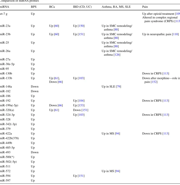
Table 1 Comparative analysis of miRNA expression profiles
Occurrence and regulation of the miRNAs, altered in BPS, BCa bladder cancer, IBD inflammatory bowel disease CD Crohn’s disease, UC ulcer-ative colitis, asthma, RA rheumatoid arthritis, SLE systemic lupus erythematosus, MS multiple sclerosis and pain
Comparison of miRNA profiles
miRNA BPS BCa IBD (CD, UC) Asthma, RA, MS, SLE Pain
let-7 g Up Up after opioid treatment [109]
Altered in complex regional pain syndrome (CRPS) [113] miR-23a Up Up [60] Up [150] Up in SMC remodeling/ asthma [88] miR-23b Up Up [60] Up [151] Up in SMC remodeling/ asthma [88] Up in neuropathic pain [110] miR-25 Up Up in SMC remodeling/ asthma [88] miR-26a Up Up in SMC remodeling/ asthma [126] miR-27a Up miR-30e-5p Up miR-95 Up miR-130b Up Down in CRPS [113] miR-133b Up Up [61], Down [66]
Up [103] Down after morphine—role in
pain [152]
miR-148a Down Up in SLE [79]
miR-182 Down miR-186 Up miR-192 Up Up [104] Down in CRPS [113] miR-199a(-5p) Up Down [66] Up [153] miR-320(a) Up Up [61] Down [151] miR-324-3p Up Up [103] Down in CRPS [113] miR-328 Up miR-342(-3p) Up miR-379 Up miR-422a Up Up in MS [94] Down in CRPS [113] miR-422b(378) Up miR-449b Up miR-485-5p Up miR-493 Down miR-500(*) Up miR-502(-5p) Up miR-511 Up miR-572 Up Up in MS [94] miR-594 Up Up [151] miR-597 Up
miR-320a, miR-145; and miRNAs, which were mostly
down-regulated in progressing tumors were 29c,
miR-29b, miR-29a, miR-361-5p, miR-203, and miR-205 [
61
].
Investigation of 322 miRNAs expressed in normal
urothe-lium from patients with high-grade urothelial cell
carcino-mas (UCC) and disease-free controls revealed that 11 % of
miRNAs were differentially expressed [
63
].
Down-regula-tion of miRNAs is a common phenomenon in low-grade
tumors, and aberrant promoter hyper-methylation
influ-ences miRNA down-regulation in low-grade UCC. Many
miRNAs down-regulated in low-grade tumors are predicted
to target FGFR3 (miRs-99a/100/214/145/30a/125b/507). In
contrast, in high-grade UCC up-regulation of many
miR-NAs including miRNA-21 can suppress p53 function [
64
].
Lin et al. performed miRNA profiling in BCa and matched
normal urothelial epithelium controls and identified 37
up-regulated and 38 down-up-regulated miRNAs. Among them,
miRNA-143 was most down-regulated, 13.7 times lower in
tumor than in the matched control [
65
]. miRNA-143 was
not expressed in the human BCa cell lines EJ and T24 and
its transfection inhibited cell proliferation and reduced RAS
protein levels [
65
]. Ichimi et al. investigated
tumor-suppres-sive miRNAs in BCa [
66
]. Upon screening of 156 miRNAs
in 14 BCas, five normal bladder epithelium (NBE)
sam-ples and three BCa cell lines, 145, 30a-3p,
miR-133a, miR-133b, miR-195, miR-125b, and miR-199a* were
shown to be significantly down-regulated in BCas [
66
].
Ker-atin 7 (KRT7) mRNA was a common predicted target for
the down-regulated miRNAs. Compared to normal bladder
epithelium, BCa samples had significantly higher mRNA
levels of KRT 7 [
66
].
Using microarray technology, Song et al. reported that
the expression profile of miRNAs was significantly altered
in bladder urothelial carcinoma tissue compared to adjacent
normal bladder tissue. Consistent with previous
observa-tions, most differentially expressed miRNAs were
down-regulated. The top ten dis-regulated miRNAs including
miR-1, miR-145, miR-143, miR-100, miR-200b, miR-708,
miR-133a, miR-133b, and mir-125b were validated by
real-time RT-PCR [
67
].
The first report of genome-wide miRNA expression
profiling in human bladder urothelial carcinoma by deep
sequencing was published in 2011 by Han et al. [
68
]. A
total of 656 differentially expressed miRNAs were detected
comprising known human miRNAs and miRNA antisense
sequences (miRNA*s). MiR-490-5p was the most
cantly down-regulated and miR-96 was the most
signifi-cantly up-regulated miRNA [
68
].
Notwithstanding a long list of miRNAs de-regulated in
BCa [
69
,
70
], there is very little overlap in the patterns of
miRNA expression between BCa and BPS (Table
1
), which
might be indicative of differential organ responses to
spe-cific diseases.
Inflammation in BPS and the role of miRNA
in immune-mediated diseases
BPS is an inflammatory disorder
Inflammation occurs frequently in the lower urinary tract, and
most often is a result of a urinary tract infection. One of the
intriguing features of BPS is the presence of inflammation,
often confirmed by biopsy, in the absence of an inflammatory
agent (toxin or microorganism) [
71
]. Many causative factors
have been suggested for BPS, including chronic or
sub-clinical infection, autoimmunity and genetic susceptibility,
which could be responsible for initiating the
inflamma-tory response. However, a central role of inflammation has
been confirmed in the pathogenesis of interstitial cystitis
[
72
]. Epithelial dysfunction, often observed during BPS,
might cause nerve sensitization, which in turn could lead to
up-regulation of neurotransmitter release (tachykinins,
glu-tamate, calcitonin gene-related peptide) [
73
]. Secretion of
inflammatory mediators such as SP from sensory nerves has
been implicated in the pathophysiology of pain triggering
mast cell secretion/neurogenic inflammation [
74
]. Indeed,
the obligatory requirement of tachykinin receptor NK1R in
cystitis and its role in mast cell degranulation and
inflam-mation have been confirmed in experiments with NK1R
knockout mice [
75
]. Recently, increased urinary NGF levels
were described in BPS/IC patients suggesting that chronic
inflammation is involved in this bladder disorder [
76
].
A recent study investigated whether bladder
inflamma-tion could directly modulate the apoptotic signaling
path-way in urothelial cells in interstitial cystitis/painful bladder
syndrome (IC/PBS, equivalent definition of BPS) [
77
]. The
levels of pro-apoptotic proteins, including phospho-p53
(Ser 15), Bad, Bax, and cleaved caspase-3 were significantly
increased in the IC/PBS bladders. These results suggested
that the tissue damage and abnormal urothelium in the IC/
PBS bladders might be regulated concurrently by
inflamma-tory signals, such as p38 mitogen-activated protein kinase
and TNF-alpha. The contents of phospho-p38α and TNF-α
in IC/PBS samples were significantly greater than in the
control. The in vitro analysis showed that the apoptotic
pro-cess could be induced by TNF-alpha treatment and
aniso-mycin stimulation in normal urothelial cells [
77
].
miRNAs as regulators of immune response
During the recent years, plentiful evidence has been amassed
pointing to the critical role of miRNAs both in the
devel-opment of immune system and its function in innate and
adaptive responses [
78
]. Several miRNAs, including miR-155,
miR-181a, miR-146a, miR-150, miR-223 and
miR-17-92 have been shown to regulate development,
differentia-tion, and function of immune cells (reviewed by [
79
,
80
]).
The innate immune response is a cellular response,
compris-ing macrophages, monocytes, granulocytes, and dendritic
cells, which is started by activation of Toll-like receptors
(TLR). TLR signaling induces numerous miRNAs
includ-ing miR-155, miR-146a, and miR-21 [
81
]. Additionally,
miR-125b and let-7 are important in controlling innate
immune responses: miR-125b targets TNF-α, while let-7
targets IL-6 mRNA [
82
,
83
]. Interestingly, although in our
study we detected an increased neutrophil infiltration in
the BPS patients [
13
], none of the classical immune
cell-specific miRNAs were significantly up-regulated. However,
we detected an increase in miR-500* and miR-511, which
are important for the dendritic cell, monocyte and
mac-rophage function [
84
,
85
]. These results indicate that the
elevated miRNAs levels might originate from the bladder
tissue remodeling during BPS, rather than the immune cell
infiltration.
Taking into account the role of inflammation in BPS and
the lack of data on miRNA regulation in the similar
inflam-matory disorders of the bladder, we compared the miRNAs
identified in other immune-mediated diseases, to the BPS
profile. These data are summarized in Table
1
and Fig.
2
.
Asthma
Asthma is a common chronic airway disease characterized
by airway remodeling (epithelial alteration, fibrosis, smooth
muscle hypertrophy) and associated with Th2 response that
stimulates eosinophile and mast cell infiltration. Asthma is a
common co-morbidity of BPS [
86
], therefore we correlated
the available information on miRNA profiling in this disorder
with our data in BPS [
13
]. In the mouse models of asthma,
miR-1 was down- and miR-21 up-regulated, along with
21 other differentially expressed miRNAs, and in another
model, miR-16, -21, and -126 were up-regulated. MiR-133,
-25, 146a- and -26a regulate human airway smooth muscle
cells in asthma models (reviewed by [
87
]). MiR-23a, -23b,
and -25 play an important role in regulating the phenotype
of airway smooth muscle via modulating the expression of
inflammatory mediators like RANTES, eotaxin, and TNF-α.
These genes are responsible for extracellular matrix
turno-ver and expression of contractile proteins (myosin heavy
chain) [
88
]. Interestingly, some of the miRNAs described in
this study appear to be important in BPS—we found
miR-23a, -23b, -25, and -320 up-regulated in BPS, arguing for
an increased smooth muscle phenotype, which is consistent
with low bladder volumes and thick bladder walls observed
in patients [
13
].
Autoimmune diseases
The role of miRNAs in autoimmune diseases including
rheumatoid arthritis (RA), systemic lupus erythematosus
(SLE), and multiple sclerosis (MS) has been postulated
and confirmed by altered levels of several important
miR-NAs (reviewed in [
79
]). Although no data support a direct
causal role of autoimmune reactivity in the pathogenesis of
BPS/IC, there is ample indirect evidence, such as the strong
female preponderance and the clinical association between
BPS and other known autoimmune diseases within patients
and families [
15
]. BPS is associated with the diagnosis of
RA [
89
], SLE, and Sjögren’s syndrome [
90
]. In RA,
miR-NAs miR-146a, miR-223, and miR-155, characteristic of
immune cell activation, were significantly up-regulated in
synovial fluid samples [
91
]. In MS, miRNAs miR-34a, -155
and -326 were elevated in active lesions [
92
]. In SLE,
miR-146a, a negative regulator of TLR signaling was profoundly
decreased, whereas miR-148a and miR-21 were increased
in T cells from patients with lupus [
93
]. Both miR-422a and
miR-22 have previously been implicated in MS [
94
]. Taken
together, miRNA profiling in autoimmune diseases revealed
a strong prevalence of immune cell-specific miRNAs and
some overlap with BPS profile established in our study [
13
].
Inflammatory bowel disease (IBD)
IBD, comprising Crohn’s disease (CD) and ulcerative
coli-tis (UC), is a gastrointestinal chronic inflammatory disorder
and a frequent co-morbidity of BPS [
95
]. Earlier work has
demonstrated that both the number of SP-positive nerve
end-ings and mast cell count were increased in patients with BPS
and IBD [
96
], implicating a similar pathogenic mechanism.
There are numerous reports indicating that patients with CD
and UC have altered miRNA profiles [
97
–
105
]. Analysis of
miRNA expression in patients with active UC, inactive UC,
CD, irritable bowel syndrome, infectious colitis, and
micro-scopic colitis revealed eight miRNAs (miR-16, miR-21,
miR-23a, miR-24, miR-29a, miR-126, miR-195, and Let-7f)
which were significantly increased in active UC tissues and
three miRNAs (miR-192, miR-375, and miR-422b) were
significantly decreased in the UC tissues. Up-regulation of
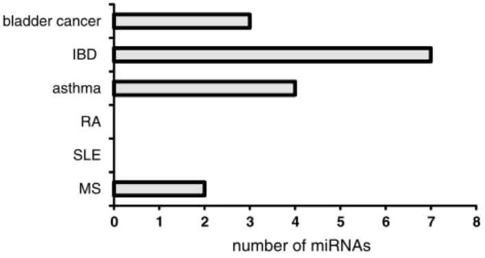
0 1 2 3 4 5 6 7 8 MS SLE RA asthma IBD bladder cancer number of miRNAs
Fig. 2 Similarly regulated miRNAs. Number of miRNAs either up- or down-regulated both in BPS and BCa, IBD, RA, SLE, or MS
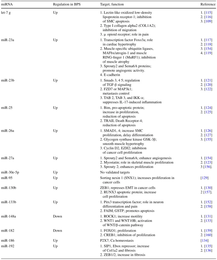
Table 2 Validated BPS miRNA targets
miRNA Regulation in BPS Target; function Reference
let-7 g Up 1. Lectin-like oxidized low-density
lipoprotein receptor-1; inhibition of SMC apoptosis
2. Type I collagen alpha2 (COL1A2); inhibition of migration
3. μ opioid receptor; role in pain
1. [115] 2. [116] 3. [109]
miR-23a Up 1. Transcription factor Foxo3a; role
in cardiac hypertrophy
2. Muscle-specific ubiquitin ligases, MAFbx/atrogin-1 and muscle RING-finger 1 (MuRF1); inhibition of muscle atrophy
3. Sprouty2 and Sema6A proteins; promote angiogenic activity. 4. E-cadherin
1. [117] 2. [118] 3. [154] 4. [119]
miR-23b Up 1. Smads 3, 4 5; regulation
of TGF-β signaling 2. FZD7 or MAP3k1;
metastasis control
3. TAB 2, TAB 3, and IKK-α;
suppresses IL-17-induced inflammation
1. [121] 2. [120] 3. [122]
miR-25 Up 1. Bim, pro-apoptotic protein;
increase in proliferation, reduction of apoptosis 2. TRAIL Death Receptor-4;
reduction of apoptosis
1. [124] 2. [125]
miR-26a Up 1. SMAD1, 4; increase SMC
proliferation, delay differentiation 2. Glycogen synthase kinase GSK-3β;
smooth muscle hypertrophy 3. Cyclin D2, EZH2; inhibition
of cancer cell proliferation
1. [126] 2. [127] 3. [155]
miR-27a Up 1. Sprouty2 and Sema6A; enhance angiogenesis
2. Myostatin; role in skeletal muscle proliferation 3. Sprouty 2; enhances proliferation
1. [154] 2. [123] 3 [156].
miR-30e-5p Up No validated targets
miR-95 Up Sorting nexin 1 (SNX1); increases proliferation in cancer cells
[129]
miR-130b Up ZEB1; represses EMT in cancer cells
2. RUNX3 apoptotic protein; increase cell proliferation
1. [130] 2 [157]. miR-133b Up 1. Pitx3 transcription factor; role in neuron
differentiation and pain
2. FAIM, GSTP; promotes apoptosis
1. [152] 2. [158]
miR-148a Down 1. ROCK1; increase motility
2. WNT1 and WNT10B; activation of WNT/β-catenin pathway
1. [131] 2. [133]
miR-182 Down 1. FOXO1; proliferation
2. CREB1; inhibition of proliferation
1. [159] 2. [160]
miR-186 Up P2X7; Ca homeostasis [134]
miR-192 Up 1. SIP1, Ebox repressor; increase
of Col1a2 and fibrosis 2. ZEB1/2; increase in fibrosis
1. [135] 2. [136]
miR-192 was observed in IBD-associated dysplasia [
104
].
UC patients show an up-regulation of immune cell-specific
miR-21 and miR-155 in inflamed tissue [
106
].
Comparing active to quiescent UC, it was reported that
four miRNAs (miR-188-5p, miR-25, miR-320a, miR-346)
were down-regulated and five miRNA (miR-29a, miR-29b,
miR-126*, miR-127-3p, miR-324-3p) were up-regulated
[
103
]. We found a strong overlap in miRNA expression
pro-files between BPS and IBD (Table
1
).
Inflammatory pain disorders
Pain is one of the hallmarks of inflammation, and chronic
inflammatory conditions are often accompanied by chronic
pain. Inflammatory pain serves as a warning that hinders
the normal bodily function until the stimulus abates or the
tissue repairs. Noxious stimuli activate numerous
recep-tors and ion channels, and signals propagate to the central
nervous system, where pain is perceived [
107
]. Epigenetic
modifications influence inflammatory cytokine metabolism,
steroid responsiveness, and opioid sensitivity thus
contrib-uting to the development of chronic pain [
108
], however,
there are little data implicating miRNA in peripheral pain.
It was shown that miRNAs might regulate the action of
opi-oid drugs: let-7 miRNA family, including let-7 g, which is
up-regulated in BPS, was found to be critical for μ opioid
receptor function. Chronic exposure to morphine caused the
reduction of MOR levels and concomitant increase in let-7
miRNA synthesis [
109
]. It is possible that miRNA
induc-tion might serve as an adaptive response, aimed at reducing
pain and inflammation: the animals with neuropathic pain
showed significant improvements after infusion of miR-23b.
miRNA Regulation in BPS Target; function Reference
miR-199a(-5p) Up 1. Cell junction proteins LIN7C, ARHGAP12, PALS1, RND1 and PVRL1; causes epithelial dysfunction
2. HIF1alpha, Sirtuin; hypertrophy in cardiomyocytes
3. Discoidin domain receptor 1; decrease cancer cell proliferation 1. [14] 2. [161] 3. [162]
miR-320(a) Up 1. β-Catenin; suppresses cancer cell proliferation 2. Neuropilin 1; suppression of cell proliferation 3. Aquaporins 1 and 4; increase of edema 4. NK1R; role in neurogenic inflammation
1. [139] 2. [140] 3. [141] 4. [13]
miR-324-3p Up Prolyl endopeptidase; promotes fibrosis [142]
miR-328 Up 1. NK1R; role in neurogenic inflammation
2. L-type calcium channel a1C, IGFR1; smooth muscle remodeling
1. [13] 2. [143]
miR-342(-3p) Up No functional data
miR-379 Up ABCC2 transporter; inhibition of efflux for
various endogenous and exogenous compounds
[163]
miR-422a Up Tumor suppressor [164]
miR-422b(378) Up 1. IGFR1; cardiac remodeling and decreased survival 2. Sufu (Suppressor of fused) and Fus-1; increased
cell survival and proliferation
1. [144] 2. [165]
miR-449b Up 1. NK1R; role in neurogenic inflammation
2. CDK6 and CDC25A; cell cycle arrest
1. [13] 2. [145]
miR-485-5p Up No functional data
miR-493 Down 1. FZD4 and RhoC; decrease bladder cancer
cell growth and migration
2. IGF1R; inhibition of cell growth and metastasis
1. [146] 2. [147]
miR-500(*) Up 1. NK1R; role in neurogenic inflammation
2. Marker of dendritic cells and monocytes, regulation of immune response
1 [13]. 2. [85]
miR-502(-5p) Up No functional data
miR-511 Up TLR4 and CD80; regulation of immune
response
[84]
miR-572 Up No functional data
miR-594 Up No functional data
miR-597 Up No functional data
This miRNA down-regulated NADPH oxidase 4 (NOX4),
a reactive oxygen species (ROS) family member
overex-pressed in neuropathic pain [
110
].
MiR-124a is expressed in the spinal cord and has been
implicated in pain. Knock-down of miRNA-124a increased
the nociceptive behavior via an up-regulation of its
pain-relevant target MeCP2 and proinflammatory marker genes
[
111
]. Another recent study identified miR-181a as a
modu-lator of GABA(Aα-1) receptor subunit down-regulation in
spinal cord following neonatal cystitis-induced chronic
vis-ceral pain in rats [
112
].
Several miRNAs were profiled from whole blood samples
of patients with complex regional pain syndrome (CRPS), a
chronic pain condition resulting from dysfunction of central
or peripheral nervous systems [
113
]. Like BPS, CRPS bears
many signs of neurogenic inflammation. MiRNA profiling
identified differential expression of 18 miRNAs in CRPS
patients. Most miRNAs were down-regulated, however
there is a significant overlap between the miRNAs, altered
in CRPS and BPS (Table
1
).
Functional information on miRNAs, altered in BPS
At the time of miRNA profiling in BPS, very few miRNA
targets were validated, making it difficult to determine their
function and potential importance for the disease
patho-genesis. Since then, there has been a surge of publications
on the miRNA-regulated proteins in cell lines and disease
states. Sometimes multiple targets have been attributed to
the same miRNA, implicating it in several, often
contradic-tory, cellular functions. This divergence reflects the
pleio-tropic activity of miRNAs, which are known to regulate
multiple mRNAs [
1
,
9
]. Nevertheless, out of 31 miRNAs
from our original study, no targets have been defined for
30e-5p, 342-3p, 485-5p, 502-5p,
miR-572, miR-594, and miR-597. In contrast, numerous proteins
have been identified as effectors of miRNAs belonging to
the well-characterized families like let-7. The information
on the validated targets of miRNAs altered in BPS is
sum-marized in Table
2
and Fig.
3
. Below, we refer in more detail
to the functional data, relevant for BPS pathogenesis.
Let-7g belongs to the Let-7 family of miRNAs. Let-7
miRNAs regulate cell differentiation, proliferation, and
neu-romuscular development [
16
,
114
]. The specific functions of
let-7g include the inhibition of smooth muscle cell (SMC)
apoptosis by targeting lectin-like oxidized low-density
lipo-protein receptor-1 [
115
]. Additionally, it targets type I
col-lagen alpha2 (COL1A2) [
116
], as well as μ opioid receptor,
and is predicted to have a role in pain perception [
109
].
23a, -23b, and -27 are members of
miR-23 ∼ 27 ∼ 24 gene cluster. miR-miR-23a is a versatile regulator
of muscle development and promotes cardiac hypertrophy
[
117
]. Ectopic expression of miR-23a was sufficient to
pro-tect muscles from atrophy in vitro and in vivo [
118
]. In
can-cer cell lines, the expression of miR-23a resulted in
inhibi-tion of E-cadherin expression [
119
]. Interestingly, we and
the others have shown a decrease of E-cadherin levels in
BPS, concomitant with an up-regulation of miR-23a.
MiR-23b has pleiotropic functions in cell proliferation, and has
been altered in many cancers [
120
]. It targets and
down-regulates Smads and consequently TGF-beta signaling
[
121
]. Recently, an important function of this miRNAs in
regulation of autoimmune responses has been highlighted:
miR-23b suppresses IL-17-associated autoimmune
inflam-mation by targeting TGF-β-activated kinase 1/MAP3K7
binding protein 2 (TAB 2), TAB 3, and inhibitor of nuclear
factor κ-B kinase subunit α (IKK-α) causing a decrease of
IL-17-, tumor necrosis factor α (TNF-α)- or IL-1β-induced
NF-κB activation and inflammatory cytokine expression
and repression of autoimmune inflammation[
122
].
miR-27 plays a role in cell survival, and its overexpression in
C2C12 cells resulted in myoblast proliferation by reducing
the expression of myostatin, a critical inhibitor of skeletal
myogenesis [
123
].
miR-25 was found up-regulated in BPS and in a mouse
model of OAB, where it has been suggested to
down-reg-ulate P2X receptors, although the authors did not proceed
beyond the in silico analysis [
56
]. miR-25 stimulates cell
proliferation in ovarian and other cancers [
124
]. It was
shown to protect cells against TNF-related
apoptosis-induc-ing ligand (TRAIL)-induced apoptosis [
125
].
miR-26a is an important regulator of smooth muscle
pro-liferation and function. miR-26a promotes vascular SMC
proliferation while inhibiting cellular differentiation and
apoptosis, and alters TGF-β pathway signaling [
126
].
Over-expression of miRNA-26a blunted SMC differentiation.
miRNA-26a influences TGF-β-pathway signaling by
target-ing Smad-1 and Smad-4. Mechanical stretch up-regulates
miR-26a expression and consequently induces SMC
prolif-eration leading to hypertrophy [
127
]. miR-26a is a regulator
of skeletal muscle: it was induced during skeletal muscle
regeneration and its inhibition de-repressed Smad activity
and inhibited differentiation [
128
].
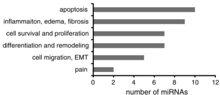
number of miRNAs
0 2 4 6 8 10 12
pain cell migration, EMT differentiation and remodeling cell survival and proliferation inflammaiton, edema, fibrosis apoptosis
Fig. 3 miRNA functions. Number of miRNAs, altered in BPS and associated with a specific function
miR-95 expression is up-regulated in many tumors.
Mechanistic studies revealed that miR-95 repressed the
expression of sorting nexin 1 (SNX1), whereas miR-95
silencing up-regulated SNX1 expression [
129
].
miR-130b targets and inhibits transcription factor ZEB1,
suppressing EMT transition. Its transcription depends on
p53, and repressed expression of miR-130b triggered
ZEB1-dependent EMT [
130
].
miR-148a is down-regulated in BPS. Rho-associated
coiled-coil containing protein kinase 1 (ROCK1) is one
of its main targets. In skeletal muscle, increase of
miR-148a helps myogenic differentiation through inhibition of
ROCK1 [
131
], and in cancer cells it leads to suppressed
tumor cell invasion and metastasis [
132
]. Similarly,
silenc-ing miR-148a in cancerous fibroblasts stimulated cell
motil-ity by de-repressing its targets WNT1 and WNT10B and
activation of WNT/β-catenin pathway [
133
].
miR-186 was shown to target P2X7 Ca channel,
impli-cated in cell survival and apoptosis [
134
].
miR-192 is highly expressed in the kidney epithelial
cells, where it controls TGF-β-induced collagen 1 a2
expres-sion via down-regulating Smad-interacting protein SIP1, a
E-box repressor [
135
]. In a separate study, it was shown to
increase collagen expression by targeting the E-box
repres-sors Zeb1/2 [
136
]. Both studies allow concluding that
miR-192 promotes fibrosis.
miR-199a-5p is an important regulator of intercellular
junctions. Upon overexpression in urothelial cells, it impairs
correct tight junction formation and leads to increased
per-meability. MiR-199a-5p directly targets mRNAs
encod-ing LIN7C, ARHGAP12, PALS1, RND1 and PVRL1 and
attenuates their expression levels to a similar extent. It is
predominantly expressed in the bladder smooth muscle, but
also detected in the mature bladder urothelium and primary
urothelial cultures [
14
].
MiR-199a-5p is up-regulated in cardiac hypertrophy
and its overexpression in cardiomyocytes leads to cell-size
increase [
137
]. A separate study claims that in cultured cell
lines, the expression of miR-199a and miR-199a*
(miR-199a/a*) is confined to fibroblasts [
138
].
miR-320a has anti-proliferative effect, which it exerts by
targeting β-catenin [
139
], neuropilin 1 (NRP-1), which is a
co-receptor of vascular epithelial growth factor [
140
], and
aquaporins 1 and 4 [
141
]. We have shown that miR-320a
down-regulates tachykinin NK1 receptor [
13
].
miR-324-3p is implicated in the pathogenesis of renal
fibrosis. A predicted target of miR-324-3p is prolyl
endo-peptidase. In cultured tubular cells, transient transfection
with a miR-324-3p mimic increased deposition of collagen
[
142
].
miR-328 is regulating the NK1R expression levels [
13
],
and also has a role in smooth muscle remodeling by
target-ing L-type calcium channel-alpha1C and the insulin growth
factor 1 receptor expression, ultimately leading to apoptosis
of pulmonary arterial smooth muscle cells [
143
].
miR-378 is a cardioabundant microRNA that targets
IGF1R. In tissues such as fibroblasts and fetal hearts, where
IGF1 levels are high, there were either absent or
signifi-cantly low miR-378 levels, suggesting an inverse
relation-ship between these two factors [
144
]. miR-449b, in addition
to influencing NK1R expression, targets and inhibits
onco-genic CDK6 and CDC25A, resulting in cell-cycle arrest
[
145
].
miR-493 is down-regulated in BPS. Interestingly, it has
been implicated in regulation of bladder cancer
tumorigene-sis: expression of miR-493 in bladder cancer (T24, J82, and
TCCSUP) cells and tissues was down-regulated. miR-493
decreased cell growth and migration by reducing the protein
expression of FZD4 and RhoC [
146
]. IGF1R was
identi-fied as a direct target of miR-493, and its inhibition partially
mimicked the anti-metastatic effects [
147
].
miR-550* was identified by us as a regulator of NK1R
expression. Recently, it was shown to be specific for
plas-macytoid dendritic cells (pDC) and monocytes [
85
].
miR-511 is an important regulator of immune cell function in
dendritic cells and macrophages: inhibition of the two
most highly up-regulated miRNAs, miR-511 and miR-99b,
resulted in reduced lower DC-specific intercellular adhesion
molecule-3-grabbing non-integrin (DC-SIGN) level.
Pre-diction of miRNA-511 targets revealed a number of genes
with known immune functions, of which TLR4 and CD80
were validated. Interestingly, under the cell-cycle arrest
conditions, miR-511 seems to function as a positive
regula-tor of TLR4 [
84
].
Conclusions and future directions
Here we conducted a comparative analysis of the miRNA
expression profile in BPS, BCa, and several inflammatory
disorders, and summarized the validated functional
infor-mation on the differentially expressed miRNAs. Despite the
existence of several miRNA profiling studies in BCa, we
found strikingly little overlap between the similarly
regu-lated miRNA species in BCa and BPS.
On the other hand, seven out of 31 miRNA, altered in
BPS showed the same regulatory pattern in IBD (Table
1
,
Fig.
2
). IBD shares many features with BPS,
includ-ing inflammation, pain, smooth muscle remodelinclud-ing, and
changes in epithelial permeability [
97
]. Disruption of
epi-thelial barrier function was identified as one of the
patho-logic mechanisms in IBD, and miR-199a-5p, which we
have recently characterized as a major regulator of
urothe-lial tight and adherens junctions [
14
], is also up-regulated
in IBD patients (Table
1
). Similarly, fibrosis is a common
feature of an advanced inflammatory disease and has been
described in both IBD [
148
] and BPS [
149
]. Interestingly,
miR-192, up-regulated in both disorders (Table
1
), has
vali-dated targets whose repression leads to increased fibrosis
(Table
2
). Based on this information, it would be interesting
to take a closer look at these miRNAs in order to determine
their therapeutic potential.
We found less overlap between BPS miRNAs and
miR-NAs de-regulated in the other inflammatory and
autoim-mune diseases, with the exception of asthma, which shared
four up-regulated miRNAs. All of them (miR-23a, -23b, -25,
and -26a) have been implicated in smooth muscle
remod-eling, and some have validated targets among the proteins
involved in the regulation of muscle growth and
differen-tiation (Table
2
). The majority of BPS patients enrolled in
our study had low-volume thick-walled bladders [
13
], and
it would be tempting to speculate on the role of these four
elevated miRNA in SMC proliferation during BPS.
Some of the functions of the miRNAs altered in BPS
could be deduced based on the information about their
vali-dated protein targets identified in other cell systems (Fig.
3
).
Due to the pleiotropic nature of miRNAs, the same miRNA
is often implicated in the regulation of opposing processes,
i.e., promoting both cell proliferation and apoptosis. Based
on the data in Table
2
, we grouped the miRNAs according
to their functions (Fig.
3
). Most of miRNAs, altered in BPS
(ten out of 31) have validated targets whose down-regulation
results in increased apoptosis. These findings fit well with
the emerging data demonstrating increased apoptosis in
BPS [
77
]. The second prominent group was miRNAs
influ-encing inflammation, edema, and fibrosis (nine out of 31).
We also identified several miRNAs whose targets include
regulators of cell proliferation and differentiation. It would
be interesting to determine which bladder layers harbors the
miRNAs belonging to these functionally opposing groups,
in order to evaluate the differential effects of inflammation
on the bladder urothelium and smooth muscle.
MicroRNA research is an exciting and challenging field,
and we are witness to its burgeoning. Recent years have
brought about a flood of publications containing both the
expression profiling and target validation data. Bringing
these results together yields helpful insights into the miRNA
function in human diseases and their potential therapeutic
applications.
Acknowledgments We gratefully acknowledge the financial support of the Swiss National Science Foundation (SNF Grant 320030_135783/1 to K. Monastyrskaya).
References
1. Bartel DP (2009) MicroRNAs: target recognition and regulatory functions. Cell 136(2):215–233. doi:10.1016/j.cell.2009.01.002
2. Farazi TA, Spitzer JI, Morozov P, Tuschl T (2011) miRNAs in human cancer. J Pathol 223(2):102–115. doi:10.1002/path.2806 3. Lagos-Quintana M, Rauhut R, Lendeckel W, Tuschl T (2001)
Identification of novel genes coding for small expressed RNAs. Science 294(5543):853–858. doi:10.1126/science.1064921 4. Orom UA, Nielsen FC, Lund AH (2008) MicroRNA-10a binds the
5′UTR of ribosomal protein mRNAs and enhances their
transla-tion. Mol Cell 30(4):460–471. doi:10.1016/j.molcel.2008.05.001 5. Henke JI, Goergen D, Zheng J, Song Y, Schuttler CG, Fehr C,
Junemann C, Niepmann M (2008) microRNA-122 stimulates translation of hepatitis C virus RNA. EMBO J 27(24):3300– 3310. doi:10.1038/emboj.2008.244
6. Tay Y, Zhang J, Thomson AM, Lim B, Rigoutsos I (2008) MicroRNAs to Nanog, Oct4 and Sox2 coding regions modulate embryonic stem cell differentiation. Nature 455(7216):1124– 1128. doi:10.1038/nature07299
7. Huang S, Wu S, Ding J, Lin J, Wei L, Gu J, He X (2010) Micro-RNA-181a modulates gene expression of zinc finger family members by directly targeting their coding regions. Nucleic Acids Res 38(20):7211–7218. doi:10.1093/nar/gkq564
8. Duursma AM, Kedde M, Schrier M, le Sage C, Agami R (2008) miR-148 targets human DNMT3b protein coding region. RNA 14(5):872–877. doi:10.1261/rna.972008
9. Thomson DW, Bracken CP, Goodall GJ (2011) Experimental strategies for microRNA target identification. Nucleic Acids Res 39(16):6845–6853. doi:10.1093/nar/gkr330
10. Lee RC, Feinbaum RL, Ambros V (1993) The C. elegans hetero-chronic gene lin-4 encodes small RNAs with antisense comple-mentarity to lin-14. Cell 75(5):843–854
11. Lu M, Zhang Q, Deng M, Miao J, Guo Y, Gao W, Cui Q (2008) An analysis of human microRNA and disease associations. PLoS ONE 3(10):e3420. doi:10.1371/journal.pone.0003420 12. Wilmott JS, Zhang XD, Hersey P, Scolyer RA (2011) The
emerg-ing important role of microRNAs in the pathogenesis, diagnosis and treatment of human cancers. Pathology 43(6):657–671. doi:10.1097/PAT.0b013e32834a7358
13. Sanchez Freire V, Burkhard FC, Kessler TM, Kuhn A, Drae-ger A, Monastyrskaya K (2010) MicroRNAs may mediate the down-regulation of neurokinin-1 receptor in chronic bladder pain syndrome. Am J Pathol 176(1):288–303. doi:10.2353/ajp ath.2010.090552
14. Monastyrskaya K, Sanchez-Freire V, Gheinani AH, Klumpp DJ, Babiychuk EB, Draeger A, Burkhard FC (2012) miR-199a-5p regulates urothelial permeability and may play a role in bladder pain syndrome. Am J Pathol. doi:10.1016/j.ajpath.2012.10.020 15. van de Merwe JP (2007) Interstitial cystitis and systemic
autoim-mune diseases. Nat Clin Pract Urol 4(9):484–491. doi:10.1038/n cpuro0874
16. Reinhart BJ, Slack FJ, Basson M, Pasquinelli AE, Bettinger JC, Rougvie AE, Horvitz HR, Ruvkun G (2000) The 21-nucleotide let-7 RNA regulates developmental timing in Caenorhabditis
elegans. Nature 403(6772):901–906. doi:10.1038/35002607 17. Vasudevan S, Tong Y, Steitz JA (2007) Switching from
repres-sion to activation: microRNAs can up-regulate translation. Sci-ence 318(5858):1931–1934. doi:10.1126/science.1149460 18. Place RF, Li LC, Pookot D, Noonan EJ, Dahiya R (2008)
Micro-RNA-373 induces expression of genes with complementary pro-moter sequences. Proc Nat Acad Sci USA 105(5):1608–1613. doi:10.1073/pnas.0707594105
19. Calin GA, Dumitru CD, Shimizu M, Bichi R, Zupo S, Noch E, Aldler H, Rattan S, Keating M, Rai K, Rassenti L, Kipps T, Negrini M, Bullrich F, Croce CM (2002) Frequent deletions and down-regulation of micro- RNA genes miR15 and miR16 at 13q14 in chronic lymphocytic leukemia. Proc Nat Acad Sci USA 99(24):15524–15529. doi:10.1073/pnas.242606799
20. Lu J, Getz G, Miska EA, Alvarez-Saavedra E, Lamb J, Peck D, Sweet-Cordero A, Ebert BL, Mak RH, Ferrando AA, Downing JR, Jacks T, Horvitz HR, Golub TR (2005) MicroRNA expres-sion profiles classify human cancers. Nature 435(7043):834– 838. doi:10.1038/nature03702
21. van Rooij E, Sutherland LB, Liu N, Williams AH, McAnally J, Gerard RD, Richardson JA, Olson EN (2006) A signature pattern of stress-responsive microRNAs that can evoke cardiac hypertro-phy and heart failure. Proc Nat Acad Sci USA 103(48):18255– 18260. doi:10.1073/pnas.0608791103
22. Schaefer A, O’Carroll D, Tan CL, Hillman D, Sugimori M, Lli-nas R, Greengard P (2007) Cerebellar neurodegeneration in the absence of microRNAs. J Exp Med 204(7):1553–1558. doi:10.1 084/jem.20070823
23. Lukiw WJ (2007) Micro-RNA speciation in fetal, adult and Alz-heimer’s disease hippocampus. NeuroReport 18(3):297–300. doi:10.1097/WNR.0b013e3280148e8b
24. Kim J, Inoue K, Ishii J, Vanti WB, Voronov SV, Murchison E, Hannon G, Abeliovich A (2007) A MicroRNA feedback circuit in midbrain dopamine neurons. Science 317(5842):1220–1224. doi:10.1126/science.1140481
25. Sonkoly E, Wei T, Janson PC, Saaf A, Lundeberg L, Teng-vall-Linder M, Norstedt G, Alenius H, Homey B, Scheynius A, Stahle M, Pivarcsi A (2007) MicroRNAs: novel regulators involved in the pathogenesis of psoriasis? PLoS ONE 2(7):e610. doi:10.1371/journal.pone.0000610
26. Dai Y, Huang YS, Tang M, Lv TY, Hu CX, Tan YH, Xu ZM, Yin YB (2007) Microarray analysis of microRNA expression in peripheral blood cells of systemic lupus erythematosus patients. Lupus 16(12):939–946. doi:10.1177/0961203307084158 27. Kota J, Chivukula RR, O’Donnell KA, Wentzel EA,
Montgom-ery CL, Hwang HW, Chang TC, Vivekanandan P, Torbenson M, Clark KR, Mendell JR, Mendell JT (2009) Therapeutic microRNA delivery suppresses tumorigenesis in a murine liver cancer model. Cell 137(6):1005–1017. doi:10.1016/ j.cell.2009.04.021
28. Krutzfeldt J, Rajewsky N, Braich R, Rajeev KG, Tuschl T, Manoharan M, Stoffel M (2005) Silencing of microRNAs in vivo with ‘antagomirs’. Nature 438(7068):685–689. doi:10.1038/ nature04303
29. Lanford RE, Hildebrandt-Eriksen ES, Petri A, Persson R, Lin-dow M, Munk ME, Kauppinen S, Orum H (2010) Therapeutic silencing of microRNA-122 in primates with chronic hepatitis C virus infection. Science 327(5962):198–201. doi:10.1126/ science.1178178
30. Carthew RW, Sontheimer EJ (2009) Origins and mechanisms of miRNAs and siRNAs. Cell 136(4):642–655. doi:10.1016/j. cell.2009.01.035
31. Chekulaeva M, Filipowicz W (2009) Mechanisms of miRNA-mediated post-transcriptional regulation in animal cells. Curr Opin Cell Biol 21(3):452–460. doi:10.1016/j.ceb.2009.04.009 32. Kim VN, Han J, Siomi MC (2009) Biogenesis of small
RNAs in animals. Nat Rev Mol Cell Biol 10(2):126–139. doi:10.1038/nrm2632
33. Rodriguez A, Griffiths-Jones S, Ashurst JL, Bradley A (2004) Identification of mammalian microRNA host genes and tran-scription units. Genome Res 14(10A):1902–1910. doi:10.1101 /gr.2722704
34. Gregory RI, Yan KP, Amuthan G, Chendrimada T, Doratotaj B, Cooch N, Shiekhattar R (2004) The Microprocessor complex mediates the genesis of microRNAs. Nature 432(7014):235– 240. doi:10.1038/nature03120
35. Wang X, Xu X, Ma Z, Huo Y, Xiao Z, Li Y, Wang Y (2011) Dynamic mechanisms for pre-miRNA binding and export by Exportin-5. RNA 17(8):1511–1528. doi:10.1261/rna.2732611
36. MacRae IJ, Zhou K, Doudna JA (2007) Structural determinants of RNA recognition and cleavage by Dicer. Nat Struct Mol Biol 14(10):934–940. doi:10.1038/nsmb1293
37. Okamura K, Hagen JW, Duan H, Tyler DM, Lai EC (2007) The mirtron pathway generates microRNA-class regulatory RNAs in Drosophila. Cell 130(1):89–100. doi:10.1016/j.cell.2007.06.028 38. Berezikov E, Chung WJ, Willis J, Cuppen E, Lai EC (2007)
Mammalian mirtron genes. Mol Cell 28(2):328–336. doi:10.1016/j.molcel.2007.09.028
39. Ruby JG, Jan CH, Bartel DP (2007) Intronic microRNA precur-sors that bypass Drosha processing. Nature 448 (7149):83-86. http://www.nature.com/nature/journal/v448/n7149/suppinfo/ nature 05983S1.html
40. Havens MA, Reich AA, Duelli DM, Hastings ML (2012) Bio-genesis of mammalian microRNAs by a non-canonical process-ing pathway. Nucleic Acids Res. doi:10.1093/nar/gks026 41. Kozomara A, Griffiths-Jones S (2011) miRBase: integrating
microRNA annotation and deep-sequencing data. Nucleic Acids Res 39(Database issue):D152–D157. doi:10.1093/nar/gkq1027 42. Griffiths-Jones S (2004) The microRNA Registry. Nucleic Acids
Res 32(suppl 1):D109–D111. doi:10.1093/nar/gkh023
43. van de Merwe JP, Nordling J, Bouchelouche P, Bouchelouche K, Cervigni M, Daha LK, Elneil S, Fall M, Hohlbrugger G, Irwin P, Mortensen S, van Ophoven A, Osborne JL, Peeker R, Richter B, Riedl C, Sairanen J, Tinzl M, Wyndaele JJ (2008) Diagnos-tic criteria, classification, and nomenclature for painful blad-der syndrome/interstitial cystitis: an ESSIC proposal. Eur Urol 53(1):60–67
44. Curhan GC, Speizer FE, Hunter DJ, Curhan SG, Stampfer MJ (1999) Epidemiology of interstitial cystitis: a population based study. J Urol 161(2):549–552
45. Fall M, Oberpenning F, Peeker R (2008) Treatment of bladder pain syndrome/interstitial cystitis 2008: can we make evidence-based decisions? Eur Urol 54(1):65–75
46. Lilly JD, Parsons CL (1990) Bladder surface glycosaminogly-cans is a human epithelial permeability barrier. Surg Gynecol Obstet 171(6):493–496
47. Moskowitz MO, Byrne DS, Callahan HJ, Parsons CL, Valder-rama E, Moldwin RM (1994) Decreased expression of a glyco-protein component of bladder surface mucin (GP1) in interstitial cystitis. J Urol 151(2):343–345
48. Birder LA, de Groat WC (2007) Mechanisms of disease: involvement of the urothelium in bladder dysfunction. Nat Clin Pract Urol 4(1):46–54. doi:10.1038/ncpuro0672
49. Parsons CL (2007) The role of the urinary epithelium in the pathogenesis of interstitial cystitis/prostatitis/urethritis. Urology 69(4 Suppl):9–16
50. Tomaszewski JE, Landis JR, Russack V, Williams TM, Wang LP, Hardy C, Brensinger C, Matthews YL, Abele ST, Kusek JW, Nyberg LM (2001) Biopsy features are associated with primary symptoms in interstitial cystitis: results from the interstitial cys-titis database study. Urology 57(6 Suppl 1):67–81
51. Slobodov G, Feloney M, Gran C, Kyker KD, Hurst RE, Culkin DJ (2004) Abnormal expression of molecular markers for blad-der impermeability and differentiation in the urothelium of patients with interstitial cystitis. J Urol 171(4):1554–1558 52. Zhang CO, Wang JY, Koch KR, Keay S (2005) Regulation of
tight junction proteins and bladder epithelial paracellular perme-ability by an antiproliferative factor from patients with intersti-tial cystitis. J Urol 174(6):2382–2387
53. Liu HT, Shie JH, Chen SH, Wang YS, Kuo HC (2012) Dif-ferences in mast cell infiltration, E-cadherin, and zonula occludens-1 expression between patients with overactive blad-der and interstitial cystitis/bladblad-der pain syndrome. Urology 80 (1):225 e213-228. doi:10.1016/j.urology.2012.01.047