A BIOCOMPATIBLE, LOCAL DRUG DELIVERY
PLATFORM FOR THE CHRONIC TREATMENT OF
NEUROLOGICAL DISORDERS OF THE BRAIN
by
MASSACI
Kevin C. Spencer OF
B.S. Materials Science and Engineering
LI
University of Illinois - Urbana Champaign, 2010LI
HUSETTS IN7IITUTE TECHN0 0Ly
R 2R
I3RARIES
ARCHIVES
SUBMITTED TO THE DEPARTMENT OF MATERIALS SCIENCE AND ENGINEERINGIN PARTIAL FULFILLMENT OF THE REQUIREMENTS FOR THE DEGREE OF DOCTOR OF PHILOSOPHY IN MATERIALS SCIENCE AND ENGINEERING AT THE
MASSACHUSSETTS INSTITUTE OF TECHNOLOGY FEBRUARY 2017
2017 Massachusetts Institute of Technology. All rights reserved.
Signature of Author: Kevin Spencer
Signature redacted
Deparment of Materials Science and Engineering December 2 0th 1 2016
Certified by:
(Sig
nature redacted
Michael J. Cima
Department of Materials Science and Engineering Thesis Supervisor
Signature redacted
Accepted by:
Donald R. Sadoway /
John F. Elliott Professor of Kiaterials Chemistry Chair, Departmental Committee on Graduate St ents
A BIOCOMPATIBLE, LOCAL DRUG DELIVERY PLATFORM FOR THE CHRONIC TREATMENT OF NEUROLOGICAL DISORDERS OF THE BRAIN
By Kevin C Spencer
Submitted to the Department of Materials Science and Engineering on December 20,
2016 in Partial Fulfilment of the Requirements for the Degree of Doctor of Philosophy
in Materials Science and Engineering
ABSTRACT
Many neurological disorders are now classified as circuit disorders, in which the underlying pathology arises from a failure in dynamic communication between anatomically distinct regions of the brain. Systemic therapies are often not effective due to their untargeted nature. The injectrode is a multifunctional probe designed to treat neurological disorders through targeted chemical and electrical stimulation directly to a focal point within the implicated neural circuit. This thesis details the characterization and biocompatibility of the injectrode for the treatment of neurological disorders on chronic timescales.
In vitro and in vivo infusion tests were conducted to validate the ability to deliver nanoliter scale volumes (10-1000 n1) of drug to targeted brain structures over the course of an eight week implantation period. Muscimol was delivered to deep brain structures to demonstrate effective modulation of neural activity and behavior. These findings highlight the utility of a local chemical delivery approach to treat circuit diseases of the brain.
Glial scar is a major barrier to neural probe function. A main objective of this thesis is focused on understanding the process of glial scar formation from a materials perspective. Micromotion and mechanical mismatch are thought to be key drivers of scar formation. This hypothesis was investigated using a novel 3D in vitro glial scar model, which replicates the magnitude and frequency of micromotions that are observed in vivo. Astrocytes were found to have a significant increase in cellular area and perimeter in response to micromotion compared to static control wells.
These findings were applied to improve the biocompatibility of the injectrode. Hydrogel coatings, with moduli matched to brain tissue, were formed to mitigate the effects of micromotion. These coatings were found to reduce local strain by up to 70%. In vivo studies were conducted to explore the impact that implant diameter and modulus have on scar formation. Hydrogel coated implants
(E=1 1.6 kPa) were found to significantly reduce scarring at 8 weeks post implantation, compared
to uncoated implants (E=70 GPa). Size effects from increasing the overall implant diameter were also observed, highlighting the importance of considering both mechanical and geometric factors when designing chronic neural implants.
Thesis Supervisor: Michael J. Cima
ACKNOWLEDGEMENTS
There are a countless number of individuals whose help and encouragement have made this thesis possible -I'd like to thank many of them here.
First of all, I'd like to thank my fantastic advisor, Michael Cima, for providing me with the opportunity of working in his lab for the past six years. Working in the Cima lab has
been a wonderful privilege, and it amazes me to think of how much I've grown, both as a scientist and as a person throughout this time. Michael provided me with both the
freedom to investigate the problems that I found interesting, as well as the tools and resources to efficiently and effectively conduct research. Michael's excitement and valuable outlook on how our work may translate to significant clinical impacts has kept me motivated throughout the long, tortuous journey of graduate school.
I am also incredibly grateful for the mentorship of Polina Anikeeva and Darrell Irvine
for the valuable guidance they have provided on my thesis committee. They have consistently provided valuable insight and great support throughout my time at MIT. I will always be appreciative of the numerous discussions during my early years at MIT, which significantly shaped the directions of my project. There is no doubt that their contributions significantly improved the outcome of my thesis.
In graduate school, you tend to spend the majority of time in lab, which quickly starts to feel like your home. I feel extremely lucky to have had the opportunity to work and interact with so many wonderful people throughout my time in graduate school. I'd like to thank all Cima lab members, current and former, for making the time spent in the lab so great. The constant lunch and trivia outings were always highlights of the day, and I've formed many lifelong friendships in lab.
foundation for this project, and Jay for basically training me in every lab technique that
I know. I'd like to thank Professor Ann Graybiel and Professor Robert Langer for their
valuable insight and suggestions provided in team meetings over the past several years. The current injectrode team is pretty great too - it has been a joy working so closely with Khalil the past couple of years. Having someone to constantly brainstorm ideas with helps make even the tallest tasks seem more achievable. I'd like to thank Helen, Canan, Pauline, and everyone in the Graybiel lab for an exciting and productive last few years as well. I'm excited to see where the project goes next! I've always been amazed at how helpful and collaborative people have been at MIT. Special thanks to Sebastien Delcasso and Howard Mak who have not only been great collaborators over the past year, but are also extremely enjoyable people to work with.
I'd also like to give a special shout out to all my friends at MIT, who have made the
hours spent outside of lab very enjoyable. My roommates Daniel, Audren, Ryan, as well as the unofficial roommate Carl, are some of the best dudes in town, and we've
certainly had our share of great times over the years. It has been great making the transitions from undergrads to full grown "adults" with these guys. Their energy and fantastic work has certainly kept me motivated over the years as well.
Thank you to my parents for making me the person that I am today. They certainly instilled their hard work ethic in me, and I am so appreciative for all that they have provided me. I would not have been able to get to this point without their unwavering support and encouragement at every point along the way. My younger brother Jack has always been an inspiration for me - it's been great growing up with such a positive and supportive brother. I don't think I've ever heard of someone not getting along with Jack.
I wish we saw each other more than a few times per year, but hopefully that'll change
Last but certainly not least, there's Beth. Beth is the most wonderful lady I've ever known, such a kind, talented, and compassionate person, and I'm so grateful to have her in my life. After finishing up her graduate work back home in Illinois, she quickly realized that I still had quite some time left. She picked up everything and came out to start a life in Boston, just to be with me. Having her by my side throughout the many highs and lows of the PhD was so important, I am eternally grateful it. As a result of our many discussions about my experiments, I highly doubt there are any architects in the world as knowledgeable in the process of glial scar formation as Beth. Can't wait to see where life takes us next!
TABLE OF CONTENTS
ACKNOWLEDGEMENTS...3
TABLE OF CONTENTS...6
LIST OF TABLES...9
LIST OF FIGURES... 10
LIST OF ABBREVIATIONS AND ACRONYMS... 13
LIST OF APPENDICES... 15
1 INTRODUCTION...16
1.1 CIRCUIT DISORDERS OF THE BRAIN: A SHIFT IN THE THERAPEUTIC TARGET FOR NEUROLOGICAL DISORDERS... 16
1.1.1 C linical R ationale: ... 16
1.1.2 Pathology of Neurological Disorders from a Circuit Perspective... 17
1.2 CURRENT STANDARDS OF CARE... 21
1.2.1 System ic D elivery ... 21
1.2.2 Convection Enhanced Delivery... 24
1.2.3 Local Electrical Modulation (Deep Brain Stimulation, DREADDs, O p togenetics) ... 25
1.3 THE NEED FOR IMPROVED LOCAL CHEMICAL DELIVERY FOR THE TREATMENT OF NEUROLOGICAL DISORDERS...27
1.4 THE INJECTRODE: COMBINED LOCAL CHEMICAL AND ELECTRICAL STIMULATION FOR THE CHRONIC TREATMENT OF NEUROLOGICAL DISORDERS... 29
1.5 THESIS STRUCTURE ... 32
2 IN VITRO AND IN VIVO CHARACTERIZATION OF THE INJECTRODE D EV IC E ... 34
2.1 INTRODUCTION AND MOTIVATION: ... 34
2 .2 M ETH O D S: ... 35
2.2.1 M aterials... . . 36
2.2.2 D evice Fabrication:... 36
2.2.3 In Vitro Device Characterization:... 39
2.2.4 Agarose Phantom Infusion... 41
2.2.5 Acute In Vivo Infusion and Diffusion Analysis via Integrative Optical Im ag ing ... . . 4 1 2.2.6 Acute Electrophysiology Experiments ... 43
2.2.7 Device Chronic Implantation Procedure: ... 44
2.2.8 Chronic Impedance Spectroscopy... 45
2.2.9 PET Imaging Studies ... 45
2.2.10 PEG-NOTA Preparation and Cu-64 Labeling... 47
2.2.11 Rodent Behavioral Studies following Unilateral Muscimol Infusion... 48
2.3 RESULTS AND D ISCUSSION ... 49
2.3.1 In Vitro Infusion ... 49
2.3.2 Agarose Phantom Infusion... 54
2.3.3 In Vivo Acute Infusion and Diffusion Measurement ... 55
2.3.4 Acute Neural Modulation through Local Infusion of Muscimol... 57
2.3.7 Rodent Behavioral Study ... 69
2.4 CONCLUSION ... 71
3 THREE DIMENSIONAL IN VITRO GLIAL SCAR MODEL TO PROBE THE EFFECTS OF MICROMOTION AROUND NEURAL IMPLANTS... 73
3.1 INTRODUCTION AND BACKGROUND ... 73
3.2 M ATERIALS AND M ETHODS ... 76
3.2.1 M aterials... 76
3.2.2 Poly-D-Lysine Coating... 77
3.2.3 Glial Cell Isolation... 77
3.2.4 3-D cell culture formation. ... 78
3.2.5 Live Dead Cell Staining... 79
3.2.6 M icromotion Apparatus Construction... 79
3.2.7 Strain Field M easurement ... 80
3.2.8 In Vitro Glial Scar Experiment ... 80
3.2.9 Immunohistochemical Analysis... 82
3.2.10 Confocal M icroscopy... 82
3.2.11 Image Analysis ... 83
3.2.12 Statistical Analysis ... 83
3.3 RESULTS: ... 84
3.3.1 M icromotion Equipment Calibration... 84
3.3.2 In Vitro Strain Field M easurements: ... 86
3.3.3 Live Dead Cell Staining... 87
3.3.4 Immunohistochemical Analysis... 89
3.4 DISCUSSION ... 91
3.5 CONCLUSION ... 94
4 MECHANICALLY MATCHED HYDROGEL TO REDUCE SCARRING ARO UND NEURAL IM PLANTS... 95
4.1 INTRODUCTION AND BACKGROUND ... 95
4.2 M ATERIALS AND M ETHODS ... 99
4.2.1 M aterials... 99
4.2.2 Synthesis of PEG-Dimethacrylate... 99
4.2.3 Formation of PEG Hydrogel on Glass Capillaries ... 100
4.2.4 M echanical Characterization of PEG Hydrogels... 101
4.2.5 In Vitro Strain Field M easurements... 103
4.2.6 Device Implantation in Rodent Brain... 103
4.2.7 Animal Euthanasia and Tissue Harvesting ... 105
4.2.8 Immunohistochemistry... 105
4.2.9 Imaging and Data Analysis ... 106
4.2.10 Statistical Analysis ... 107
4.3 RESULTS... 107
4.3.1 Hydrogel Formation and Characterization... 107
4.3.2 In Vitro Strain Field Reduction... 110
4.3.3 In Vivo Animal Study Results... 114
4.3.4 D iscussion... 126
4.4 CONCLUSION ... 133
5 CONCLU SIONS AND FUTURE W ORK ... 135
5.2.1 Device Performance in Animal Disease Models... 137
5.2.2 PET Imaging to Assess Glial Scar Characteristics: ... 141
5.2.3 Biocom patibility Studies ... 142
6 REFERENCES ... 148
7 APPENDICES... 160
APPENDIX A. MICROMOTION EQUIPMENT SETUP: ... 161
LIST OF TABLES
TABLE 1-1 DESIGN FEATURES OF THE INJECTRODE PLATFORM ... 31
TABLE 2-1 QUANTIFIED INFUSION PARAMETERS FROM ACUTE IN VIVO INFUSION ... 56
TABLE 2-2 SUMMARY OF MEASURED INFUSION VOLUMES FOR CU64 INFUSION
E XPERIM ENTS... 66 TABLE 2-3 CU64 LABELING EFFICIENCY OF 4-ARM PEG-NOTA... 68
TABLE 3-1. EXAMPLE EXPERIMENTAL SETUP FOR IN VITRO GLIAL SCAR EXPERIMENT.. 82
LIST OF FIGURES
FIGURE 1-I SCHEMATIC DIAGRAM OF THE BASAL GANGLIA CIRCUIT IN THE BRAIN...20
FIGURE 1-2 THE BLOOD BRAIN BARRIER... 23
FIGURE 1-3 CONVECTION ENHANCED DELIVERY TO THE BRAIN ... 25
FIGURE 1-4 SEM MICROGRAPH OF THE INJECTRODE DEVICE ... 31
FIGURE 2-1 SCHEMATIC OF INJECTRODE DEVICE FABRICATION PROCEDURE ... 39
FIGURE 2-2 FOUR ARM PEG THIOL USED IN CU64 LABELING STUDIES ... 48
FIGURE 2-3 IN VITRO INFUSION DATA ... 50
FIGURE 2-4 MUSCIMOL IN VITRO INFUSION EXPERIMENT ... 52
FIGURE 2-5 MUSCIMOL INFUSION DELAY EXPERIMENT... 52
FIGURE 2-6 IN VITRO WIRELESS PUMP CHARACTERIZATION ... 53
FIGURE 2-7 IN VITRO BRAIN PHANTOM INFUSION... 55
FIGURE 2-8 ACUTE IN VIVO INFUSION OF ICG INTO THE RODENT STRIATUM... 56
FIGURE 2-9 INTEGRATIVE OPTICAL IMAGING TO QUANTIFY DIFFUSION IN THE BRAIN ... 57
FIGURE 2-10 LOCAL NEURAL ACTIVITY SILENCING FOLLOWING LOCAL INFUSION OF M U SC IM O L ... 58
FIGURE 2-11 SALINE CONTROL INFUSION EXPERIMENT ... 59
FIGURE 2-12 SPIKE SORTING AND ISOLATED WAVEFORMS OF LOCAL NEURAL ACTIVITY BEFORE AND AFTER SALINE INFUSION... 59
FIGURE 2-13 IMPEDANCE SPECTROSCOPY RESULTS ... 62
FIGURE 2-14 NYQUIST PLOT BEHAVIOR AT ACUTE AND CHRONIC TIMEPOINTS ... 62
FIGURE 2-15 IN VITRO SALINE IMPEDANCE MEASUREMENTS ... 63
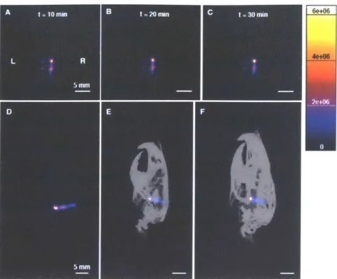
FIGURE 2-16 PRELIMINARY PET/CT IMAGING EXPERIMENT TO IDENTIFY BRAIN LOCATION IN THE FIELD OF VIEW. ... 64
FIGURE 2-17 PET IMAGING OF 1.0 ML INFUSION OF CU64 INTO THE RODENT BRAIN...65
FIGURE 2-18 PET IMAGING OF 167 NL INFUSION INTO THE SN. ... 65
FIGURE 2-19 LONGITUDINAL ASSESSMENT OF DEVICE FUNCTION ... 67
FIGURE 2-20 REPRESENTATIVE INFUSION PROFILE FOR PET-NOTA INFUSION IN THE R ODENT B RA IN ... 68
FIGURE 2-21 PEG-NOTA INFUSION SITE ROI ANALYSIS ... 69
FIGURE 2-22 RODENT BEHAVIORAL STUDY RESULTS ... 70
FIGURE 3-1 PHOTOGRAPH OF IN VITRO GLIAL SCAR MODEL EXPERIMENT IN INCUBATOR ... 8 1 FIGURE 3-2 MICROMOTION SIMULATION DEVICE... 84
FIGURE 3-4 SIDE TO SIDE CALIBRATION CURVE ... 85
FIGURE 3-5 STRAIN FIELD MAP AROUND DEVICE INSERTED INTO A COLLAGEN GEL... 86
FIGURE 3-6 LINE PROFILES FROM STRAIN FIELD MAPPING... 87
FIGURE 3-7 REPRESENTATIVE LIVE/DEAD STAIN AFTER ONE WEEK IN CULTURE ... 88
FIGURE 3-8 CALCULATED CELL VIABILITY FROM LIVE/DEAD STAIN... 88
FIGURE 3-9 CHARACTERISTIC CONFOCAL IMAGE OBTAINED FOLLOWING GLIAL SCAR E X PERIM ENT... 90
FIGURE 3-10 QUANTIFIED ASTROCYTE MORPHOLOGY FROM IMAGE ANALYSIS... 91
FIGURE 4-1 REACTION SCHEME TO FORM HYDROGEL COATINGS ON BOROSILICATE G LASS C APILLARIES ... 101
FIGURE 4-2 CHARACTERISTIC FORCE DISPLACEMENT CURVE FOR ELASTIC MODULUS M EA SUREM ENT ... 103
FIGURE 4-3 HYDROGEL COATING SWELLING KINETICS... 109
FIGURE 4-4 HYDROGEL MECHANICAL CHARACTERIZATION... 110
FIGURE 4-5 IN VITRO STRAIN FIELD MAGNITUDE MAPS FOR SIDE TO SIDE MICROMOTION ... 1 12 FIGURE 4-6 HYDROGEL STRAIN FIELD LINE PROFILES FOR SIDE TO SIDE MICROMOTION ... 1 12 FIGURE 4-7 IN VITRO STRAIN FIELD MAGNITUDE MAPS FOR AXIAL MICROMOTION... 113
FIGURE 4-8 HYDROGEL STRAIN FIELD LINE PROFILES FOR AXIAL MICROMOTION ... 113
FIGURE 4-9 EFFECT OF IMPLANT MODULUS ON SCARRING. ... 115
FIGURE 4-10 LINE PROFILE ANALYSIS FOR SIZE CONTROL HYDROGEL STUDY ... 116
FIGURE 4-11 GLASS CAPILLARY STUDY - ONE WEEK GFAP ANALYSIS ... 118
FIGURE 4-12 GLASS CAPILLARY STUDY - 4 WEEK GFAP ANALYSIS ... 118
FIGURE 4-13 REPRESENTATIVE IMAGES OF GFAP REACTIVITY AROUND GLASS CAPILLARY IMPLANTS AT 8 WEEKS POST IMPLANTATION ... 119
FIGURE 4-14 GLASS CAPILLARY STUDY -EIGHT WEEK GFAP ANALYSIS ... 119
FIGURE 4-15 EFFECT OF IMPLANT DIAMETER ON BLOOD BRAIN BARRIER PERMEABILITY AT EIGHT W EEKS POST IMPLANTATION ... 121
FIGURE 4-16 EFFECT OF IMPLANT DIAMETER ON ACTIVATED MACROPHAGE DENSITY AT EIGHT W EEKS POST IMPLANTATION ... 122
FIGURE 4-17 EFFECT OF IMPLANT DIAMETER ON NEURAL DENSITY AT EIGHT WEEKS POST IM PLANTATION. ... 123
FIGURE 4-18 HYDROGEL COATING STUDY - GFAP REACTIVITY AT EIGHT WEEKS POST IM PLAN TATION ... 124
FIGURE 4-19 HYDROGEL COATING STUDY -BLOOD BRAIN BARRIER PERMEABILITY AT EIGHT W EEKS POST IMPLANTATION ... 125
FIGURE 4-20 HYDROGEL COATING STUDY - ACTIVATED MACROPHAGE DENSITY AT EIGHT W EEKS POST IMPLANTATION ... 125
FIGURE 4-21 HYDROGEL COATING STUDY - NEURAL BODY DENSITY AT EIGHT WEEKS POST IM PLANTATION ... 126 FIGURE 4-22 PEG-DMA HYDROGEL COATINGS FORMED BY SPRAY COATINGS AND DIP
C O A TIN G ... 12 8 FIGURE 4-23 AVERAGE IMPLANT DIAMETER FOR HYDROGEL COATED SAMPLES AT ONE,
FOUR, AND EIGHT W EEKS POST IMPLANTATION... 129
FIGURE 5-1 6-OHDA RODENT PARKINSON'S MODEL ... 139
FIGURE 5-2 INITIAL DEMONSTRATION OF NEURAL SILENCING IN NHP MODEL ... 140
FIGURE 5-3 SCHEMATIC DIAGRAM OF Two POSSIBLE HYDROGEL DRUG DELIVERY
M ECHAN ISM S ... 145 FIGURE 7-1 ELECTRICAL SCHEMATIC DIAGRAM FOR THE MICROMOTION MODEL ... 161
PD MDD TRD DBS L-dopa CNS BBB MTD CED GBM STN GPi DREADD IV IP DA PET CT NOTA PEG-DMA BS CNC HPLC PBS ICG II ECS ROI PCA GFAP PDL LPS
LIST OF ABBREVIATIONS AND ACRONYMS
Parkinson's disease Major Depressive Disorder Treatment resistant despression Deep Brain Stimulation
Levadopa
Central Nervous System Blood brain barrier Maximum tolerated dose Convection enhanced delivery Glioiblastoma multiforme Subthalamic Nucleus Global pallidus
Designer receptor exclusively activated by designer drugs Intravenous
Intraperitoneal Dopamine
Positron emission tomography Computerized tomography
1,4,7-triazacyclononane-1,4,7-trisacetic acid Polyethylene glycol dimethacrylate
Borosilicate
Computer numerical control
High pressure liquid chromatography Phosphate buffered saline
Indocyanine green
Integrative optical imaging Extracellular space
Region of interest
Principal component analysis Glial fibrillary acidic protein Poly-d-lysine
bFGF Basic fibroblast growth factor
PIV Particle image velocimetry
ROIs Reactive oxygen intermediates
FEA Finite element analysis
TPM 3-(Trichlorosilyl) propyl methacrylate
BF Bright field
IF Immunofluorescent
GC Glass capillary
6-OHDA 6-hydroxydopamine
LIST OF APPENDICES
1
INTRODUCTION
1.1 Circuit Disorders of the Brain: A Shift in the Therapeutic
Target for Neurological Disorders
1.1.1 Clinical Rationale:
Neurological disorders are among the most prevalent and debilitating diseases in the world, contributing to approximately 10% of the global disease burden (1). Many neurological disorders are severely debilitating and there are often no effective treatments available to large subsets of the patient populations.
One example is Parkinson's disease (PD). PD is the second most common
neurodegenerative disorder (behind Alzheimer's disease), affecting approximately 645,000 patients in the USA (2). Parkinson's disease affects approximately 0.3% of the total population, with incidence rates increasingly drastically beyond the age of 60 (2). PD is known for its very noticeable movement symptoms including resting tremor, bradykinesia (slowness or absence of movement), and muscle rigidity. Non-movement symptoms of PD include cognitive impairment, depression, and personality changes. PD is severely debilitating and carries a large cost of treatment. The cost burden of PD is estimated at $23 billion dollars annually, with approximately 50% of this cost due to productivity loss of the patient. While the origin of PD is still not fully understood, it is known that the symptoms occur as a result of the selective loss of dopaminergic neurons in the substantia nigra (SN). These neurons project to the striatum where they stimulate the release of dopamine to control movement (3). The loss of these neurons leads to less
dopamine in the striatum, disrupting the neural control of movement and behavior within the basal ganglia circuit (4).
Anxiety and mood disorders are another class of neurological disorder which are very common and extremely debilitating. Nearly half of all Americans will suffer from a psychiatric disorder in their lifetime (5), with the two most prevalent being anxiety disorders (29%) and major depressive disorder (MDD) (17%). MDD is ranked as the top contributor to years lived with disability, according to the World Health
Organization (6). A key reasons for this is that MDD is often resistant to current
treatments (7). It is estimated that three percent of Americans a year (up to 4 million) suffer from treatment resistant depression (TRD), in which traditional therapies provide no improvement to the patient's quality of life (8). TRD patients often fail several rounds of antidepressant drug therapies before being referred to electroconvulsive therapy. Patients with TRD are twice as likely to be hospitalized due to their illness compared to non-TRD patients. TRD patients also bear a 6 fold higher heath care cost per year as well (8). There is a clear need for improved treatment strategies for patients with TRD.
1.1.2 Pathology of Neurological Disorders from a Circuit Perspective
Recent efforts in neuroscience have greatly expanded our understanding of the underlying pathology of many of these neurological disorders. Neurons in the brain function within circuits to carry out brain functions such as the processing of information, controlling movement, and decision making (9). Recent advances in neuroscience imaging and recording technologies have identified abnormal activity across key neural circuits which underlies the pathology of many neurological
termed circuit disorders. Unlike conventional diseases in which the pathology is
localized to a certain tissue or cell type, the pathology of these circuit disorders exists at the systems level. The symptoms of these diseases arise as a result of failure in the dynamic communication between anatomically distinct areas of the brain (4). Many diseases have now been classified as circuit disorders including Parkinson's disease
(10), depression (4, 11), and obsessive compulsive disorder (12). In the case of
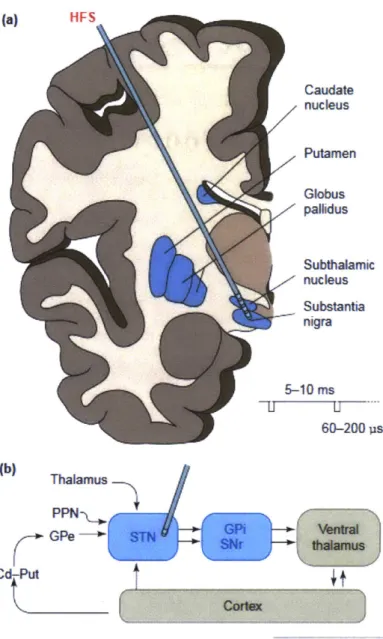
Parkinson's disease it has been suggested that many of the behavioral symptoms are explained by irregular neural activity in the basal ganglia-thalamocortical circuit (13). Figure 1-1 shows a schematic drawing of the basal-ganglia circuit that is implicated in PD. One can see that the regions implicated in the disease, are small brain structure which are physically separated from each other.
Recent advances in imaging and neural modulation technologies (e.g. fMRI, dMRI, optogenetics) have begun to further refine our understandings of these diseases, by resolving connections within individual brain structures, such as the striatum (14). This work has defined smaller regions referred to as microcircuits. Microcircuitry that is implicated in many disease has been identified including PD (15) and mood disorders (16).
The focal points within microcircuits have been described as regions with high density of neural connections, with dimensions ranging from 300 gm (15) to 1-2 mm in
diameter (17). This concept presents a major shift in the therapeutic target. In contrast to delivering drug to an entire pathological tissue or organ, as in cancer, the goal is to normalize abnormal activity across the malfunctioning circuit. This can be achieved by modulating neural activity via a stimuli at any link within the communication chain, not just the region of original deficiency. One example of such an approach is high
frequency deep brain stimulation (DBS), which will be discussed in more detail in the following section.
Recent work in animal models has identified specific anatomical regions of the brain that are implicated with many of these movement and mood/behavioral disorders (15-18). While the symptoms of these disorders are often very different in appearance, the underlying approach to treatment is very similar between the two from a circuit perspective. The objective is to deliver normalizing stimuli to one focal point in the circuit, which will normalize activity throughout the entire circuit and thus improve the patient's condition. Despite the new insights into the underlying pathology of these diseases, there is a clear need for improved technologies to establish suitable long term treatment in a clinical setting.
(a) [IFS Caudate nucleus Putamen Globus palhdus Subthalamic nucleus Substantia nigra 5-40 ms 60-200 ps (b) Thalamus PPN-GPe STN GA entral SSNr thaamus Cd A I4
K
Corde=3
WRENDS i NeswosaincesFigure 1-1 Schematic Diagram of the Basal Ganglia Circuit in the Brain.
Disfunction in the communication of anatomically distinct regions of the brain underlies the pathology of many neurological disorders, termed circuit disorders. The regions of the brain associated with the Basal ganglia circuit are shaded blue in this figure. The degeneration of neurons in the Substantia nigra leads to a decrease in dopamine concentrations in the striatum (Globus pallidus and Putamen). This reduced concentration leads to the mood and behavioral symptoms associated with the disease. Effective treatment for neurological disorders may be achieved by focusing treatment to any point within the implicated circuit. A potential location for effective high frequency deep brain stimulation (DBS) is indicated in the picture. Figure
1.2 Current Standards of Care
1.2.1 Systemic Delivery
The first pass at therapy for neurological disorders is nearly always some sort of systemic pharmacologic treatment (intravenous or oral), due to the low barrier of administration. When thinking of these diseases from a circuit perspective, however, it is not surprising that these treatments are often not successful.
The current standard of care of PD patients is systemic levodopa (Dopa) therapy. L-dopa is a L-dopamine precursor that is metabolized by the enzyme L-dopa-decarboxylase to form dopamine in the body. L-dopa is administered as it enters the brain tissue much more effectively than dopamine. L-dopa is metabolized to form dopamine once in the
brain. L-dopa is typically administered with a peripheral dopa-decarboxylase inhibitor, carbidopa, which increases the bioavailability of L-dopa 4-fold (20). Carbidopa is prevented from entering the CNS by the blood brain barrier (BBB). Dosing regimens of L-Dopa therapy inevitably become less effective and increasingly complex over time.
Up to half of all patients taking L-dopa for PD treatment experience motor symptoms
and drug induced side effects such as dyskinesia after five years of treatment (20).
Several factors contribute to systemic delivery failing for many neurological disorders: off target effects, poor tissue penetration due to the blood brain barrier, and improper delivery kinetics.
Systemic drug delivery (i.e. oral, intravenous or intramuscular) of neuromodulatory drugs is a very inefficient mode of delivery from a circuit perspective. A large portion of the administered drug is going to other tissues and parts of the brain where it is not needed. Furthermore, the receptors located on cellular membranes that are the targets of these drugs are found throughout the central nervous system and peripheral nervous
system. This leads to dose limiting side effects and further limits the therapeutic efficacy of the drug treatment (21). Many potentially novel and useful treatments have been thrown out for this reason alone. Secondly, the blood brain barrier (BBB) prohibits many potentially viable therapeutics from being considered due to poor penetration into the brain. The BBB is composed of tight endothelial junctions formed by cerebral microvessels which separate systemic blood flow from the brain microenvironment. The tight junctions in the BBB form a seamless surface preventing a majority of molecules from entering the brain vasculature (22). The BBB leads to an 8 log
difference between the permeability of the liver and brain capillaries (Figure 1-2) (23).
The BBB is a rigorous barrier through which molecules only pass through specialized mechanisms. Highly lipid, low molecular weight molecules can pass by diffusion through lipid membranes. Other vital nutrient molecules such as glucose or amino acids pass the BBB via specialized transport mechanisms (22). Strategies of improving a
systemically administered drug's ability to cross the BBB include receptor mediated transport (24), chemical modification of drugs (25), and hyperosmotic BBB disruption
(26). Even in ideal cases, systemic approaches are still hindered by the fact that the entire body acts as a sink and systemically administered drugs go to all tissues. Only approximately 1% of an ideal drug which readily crosses the BBB will end up reaching tissue target tissue in the brain due to this sink effect (23). Any effort in improving the
delivery of drugs to the brain through systemic approaches will have to accept this inherent obstacle. The maximum tolerated dose (MTD) of many drugs is often limited
by the toxicity on normal tissues outside of central nervous system.
Systemic administration of drugs to treat neurological disorders provides course temporal resolution which does not adequately match the rapid dynamics of neural circuits. It is difficult to achieve and maintain the proper therapeutic concentrations for
extended periods of time. Over 60% of PD patients experience issues in treatment due to delays in the drug reaching effective concentrations in the striatum after ingesting a dose of levodopa (27). Many patients also experience movement side effects, such as diskenesia, due to the drug concentration half-life shortening overtime (27). Similarly,
systemic administration of drugs to treat mood disorders by modulating
neurotransmitter concentrations has only been moderately successful and is typically accompanied with a wide range of debilitating side effects (28).
In summary, it is very challenging to achieve the proper therapeutic concentrations from a systemic approach, while simultaneously avoiding side effects, for complex diseases
such as circuit disorders. Local treatment strategies are a more viable approach due to their precise nature of delivery. The next two sections will highlight two local
intervention strategies for the treatment of brain disorders: convection enhanced delivery, and deep brain stimulation.
a B &IS10-31 -
-10 20 30 40 50 60
Moleculr Radius, Angstroms Figure 1-2 The Blood Brain Barrier
A) Illustration of the dramatic effect of the BBB. A mouse was injected with a small radiolabled
dye and imaged after 30 minutes. The dye had distributed in almost all parts of the body besides the brain (24). B) The relationship between molecular size and capillary permeability for various tissues in the body. Brain tissue as an 8-log difference in permeability compared to liver tissue for a 50 angstrom molecular radius (23).
1.2.2 Convection Enhanced Delivery
Convection Enhanced Delivery (CED) is a method that was first described by Bobo et al. to bypass the BBB and improve the poor passive distribution profiles of drugs in the brain interstitium (29). CED involves inserting a catheter directly into the brain and
infusing drug solutions which are driven by an external pump. The convection from the infusion greatly supplements diffusion resulting in larger distribution. CED can produce concentrations of drug that are 100 fold greater than systemic administration (29). The use of CED to treat CNS disorders including glioblastoma multiforme (GBM) has been thoroughly investigated over the past 15 years (30-32). These studies have reinforced the main advantage of CED which is to provide an increased distribution profile compared to systemic approaches. Catheter based delivery provides much finer
temporal control compared to systemic approaches. Control of the infusion parameters (flow rate, duration, location, infusate viscosity etc.) provides adjustable control over the total drug distribution achieved by CED.
CED has not yet been readily adopted for clinical use due to several critical drawbacks
of the approach. The greatest issue concerning CED is it that distribution profiles are often non-uniform (Figure 1-3). This is largely a result of the brain tissue being
heterogeneous and having anisotropic hydraulic resistance properties. The drug tends to follow paths which provide less resistance to fluid flow such as white matter tracts, previous infusion paths, or back along the catheter (33). The high pressure associated with CED has a tendency to disrupt tissue around the catheter increasing the risk of backflow along the catheter. Other complications include brain edema resulting from the high volumes and pressures associated with the technique (33). Despite the drawbacks associated with CED, catheter based drug delivery remains a potential approach to deliver therapeutics directly to the CNS. The external control that comes
with catheter drug delivery can achieve complex release regimens that are not
achievable with controlled release polymer implants. Control of the infusion parameters provides robust control over the region that receives therapeutic dose of chemical. Stereotactic implantation procedures make accurate targeting of precise brain regions possible. These potential benefits make catheter based delivery a viable strategy to treat disorders which require therapeutic drug exposures in targeted regions of the brain. Circuit disorders are one class of diseases which could benefit from a drug delivery strategy with precise temporal and spatial control. Recent studies have shown that small CED infusions of non-diffusible excitatory neurotransmitters can provide long-lasting seizure protection for periods of weeks to months (34).
Figure 1-3 Convection Enhanced Delivery to the Brain
A. Sketch illustrating the irregular distribution resulting from CED infusion. The high pressure associated with the technique influences the drug to preferentially follows white matter tracts (illustrated in orange). B, C) Ti weighted MRI image after infusion of Gd-DTPA into pig brain. The infusion pattern has an irregular shape due to motion along lower resistance white matter tracts. (33)
1.2.3 Local Electrical Modulation (Deep Brain Stimulation, DREADDs, Optogenetics)
Deep brain stimulation (DBS) is a treatment strategy in which an electrode is implanted in a specific region of the brain, and high frequency stimulation is applied to modulate the local neural activity. The electrode is targeted to a specific region of the brain, which is implicated in the circuit disorder. The objective is to normalize the activity across the entire circuit through the local activation of neural cell bodies and fibers of
passage. DBS was approved by the FDA in 2003 for electrical stimulation to the STN and GPi for the treatment of PD. While the exact mechanism of action for DBS is still being elucidated, the efficacy of DBS is well documented (35). Over 20000 patients worldwide have been implanted with stimulators for the treatment of PD, with a vast majority experiencing a drastic improvement to their quality of life. The now
widespread use of DBS in the treatment of PD demonstrates the value in localized, focal treatment strategies for the treatment of circuit disorders. The use of DBS for the
treatment of other neurological disorders, including mood disorders, has also shown great potential. Studies have suggested DBS may be beneficial for patients suffering from other severe neurologic disorders including TRD (4, 11), severe anxiety (17), and obsessive compulsive disorder (12).
In the past several years, techniques such as optogenetics and designer receptors exclusively activated by designer drugs (DREADD) have been established to provide enhanced control on the local activation of neural activity compared with electrical stimulation. Optogenetics uses genetic engineering techniques to express light responsive ion channels on the surface a targeted neuron population (36). When exposed to a specific wavelength of light, the ion channels are activated and thus the local neural activity of the cells is modulated. DREADDS use similar genetic engineering techniques so that certain neural populations express G-protein coupled receptors. These receptors are responsive to otherwise biologically inert small
molecules which readily cross the BBB (37). Administration of these chemicals IV or IP results in the selective activation of the neurons which express the designer receptor. Both of these techniques provide greater targeting of specific cell populations in that only the neural cells transfected are controlled by the stimuli. Some obstacles associated
with implementing these techniques clinically include the many challenges associated with modifying the genome in in human patient populations (38).
1.3 The Need for Improved Local Chemical Delivery for the
Treatment of Neurological Disorders
The systems level disruptions that occur as a result of neurodegenerative disorders, such as Parkinson's disease (PD), arise as a result of the loss of a specific population of neurons.. The clinical diagnosis of PD is based on the development of motor symptoms such as resting tremor and bradykinesia which occurs as the result of the loss of dopaminergic neurons projecting from the substantia nigra (SN) to the striatum (39). It is estimated that approximately 50%-70% of the dopaminergic neurons in the SN are lost before the patients experience any detectable symptoms (39). This corresponds with an 80% decrease in striatal dopamine concentration at symptom onset (40). This
depression in striatal dopamine concentration manifests itself by disrupting motor and behavior circuits in the patient (41).
The first course of action for most Parkinson's patients in Levodopa therapy. L-DOPA, a DA precursor, is delivered systemically along with carbidopa. Carbidopa prevents the metabolism of L-DOPA to DA until L-DOPA enters the central nervous system (CNS).
Levodopa therapies are successful in the short term, as they replenish the dopamine supply, however the CNS-wide delivery induces tolerance and off target effects with time (39, 42).
The objective of Parkinson's therapy is to restore the deficit in dopamine concentration in the striatum, thus normalizing the abnormal activity across the implicated neural circuits. However, the lack of precise targeting and dose-limiting side effects associated with L-Dopa therapy limit the effectiveness of this approach over time (27). Local
neural stimulation techniques such as DBS and optogenetics aim to use electricity and/or light to selectively excite certain neural cell populations (43). Due to the loss of the majority of the dopaminergic neurons, successful treatment of neurodegenerative diseases such as PD via electrical stimulation alone may not be successful in severe cases. Considering that dopaminergic neuron loss progresses overtime, long term treatment would likely require the engineering of another cell type which projects to the striatum to achieve adequate dopamine production. Another option would be to
selectively intervene at other specific regions within the circuit. However this may require multiple implantations and viral injections to successfully normalize all essential elements of the neural circuit.
A more direct approach to successful treatment of these diseases should involve precise
chemical delivery directly to a critical point within the circuit, such as the striatum. Neurons in the brain use a combination of chemical and electrical signals to communicate. Combining electrical and chemical therapeutic approaches would therefore be expected to have great potential advantages compared to either treatment alone in normalizing pathogenic neural circuits. Successful treatment here would require a device that has both precise spatial and temporal resolution to mimic the dopamine concentration dynamics of healthy patient's populations. The device should have be scalable in size so that it can be readily translated from animal models to the clinic.
1.4 The Injectrode: Combined Local Chemical and Electrical
Stimulation for the Chronic Treatment of Neurological
Disorders
The injectrode is a cannula based device developed in our lab which is designed to provide greater precision in the treatment of circuit diseases by combining local chemical delivery of multiple drugs with simultaneous electrical recording/stimulation. Local treatment is expected to reduce non-specific side effects compared to systemic administration. Local delivery enables a much wider range of drugs and concentrations to be delivered by bypassing the BBB, and avoiding off target effects.
A combined chemical/electrical approach is expected to improve the efficacy of
treatment compared to DBS alone. A drug could be administered to increase the excitability of a brain structure, effectively priming the neurons for electrical
stimulation and reducing the currents required to stimulate a given area. This reduces the risk of side effects such as unintended neuron activation and tissue damage from high currents (44). Drugs with opposing effects (e.g. bicuculline/muscimol to
increase/decrease neuron activity respectively) can be administered to normalize circuit activity based on patient need. The recording electrode provides real time feedback on the treatment progress.
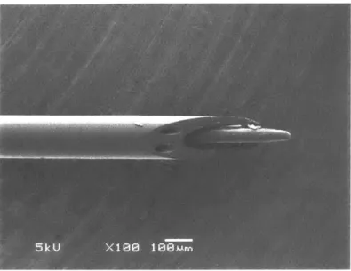
The core of the injectrode device consists of a 150 gm multi-lumen borosilicate glass tubing, which is implanted directly in the region of interest in the brain (Figure 1-4). Two lumens are designated for delivery of neuromodulatory drugs with differing effects. The third lumen houses an electrode for stimulation/recording of neural activity.
A manifold rests on the skull and facilitates the connection between the individual
lumens and wirelessly controlled drug pumps for the precise, on demand delivery of therapeutics. The manifold construction procedure will be described in the next chapter
(section 2.2.2). The injectrode allows for the precise delivery of potent therapeutics directly to the desired site of action. Driving flow via wirelessly controlled precision micropumps enables complex dosing regimens based on patient response and activity. The injectrode platform was developed to improve upon existing chemical/electrical
stimulation devices which were limited in utility due to their design (33, 45, 46). One class of devices consists of single-lumen devices in which an electrode is centered within a cannula. These platforms are limited in their ability to only administer one
fluid at a time. Switching solutions requires expelling the entire dead-volume of the device into the tissue, potentially leading to overdosing and off target effects. The
second class of devices are constructed using microfabrication techniques, and can have multiple lumen and onboard electrodes. The merit of these primarily silicon-based devices is the wide range of functions (multiple solution infusion and
electrophysiology) and dimensions that are possible with a single device. The brittle nature of the substrate limits the aspect ratios that can be achieved (5 mm: 200 gm).
Silicon-based devices are therefore limited to use in rodent models and are not directly scalable to larger animal models or to clinical applications (47).
The injectrode combines the advantages of both classes of devices; the three lumens can be used for any combination of multiple drug delivery, neurochemical sensing,
electrical recording, and stimulation. The high aspect ratio and micron scale dimensions of the borosilicate channel ensure that minimal diffusion occurs in the "off' state and that there is a low amount of dead-volume within the device. A summary of the design
Figure 1-4 SEM Micrograph of the Injectrode Device
The core of the device consists ofmultilumen borosilicate glass tubing which is implanted to interface with the nodes of neural circuits. The two smaller 35 Pm channels are designated for drug delivery, while the third channel houses an electrode for monitoring and stimulation of the local neural activity.
High Spatial + Temporal a Rigid fluidic channels 9 Targeted delivery to circuit
Resolution nodes.
* Micron-scale channels
& Limited diffusionleak from
the device.
E Enables on" and Wor state Translatable Design * BS channel has scalable * Sufficiently rigid to target
length deep brain structures
* Small and large animal
models without change in design
u d
l mlathn 0 Multiple lumens In device * Drugs with opposing
eflects can be delivered to * Electrode for electrical fine tune neural activity
stimulation
* Synergies between electrical and chemical
delivery
Guided treatment On board electrode * Local neural activity to
guide treatment and assess drug function
Chronic Junctlendng * Mechanically matched Minimize scar fbrmation to
hydrogel coatings, ensure long term device
functia. * Wireless pumps
* Wireless pumps enable treatment in awake, behaving rodents
Table 1-1 Design Features of the Injectrode Platform
This table highlights the critical design features of the injectrode platform and a comment on their utility.
1.5 Thesis Structure
The work presented in this thesis will describe the progress that has been made towards establishing a chronic local treatment strategy for the treatment of circuit disorders. It is essential to have a chronic functioning system capable of high resolution delivery of therapeutics on demand to effectively treat neurological diseases. The work presented in this thesis investigates several key materials issues associated with establishing a
chronic functioning platform in the brain. The first portion of the thesis will characterize the fluidic and electrical operation of the device on acute and chronic timescales. The ability to modulate animal behavior through the local intervention of neural circuits will be presented.
Additionally, a successful implantable treatment device must operate as intended for months to years following implantation. The second portion of the thesis will address the topic of glial scarring, the biological response to implants in the brain from a materials perspective. The work presented in this portion of the thesis first seeks to understand mechanical factors that are thought to drive scar formation response around neural implants, and then proposes a potential solution to modulate this response. These findings will provide a strategy to improve the biocompatibility, not only for the injectrode platform, but for all conventional neural probes going forward.
Chapter 2 will focus on the validation of the drug delivery aspects of the device design
in both in vitro and in vivo animal models. PET imaging, chronic impedance
spectroscopy, and rodent behavioral testing are used to validate proper device function following chronic implantation.
Chapter 3 will explore the effect that mechanical micromotion has on glial cells in the
central nervous system. This was conducted by establishing a novel 3D in vitro glial scar model.
Chapter 4 investigates the ability to reduce scarring around the device using a materials
based approach. Soft hydrogel coatings are used to improve the biocompatibility of neural implants, by mitigating the effects of micromotion induced strain around the device. Experiments are highlighted which characterize the mechanical and geometric effects of these coatings in tissue phantoms, as well as in a rodent implantation model.
Chapter 5 summarizes the findings presented in this thesis and proposes potential future directions for the project.
2
IN VITRO AND IN VIVO CHARACTERIZATION OF
THE INJECTRODE DEVICE
2.1 Introduction and Motivation:
As discussed in Chapter 1, the injectrode is a multimodal device designed to treat circuit disorders of the brain through combined local chemical and electrical stimulation. The device must be able to dose nanoliter scale volumes of drug, modulate neural behavior, and operate effectively on chronic timescales in order to maximize the effectiveness of ~Infusions need to be reliable and controllable in order to target small regions of brain tissue on demand. The neural circuit nodes that we are targeting with this device are on the order of 0.5 - 2.5 mm in diameter (17). Often times neurons in neighboring regions are known to produce different or opposing effects (17). The approximate target drug infusion volume may be determined according to the equation:
4 d
VDrug = a * VTissue= a * 3 *
where a is the ECS space volume fraction (typically assumed to be ~O.2 for healthy brain tissue (48)), d is the diameter of the neural target region in the tissue. This
equation suggests that 13 nl - 1.6 gl scale volumes of drug should be infused to
successfully target these regions (0.5-2.5 mm in diameter). This is desired so that the appropriate neural circuit nodes are targeted, without producing unwanted side effects in nearby tissue regions. The dynamics of neural circuit dynamics are highly dynamic. Therefore it is also critical to be able to have high temporal resolution in the drug delivery process in addition to precise spatial control. There should be minimal effects from drug passive release (e.g. diffusion from the exposed tip) when the device is in the "off' state.
This chapter highlights the work that has been completed to validate the device's ability to accomplish these tasks on acute and chronic timescales. Preliminary infusion tests were conducted in vitro and in vivo to assess the spatial and temporal characteristics of typical infusion parameters. Electrophysiology experiments were conducted to
demonstrate these nanoliter scale volumes of drugs are capable of locally modulating neural activity in a rodent model.
The final portion of this chapter will focus on the chronic operation of the device in a rodent model. Positron emission tomography (PET) imaging was used to quantify
infusion volumes and assess device operation at various time points after chronic implantation. Changes in electrical properties of the recording electrode and the local tissue environment were assessed via chronic impedance spectroscopy. As a last proof of concept for the potential of the injectrode platform, we demonstrate behavioral modification through infusion of microliter volumes of drug in a rodent behavioral model.
Throughout these experiments muscimol was used as a model drug. Muscimol is a potent GABAA agonist, which silences neural activity by selectively binding to the GABAA receptors in the brain (49).
2.2
Methods:
Many established techniques were employed to characterize the operation of the injectrode platform in vitro and in vivo, on both acute and chronic timescales. Background and procedures related to these techniques are provided in this section.
2.2.1 Materials
Muscimol, heptafluorobutric acid (HPTA), agarose, indocyanine green, and Tris(2-carboxyethyl)phosphine hydrochloride (TCEP) were purchased from Sigma Aldrich (St. Louis, MO, USA). 4-arm Polyethylene glycol was purchased from Creative PEGworks (Chapel Hill, NC, USA). Maleimido-mono-amide-NOTA was purchased from
Macrocyclics Inc (Plano, TX, USA). Cu-64 was purchased from the cyclotron facility at Washington University in St. Louis.
2.2.2 Device Fabrication:
The core of the injectrode platform consists of multiple micron scale borosilicate (BS) channels for fluid flow, as well as a larger lumen to house an electrode for neural recording and stimulation. The 1st and 2nd generation fabrication procedures are
described below. The implanted core of both device designs are largely identical, with the mode of upstream fluidic connections varying between the two. The 2nd generation
device design was implemented to improve device yield and fluidic reliability of the device.
Ist Generation Device:
The device design consists of four components: borosilicate glass tubing (Vitrocom, Inc, Mountain Lakes, NJ), a five-piece custom machined manifold, an electrode, and upstream drug reservoirs connected to a microsyringe pump (Figure 2-1A). The borosilicate (BS) tubing is 150 pm in diameter and contains three lumens. Two of the lumens are 30 pm in diameter and are intended for the delivery of neuromodulating drug solutions to the region of interest. The third lumen is larger in diameter (90 im) and houses an electrode to record and stimulate neural activity, or a carbon fiber electrode to measure local neurotransmitter concentrations.
The manifold is designed to establish airtight fluid connections between the upstream syringe pump and the specific lumens in the glass tube. The components of the manifold are designed and fabricated in house using a micro-computer numerical control (CNC, Cameron Micromaching Center, Sonora, CA, USA) machine. The chambers are separated by the septa. Two of the chambers connect a fluid port to a specific lumen and the middle lumen is designed to prevent leaking between the two fluid channels. Access ports are cut in the glass tubing so that each lumen aligns with one compartment in the manifold. Access ports are made with a precision milling machine and diamond tipped engraver tool so that they intersect with one lumen along the length of the glass.
PE20 tubing (Becton Dickinson, Franklin Lakes, NJ), is connected to each infusion chamber to interface with syringe pumps to drive fluid flow. Devices were tested to ensure proper function prior to experimental use. Quality testing includes infusion of a controlled amount of dye through each lumen and comparison to a calibration curve and visual inspection for any minor leaks.
2nd Generation Device:
Second generation injectrode devices were made by employing microfabrication techniques to improve the production yield and fluidic reliability of the device (Figure
2-1C). The 2nd generation device consisted of a stainless steel guide tube (200 gm O.D., Hamilton Company), two borosilicate fluidic channels (30 gm OD, 20 gm ID,
Vitrocom), a tungsten electrode/tetrode for recording and electrical stimulation (FHC, Inc, Bowdoin, ME, USA), and the 150 gm multichannel BS glass tip. Briefly, all components were aligned utilizing a polyimide template constructed in the Harvard cleanroom facility. The components were fixed in place with UV curable epoxy, and then threaded down the guide tube. The tips of the fluidic channels were inserted into a
short 150 pm borosilicate multichannel glass tube mentioned in the previous section that served to align the tips at the end of the device. The two fluidic channels were placed in an individual 35 gm channel and the tungsten electrode/tetrode was placed in the larger channel. The glass tips typically extended approximately 2-3 mm from the guide tube, but is customizable based on the intended application. The length and electrical components (e.g. tungsten electrode, carbon fiber, or tetrode) of the device were chosen based on the intended application and targeted brain structure. Flow from the devices was driven by syringe pump (Quintessential Stereotactic Injector, Stoelting) or wirelessly controlled implantable pumps (iPrecio SMP-300 pump Primetech Corp., Japan). The device fabrication procedure and assembly was established and carried out
I
1St Generation Injectrode A A (UarUpstream pump and drug reservoirsBorosilicate Target neural region (15tu dia.) (almm dia.)
Drug I
//
Orug2
[
Manifold Electro for
A
e
I P
(2mm dia. ) recording/
Access orts stimulation
B
two 30 pm lumens for chemical infusion 90 pm bore for microelectrode -- IL SOL b 1111 pm1 2nd Gen Injectrode BS glass tip ... :
Figure 2-1 Schematic of Injectrode Device Fabrication Procedure
The injectrode core consists of a multilumen borosilicate glass tube. Two lumens are designated for drug delivery to the target neural region, while the larger channel houses an electrode for
electrical recording and stimulation. An SEM micrograph of the glass tube is shown in panel B. Fluidic connections were established through a custom machined delrin manifold in Is' generation devices (A), while fluidic connections to radel tubing were established through microfabrication techniques in the 2"d generation devices (CD). The injectrode devices were
threaded through a steel guide tube prior to implantation.
2.2.3 In Vitro Device Characterization:
Injectrode devices were attached to a syringe pump and primed with dye/drug
containing solution. Devices were primed before beginning the experiment by infusing approximately 1-2 ml of solution through the device. All fluidic lines were visually
inspected for air bubbles prior to beginning the experiment. The tips of the devices were lowered into a water bath, and a set amount of dye was infused. Programmed infusion
D Electrical
conection--..,-g6O~ Fluidic channels
am OM
IMMOMM - I
volumes ranged from 50 nL to 300 nL, as these are volumes we anticipate are necessary to target focal points in neural circuits. The amount of dye which was infused was determined by measuring of the peak absorbance at a given wavelength using a plate reader (Biotek Synergy 2, Winooski, VT, USA) and comparison to a calibration curve. This procedure was conducted with two dyes with distinct absorbance spectra, one delivered through each lumen. The volume of each dye (both the dye infused and the dye in the other lumen) was measured to detect any cross contamination between fluidic channels. Each infusion study was repeated in triplicate.
A similar protocol was used for drug infusion studies. Muscimol (5 mg/ml) was primed
in the device, and the total amount of drug infused was quantified via high pressure liquid chromatography (HPLC) (50). Briefly, 20 gl of sample was injected into a 4.6 mm (ID) x 25 cm (L) ODS-2 column (Spherisorb, Column Engineering, Ontario, CA,
U.S.A) on an Agilent 1200 LC system. The column was eluted with a HPTA 0.5%
(V/V%) running buffer at a flow rate of 1 ml/min. The muscimol peak was detected at
230 nm, with a reference wavelength of 360 nm, at an elution time of approximately 8.4
minutes. Known concentrations of muscimol were analyzed to produce a calibration curve. Comparison to this curve enabled the concentration of the drug infusion bath to be determined.
The rate at which muscimol diffuses from the tip of the injectrode when no infusion is programmed was also measured. Devices were primed with muscimol solution (5 mg/ml) and were placed into a water bath. Samples were collected at set time (0-48 hours) points and the drug concentration was quantified.
In Vitro infusion studies were also conducted to assess the capabilities of the iPrecio wirelessly controlled pump. Devices were attached to a pump and placed in a water bath on a precision microbalance (Mettler Toledo, Columbus, OH, USA). The water bath