REVIEW
Tissue engineering on matrix: future of autologous
tissue replacement
Benedikt Weber&Maximilian Y. Emmert&
Roman Schoenauer&Chad Brokopp&
Laura Baumgartner&Simon P. Hoerstrup
Received: 13 October 2010 / Accepted: 19 January 2011 / Published online: 29 January 2011 # Springer-Verlag 2011
Abstract Tissue engineering aims at the creation of living neo-tissues identical or close to their native human counter-parts. As basis of this approach, temporary biodegradable supporter matrices are fabricated in the shape of a desired construct, which promote tissue strength and provide functionality until sufficient neo-tissue is formed. Besides fully synthetic polymer-based scaffolds, decellularized biological tissue of xenogenic or homogenic origin can be used. In a second step, these scaffolds are seeded with autologous cells attaching to the scaffold microstructure. In order to promote neo-tissue formation and maturation, the seeded scaffolds are exposed to different forms of stimula-tion. In cardiovascular tissue engineering, this “condition-ing” can be achieved via culture media and biomimetic in vitro exposure, e.g., using flow bioreactors. This aims at adequate cellular differentiation, proliferation, and extracel-lular matrix production to form a living tissue called the construct. These living autologous constructs, such as heart valves or vascular grafts, are created in vitro, comprising a viable interstitium with repair and remodeling capabilities already prior to implantation. In situ further in vivo remodeling is intended to recapitulate physiological vascu-lar architecture and function. The remodeling mechanisms were shown to be dominated by monocytic infiltration and chemotactic host-cell attraction leading into a multifaceted inflammatory process and neo-tissue formation. Key
mol-ecules of these processes can be integrated into the scaffold matrix to direct cell and tissue fate in vivo.
Keywords Tissue engineering . Cardiovascular . Heart valve . Biocompatibility . Matrix . Remodeling
Introduction
The ultimate goal of any tissue engineering approach is the creation of autologous living neo-tissues similar in archi-tecture and function to native human structures. Therefore, an accurate understanding of the fundamentals of native tissue—representing the “gold standard”—constitutes a prerequisite to a successful development of native analo-gous tissue-engineered substitutes. Interestingly, it is the research on tissue engineering of recent years, which has fundamentally stipulated a novel interest in embryology, native tissue architecture, and development. In cardiovas-cular medicine, the in vitro fabrication of heart valves represents an example of how tissue engineering solutions aim to overcome obvious clinical limitations of currently available treatment options (Fig.1). Native heart valves are composed of living, dynamic tissue capable of continuous remodeling to adapt to the constantly alternating hemody-namic environment [1]. None of the currently available valvular replacements are capable of fully restoring the native function due to insufficient adaptive capacity. State-of-the-art prostheses in today's clinical use show consider-able limitations. These include the lack of growth, repair and remodeling capabilities, once they are implanted into the body. Additionally, mechanical valve substitutes are inherently susceptible to thromboembolic events due to high shear stress, nonphysiological flow profiles, and blood damage necessitating lifelong anticoagulation therapy [2,
This article is published as part of the Special Issue on“Implant
devices: Biocompatibility, Tissue Engineering and Infection”.
B. Weber
:
M. Y. Emmert:
R. Schoenauer:
C. Brokopp:
L. Baumgartner
:
S. P. Hoerstrup (*)Swiss Center for Regenerative Medicine and Clinic for Cardiovascular Surgery, University Hospital Zurich, Raemistrasse 100,
8091 Zurich, Switzerland
3]. Bioprostheses from xenogenic or homogenic origin are inherently prone to structural degeneration, and the associ-ated need for repeat reoperations makes them less suitable for many patients [4,5]. Tissue engineering of heart valves represents a technology with the potential to overcome these limitations by creating a living autologous valve replacement that prevents an immune response, clotting activation, and valvular degeneration on the one hand, and allows for growth, remodeling, and repair throughout the patient's lifetime on the other hand.
The basis of most tissue engineering approaches is the fabrication of temporary supporter matrices in the shape of a desired construct. These biodegradable matrices promote tissue strength and provide functionality during the engi-neering process until sufficient neo-tissue is formed to restore adequate physiological function. In cardiovascular tissue engineering, fully synthetic polymer-based scaffolds, as well as decellularized biological tissue of xenogenic or homogenic origin, can be used. After in vitro tissue formation, the living autologous constructs are implanted into the patient, where further in vivo remodeling is intended to recapitulate physiological vascular architecture and function. This step of in situ remodeling represents an essential part of the tissue engineering concept, as it will fundamentally influence the fate and success of the substitute. The mechanisms involved in this remodeling process were shown to be mainly dominated by monocytic infiltration and host-cell attraction to the construct; however, little is known about the actual molecular and cellular pathways involved, which are central for this essential reorganization of bioengineered implants.
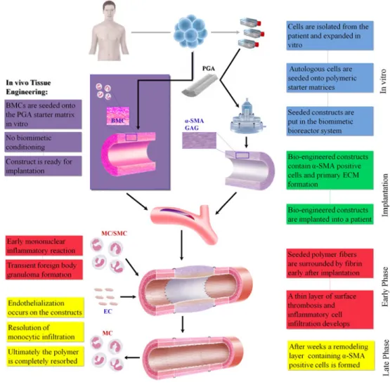
Strategies in tissue engineering: in vivo or ex vivo? Tissue engineering is defined as an interdisciplinary field, applying the principles and methods of engineering to the
development of biological substitutes that can restore, maintain, or improve tissue formation [6]. According to this predefinition, two principle strategies have been developed to generate living autologous replacements: The in vitro as well as the in vivo approach. The first approach, requires an ex vivo phase generating the optimal native-like substitute in vitro. This traditional tissue engineering paradigm comprises the isolation and expan-sion of cells from the patient, subsequent seeding onto an appropriate scaffold material, in vitro tissue formation and finally, implantation into the patient from whom the cells were taken (autologous approach). This paradigm, further referred to as the in vitro tissue engineering approach, being employed as the principal approach for heart valve tissue engineering and is aimed at full development of the tissue substitute ex vivo (see Fig.1). Several different cell sources serve as the basis for the generation of these constructs, where minimally invasively accessible stem and progenitor cells have shown tremendous potential [7–9].
The second approach of in situ heart valve tissue engineering circumvents the in vitro tissue culture phase by straight implantation of natural tissue-derived heart valve matrices, aiming at potential cell in-growth and remodeling in vivo [10]. In recent years, a further approach has emerged—mainly driven by the advances in stem cell technology and signaling. By seeding the construct with autologous cells, such as progenitor and/or mononuclear cells, using a cell carrier matrix, host-cells can be attracted to the implant site via chemo-attractive paracrine pathways. These attracted immune cells then support a distinct remodeling process, resulting in enhanced extracellular matrix and collagen formation [11].
The in vitro fabrication of an autologous construct: steps in cardiovascular tissue engineering
According to the approach of in vitro tissue engineering techniques, such as heart valve tissue engineering, the successful fabrication of autologous living replacements similar to the native benchmark is supported by three main elements: (1) autologous cells that resemble their native counterparts in phenotype and functionality are isolated and expanded using standard cell culture methods. For this purpose, several different sources are available, ranging from mature vascular-derived cells to prenatally harvested fetal progenitor cells. (2) The cells are seeded onto a temporary biodegradable supporter matrix fabricated in the shape of a trileaflet heart valve, termed the scaffold, which promotes tissue strength until the produced ECM (extracel-lular matrix) provides functionality on its own. Several different matrix materials, including synthetic as well as biologic materials, have been assessed for these purposes
Fig. 1 Tissue-engineered heart valve. Autologous living tissue-engineered heart valve before implantation after in vitro conditioning in a bioreactor system
[12]. (3) In order to promote tissue formation and maturation, the seeded scaffolds are exposed to mechanical stimulation transmitted via a culture medium (biological stimuli) or via“conditioning” of the tissue in a bioreactor. This bioreactor phase targets at the in vitro generation of a matured, high-quality extracellular matrix, having the capacity to grow as well as being able to respond to varying physiological needs and to repair structural injury by remodeling [13,14; Fig.2].
Cell sources for cardiovascular tissue engineering: from vascular cells to stem cells
The in vitro formation of a durable, well-structured and viable tissue is crucial for the in vivo functionality of a tissue-engineered construct. In this context, the choice of the optimal cell source is critical for the quality and long-term success of heart valve tissue engineering [1, 15; Fig. 2]. An established approach for heart valve tissue
engineering uses cells originating from aortic, saphenous vein, or peripheral artery biopsies [15]. Out of these vessels, two cell types can be isolated: Endothelial cells (ECs) with antithrombogenic properties and myofibroblasts capable of ECM development [16–18]. After preliminary studies, mainly in sheep [16–21], the potential of human vascular-derived cells was evaluated by seeding on biode-gradable scaffolds and revealed excellent tissue formation [15, 22, 23]. A promising alternative cell source for regenerative medicine is bone marrow-derived stem cells (BMSCs) [3,24,25]. BMSCs were successfully used for in vitro production of heart valves [24, 26], and have been implanted in vivo demonstrating adequate functionality [27].
To improve the functional capacities and to reduce the risk for complications [28] tissue-engineered heart valves are usually covered with autologous human ECs [18]. Differentiated ECs have been isolated from vascular sources exhibiting promising results in heart valve tissue engineering [16, 19, 20, 22]. Furthermore, endothelial
Fig. 2 Concept of cardiovascular tissue engineering. Autologous cells are harvested from the patient and expanded in vitro. When sufficient numbers are reached, cells are seeded onto a biodegrad-able scaffold. Constructs are either positioned in a bioreactor and conditioned (in vitro approach) or directly implanted into the patient (in vivo approach). After implan-tation of the tissue-engineered construct, the proposed mecha-nism of vascular remodeling comprises an early monocyte recruitment to the scaffold with the release of multiple angiogenic cytokines and growth factors. These factors (i.e., VEGF) cause recruitment of host cells, such as MC, SMCs, and ECs, to the scaffold. The invading host cells originate from circulating progenitors and (trans-anastomotic) migration/in-growth of mature vascular cells from adjacent vessel segments. Incoming ECs and SMCs appropriately organize into a mature blood vessel structure on the luminal surface of the scaffold (with the remaining scaffold in the center of the construct). As the scaffold degrades, early monocytes migrate away, leaving behind a remodeled, completely autologous neo-vessel
progenitor cells (EPCs), discovered in human blood [29], have been established as source of ECs [30, 31]. Since they are easily accessible, current research aims at their trans-differentiation into myofibroblast-like cells to estab-lish blood as single cell source for heart valve tissue engineering.
Another interesting cell source is the umbilical cord, containing several cell types that can be used for heart valve tissue engineering: (1) umbilical cord vein-derived and artery-derived cells, (2) Wharton's Jelly-derived MSCs, and (3) umbilical cord blood-derived EPCs. These cells demonstrated excellent growth properties and tissue forma-tion [32–35]. Vascular neo-tissues were produced using human umbilical cord blood-derived EPCs seeded on vascular scaffolds [36]. By using umbilical cord-derived cells several different cardiovascular replacements could be generated [37–40].
The ideal pediatric tissue engineering paradigm com-prises a prenatal fetal cell harvest allowing for tissue engineering processes during pregnancy followed by the implantation of the autologous tissue-engineered construct directly after birth. A new concept, using human prenatal progenitor cells derived from chorionic villi and umbilical cord blood for the production of autologous heart valve leaflets, has been introduced by Schmidt et al. [9]. Furthermore, human amniotic fluid-derived cells, as an easily accessible cell source, have been used as a sole cell source for the fabrication of living autologous heart valves prior to birth [8,41].
Human adipose tissue contains mesenchymal stem cells with the potential to differentiate into various phenotypes in vitro [42,43] and in vivo [44]. Due to the high availability and the ease of harvest, adipose-derived stem cells (ADSCs) represent a potential alternative stem cell source to BMSCs [45].
Biocompatible starter matrices: the optimal scaffold for cardiovascular tissue engineering
The development of scaffolds for heart valve tissue engineering has proceeded along two fronts: a biological matrix material and a fully synthetic scaffold [46]. Regardless of the material of the scaffold matrix, the design of a scaffold capable of supporting cellular growth and of withstanding mechanically complex cardiovascular envi-ronment is critical to the success of the tissue-engineered construct. In addition to meeting all the standard design criteria of traditional tissue valves, in which durability and biocompatibility are effectively passive attributes of the underlying materials, and selecting the best scaffold material, it requires consideration of the active behavior of the cells in the regulation of tissue growth, remodeling, and
homeostasis. These matrices must be able to support cell growth and cell-to-cell interaction guiding tissue formation into a functional organ with organotypic ECM. The surfaces of these starter vehicles must be biocompatible, allowing cellular ingrowth and the formation of antithrombogenic cell linings, and biodegradable, providing an optimized degrada-tion rate for cellular expansion [13]. These specific require-ments entailed the development of various approaches to identify the optimal scaffold material, including the creation of synthetic [12] and biological scaffold materials [47]. These can be further subdivided into native tissue-derived ECM scaffolds [48], polymeric scaffolds [49–53], biological-polymeric hybrid scaffolds [54–56], and collagen or fibrin gel scaffolds [57–60]. Although significant advan-ces have been made in all these approaches, the polymeric scaffolds have, to date, received most attention regarding heart valve tissue engineering applications.
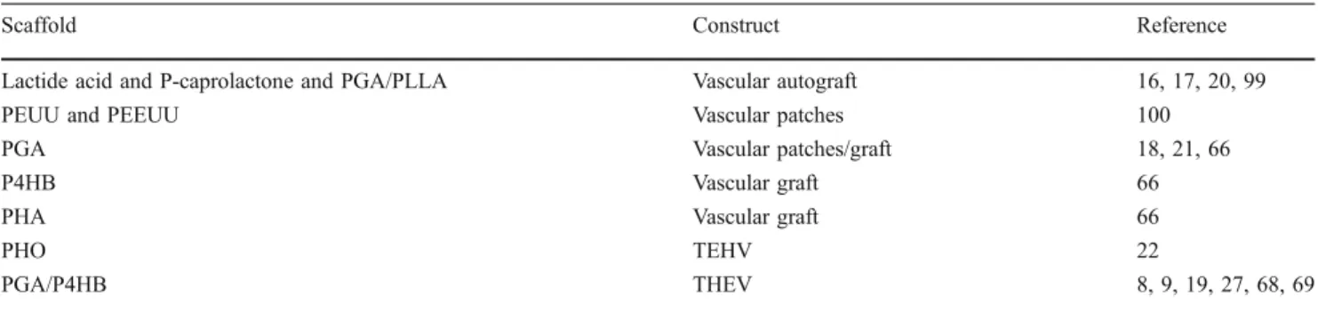
Polymeric starter matrices for cardiovascular tissue engineering: the future of autologous tissue replacement The use of polymeric scaffold materials for different tissue engineering approaches has already been broadly demon-strated [12]. The ideal scaffold matrix for heart valve tissue engineering has to be at least 90% porous [61], and comprises an interconnected pore network, as this is essential for cell growth, nutrient supply, and removal of metabolic waste products. Besides being biodegradable, biocompatible, and reproducible, the scaffold material should also display a cell-favorable surface chemistry and match the bio-mechanical properties of the native heart valve tissue [12]. In addition, the rate of matrix degradation should be controllable and commensurate with the rate of novel tissue formation in order to provide a sufficient but reducing mechanical stability of the construct over time [1, 62]. Several synthetic biodegradable polymers have been investigated as potential starter matrices for heart valve tissue engineering that vary in their manufacturing possi-bilities and degradation rates (Table1).
Aliphatic polyesters, including polyglactin (PG), poly-glycolic acid (PGA), and polylactic acid (PLA), degrade by cleavage of the polymer chains due to hydrolysis of their ester bonds. The resulting monomer is either excreted via urinal secretion or enters the tricarboxylic acid cycle [61]. In order to fabricate single heart valve leaflets, the creation of scaffolds was initially based on combinations of aliphatic polyesters, including PG non-woven PGA meshes with layers of PGLA and non-woven PGA meshes. The major limitations of aliphatic polyesters, when used as a sole material, are their thickness, initial stiffness, and non-pliability, making the fabrication of trileaflet heart valves a difficult process.
A further group of widely used polymers is the polyhydrox-yalcanoates (PHA) family, which is composed of polyesters built up from hydroxyacids that are produced as intracellular granules by various bacteria [63]. PHAs, as well as poly-4-hydroxybutyrate, have been used to create trileaflet heart valve conduits [22, 64]. These materials possess thermoplastic properties and can be molded into any desired shape using stereolithography [22,65]. A limitation of PHAs can be found in their slow degradation. Combinations of aliphatic polyesters and PHAs have also been tested as alternative composite materials [19,66]. Particularly, the use of PGA coated with P4HB (Fig. 3), combining the thermoplastic properties of P4HB and the high porosity of PGA, for the fabrication of complete trileaflet heart valves revealed promising results in a rapidly growing sheep model [8,19,26,66,67].
Decellularized tissue-derived matrices: the biologic counterpart
In principal, donor heart valves (homografts) or animal-derived heart valves (xenografts) are among the most
obvious choices for scaffold materials. They are fixed and depleted of cellular antigens, which makes them less immunogenic, and/or thus eligible to be used as a scaffold material in tissue engineering. The removal of cellular components results in a template composed of extracellular matrix proteins that serve as an intrinsic medium for subsequent cell attachment. Nevertheless, they still possess a native-like geometry and architecture with bio-mechanical and hemodynamic properties similar to their native counterpart [8,13,67].
Various decellularization techniques have been exten-sively investigated in order to minimize the residual immunologic potential of biological matrices. Although it is essential to remove all cellular components, the decellu-larization treatment should avoid any harm or alteration of the ECM properties. This preservation of matrix integrity, as well as the efficiency of cell removal is highly dependent on the method used for decellularization [68]. Several different decellularization methodologies for heart valve scaffold fabrication have been reported, including trypsin/ EDTA [68–71], freeze drying [72], osmotic gradients [73], non-enzymatic detergent treatment [68, 74], and multistep enzymatic procedures [75]. The use of non-enzymatic detergent-based techniques has been shown to result in a much more efficient cell removal, while preserving the overall matrix integrity of the scaffold, when compared to other more aggressive decellularization methods such as trypsin/EDTA [68, 76, 77]. In order to avoid this impairment of the matrix integrity and function due to tissue-derived protease activation, the use of suitable protease inhibitors has been recommended [78]. Accesso-rily, nuclease digestion steps should be embedded into the decellularization procedure to remove any residual RNA or DNA within the scaffold.
Several in vivo studies have proven the feasibility to use decellularized scaffolds as starter matrix for cardiovascular tissue engineering [79, 80]. Moreover, first clinical trials have been initiated [81]. However, using xenogenic materials, serious complications have been reported due
Table 1 Examples of polymeric starter matrices used for cardiovascular tissue engineering
Scaffold Construct Reference
Lactide acid and P-caprolactone and PGA/PLLA Vascular autograft 16, 17, 20, 99
PEUU and PEEUU Vascular patches 100
PGA Vascular patches/graft 18, 21, 66
P4HB Vascular graft 66
PHA Vascular graft 66
PHO TEHV 22
PGA/P4HB THEV 8, 9, 19, 27, 68, 69
PGA polyglycolic acid; PHA polyhydroxyalkanoate; PHO polyhydroxyoctanoate; PEUU poly(ester–urethane)urea; PEEUU poly(ether–ester–
urethane)urea; PLLA polylactic acid; P4HB poly-4-hydroxybutyrate; TEHV tissue-engineered heart valve
Fig. 3 The matrix for cardiovascular tissue engineering. SEM images of the PGA mesh coated with P4HB at low (a) and high (b) magnification
to residual α-Gal-mediated immunogenicity [81, 82]. Furthermore, the use of xenografts principally involves the risk of zoonoses and prionic diseases, in terms of human diseases caused by animal-derived infectious agents, which has given rise to widespread concern [83, 84]. When the matrix material is from homogenic origin, the limited availability of donor valves and associated ethical concerns represent a considerable shortcoming. Moreover, the lack of evidence of growth and remodeling capacities of valve replacements when using decellular-ized scaffolds seems to be a further drawback, especially with regard to the pediatric field [9]. These drawbacks and uncertainties raise a common concern associated with the use of decellularized starter matrices, also fuelling the search for synthetic scaffold alternatives.
Implant-mediated inflammation: the key to optimal tissue remodeling?
Healing of a tissue-engineered constructs in vivo has to be seen as a continuous but multifaceted process. Following the initial blood-material interactions, inflam-matory processes occur around the implanted construct. The extent of this response depends upon the degree of maturation of the tissue-engineered construct and the extent of surgical injury, which directs a physiological healing reaction, consisting first of an acute inflamma-tion, followed by repair processes. Immediately after implantation, phagocytic cells (predominantly neutrophils and monocytes) migrate from the microcirculation to the interface between the implant surface and the injured tissue. The inflammatory phase, (which lasts up to several weeks in humans) consists of phagocytic removal of debris due to trauma, and then, provides the appropriate signals for the shift from inflammation to repair and remodeling of the tissue. Taken as a whole, two main processes seem to be indispensable for a successful remodeling of tissue-engineered constructs in vivo: (1) The formation of an atypical vascular response to injury at the luminal surface, including intimal thickening, pannus formation, and neointima develop-ment, and (2) deep tissue biomaterial-associated effects of foreign body reaction, granulation, tissue formation, and fibrosis forming a media-like structure [85].
Upon resolution of the acute inflammatory reaction, monocytes are observed within the implanted construct. Indeed, it has been demonstrated that monocyte chemotac-tic protein (MCP-1), a potent monocyte-attracting cytokine, is expressed by activated neutrophils at the implant site, and thus, seems to play a central role within the early remodeling phase, which is mainly characterized by chemotactic immune cell infiltration [86]. While the exact
roles recruited monocytes play in the implant transforma-tion remains largely conjectural, their role in tissue repair is better understood. Recent findings suggest that monocytes remain within the implant scaffold until it fully degrades, and therefore, may play a role in the entire remodeling process. At 100-week follow-up, diffuse mononuclear inflam-mation, particularly in areas of residual polymer fibers, has been observed [87]. In implanted tissue-engineered vascular grafts, recent evidence suggests that monocytes produce important cytokines (i.e., MCP-1/CCL-2, IL-6, IP-10), growth factors, and proteases necessary for vascular cell proliferation/migration and appropriate vascular remodeling [85,88,89].
Monocytes/macrophages at the site of long-term implan-tation express ECM remodeling proteases (i.e., MMPs), cytokines characteristic of the innate immune response (IL-1α, IL-1β, IL-6, IL-10, and TNFα) and cell adhesion (ICAM-1, VCAM-1). Moreover, monocytes involved in a pro-fibrotic foreign body response have been shown mainly properties of classically activated macrophages (1β, IL-6, TNF, Ccl20, Cxcl10), and to a lesser extent, wound-healing (factor XIII-A) and regulatory macrophages (IL-10; [90, 91]). While these monocyte properties may affect implant integration (foreign body response vs. tissue integration) via paracrine signaling, the precise role of monocytes/macrophages in tissue remodeling is not under-stood. However, expression of angiogenic cytokines (VEGF) from implant-associated monocytes/macrophages has lead to a more descriptive understanding of their role in implant neovascularization.
In vivo animal studies have revealed that arteriogenesis depends on circulating monocytes and macrophage accu-mulation. This finding has been confirmed by recent studies, which demonstrate that monocyte recruitment is central to postnatal blood vessel formation [92–94]. This mechanism has, in part, been attributed to the critical role of VEGF in adult neovascularization, and prevention of neo-vessel regression [95–97]. Indeed, monocyte/macrophage infiltration associates with VEGF expression with the tissue-engineered constructs [98]. In addition to VEGF, recruited monocytes likely release multiple cytokines, which then orchestrate the proper vascular neovascularization of biodegradable implants.
Besides the involvement of MCP-1 for monocytic attraction and VEGF expression, the molecular pathways of these remodeling phenomena remain largely unknown.
The discovery of these underlying pathways seems indispensable for the development of strategies to modulate early inflammatory reactions and enhance remodeling [99]. First success has been achieved by attaching MCP-1-releasing biodegradable microparticles to the scaffold matrix in order to mimic the chemo-attractive properties of seeded cells in vivo [98].
Conclusions
Cardiovascular tissue engineering is a promising approach aiming at the creation of living functional autologous replacements and holds exciting potential for improving therapy of many diseases. However, before clinical appli-cation of the tissue engineering concept becomes routine, numerous steps must be overcome in the laboratory. Primary amongst these is the limited knowledge about the mechanisms involved in the in vivo remodeling process of tissue-engineered constructs after implantation. Although having first indications as to the influence of cytokine-mediated monocyte-attraction and neovascularization, the underlying exact molecular pathways remain unknown. Another important consideration concerns the definition of the ideal matrix material for engineering, providing a template for directing new tissue growth and organization, as well as for regulating cellular adhesion, migration, and differentiation. The mechanism involved in neo-vascular formation displays many parallels to natural neovasculari-zation and may provide further insights into the biology of these processes. However, although similarities to natural processes exist, the development of a tissue-engineered construct does appear to be a distinct process of vascular formation in itself. A better understanding of how the different steps of in vivo remodeling start and are controlled will lead to a new class of implants. Moreover, the identification and attachment of important regulatory molecules to the scaffold matrices, such as VEGF, may provide engineers with the key to enhance the body's innate ability to regenerate implanted constructs. Eventually, this could allow for the fabrication of “intelligent” scaffold materials incorporating specific signaling molecules that direct tissue fate within the implanted constructs.
Acknowledgments This work was supported by grants from the
Swiss National Science Foundation [32-122273], Swiss South African Joint Research Programme of the State Secretariat for Education and Research, Swiss Government [EX25-2010] as well as the 7th Framework Programme, Life Valve, European Commission [242008].
Conflicts of interest The authors declare that they have no conflict
of interest.
References
1. Schoen FJ (2008) Evolving concepts of cardiac valve dynamics: the continuum of development, functional structure, pathobiology,
and tissue engineering. Circulation 118:1864–1880
2. Yoganathan AP, He Z et al (2004) Fluid mechanics of heart
valves. Annu Rev Biomed Eng 6:331–362
3. Dasi LP, Simon HA, Sucosky P et al (2009) Fluid mechanics
of artificial heart valves. Clin Exp Pharmacol Physiol 36:225–
237
4. Yacoub MH, Takkenberg JJ (2005) Will heart valve tissue engineering change the world? Nat Clin Pract Cardiovasc Med
2:60–61
5. Zilla P, Brink J, Human P et al (2008) Prosthetic heart valves:
catering for the few. Biomaterials 29:385–406
6. Langer R, Vacanti JP (1993) Tissue engineering. Science
260:920–926
7. Sutherland FW, Perry TE, Yu Y, Sherwood MC, Rabkin E, Masuda Y, Garcia GA, McLellan DL, Engelmayr GC Jr, Sacks MS, Schoen FJ, Mayer JE Jr (2005) From stem cells to viable autologous semilunar heart valve. Circulation 31(111):2783– 2791
8. Schmidt D, Achermann J, Odermatt B et al (2007) Prenatally fabricated autologous human living heart valves based on amniotic fluid derived progenitor cells as single cell source.
Circulation 116:I64–I70
9. Schmidt D, Mol A, Breymann C et al (2006) Living autologous heart valves engineered from human prenatally harvested
progen-itors. Circulation 4(114):I125–I131
10. Matheny RG, Hutchison ML, Dryden PE et al (2000) Porcine small intestine submucosa as a pulmonary valve leaflet substitute. J Heart
Valve Dis 9:769–775
11. Brennan MP, Dardik A, Hibino N, Roh JD, Nelson GN, Papademitris X, Shinoka T, Breuer CK (2008) Tissue-engineered vascular grafts demonstrate evidence of growth and develop-ment when implanted in a juvenile animal model. Ann Surg 248:370–377
12. Schmidt D, Stock UA, Hoerstrup SP (2007) Tissue engineering of heart valves using decellularized xenogeneic or polymeric starter matrices. Philos Trans R Soc Lond B Biol Sci 29(362):1505–1512 13. Brody S, Pandit A (2007) Approaches to heart valve tissue engineering scaffold design. J Biomed Mater Res B Appl
Biomater 83:16–43
14. Sacks MS, Schoen FJ, Mayer JE (2009) Bioengineering challenges
for heart valve tissue engineering. Annu Rev Biomed Eng 11:289–
313
15. Schmidt D, Hoerstrup SP (2007) Tissue engineered heart valves
based on human cells. Swiss Med Wkly 155:80–85
16. Shin’oka T, Ma PX, Shum-Tim D et al (1996) Tissue-engineered
heart valves. Autologous valve leaflet replacement study in a lamb
model. Circulation 1(94):II164–II168
17. Shin'oka T, Shum-Tim D, Ma PX (1997) Tissue-engineered heart valve leaflets: does cell origin affect outcome? Circulation
96:II-102–II-107
18. Hoerstrup SP, Zund G, Schoeberlein A et al (1998) Fluorescence activated cell sorting: a reliable method in tissue engineering of a bioprosthetic heart valve. Ann Thorac Surg 66:1653–1657 19. Hoerstrup SP, Sodian R, Daebritz S et al (2000) Functional
living trileaflet heart valves grown in vitro. Circulation 102:
III44–III49
20. Shin'oka T, Breuer CK, Tanel RE (1995) Tissue engineering heart valves: valve leaflet replacement study in a lamb model. Ann
Thorac Surg 60:513–516
21. Sodian R, Hoerstrup SP, Sperling JS et al (2000) Early in vivo experience with tissue-engineered trileaflet heart valves.
Circula-tion 102:22–29
22. Zund G, Hoerstrup SP, Schoeberlein A et al (1998) Tissue engineering: a new approach in cardiovascular surgery: seeding of human fibroblasts followed by human endothelial cells on
resorbable mesh. Eur J Cardiothorac Surg 13:160–164
23. Schnell AM, Hoerstrup SP, Zund G et al (2001) Optimal cell source for cardiovascular tissue engineering: venous vs. aortic
human myofibroblasts. Thorac Cardiovasc Surg 49:221–225
24. Kadner A, Hoerstrup SP, Zund G et al (2002) A new source for cardiovascular tissue engineering: human bone marrow stromal cells. Eur J Cardiothorac Surg 21:1055–1060
25. Perry TE, Kaushal S, Sutherland FW et al (2003) Thoracic surgery directors association award. Bone marrow as a cell source for tissue
engineering heart valves. Ann Thorac Surg 75:761–767
26. Hoerstrup SP, Kadner A, Melnitchouk S et al (2002) Tissue engineering of functional trileaflet heart valves from human marrow stromal cells. Circulation 106:I143–I150
27. Schmidt D, Dijkman PE, Driessen-Mol A, Stenger R, Mariani C, Puolakka A, Rissanen M, Deichmann T, Odermatt B, Weber B, Emmert MY, Zund G, Baaijens FPT, Hoerstrup SP (2010) Minimally invasive implantation of living tissue engineered heart
valves—a comprehensive approach from autologous vascular
cells to stem cells. J Am Col Cardiol 3(56):510–520
28. Tanaka KA, Key NS, Levy JH (2009) Blood coagulation:
hemostasis and thrombin regulation. Anesth Analg 108:1433–
1446
29. Asahara T, Murohara T, Sullivan A et al (1997) Isolation of putative progenitor endothelial cells for angiogenesis. Science
275:964–967
30. Kim S, von Recum HA (2008) Endothelial stem cells and precursors for tissue engineering: cell source, differentiation,
selection, and application. Tissue Eng B Rev 14:133–147
31. Kim S, von Recum HA (2009) Endothelial progenitor populations in differentiating embryonic stem cells I: identification and differentiation kinetics. Tissue Eng A 15:3709–3718
32. Hoerstrup SP, Kadner A, Breymann CI et al (2002) Living, autologous pulmonary artery conduits tissue engineered from
human umbilical cord cells. Ann Thorac Surg 74:46–52
33. Kadner A, Hoerstrup SP, Tracy J et al (2002) Human umbilical cord cells: a new cell source for cardiovascular tissue engineering.
Ann Thorac Surg 74:S1422–S1428
34. Kadner A, Zund G, Maurus C et al (2004) Human umbilical cord cells for cardiovascular tissue engineering: a comparative study.
Eur J Cardiothorac Surg 25:635–641
35. Sipehia R, Martucci G, Lipscombe J (1996) Transplantation of human endothelial cell monolayer on artificial vascular prosthesis: the effect of growth-support surface chemistry, cell seeding density, ECM protein coating, and growth factors. Artif Cells
Blood Substit Immobil Biotechnol 24:51–63
36. Schmidt D, Breymann C, Weber A et al (2004) Umbilical cord blood derived endothelial progenitor cells for tissue engineering of vascular grafts. Ann Thorac Surg 78:2094–2098
37. Schmidt D, Mol A, Neuenschwander S et al (2005) Living patches engineered from human umbilical cord derived fibroblasts and endothelial progenitor cells. Eur J Cardiothorac Surg 27:795–800 38. Schmidt D, Asmis LM, Odermatt B (2006) Engineered living
blood vessels: functional endothelia generated from human
umbilical cord-derived progenitors. Ann Thorac Surg 82:1465–
1471
39. Schmidt D, Mol A, Odermatt B et al (2006) Engineering of biologically active living heart valve leaflets using human umbilical
cord-derived progenitor cells. Tissue Eng 12:3223–3232
40. Breymann C, Schmidt D, Hoerstrup SP (2006) Umbilical cord cells as a source of cardiovascular tissue engineering. Stem Cell
Rev 2:87–92
41. Parolini O, Soncini M, Evangelista M, Schmidt D (2009) Amniotic membrane and amniotic fluid-derived cells: potential
tools for regenerative medicine? Regen Med 4:275–291
42. Pansky A, Roitzheim B, Tobiasch E (2007) Differentiation potential of adult human mesenchymal stem cells. Clin Lab 53:81–84
43. Tuan RS, Boland G, Tuli R (2003) Adult mesenchymal stem cells
and cell-based tissue engineering. Arthritis Res Ther 5:32–45
44. Cao Y, Sun Z, Liao L et al (2005) Human adipose tissue-derived stem cells differentiate into endothelial cells in vitro and improve postnatal neovascularization in vivo. Biochem Biophys Res
Commun 332:370–379
45. DiMuzio P, Tulenko T (2007) Tissue engineering applications to vascular bypass graft development: the use of adipose-derived
stem cells. J Vasc Surg 45:A99–A103
46. Breuer CK, Mettler BA, Anthony T et al (2004) Application of tissue-engineering principles to-ward the development of a semilunar heart valve substitute. Tissue Eng 10:1725–1736 47. Lichtenberg A, Cebotari S, Tudorache I et al (2007) Biological
scaffolds for heart valve tissue engineering. Methods Mol Med 140:309–317
48. Sales VL, Engelmayr GC Jr, Johnson JA Jr et al (2007) Protein precoating of elastomeric tissue-engineering scaffolds increased cellularity, enhanced extracellular matrix protein production, and differentially regulated the phenotypes of circulating endothelial
progenitor cells. Circulation 116:I55–I63
49. Affonso da Costa FD, Dohmen PM, Lopes SV et al (2004) Comparison of cryopreserved homografts and decellularized
porcine heterografts implanted in sheep. Artif Organs 28:366–370
50. Allen BS, El-Zein C, Cuneo B et al (2002) Pericardial tissue valves and Gore-Tex conduits as an alternative for right ventricular outflow tract replacement in children. Ann Thorac
Surg 74:771–777
51. Bielefeld MR, Bishop DA, Campbell DN et al (2001) Reoperative homograft right ventricular outflow tract reconstruction. Ann Thorac Surg 71:482–487
52. Carr-White GS, Glennan S, Edwards S et al (1999) Pulmonary autograft versus aortic homograft for rereplacement of the aortic valve: results from a subset of a prospective randomized trial.
Circulation 100:II103–II106
53. Grauss RW, Hazekamp MG, van Vliet S et al (2003) Decellula-rization of rat aortic valve allografts reduces leaflet destruction and extracellular matrix remodeling. J Thorac Cardiovasc Surg
126:2003–2010
54. Stamm C, Khosravi A, Grabow N et al (2004) Biomatrix/polymer composite material for heart valve tissue engineering. Ann Thorac
Surg 78:2084–2092
55. Hong H, Dong GN, Shi WJ et al (2008) Fabrication of biomatrix/ polymer hybrid scaffold for heart valve tissue engineering in vitro.
ASAIO J 54:627–632
56. Hong H, Dong N, Shi J et al (2009) Fabrication of a novel hybrid heart valve leaflet for tissue engineering: an in vitro study. Artif Organs 33:554–558
57. Neidert MR, Tranquillo RT (2006) Tissue-engineered valves with commissural alignment. Tissue Eng 12:891–903
58. Williams C, Johnson SL, Robinson PS et al (2006) Cell sourcing and culture conditions for fibrin-based valve constructs. Tissue
Eng 12:1489–1502
59. Robinson PS, Johnson SL, Evans MC et al (2008) Functional tissue-engineered valves from cell-remodeled fibrin with commissural
alignment of cell-produced collagen. Tissue Eng A 14:83–95
60. Syedain ZH, Weinberg JS, Tranquillo RT (2008) Cyclic distension of fibrin-based tissue constructs: evidence of adaptation during growth of engineered connective tissue. Proc Natl Acad Sci 6
(105):6537–6542
61. Agrawal CM, Ray RB (2001) Biodegradable polymeric scaffolds for musculoskeletal tissue engineering. J Biomed Mater Res
55:141–150
62. Hutmacher DW, Schantz T, Zein I et al (2001) Mechanical properties and cell cultural response of polycaprolactone scaffolds designed and fabricated via fused deposition modeling. J Biomed Mater Res 55:203–216
63. Kessler B, Witholt B (2001) Factors involved in the regulatory network of polyhydroxyalkanoate metabolism. J Biotechnol
86:97–104
64. Sodian R, Hoerstrup SP, Sperling JS et al (2000) Evaluation of biodegradable, three-dimensional matrices for tissue engineering
65. Schaefermeier PK, Szymanski D, Weiss F et al (2009) Design and fabrication of three-dimensional scaffolds for tissue engineering of
human heart valves. Eur Surg Res 42:49–53
66. Rabkin E, Hoerstrup SP, Aikawa M et al (2002) Evolution of cell phenotype and extracellular matrix in tissue-engineered heart valves during in-vitro maturation and in-vivo remodeling. J Heart Valve Dis 11:308–314
67. Mol A, Rutten MC, Driessen NJ et al (2006) Autologous human tissue-engineered heart valves: prospects for systemic application.
Circulation 114:I152–I158
68. Kasimir MT, Rieder E, Seebacher G et al (2003) Comparison of different decellularization procedures of porcine heart valves. Int J
Artif Organs 26:421–427
69. Steinhoff G, Stock U, Karim N et al (2000) Tissue engineering of pulmonary heart valves on allogenic acellular matrix conduits: in
vivo restoration of valve tissue. Circulation 7(102):50–55
70. Leyh RG, Wilhelmi M, Rebe P et al (2003) In vivo repopulation of xenogeneic and allogeneic acellular valve matrix conduits in
the pulmonary circulation. Ann Thorac Surg 75:1457–1463
71. Metzner A, Stock UA, Iino K, Fischer G, Huemme T, Boldt J, Braesen JH, Bein B, Renner J, Cremer J, Lutter G (2010) Percutaneous pulmonary valve replacement: autologous tissue-engineered valved stents. Cardiovasc Res 88:453–461
72. Curtil A, Pegg DE, Wilson A (1997) Repopulation of freeze-dried porcine valves with human fibroblasts and endothelial cells. J Heart Valve Dis 6:296–306
73. Wilson GJ, Courtman DW, Klement P et al (1995) Acellular matrix: a biomaterials approach for coronary artery bypass and
heart valve replacement. Ann Thorac Surg 60:353–358
74. Bertiplaglia B, Ortolani F, Petrelli L et al (2003) Cell character-ization of porcine aortic valve and decellularized leaflets repopu-lated with aortic valve interstitial cells: the VESALIO project.
Ann Thorac Surg 75:1274–1282
75. Zeltinger J, Landeen LK, Alexander HG et al (2001) Development and characterization of tissue-engineered aortic valves. Tissue Eng
7:9–22
76. Rieder E, Kasimir MT, Silberhumer G et al (2004) Decellulariza-tion protocols of porcine heart valves differ importantly in efficiency of cell removal and susceptibility of the matrix to re-cellularization with human vascular cells. J Thorac Cardiovasc Surg 127:399–405
77. Tudorache I, Cebotari S, Sturz G et al (2007) Tissue engineering of heart valves: biomechanical and morphological properties of decellularized heart valves. J Heart Valve Dis 16:567–573 78. Booth C, Korossis SA, Wilcox HE et al (2002) Tissue engineering
of cardiac valve prostheses I: development and histological characterization of an acellular porcine scaffold. J Heart Valve
Dis 11:457–462
79. Knight RL, Booth C, Wilcox HE et al (2005) Tissue engineering of cardiac valves: re-seeding of acellular porcine aortic valve matrices with human mesenchymal progenitor cells. J Heart Valve
Dis 14:806–813
80. Kim SS, Lim SH, Hong YS et al (2006) Tissue engineering of heart valves in vivo using bone marrow-derived cells. Artif
Organs 30:554–557
81. Simon P, Kasimir MT, Seebacher G, Weigel G, Ullrich R, Salzer-Muhar U, Rieder E, Wolner E (2003) Early failure of the tissue engineered porcine heart valve SYNERGRAFT in pediatric patients. Eur J Cardiothorac Surg 23:1002–1006
82. Dohmen PM, Konertz W (2009) Tissue-engineered heart valve scaffolds. Ann Thorac Cardiovasc Surg 15:362–367
83. Takeuchi Y (2000) Risk of zoonosis in xenotransplantation.
Transplant Proc 32:2698–2700
84. Weiss RA, Magre S, Takeuchi Y (2000) Infection hazards of
xenotransplantation. J Infect 40:21–25
85. Mendelson K, Aikawa E, Mettler BA, Sales V, Martin D, Mayer JE, Schoen FJ (2007) Healing and remodeling of bioengineered pulmonary artery patches implanted in sheep. Cardiovasc Pathol 16(5):277–282
86. Burn TC, Petrovick MS, Hohaus S, Rollins BJ, Tenen DG (1994) Monocyte chemoattractant protein-1 gene is expressed in activated neutrophils and retinoic acid-induced human myeloid cell lines.
Blood 15(84):2776–2783
87. Hoerstrup SP, Cummings Mrcs I, Lachat M et al (2006) Functional growth in tissue-engineered living, vascular grafts: follow-up at 100 weeks in a large animal model. Circulation 4
(114):59–66
88. Moldovan NI, Goldschmidt-Clermont PJ, Parker-Thornburg J, Shapiro SD, Kolattukudy PE (2000) Contribution of monocytes/ macrophages to compensatory neovascularization: the drilling of metalloelastase-positive tunnels in ischemic myocardium. Circ
Res 87:378–384
89. Rehman J, Li J, Orschell CM, March KL (2003) Peripheral
blood “endothelial progenitor cells” are derived from monocyte/
macrophages and secrete angiogenic growth factors. Circulation 107:1164–1169
90. Mosser DM, Edwards JP (2008) Exploring the full spectrum of
macrophage activation. Nat Rev Immunol 8:958–969
91. Martinez FO, Gordon S, Locati M, Mantovani A (2006) Transcriptional profiling of the human monocyte-to-macrophage differentiation and polarization: new molecules and patterns of
gene expression. J Immunol 177:7303–7311
92. Arras M et al (1998) Monocyte activation in angiogenesis and
collateral growth in the rabbit hindlimb. J Clin Invest 101:40–
50
93. Heil M et al (2002) Blood monocyte concentration is critical for enhancement of collateral artery growth. Am J Physiol Heart Circ
Physiol 283:2411–2419
94. Bergmann CE et al (2006) Arteriogenesis depends on circulating monocytes and macrophage accumulation and is severely de-pressed in op/op mice. J Leukoc Biol 80:59–65
95. Grunewald M et al (2006) VEGF-induced adult neovasculariza-tion: recruitment, retention, and role of accessory cells. Cell 124:175–189
96. Alon T et al (1995) Vascular endothelial growth factor acts as a survival factor for newly formed retinal vessels and has
implications for retinopathy of prematurity. Nat Med 1:1024–1028
97. Asahara T et al (1999) Bone marrow origin of endothelial progenitor cells responsible for postnatal vasculogenesis in physiological and pathological neovascularization. Circ Res
85:221–228
98. Roh JD, Sawh-Martinez R, Brennan MP, Jay SM, Devine L, Rao DA, Yi T, Mirensky TL, Nalbandian A, Udelsman B, Hibino N, Shinoka T, Saltzman WM, Snyder E, Kyriakides TR, Pober JS, Breuer CK (2010) Tissue-engineered vascular grafts transform into mature blood vessels via an inflammation-mediated process
of vascular remodeling. Proc Natl Acad Sci USA 9(107):4669–
4674
99. Piemonti L, Guidotti LG, Battaglia M (2010) Modulation of early inflammatory reactions to promote engraftment and function of transplanted pancreatic islets in autoimmune diabetes. Adv Exp Med Biol 654:725–747