https://doi.org/10.1177/1179069518789149
Creative Commons Non Commercial CC BY-NC: This article is distributed under the terms of the Creative Commons Attribution-NonCommercial 4.0 License (http://www.creativecommons.org/licenses/by-nc/4.0/) which permits non-commercial use, reproduction and distribution of the work without further permission provided the original work is attributed as specified on the SAGE and Open Access pages (https://us.sagepub.com/en-us/nam/open-access-at-sage).
Journal of Experimental Neuroscience Volume 12: 1–7
© The Author(s) 2018 Reprints and permissions:
sagepub.co.uk/journalsPermissions.nav DOI: 10.1177/1179069518789149
Introduction
Major depressive disorder (MDD) is a complex neuropsychiat-ric illness characterized by diverse neuropathological and phys-iological symptoms, involving a broad array of neuronal circuits.1 Prolonged exposure to stress can precipitate into
depression in the individuals who are predisposed to stress-related mental illnesses. However, the predisposing factors as well as the mechanisms underlying the pathophysiology of these disorders are yet to be well-understood. Brain imaging studies have revealed marked structural changes in several brain regions in patients having stress-related neuropsychiatric illnesses. Most notable of these changes are severe volume reductions observed in the hippocampus and medial prefrontal cortex (mPFC), particularly in MDD.2–4 Although neuronal
atrophy has been thought to underlie the structural and cogni-tive manifestations of MDD,4 growing evidence supports a
major role for loss of nonneuronal (glial) cells in the cognitive dysfunction observed in stress-related disorders.5–11 Studies in
postmortem brain samples from depressed patients have shown lowered density and number of glial cells in cortical regions, most prominently in prefrontal and cingulate areas.6,7,12 The
density of astrocytes, the major glial cell type in the human brain, as measured by the glial fibrillary acidic protein immu-nopositivity, was found to be significantly lower selectively in the PFC of young patients with MDD.7 In addition,
expres-sion of glial high-affinity glutamate transporters and glutamine
synthetase is decreased in depressed patients.13,14 In agreement
with a major role for the astrocyte dysfunction in MDD, phar-macologic ablation of astrocytes in the mPFC is sufficient to induce depressive-like symptoms in experimental rats.9
Moreover, chronic administration of fluoxetine, a selective ser-otonin (5-HT) reuptake inhibitor (SSRI) reverses stress-induced decline in glial cell number in the hippocampus of tree shrews.10 These results suggest that antidepressant-mediated
changes in astrocytes may lie at the heart of their behavioral effects. However, the mechanism underlying the modulation of behavior by astrocytes is yet to be uncovered.
Despite being the most abundant cell type in the brain,15
glial cells were assumed to be just the glue that held more important brain cells (neurons) together.16 Over time, it has
become clear that glia, and particularly astrocytes, are not the merely silent spectators of neuronal activity as once thought, but play a more active role in brain function.16 Molecules
secreted by astrocytes have been shown to be essential for neu-ronal survival, neurite outgrowth, and synaptogenesis during development.17 Astrocytes also provide trophic and metabolic
support to neurons, which is essential for their normal func-tioning.17 Moreover, astrocytes are closely associated with
syn-aptic compartments in the adult brain and their proximity to synapses and calcium signaling in perisynaptic astrocytic pro-cesses (PAPs) seems to be a function of neuronal activity and plasticity.18 Astrocytes can sense the neurotransmitter release
Effects of Monoamines and Antidepressants on
Astrocyte Physiology: Implications for Monoamine
Hypothesis of Depression
Swananda V Marathe
1, Priyal L D’almeida
1, Garima Virmani
1,
Praveen Bathini
2,3and Lavinia Alberi
2,31Centre for Neuroscience, Indian Institute of Science, Bangalore, India. 2Department of Medicine
University of Fribourg, Fribourg, Switzerland. 3Swiss Integrative Center for Human Health SA
(SICHH), Fribourg, Switzerland.
ABSTRACT: Major depressive disorder (MDD) is one of the most common neuropsychiatric disorders affecting over one-fifth of the population worldwide. Owing to our limited understanding of the pathophysiology of MDD, the quest for finding novel antidepressant drug targets is severely impeded. Monoamine hypothesis of MDD provides a robust theoretical framework, forming the core of a large jigsaw puzzle, around which we must look for the vital missing pieces. Growing evidence suggests that the glial loss observed in key regions of the limbic system in depressed patients, at least partly, accounts for the structural and cognitive manifestations of MDD. Studies in animal models have subsequently hinted at the possibility that the glial atrophy may play a causative role in the precipitation of depressive symptoms. Antidepressants as well as monoamine neurotransmitters exert profound effects on the gene expression and metabolism in astrocytes. This raises an intriguing possibility that the astrocytes may play a central role alongside neurons in the behavioral effects of antidepressant drugs. In this article, we discuss the gene expression and metabolic changes brought about by antidepressants in astrocytes, which could be of relevance to synaptic plasticity and behavioral effects of antidepressant treatments.
KEywoRDS: Astrocyte, monoamine hypothesis, antidepressants, noradrenaline, norepinephrine, BDNF
RECEIVED: March 30, 2018. ACCEPTED: June 19, 2018. TyPE: Mini Review
FunDIng: The author(s) disclosed receipt of the following financial support for the research,
authorship and/or publication of this article: This work was supported by INSPIRE faculty grant to S.V.M. Further support came from the Swiss National Foundation Project n. 31003A_163470 granted to L.A., the Indo-Swiss Excellence Doctoral Fellowship to P.B., and CSIR-NET Junior Research fellowship to G.V.
DEClARATIon oF ConFlICTIng InTERESTS: The author(s) declared no potential
conflicts of interest with respect to the research, authorship, and/or publication of this article.
CoRRESPonDIng AuTHoR: Swananda V Marathe, Centre for Neuroscience, Indian
Institute of Science, Old TIFR Building, Gulmohar Road, Bangalore 560012, India. Email: [email protected]
in the synaptic cleft through the neurotransmitter receptors and transporters expressed at the PAPs and can secrete gli-otransmitters in response, to modulate postsynaptic responses.19
Astrocytes seem to be highly responsive to changes in extracel-lular monoamine concentrations,20 and hence they can directly
link the changes in synaptic monoamine release to synaptic plasticity. In this mini review, we aim to reconcile the data showing the effects of monoamine-mediated gene expression and metabolic changes, which may be relevant to the patho-physiology of MDD and to the behavioral effects of antide-pressant drugs which act via monoamine system. With this article, we hope to integrate astrocytes in the classical mono-amine hypothesis model, which will provide a theoretical framework to look for alternate drug targets influencing astro-cyte-neuron interactions.
Classical Monoamine Hypothesis of Depression Monoamine hypothesis of major depression was formulated almost half a century ago,21 which stated that deficiency of
monoamine neurotransmitters, namely, norepinephrine and/or serotonin, underlies clinical depression.22 Although this
hypothesis mainly originated from the mechanism of action of serendipitously discovered antidepressant drugs, it has resulted in the design of SSRI class of antidepressants that have proved quite useful in treating clinical depression.23 However, classical
antidepressant drugs of the classes SSRI (eg, fluoxetine), tricy-clics (eg, imipramine, desipramine, and amitriptyline), mono-amine oxidase inhibitors (eg, iproniazid) etc, that act via increasing central monoamine levels, are marred by various drawbacks such as delayed effects on behavior, low efficacy, and frequent relapse.1 Although there seems to be a consensus on
the notion that increased monoamine levels can alleviate depressive symptoms,22 a fuller understanding of monoamine
hypothesis and novel drug targets targeting the central mono-amine system are under investigation.
Mechanisms Underlying Behavioral Effects of Antidepressant Drugs
It has become evident that there is no single isolated mecha-nism of action for antidepressant treatments and that the ther-apies must exert multipronged effects on various cellular and molecular systems to effectively alleviate the depressive symp-toms.1 The success of NMDA receptor (NMDAR)
antago-nists as effective and rapid-action antidepressant drugs has shown that excitatory synapses may be the prime targets for effective antidepressant action.24 Indeed, antidepressant-like
effects of ketamine rely on protein synthesis–dependent increase in new excitatory synapses and increased excitatory neurotransmission.25 Furthermore, it was shown that acute
treatment with an antidepressant imipramine enhances long-term potentiation (LTP).26 Interestingly, induction of LTP
within the dentate gyrus (DG) of the hippocampus by stimu-lating perforant pathway is sufficient to induce antidepressant-like behavioral effects in rats.27 These results indicate that
antidepressant drugs may have to aid the formation, stabiliza-tion, and potentiation of excitatory synapses to produce antide-pressant-like effects.
Various classes of antidepressant drugs are known to enhance adult hippocampal neurogenesis.28,29 Adult
hip-pocampal neurogenesis is a remarkable form of structural plas-ticity exhibited by adult mammalian brain, which involves birth, maturation, and integration of new neurons throughout adulthood.30 Subgranular zone of the hippocampal DG
har-bors a neurogenic niche containing neural progenitor cells which keep giving rise to new neurons throughout life.30 These
neural progenitor cells and resulting newborn neurons seem to be highly responsive to monoamines and antidepressant drugs.29 Intriguingly, enhancement of adult neurogenesis is
necessary for behavioral effects of antidepressant drugs.31
Adult hippocampal neurogenesis is a fairly protracted process encompassing progenitor proliferation, differentiation, matu-ration, and eventual integration of newly born neuron into the functional circuitry.30 Hence, the delay involved in the onset of
behavioral effects of chronic antidepressant treatment may be, at least partially, attributed to the time it takes for the newly born neurons to get integrated into the functional circuitry. In conclusion, the enhancement of adult hippocampal neurogen-esis appears to be an integral aspect of antidepressant effects on behavior.
Finally, antidepressant drugs have been shown to increase the expression of various neurotrophic factors such as BDNF (brain-derived neurotrophic factor),32 VEGF (vascular
endothelial growth factor),33 and VGF (nonacronymic).34
These neurotrophic factors provide vital trophic support neces-sary for synapse stability and function. Furthermore, overex-pression of each of these trophic factors produces antidepressant-like neurogenic and behavioral effects.33-35
In this article, we will discuss the potential role astrocytes could play in aiding these different mechanisms of action of antidepressant drugs.
Monoamine Receptors and Transporters on Astrocytes
It is important to note that astrocytes express transporters for both norepinephrine (NET)36 and serotonin (SERT),37 which
are the targets of several classical antidepressant drugs. This raises a possibility that antidepressants can have direct effects on astrocytes by blocking the reuptake of monoamines by astrocytes. Moreover, astrocytes abundantly express α2A and β1
adrenoreceptors, with α1A expressed at much lower levels.38
Astrocytes also express 5-HT1A, 5-HT2A, and 5-HT2B
recep-tors of serotonin, in addition to 5-HT5A, which is a
predomi-nantly astrocyte-specific receptor.39 Several studies have
revealed that these receptors respond to physiologically rele-vant stimuli such as calcium influx and cyclic adenosine monophosphate (cAMP) concentrations, suggesting that these receptors could play an important role in antidepressant-medi-ated changes in monoamine concentrations.
Neurotrophic Factors
It has long been accepted that astrocytes provide trophic sup-port to neurons by secreting various trophic factors. Particularly, they have been shown to secrete BDNF, nerve growth factor (NGF), neurotrophin 3 (NT3), ciliary neurotrophic factor (CNTF), fibroblast growth factor 2 (FGF2), glial cell line– derived neurotrophic factor (GDNF), insulin-like growth fac-tor 1 (IGF-1), VGF, and VEGF.40–44 Out of these, secretion of
BDNF, VEGF, and VGF is regulated by monoamines and/or by antidepressant drugs that act via monoamine system.45
Moreover, these are involved in regulating various aspects of depressive pathophysiology or antidepressant action.33–35 In
this section, we summarize the results showing the regulation of trophic factor expression by monoamines and their effects on synaptic plasticity.
Brain-Derived Neurotrophic Factor
Antidepressants are known to induce the expression of BDNF, which is essential for their behavioral effects.32 Pyramidal
neu-rons, but not interneuneu-rons, in rodent cerebral cortex and hip-pocampus express BDNF.46,47 Interestingly, fast-acting
antidepressant treatments such as electroconvulsive therapy (ECT)32 and a combination treatment of yohimbine, an
alpha-2 noradrenergic receptor antagonist, and imipramine, a norepinephrine reuptake inhibitor (Y+I),48 seem to increase
the expression of BDNF transcripts in the inner molecular layer (ML) of DG. Although the subcellular localization of these transcripts is not very clear, it is interesting to note that the ML is enriched in astrocytes, and thus, at least a portion of those transcripts may be contributed by astrocytes. The ML of the DG harbors the perforant path terminals that form the inputs from entorhinal cortex to the DG, and the astrocytes present in ML are in a perfect position to modulate the entorhinal inputs to the hippocampus. Indeed, cultured astro-cytes have also been shown to secrete BDNF in vitro.45
Interestingly, a recent report suggests that chronic unpredicta-ble mild stress decreases the expression of BDNF in hippocam-pal astrocytes and this is reversed by the administration of 3,5,6,7,8,3ʹ,4ʹ-heptamethoxyflavone, a compound present in citrus fruits that exerts antidepressant-like behavioral effects.49
It would be interesting to study whether the BDNF expression induced by fast-acting antidepressant treatments such as ECT and Y+I is, at least in part, contributed by astrocytes. The SSRI antidepressants, namely, fluoxetine and paroxetine,45 as well as
TCAs, namely, imipramine and amitriptyline,50,51 induce
BDNF overexpression in cultured primary astrocytes. Interestingly, overexpression of BDNF specifically in mouse hippocampal astrocytes is sufficient to induce neurogenesis and to produce anxiolytic behavior.52 Hence, it is tempting to
spec-ulate that homeostatic secretion of BDNF by astrocytes may be necessary for mounting effective stress response and any dys-regulation in astrocytic BDNF may precipitate into mood disorders.
It was shown that the BDNF induction by fluoxetine in vitro is not mimicked by application of serotonin,45
suggest-ing that this phenomenon could be independent of the block-ade of serotonin transporters. It is important to note that fluoxetine concentration builds up in the brain to about 1 to 25 µM during MDD treatment as measured by fluorine mag-netic resonance spectroscopy.53,54 These concentrations are
much higher than those required for serotonin reuptake inhi-bition at nerve terminals, where Ki is only about 0.07 µM.55 This suggests that the BDNF induction by fluoxetine may employ additional targets, possibly the inhibition of astrocytic inward rectifying potassium channels Kir4.1.56 Astrocytic
Kir4.1 channels regulate neuronal firing by spatial K+
buffer-ing.57 Astrocytic Kir4.1 channels are blocked by
antidepres-sants and it was recently shown that small interfering RNA–mediated knockdown of Kir4.1 channels in cultured astrocytes is sufficient to increase BDNF expression.58
However, direct effects of monoamines through their recep-tors on BDNF expression in astrocytes may not be ruled out just yet. It is shown that dopamine as well as norepinephrine induces BDNF expression in cultured astrocytes.59 The
effects of dopamine are brought about by its cross-reactivity with norepinephrine receptors.59 Hence, astrocytic
norepi-nephrine receptors may cell autonomously induce BDNF secretion in response to norepinephrine-enhancing antide-pressant drugs. Such mechanisms need more thorough inves-tigation as they may lie at the heart of mechanism of action of antidepressant drugs.
One study found that the BDNF induction by norepineph-rine or dopamine is brought about by β-noradrenergic recep-tors, whereas the α1 receptors contribute to a much lesser
extent59; another study found that β as well as α
1-noradrenergic
receptors contributes to norepinephrine-mediated BDNF induction.60 Moreover, activation of adenylate cyclase, protein
kinase A (PKA) or protein kinase C (PKC) could mimic BDNF increase.60 Hence, increased cAMP levels following
β-adrenergic receptor stimulation could increase CRE-binding protein (CREB)-dependent BDNF transcription via activa-tion of PKA. However, this hypothesis warrants a direct in vivo validation.
The BDNF secreted from astrocytes in response to chronic antidepressant treatments may help boost synaptic plasticity at the presynaptic terminals by increasing quantal neurotransmit-ter release, aiding vesicle docking and by increasing the expres-sion of synaptic vesicle proteins.61 Postsynaptically, BDNF may
regulate actin polymerization at dendritic spines,62 increase the
expression and phosphorylation of NR2B subunits,63,64 and
upregulate NR2A and NR1 protein levels.64 In addition,
BDNF secreted by astrocytes can boost adult hippocampal neurogenesis.52 Such synaptic and structural plasticity events
are necessary to induce long-lasting behavioral effects of anti-depressant drugs, and astrocytic BDNF may play a vital role in these processes.
Vascular Endothelial Growth Factor
Vascular endothelial growth factor is an important regula-tor of the adult hippocampal neurogenesis.33 It has been
shown to enhance progenitor proliferation33 and promote
neurite outgrowth.65 Moreover, VEGF also enhances
syn-aptic plasticity by increasing LTP in the DG, whereas blockade of VEGF completely abolishes LTP,66 suggesting
that it is necessary for LTP induction under physiological conditions. Interestingly, VEGF has been shown to be nec-essary for neurogenic and behavioral effects of chronic
anti-depressant treatments.33 Furthermore, chronic
intracerebroventricular infusion of VEGF is sufficient to produce neurogenic and antidepressant-like behavioral effects showing that it is both necessary and sufficient to produce antidepressant action.33
Cultured astrocytes upregulate the expression of VEGF in response to antidepressants such as fluoxetine, paroxetine, and amitriptyline.45,67 Intriguingly, lithium, a mood stabilizer used
in the treatment of bipolar disorders, induces VEGF expres-sion in the cortical astrocytes as well.68 Together, these results
indicate that astrocyte-derived VEGF may be an important contributor to the enhancement of synaptic plasticity, adult hippocampal neurogenesis, and behavioral effects of chronic antidepressant treatments.
VGF
VGF, a secreted neuropeptide, is a key modulator of depres-sive-like behavior. VGF levels are downregulated in animal models of depression and are upregulated by various antide-pressant treatments in rat hippocampus.34 Interestingly,
hip-pocampal infusions of VGF produce antidepressant-like behavioral phenotype in experimental animals.34 Moreover,
VGF +/− heterozygous mice that have reduced levels of VGF expression show depressive-like behavior.69 VGF has been
shown to enhance proliferation of adult hippocampal pro-genitors,34 suggesting that neurogenesis may contribute to its
antidepressant-like effects. VGF also increases dendritic growth,70 suggesting that VGF may even reverse the
volu-metric loss seen in MDD. It has been shown that fluoxetine and paroxetine increase VGF expression in cultured mouse astrocytes.45 These results must be verified in vivo;
neverthe-less, they do indicate that astrocytic VGF may contribute to the neurogenic and behavioral effects of chronic antidepres-sant treatments.
It is interesting to note that serotonin on its own does not mimic the effects of fluoxetine on astrocytic VEGF and VGF levels, suggesting that these may also be brought about by addi-tional targets such as Kir4.1. However, any such possible links need further investigation. Alternatively, BDNF by itself is shown to upregulate the expression of VEGF71 and VGF.72
Thus, antidepressants and monoamines may directly affect BDNF levels, which can in turn induce VEGF and VGF expression, thus acting as a master regulator of trophic support provided by astrocyte.
FGF2
FGF2 regulates neurogenesis and is known to be involved in pathophysiology of depression as well as in antidepressant action. Postmortem analysis in depressed patients has revealed decreased expression of FGF2 in the hippocampus and pre-frontal areas.73 Interestingly, FGF2 overexpression has been
shown to be sufficient to elicit antidepressant-like effects in various animal and cellular models.74,75 Desipramine and
fluox-etine upregulated FGF2 expression both in neurons and in astrocytes76 suggesting that astrocytic FGF2 can contribute to
their neurogenic and behavioral effects. Whether antidepres-sants can directly act on astrocytes to induce FGF2 expression is debatable as FGF2 upregulation has been observed in cul-tured astrocytes with amitriptyline, clomipramine, fluvoxam-ine, and duloxetfluvoxam-ine,67 whereas no such upregulation was
observed with fluoxetine, paroxetine, imipramine, and desipra-mine.45 Hence, it is plausible that this regulation is brought
about through off-target effects independent of monoamine system. Indeed, FGF2 induction by amitriptyline is not medi-ated via α1 or β-noradrenergic receptors.67 Hence, more work
is needed to assess whether FGF2 can contribute to astrocyte mediated behavioral effects of antidepressants.
Effects of Monoamines and Antidepressants on Metabolism in Astrocytes
Astrocytes provide metabolic support to neurons. In the events of elevated neuronal activity, astrocytes increase their glucose uptake and convert it to lactate which is used by neurons to derive energy.77,78 In addition, astrocytes also contain glycogen
reserves, which can be mobilized in a process known as glycog-enolysis, to obtain additional glucose to be metabolized.79
Interestingly, it has been recently shown that astrocyte-derived lactate is not just an energy metabolite but also plays an impor-tant role in synaptic plasticity.77,80 In addition, peripheral
administration of lactate also exerts antidepressant-like behav-ioral effects.81 These behavioral effects of lactate may be
brought about by its ability to increase plasticity at the excita-tory synapses. Indeed, lactate is known to potentiate NMDAR currents, promote LTP, and induce expression of plasticity genes such c-Fos and BDNF.80,82 It has been shown that
noradrenaline, serotonin, as well as antidepressant fluoxetine and paroxetine decrease glycogen levels in astrocytes indicating that they induce glycogenolysis.45,83 The glycogenolytic effects
of norepinephrine are brought about mainly by β-adrenoreceptors, although α2-adrenoreceptors also contribute
to some extent.84 A complete inhibition of
norepinephrine-induced glycogenolysis can only be achieved by simultaneous inhibition of β- and α2-adrenoreceptors.84 Glycogenolysis
results in increased glycolytic drive in astrocytes, which in turn could provide antidepressant-like effects by stimulating extra-cellular secretion of lactate. These results indicate that glycog-enolysis and subsequent lactate release stimulated by certain antidepressant drugs may play an important role in their behavioral effects.
Concluding Remarks
There is plenty of evidence suggesting a direct role for astrocytes in pathophysiology of depression and in antidepressant effects. Here, we formulate a theoretical model based on published lit-erature, which incorporates astrocytes into the classical mono-amine hypothesis for antidepressant action (Figure 1). Stimulation of β-adrenoreceptors by norepinephrine activates the cAMP/PKA pathway to activate CREB signaling. This is known to result in increased BDNF transcription. Increased BDNF may in turn increase the expression of VEGF and VGF. Various antidepressants through yet unknown mechanisms also mediate increased expression of trophic factors such as BDNF, VEGF and VGF in astrocytes. These trophic factors are known to produce antidepressant-like effects, at least in part, by enhanc-ing adult hippocampal neurogenesis. Brain-derived neurotrophic factor is known to promote excitatory synaptic transmission by various mechanisms including dendritic growth and synap-togenesis, aiding in neurotransmitter vesicle docking, phospho-rylation of NR2B subunit of the NMDARs, actin polymerization at the postsynapses and promoting LTP. Increased excitatory synaptic transmission is crucial for antidepressant’s behavioral
effects and has been proposed as an important mediator of rapid antidepressant-like effects of NMDAR antagonists. Taken together, we postulate that astrocytes play an integral part in mediating the behavioral effects of antidepressant drugs, mainly by releasing plasticity-enhancing growth factors. The role of astrocytes in modulating depressive-like behavior needs more thorough investigation to improve our understanding of the monoamine system in pathophysiology of depression.
Author Contributions
SVM conceptualized and wrote the article, PLD and GV pre-pared the figure, all authors discussed and edited the manu-script. PLD and GV contributed equally to this article. ORCID iD
Swananda V Marathe https://orcid.org/0000-0002-2539- 366X
REFERENCEs
1. Nestler EJ, Barrot M, DiLeone RJ, Eisch AJ, Gold SJ, Monteggia LM. Neuro-biology of depression. Neuron. 2002;34:13–25.
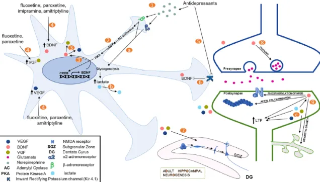
Figure 1. A schematic representation showing effects of monoamines and antidepressants on astrocytes, which are relevant to their antidepressant action. Classical antidepressants lead to increase in synaptic concentrations of monoamines, depicted here is norepinephrine (1). Activation of β-adrenoreceptors by norepinephrine can lead to upregulation of CREB-mediated transcription through adenylyl cyclase/PKA activation (2). This increases the expression of BDNF (3), which can, in turn, increase the expression of other trophic factors, namely, VGF and VEGF. Antidepressant may also increase trophic factor expression in astrocytes through yet unknown mechanisms (4). For instance, antidepressants are known to decrease the activity of an inward rectifying potassium channel (Kir4.1) (5), and a decrease in Kir4.1 activity or expression decreases BDNF expression (6). These trophic factors can then enhance adult hippocampal neurogenesis, thus aiding the behavioral effects of chronic antidepressant treatments (7). BDNF also enhances excitatory transmission by increasing vesicle docking and enhancing quantal release from glutamatergic presynaptic terminals (8). Trophic factors also enhance excitatory postsynaptic responses through various mechanisms (9). BDNF increases the expression of various NMDA receptor subunits and mediates the phosphorylation of NR2B. It is also known to enhance actin polymerization. In addition, both BDNF and VEGF are known to promote LTP. Moreover, BDNF and VGF mediate dendritic outgrowth which can rescue volumetric loss observed in depressive disorders. Norepinephrine and several antidepressants are also shown to induce breakdown of glycogen through α2- and β-adrenoreceptors in astrocytes (a). This results in increase in
glycolytic activity and production and secretion of lactate by astrocytes (b). Lactate, apart from acting as an energy substrate in neurons, is also known to increase NMDA currents, LTP, and plasticity-related gene expression, including expression of BDNF (c). BDNF indicates brain-derived neurotrophic factor; CREB, CRE-binding protein; LTP, long-term potentiation; PKA, protein kinase A; VEGF, vascular endothelial growth factor.
2. Drevets WC. Functional anatomical abnormalities in limbic and prefrontal cor-tical structures in major depression. Prog Brain Res. 2000;126:413–431. 3. Bremner JD, Vythilingam M, Vermetten E, et al. Reduced volume of
orbitofron-tal cortex in major depression. Biol Psychiatry. 2002;51:273–279.
4. Sheline YI. Neuroimaging studies of mood disorder effects on the brain. Biol
Psychiatry. 2003;54:338–352.
5. Etiévant A, Lambás-Señas L, Scarna H, Lucas G, Haddjeri N. Astrocytes and gliotransmitters: new players in the treatment of major depression? Curr Drug
Targets. 2013;14:1295–1307.
6. Cotter D, Mackay D, Landau S, Kerwin R, Everall I. Reduced glial cell density and neuronal size in the anterior cingulate cortex in major depressive disorder.
Arch Gen Psychiatry. 2001;58:545–553.
7. Miguel-Hidalgo JJ, Baucom C, Dilley G, et al. Glial fibrillary acidic protein immunoreactivity in the prefrontal cortex distinguishes younger from older adults in major depressive disorder. Biol Psychiatry. 2000;48:861–873.
8. Banasr M, Chowdhury G, Terwilliger R, et al. Glial pathology in an animal model of depression: reversal of stress-induced cellular, metabolic and behavioral deficits by the glutamate-modulating drug riluzole. Mol Psychiatry. 2010;15:501–511. 9. Banasr M, Duman RS. Glial loss in the prefrontal cortex is sufficient to induce
depressive-like behaviors. Biol Psychiatry. 2008;64:863–870.
10. Czéh B, Simon M, Schmelting B, Hiemke C, Fuchs E. Astroglial plasticity in the hippocampus is affected by chronic psychosocial stress and concomitant fluoxetine treatment. Neuropsychopharmacology. 2005;31:1616–1626.
11. Czéh B, Müller-Keuker JIH, Rygula R, et al. Chronic social stress inhibits cell proliferation in the adult medial prefrontal cortex: hemispheric asymmetry and reversal by fluoxetine treatment. Neuropsychopharmacology. 2007;32:1490–1503. 12. Rajkowska G, Miguel-Hidalgo JJ, Wei J, et al. Morphometric evidence for
neu-ronal and glial prefrontal cell pathology in major depression. Biol Psychiatry. 1999;45:1085–1098.
13. Choudary PV, Molnar M, Evans SJ, et al. Altered cortical glutamatergic and GABAergic signal transmission with glial involvement in depression. Proc Natl
Acad Sci U S A. 2005;102:15653–15658.
14. Miguel-Hidalgo JJ, Waltzer R, Whittom AA, Austin MC, Rajkowska G, Stockmeier CA. Glial and glutamatergic markers in depression, alcoholism, and their comorbidity. J Affect Disord. 2010;127:230–240.
15. Volterra A, Meldolesi J. Astrocytes, from brain glue to communication elements: the revolution continues. Nat Rev Neurosci. 2005;6:626–640.
16. Elsayed M, Magistretti PJ. A new outlook on mental illnesses: glial involvement beyond the glue. Front Cell Neurosci. 2015;9:468.
17. Eroglu C, Barres BA. Regulation of synaptic connectivity by glia. Nature. 2010;468:223–231.
18. Bernardinelli Y, Randall J, Janett E, et al. Activity-dependent structural plastic-ity of perisynaptic astrocytic domains promotes excitatory synapse stabilplastic-ity. Curr
Biol. 2014;24:1679–1688.
19. Zorec R, Araque A, Carmignoto G, Haydon PG, Verkhratsky A, Parpura V. Astroglial excitability and gliotransmission: an appraisal of Ca2+ as a signalling
route. ASN Neuro. 2012;4:e00080.
20. Magistretti PJ. Regulation of glycogenolysis by neurotransmitters in the central nervous system. Diabet Metab. 1988;14:237–246.
21. Schildkraut JJ. The catecholamine hypothesis of affective disorders. A review of supporting evidence. Int J Psychiatry. 1967;4:203–217.
22. Hirschfeld RM. History and evolution of the monoamine hypothesis of depres-sion. J Clin Psychiatry. 2000;61:4–6.
23. Lemberger L, Fuller RW, Zerbe RL. Use of specific serotonin uptake inhibitors as antidepressants. Clin Neuropharmacol. 1985;8:299–317.
24. Williams NR, Schatzberg AF. NMDA antagonist treatment of depression. Curr
Opin Neurobiol. 2016;36:112–117.
25. Li N, Lee B, Liu R-J, et al. mTOR-dependent synapse formation underlies the rapid antidepressant effects of NMDA antagonists. Science. 2010;329:959–964. 26. Birnstiel S, Haas HL. Acute effects of antidepressant drugs on long-term
poten-tiation (LTP) in rat hippocampal slices. Naunyn Schmiedebergs Arch Pharmacol. 1991;344:79–83.
27. Kanzari A, Bourcier-Lucas C, Freyssin A, Abrous DN, Haddjeri N, Lucas G. Inducing a long-term potentiation in the dentate gyrus is sufficient to produce rapid antidepressant-like effects. Molec Psychiatry. 2018;23:587–596.
28. Warner-Schmidt JL, Duman RS. Hippocampal neurogenesis: opposing effects of stress and antidepressant treatment. Hippocampus. 2006;16:239–249. 29. Malberg JE, Eisch AJ, Nestler EJ, Duman RS. Chronic antidepressant
treat-ment increases neurogenesis in adult rat hippocampus. J Neurosci. 2000;20:9104–9110.
30. Ming G, Song H. Adult neurogenesis in the mammalian central nervous system.
Annu Rev Neurosci. 2005;28:223–250.
31. Santarelli L, Saxe M, Gross C, et al. Requirement of hippocampal neurogenesis for the behavioral effects of antidepressants. Science. 2003;301:805–809. 32. Nibuya M, Morinobu S, Duman RS. Regulation of BDNF trkB mRNA in rat
brain by chronic electroconvulsive seizure antidepressant drug treatments. J
Neu-rosci. 1995;15:7539–7547.
33. Warner-Schmidt JL, Duman RS. VEGF is an essential mediator of the neuro-genic and behavioral actions of antidepressants. Proc Natl Acad Sci USA. 2007;104:4647–4652.
34. Thakker-Varia S, Krol JJ, Nettleton J, et al. The neuropeptide VGF produces antidepressant-like behavioral effects and enhances proliferation in the hippo-campus. J Neurosci. 2007;27:12156–12167.
35. Shirayama Y, Chen AC, Nakagawa S, Russell DS, Duman RS. Brain-derived neurotrophic factor produces antidepressant effects in behavioral models of depression. J Neurosci. 2002;22:3251–3261.
36. Inazu M, Takeda H, Matsumiya T. Functional expression of the norepi-nephrine transporter in cultured rat astrocytes. J Neurochem. 2003;84: 136–144.
37. Hirst WD, Price GW, Rattray M, Wilkin GP. Serotonin transporters in adult rat brain astrocytes revealed by [3H]5-HT uptake into glial plasmalemmal vesi-cles. Neurochem Int. 1998;33:11–22.
38. Hertz L, Lovatt D, Goldman SA, Nedergaard M. Adrenoceptors in brain: cel-lular gene expression and effects on astrocytic metabolism and [Ca(2+)]i.
Neuro-chem Int. 2010;57:411–420.
39. Berumen LC, Rodríguez A, Miledi R, García-Alcocer G. Serotonin receptors in hippocampus. Scientificworldjournal. 2012;2012:823493.
40. Eckenstein FP. Fibroblast growth factors in the nervous system. J Neurobiol. 1994;25:1467–1480.
41. Rudge JS, Alderson RF, Pasnikowski E, McClain J, Ip NY, Lindsay RM. Expression of ciliary neurotrophic factor and the neurotrophins-nerve growth factor, brain-derived neurotrophic factor and neurotrophin 3-in cultured rat hip-pocampal astrocytes. Eur J Neurosci. 1992;4:459–471.
42. Schaar DG, Sieber BA, Dreyfus CF, Black IB. Regional and cell-specific expres-sion of GDNF in rat brain. Exp Neurol. 1993;124:368–371.
43. Ballotti R, Nielsen FC, Pringle N, et al. Insulin-like growth factor I in cultured rat astrocytes: expression of the gene, and receptor tyrosine kinase. EMBO J. 1987;6:3633–3639.
44. Ijichi A, Sakuma S, Tofilon PJ. Hypoxia-induced vascular endothelial growth factor expression in normal rat astrocyte cultures. Glia. 1995;14:87–93. 45. Allaman I, Fiumelli H, Magistretti PJ, Martin J-L. Fluoxetine regulates the
expression of neurotrophic/growth factors and glucose metabolism in astrocytes.
Psychopharmacology (Berl). 2011;216:75–84.
46. Schmidt-Kastner R, Wetmore C, Olson L. Comparative study of brain-derived neurotrophic factor messenger RNA and protein at the cellular level suggests multiple roles in hippocampus, striatum and cortex. Neuroscience. 1996;74:161–183.
47. Gorba T, Wahle P. Expression of TrkB and TrkC but not BDNF mRNA in neu-rochemically identified interneurons in rat visual cortex in vivo and in organo-typic cultures. Eur J Neurosci. 1999;11:1179–1190.
48. Yanpallewar SU, Fernandes K, Marathe SV, et al. α2-adrenoceptor blockade accelerates the neurogenic, neurotrophic, and behavioral effects of chronic anti-depressant treatment. J Neurosci. 2010;30:1096–1109.
49. Sawamoto A, Okuyama S, Amakura Y, et al. 3,5,6,7,8,3′,4′-heptamethoxyfla-vone ameliorates depressive-like behavior and hippocampal neurochemical changes in chronic unpredictable mild stressed mice by regulating the brain-derived neurotrophic factor: requirement for ERK activation. Int J Mol Sci. 2017;18:E2133.
50. Kittel-Schneider S, Kenis G, Schek J, et al. Expression of monoamine transport-ers, nitric oxide synthase 3, and neurotrophin genes in antidepressant-stimulated astrocytes. Front Psychiatry. 2012;3:33.
51. Takano K, Yamasaki H, Kawabe K, Moriyama M, Nakamura Y. Imipramine induces brain-derived neurotrophic factor mRNA expression in cultured astro-cytes. J Pharmacol Sci. 2012;120:176–186.
52. Quesseveur G, David DJ, Gaillard MC, et al. BDNF overexpression in mouse hippocampal astrocytes promotes local neurogenesis and elicits anxiolytic-like activities. Transl Psychiatry. 2013;3:e253.
53. Bolo NR, Hodé Y, Nédélec JF, Lainé E, Wagner G, Macher JP. Brain pharmacoki-netics and tissue distribution in vivo of fluvoxamine and fluoxetine by fluorine mag-netic resonance spectroscopy. Neuropsychopharmacology. 2000;23:428–438. 54. Henry ME, Schmidt ME, Hennen J, et al. A comparison of brain and serum
pharmacokinetics of R-fluoxetine and racemic fluoxetine: a 19-F MRS study.
Neuropsychopharmacology. 2005;30:1576–1583.
55. Wong DT, Perry KW, Bymaster FP. Case history: the discovery of fluoxetine hydrochloride (Prozac). Nat Rev Drug Discov. 2005;4:764–774.
56. Furutani K, Ohno Y, Inanobe A, Hibino H, Kurachi Y. Mutational and in silico analyses for antidepressant block of astroglial inward-rectifier Kir4.1 channel.
Mol Pharmacol. 2009;75:1287–1295.
57. Larsen BR, MacAulay N.Kir4.1-mediated spatial buffering of K+: experimental challenges in determination of its temporal and quantitative contribution to K+ clearance in the brain. Channels (Austin). 2014;8:544–550.
58. Kinboshi M, Mukai T, Nagao Y, et al. Inhibition of inwardly rectifying potas-sium (Kir) 4.1 channels facilitates brain-derived neurotrophic factor (BDNF) expression in astrocytes. Front Mol Neurosci. 2017;10:408.
59. Koppel I, Jaanson K, Klasche A, et al. Dopamine cross-reacts with adrenorecep-tors in cortical astrocytes to induce BDNF expression, CREB signaling and morphological transformation. Glia. 2018;66:206–216.
60. Juric DM, Loncar D, Carman-Krzan M. Noradrenergic stimulation of BDNF synthesis in astrocytes: mediation via alpha1- and beta1/beta2-adrenergic recep-tors. Neurochem Int. 2008;52:297-306.
61. Tyler WJ, Pozzo-Miller LD. BDNF enhances quantal neurotransmitter release and increases the number of docked vesicles at the active zones of hippocampal excitatory synapses. J Neurosci. 2001;21:4249-4258.
62. Hu B, Nikolakopoulou AM, Cohen-Cory S. BDNF stabilizes synapses and maintains the structural complexity of optic axons. Development. 2005;132: 4285-4298.
63. Carreño FR, Walch JD, Dutta M, Nedungadi TP, Cunningham JT. BDNF-TrkB pathway mediates NMDA receptor NR2B subunit phosphorylation in the supraoptic nuclei following progressive dehydration. J Neuroendocrinol. 2011;23:894-905.
64. Caldeira MV, Melo CV, Pereira DB, Carvalho RF, Carvalho AL, Duarte CB. BDNF regulates the expression and traffic of NMDA receptors in cultured hip-pocampal neurons. Mol Cell Neurosci. 2007;35:208-219.
65. Jin K, Mao XO, Greenberg DA. Vascular endothelial growth factor stimulates neurite outgrowth from cerebral cortical neurons via Rho kinase signaling. J
Neurobiol. 2006;66:236-242.
66. Licht T, Goshen I, Avital A, et al. Reversible modulations of neuronal plasticity by VEGF. Proc Natl Acad Sci U S A. 2011;108:5081-5086.
67. Kajitani N, Hisaoka-Nakashima K, Morioka N, et al. Antidepressant acts on astrocytes leading to an increase in the expression of neurotrophic/growth factors: differential regulation of FGF-2 by noradrenaline. PLoS ONE. 2012;7:e51197. 68. Guo S, Arai K, Stins MF, Chuang D-M, Lo EH. Lithium upregulates vascular
endothelial growth factor in brain endothelial cells and astrocytes. Stroke. 2009;40:652-655.
69. Hunsberger JG, Newton SS, Bennett AH, et al. Antidepressant actions of the exercise-regulated gene VGF. Nat Med. 2007;13:1476-1482.
70. Sato H, Fukutani Y, Yamamoto Y, et al. Thalamus-derived molecules promote survival and dendritic growth of developing cortical neurons. J Neurosci. 2012;32:15388-15402.
71. Lin C-Y, Hung S-Y, Chen H-T, et al. Brain-derived neurotrophic factor increases vascular endothelial growth factor expression and enhances angiogenesis in human chondrosarcoma cells. Biochem Pharmacol. 2014;91:522-533.
72. Alder J, Thakker-Varia S, Bangasser DA, et al. Brain-derived neurotrophic fac-tor-induced gene expression reveals novel actions of VGF in hippocampal synap-tic plassynap-ticity. J Neurosci. 2003;23:10800-10808.
73. Evans SJ, Choudary PV, Neal CR, et al. Dysregulation of the fibroblast growth fac-tor system in major depression. Proc Natl Acad Sci USA. 2004;101:15506-15511. 74. Turner CA, Gula EL, Taylor LP, Watson SJ, Akil H. Antidepressant-like effects
of intracerebroventricular FGF2 in rats. Brain Res. 2008;1224:63-68. 75. Elsayed M, Banasr M, Duric V, Fournier NM, Licznerski P, Duman RS.
Anti-depressant effects of fibroblast growth factor-2 in behavioral and cellular models of depression. Biol Psychiatry. 2012;72:258-265.
76. Bachis A, Mallei A, Cruz MI, Wellstein A, Mocchetti I. Chronic antide-pressant treatments increase basic fibroblast growth factor and fibroblast growth factor-binding protein in neurons. Neuropharmacology. 2008; 55:1114-1120.
77. Magistretti PJ. Neuron-glia metabolic coupling and plasticity. J Exp Biol. 2006;209:2304-2311.
78. Pellerin L, Magistretti PJ. Sweet sixteen for ANLS. J Cereb Blood Flow Metab. 2012;32:1152-1166.
79. Swanson RA, Morton MM, Sagar SM, Sharp FR. Sensory stimulation induces local cerebral glycogenolysis: demonstration by autoradiography. Neuroscience. 1992;51:451-461.
80. Yang J, Ruchti E, Petit J-M, et al. Lactate promotes plasticity gene expression by potentiating NMDA signaling in neurons. PNAS. 2014;111:12228-12233. 81. Carrard A, Elsayed M, Margineanu M, et al. Peripheral administration of
lac-tate produces antidepressant-like effects. Mol Psychiatry. 2016;23:392-399. 82. Suzuki A, Stern SA, Bozdagi O, et al. Astrocyte-neuron lactate transport is
required for long-term memory formation. Cell. 2011;144:810-823.
83. Magistretti PJ, Manthorpe M, Bloom FE, Varon S. Functional receptors for vasoactive intestinal polypeptide in cultured astroglia from neonatal rat brain.
Regul Pept. 1983;6:71-80.
84. Subbarao KV, Hertz L. Effect of adrenergic agonists on glycogenolysis in pri-mary cultures of astrocytes. Brain Res. 1990;536:220-226.