HAL Id: hal-01435010
https://hal-univ-rennes1.archives-ouvertes.fr/hal-01435010
Submitted on 6 Mar 2017HAL is a multi-disciplinary open access
archive for the deposit and dissemination of sci-entific research documents, whether they are
pub-L’archive ouverte pluridisciplinaire HAL, est destinée au dépôt et à la diffusion de documents scientifiques de niveau recherche, publiés ou non,
Altered Expression of the Transcription Factor Forkhead
Box A1 (FOXA1) Is Associated With Poor Prognosis in
Urothelial Carcinoma of the Upper Urinary Tract
Jay D. Raman, Joshua I. Warrick, Carla Caruso, Zhaohai Yang, Lauren
Shuman, Richard D. Bruggeman, Shahrokh Shariat, Jose A. Karam,
Christopher Wood, Alon Z. Weizer, et al.
To cite this version:
Jay D. Raman, Joshua I. Warrick, Carla Caruso, Zhaohai Yang, Lauren Shuman, et al.. Altered Expression of the Transcription Factor Forkhead Box A1 (FOXA1) Is Associated With Poor Prognosis in Urothelial Carcinoma of the Upper Urinary Tract. Urology, Elsevier, 2016, 94, pp.314.e1-314.e7. �10.1016/j.urology.2016.05.030�. �hal-01435010�
Altered Expression of the Transcription Factor Forkhead Box A1 (FOXA1) is Associated with Poor Prognosis in Urothelial Carcinoma of the Upper Urinary Tract
Jay D. Raman1, Joshua I. Warrick1, Carla Caruso1, Zhaohai Yang1, Lauren Shuman1, Richard D. Bruggeman1, Shahrokh Shariat2, Jose A. Karam3, Christopher Wood3, Alon Z. Weizer4, Mesut Remzi2, Andrea Haitel2, Karim Bensalah5, Nathalie Rioux-Leclerq5, Christian Bolenz6, Marco Roscigno7, Laura-Maria Krabbe8, Payal Kapur8, Yair Lotan8, Vitaly Margulis8, David J. DeGraff1
1Penn State Milton S. Hershey Medical Center, Hershey, PA, USA 2Medical University of Vienna, General Hospital, Vienna, Austria 3M.D. Anderson Cancer Center, Houston, TX, USA
4University of Michigan, Ann Arbor, MI, USA 5University of Rennes, Rennes, France 6University of Ulm, Ulm, Germany 7AO Papa Giovanni XXIII, Bergamo, Italy
8UT Southwestern Medical Center, Dallas, TX, USA Running head: FOXA1 in upper tract urothelial carcinoma
Funding/Research support: Ken and Bonnie Shockey Fund for Urologic Research Disclosures: none
Keywords: biomarker, molecular marker, FOXA1, upper urinary tract, urothelial carcinoma, carcinoma in situ
Address for correspondence: David J. DeGraff, Ph.D.
Pennsylvania State University College of Medicine
Departments of Pathology, Biochemistry and Molecular Biology and Surgery, Division of Urology 500 University Drive Hershey, PA 17033 Phone: 717-531-0001 Ext 281295 Fax: 717-531-5021 ddegraff@hmc.psu.edu Text: 3,477 Abstract: 249 Abstract Objective:
To determine the prognostic significance of Forkhead Box A1 (FOXA1) expression in patients with upper tract urothelial carcinoma (UTUC) undergoing radical nephroureterectomy (RNU).
Materials and Methods:
A retrospective analysis of 566 patients undergoing RNU at 7 academic medical centers was performed. Tissue microarrays were subjected to immunohistochemistry using a commercially available polyclonal FOXA1 antibody. Logistic regression determined the association of FOXA1 expression with pathologic features and survival outcomes.
Results:
322 men and 244 women were included. The pathologic distribution of specimens included 53% muscle invasive or greater (≥ pT2), 74% high grade, 16% with flat architecture, 13% with necrosis, 21% with lymphovascular invasion, 18% with concomitant carcinoma in situ (CIS), and 8% with positive lymph nodes. Median FOXA1 score was 5.0 (range 0 - 8). Lower FOXA1 expression was significantly correlated with advanced pathologic stage (≥ pT3) (p=0.02), concomitant CIS (p=0.006) and renal pelvis (vs. ureter) location (p<0.0001). At a median follow-up of 27.0 months (range 3 - 196), 139 patients (25%) experienced a disease recurrence and 121 (21%) died from disease. In a multivariate model, lower FOXA1 expression was independently associated with disease recurrence (HR 1.11, 95% CI 1.05 - 1.62, p=0.04), cancer-specific mortality (HR 1.17, 95% CI 1.03 - 1.92, p=0.04), and all-cause mortality (HR 1.08, 95% CI 1.02 - 1.18, p=0.05).
Conclusions:
Lower FOXA1 expression is associated with adverse pathologic features and inferior survival outcomes for UTUC patients undergoing RNU. These data indicate lower FOXA1 expression may be a marker of aggressive disease in UTUC.
Introduction:
Upper-tract urothelial carcinoma (UTUC) is defined as urothelial carcinoma occurring in the kidney or the ureter, and accounts for 5% of urothelial tumors and 10% of renal neoplasms [1]. At present, the standard of care for the management of bulky, high-grade, or multifocal UTUC is radical nephroureterectomy (RNU) with an ipsilateral bladder cuff. Contemporary oncologic outcomes with this surgical approach remain durable for organ-confined, non-metastatic disease [2].
The relatively uncommon occurrence of UTUC renders it a difficult entity to manage clinically. Specifically, accurate identification of patients who would benefit from systemic chemotherapy (either before or after radical surgery), regional or extended lymph node dissection, or endoscopic (vs. extirpative) treatment of UTUC tumors remains vexing. Moreover, even in patients with similar disease on final pathology, clinical outcomes are somewhat variable thereby precluding tailored surveillance
regimens that are patient-specific [3]. Therefore, development of molecular markers that identify patients in need of the most aggressive clinical management and/or risk stratify patients in terms of clinical outcome have the potential to benefit treatment and reduce the cost of managing this disease [4].
The nuclear transcription factor Forkhead Box A1 (FOXA1) is normally expressed in urothelium of the bladder, ureter and renal pelvis [5] and expression of FOXA1 is required for the maintenance of normal urothelial differentiation in the bladder [6, 7]. In urothelial carcinoma of the bladder, FOXA1 expression can be either absent, increased, or altered. FOXA1 abnormalities are correlated with the molecular subtype of disease in bladder cancer [8], as well as squamous differentiation. In addition, we previously showed that lower FOXA1 expression is an independent predictor of reduced overall survival in bladder cancer and that Foxa1 knockout in the murine bladder induces urothelial hyperplasia and squamous metaplasia [7].
While FOXA1 is expressed in the urothelium of the renal pelvis and ureter, it is unknown if FOXA1 expression is altered in UTUC or whether the changes in FOXA1 expression correlate with oncologic outcomes. To investigate this, we determined the expression status and prognostic significance of FOXA1 in a large, multicenter,
Patients and Methods:
Patient selection
This study includes a previously described multi-institutional cohort of patients with UTUC who underwent RNU for curative treatment of their disease [2]. Institutional Review Board (IRB) approval was obtained prior to initiation of this study at all sites. The initial study cohort comprised 753 patients who underwent RNU for non-metastatic UTUC (pTa-pT4 N0/Nx M0) between March 1990 and May 2008. Exclusion criteria included the use of neoadjuvant chemotherapy and/or radiotherapy due to potential impact of such therapies on expression of FOXA1 in tumors. Additionally, patients with a follow-up less than three months and those who did not have evaluable tissue
specimens in triplicate were excluded. With such criteria, the final study cohort for this investigation comprised 566 patients.
Data collection and pathological evaluation
A computerized database was used to collect patient and tumor characteristics in conjunction with operative and outcomes data. Data was integrated amongst sites in a de-identified manner following approved data and material transfer agreements with the final data set frozen prior to analysis. Multiple data controls and quality assurance checks were done to ensure validity and completeness of the data.
Standard procedures were used to process pathological specimens. All specimens were re-reviewed by an institutional genitourinary pathologist blinded to clinical outcomes. Staging was assigned according to the 2002 American Joint Committee on Cancer – Union Internationale Contre le Cancer (AJCC-UICC)
Tumor-Node-Metastasis (TNM) classification. The 1998 WHO/International Society of Urologic Pathology (ISUP) consensus classification was used to assess tumor grade.
Furthermore, the following variables were collected for each specimen: tumor location, architecture (papillary vs. flat), lymphovascular invasion (LVI), tumor necrosis (defined as microscopic coagulative necrosis in > 10% of the tumor area) and presence of concomitant carcinoma in situ (CIS).
Creation of TMA and Immunohistochemistry for FOXA1
A tissue microarray (TMA) was created at the University of Texas Southwestern Medical Center for the 753 RNU patients with all tumors represented in triplicate as previously described [9]. Immunohistochemical analysis for FOXA1 (SC-6553 (C-20)); Santa Cruz Biotechnology) was performed using a Ventana Discovery XT stainer (Ventana Medical Systems, Tucson, AZ). Staining protocol consisted of pretreatment with CC2 (Ventana Medical Systems, Tucson, AZ) followed by incubation in the primary antibody for 60 minutes at 1:1000 dilution. The specificity of this antibody for FOXA1 has been previously determined through several studies, including through genetic knockout studies [7, 10-12]. Detection of primary antibody was performed with Ventana OmniMap DAB anti-goat detection kit (Ventana Medical Systems, Tucson,
AZ). Sections were counterstained with Mayer’s hematoxylin, dehydrated, cleared and coverslipped. Formalin-fixed, paraffin embedded samples of prostate and normal upper-tract urothelial tissue were used as positive controls for FOXA1 staining. In addition, preliminary analysis of whole 10 slides whole UTUC following
distribution was uniform throughout the specimen (data not shown). The TMA was scored by a single genitourinary pathologist blinded to the clinical and disease outcomes for patients. The Allred scoring system, which exhibits high-level
reproducibility [13], likely in response to its daily clinical use was chosen to quantify FOXA1 expression. This approach (total score of 0-8) is a summation of intensity (0 to 3 scale) and percentage (0 to 5 scale) [15]. The mean Allred score among the triplicate specimens was used for each tumor with those lacking three interpretable specimens excluded from analysis. With such criteria, the final cohort was 566 patients with evaluable TMA staining.
Surgical management and follow-up
There was no standardized approach for surgical RNU technique, management of the distal ureter, performance and extent of lymph node dissection, nor incorporation of adjuvant chemotherapy following intervention.
Follow-up after surgery was also not standardized, but in general patients were followed every 3-4 months in the first year after surgery, biannually in the second and annually in subsequent postoperative years. Follow-up included a personal history, physical examination, laboratory measurements, urinary cytology, cystoscopic evaluation of the urinary bladder and imaging of the contralateral upper urinary tract. Recurrences in the renal bed, retroperitoneal lymph nodes or distant sites were coded as disease relapse, whereas recurrences in the urinary bladder were coded as
secondary primaries. In case of death, the cause was determined on the basis of the death certificate only or in association with the patient’s medical record [16].
Statistical analysis
Outcomes of interest included the association of FOXA1 expression with
clinicopathologic variables as well as the association with oncologic survival outcomes. The Chi-square test was used to assess FOXA1 expression amongst categorical variables, whilst differences in continuous variables were analyzed using the Kruskal-Wallis H test. The Kaplan-Meier method was used to estimate the recurrence-free (RFS) and cancer-specific survival (CSS), as well as overall survival. Univariate and multivariate Cox proportional hazards models addressed associations of RFS and CSS with potential prognostic factors. All reported p-values are two-sided and statistical significance was set at ≤0.05. Statistical analysis was performed with S-Plus Professional version 4.5 (MathSoft Inc., Seattle, Washington).
Results
322 men and 244 women with a median age of 69 years were included. There was a roughly equal distribution of left (53%) and right (47%) sided UTUC cases with 434 (77%) managed via open and 132 (23%) by minimally invasive techniques. Distal ureter management was performed intravesically (n=270, 48%), extravesically (n=256, 45%), or endoscopically (n=40, 7%). Overall, 125 patients (22%) underwent a regional lymphadenectomy (median 6 nodes, range 1-24) with 44 (35%) having positive LN on final pathology.
The pathologic distribution of specimens included 53% muscle-invasive
(including 36% > pT3), 74% high grade, 16% with flat architecture, 13% with necrosis, 21% with LVI, and 18% with concomitant CIS. Median FOXA1 Allred score was 5.0 (IQR 4 - 6, range 0 - 8). When dichotomized by median FOXA1 expression, 253 (45%) patients had high (Allred > 5) while 313 (55%) had low FOXA1 (Allred < 5) expression. Patients with lower FOXA1 expression were more likely to have tumors with advanced pathologic stage (≥ pT3) (p=0.02), concomitant CIS (p=0.006) and renal pelvis (vs. ureter) location (p<0.0001). (Supplementary Figure 1 and Table 1)
Median follow-up for this study cohort was 27.0 months (IQR 12 – 52). 55 (10%) and 12 (2%) patients received adjuvant chemotherapy and radiotherapy, respectively. At last follow-up, 139 patients (25%) developed disease recurrence, 121 (21%) died of disease, and 196 (35%) died of any cause. For those with recurrence, median time to recurrence was 9.0 months (IQR 4 -19). The recurrence-free survival estimates in all patients at 3, 5, and 10 years were 79% (SE±2%), 63% (SE±3%), and 51% (SE±3%).
The cancer-specific survival estimates in all patients at 3, 5, and 10 years were 81% (SE±2%), 68% (SE±3%), and 60% (SE±3%). The overall survival estimates in all patients at 3, 5, and 10 years were 74% (SE±2%), 61% (SE±3%), and 49% (SE±3%). Lower FOXA1 Allred score (0-4) was associated with disease recurrence (p<0.005), death from UTUC (p=0.02), and death from any cause (p=0.04). (Figure 1A-C)
Table 2 highlights the univariate analysis. Non-organ confined UTUC (> pT3, p<0.001), concomitant CIS (p=0.04), tumor multifocality (p=0.04), and LN involvement (p<0.001) were all associated with UTUC recurrence. When considering cancer-specific survival, > pT3 UTUC (p<0.001), multifocality (p=0.003), LN involvement (p<0.001) as well as patient age (p=0.008) were associated with adverse outcomes. In addition, FOXA1 expression was associated with outcomes of UTUC. Specifically, lower FOXA1 expression was associated with disease recurrence (OR 1.16, 95% CI 1.06 - 1.27, p=0.03), cancer-specific mortality (HR 1.15, 95% CI 1.05 - 1.25, p=0.03), and overall mortality (HR 1.15, 95% CI 1.04 - 1.22, p=0.03).
A multivariate model was constructed incorporating clinicopathologic features associated with disease recurrence and cancer survival identified in the univariate analysis. (Table 3) Here, we identified that in addition to tumor stage, multifocality, and LN involvement, lower FOXA1 expression was independently associated with disease recurrence (OR 1.11, 95% CI 1.05 - 1.62, p=0.04), cancer-specific mortality (HR 1.17, 95% CI 1.03 - 1.92, p=0.04), and all-cause mortality (HR 1.08, 95% CI 1.02 - 1.18, p=0.05). Incorporation of FOXA1 into a base model including stage, LN status, and tumor focality improved the predictive accuracy for disease recurrence (AUC 72.2% to 73.3%, p=0.04) and UTUC mortality (AUC 74.6% to 75.5%, p=0.05).
Discussion:
Accurate staging of UTUC at the time of diagnosis has the potential to significantly improve the quality of care provided to these patients. This is because individuals with highly similar tumor characteristics often exhibit variable clinical
outcomes, indicating conventional pathologic assessment lacks specificity for diagnostic and prognostic purposes in regard to UTUC. Therefore, identification of molecular markers to more accurately stage disease, as well as enhance risk stratification of patients with UTUC can have a significant impact on clinical management for this understudied malignancy.
FOXA1 is a key transcriptional regulator of urothelial differentiation in the
bladder, and is directly implicated in bladder cancer [5-8, 17, 18]. However, the role of FOXA1 in the differentiation of upper-tract urothelium, as well as the degree to which altered FOXA1 expression is implicated in development and progression of UTUC is unknown. In the current study, we show lower FOXA1 expression is significantly associated with advanced tumor stage, presence of concomitant CIS and urothelial tumors of the renal pelvis. In addition, we show that loss of FOXA1 expression is an independent predictor of disease recurrence, cancer-specific mortality and overall mortality. These results provide strong evidence suggesting an important role for FOXA1 in UTUC, and serve as strong rationale to examine the function of this transcription factor in the biology of upper-tract urothelium and UTUC.
With the exception of RB1, which is rarely mutated in UTUC, a recent landmark next generation, targeted sequencing study revealed the mutational landscape of UTUC
and bladder cancer are similar [19]. However, this study reported significant differences in the prevalence of mutations in genes in UTUC compared to bladder cancer. For example, mutations in FGFR3, HRAS and CDKN2B were shown to be more common in UTUC, while TP53 and ARID1A mutations were more common in bladder cancer. In addition, while FOXA1 is mutated in approximately 5% of bladder cancers, the previous UTUC sequencing study identified only one case with a missense mutation, and two cases with an amplification of the FOXA1 gene ([19] and Drs. Hikmat Al-Ahmadie and Gopa Iyer, personal communications). One potential reason for the smaller number of mutations associated with FOXA1 in UTUC relative to bladder cancer could be a sampling issue, as frozen samples were used in the study focused on the genetic characterization of UTUC, and may have represented the superficial component of the analyzed tissue. This observation is pertinent because the majority of studies
describing mutations in FOXA1 in bladder cancer were focused on cystectomy samples for locally advanced disease. In addition, while amplifications in the FOXA1 gene also suggest the existence of molecular subtypes of UTUC similar to bladder cancer,
additional work is required to determine the mechanism(s) by which FOXA1 expression is altered in UTUC, and how alterations in FOXA1 expression potentially contribute to adverse clinical outcomes in patients with UTUC.
In bladder cancer, decreased FOXA1 expression is associated with the presence of a basal molecular subtype. The basal molecular subtype of bladder cancer is the most aggressive subtype identified to date [20]. The fact that our current studies show an association between reduced FOXA1 expression and reduced cancer-specific and overall mortality, as well as increased risk of recurrence suggest that UTUC may follow
a similar pattern of behavior, with tumors expressing diminished FOXA1 expression behaving in an aggressive manner akin to basal bladder cancer. Additional effort is required to identify molecular subtypes of UTUC, and determine how similar or different they are relative to bladder cancer. This is especially true as basal bladder cancers exhibit differential response to neoadjuvant chemotherapy [20], as well as potential susceptibility to targeted therapies [18], suggesting the same may potentially be true for UTUC.
We acknowledge some limitations in this study. Firstly, we restricted our analysis to tumor specimens replicated in triplicate in our TMA thereby reducing our overall cohort from over 750 patients to 566. Although our sample size declined, we do believe that this strict criterion improved our confidence of the reproducibility of staining and interpretation of FOXA1. Secondly, although tumor slides were reviewed by institutional genitourinary pathologists, no centralized pathologic review was performed. Thirdly, our results are based on use of the Allred scoring system [21] for interpretation of
immunohistochemical staining and different scoring systems may potentially yield different observations. Finally, our cohort is composed of patients managed at tertiary care academic medical centers which may introduce bias in the pathologic distribution presenting at such facilities. Despite these limitations, this work is certainly novel being the first to explore the expression of FOXA1 in any UTUC cohort, let alone a large international UTUC cohort.
Conclusions:
In a large, multi-institutional international cohort, FOXA1 expression is
significantly associated with advanced tumor stage, presence of concomitant CIS and urothelial tumors of the renal pelvis in UTUC. In addition, lower FOXA1 expression is an independent predictor of disease recurrence, cancer-specific mortality, and overall mortality in patients with UTUC. While statistically significant, increases in overall predictive accuracy when incorporating FOXA1 expression into a multivariate model are marginal. However, these results indicate FOXA1 potentially plays an important role in UTUC pathogenesis.
References
1. Lucca I, Leow JJ, Shariat SF, Chang SL: Diagnosis and management of upper tract
urothelial carcinoma. Hematology/oncology clinics of North America 2015, 29(2):271-288, ix.
2. Margulis V, Shariat SF, Matin SF, Kamat AM, Zigeuner R, Kikuchi E, Lotan Y, Weizer A, Raman JD, Wood CG et al: Outcomes of radical nephroureterectomy: a series
from the Upper Tract Urothelial Carcinoma Collaboration. Cancer 2009, 115(6):1224-1233.
3. Verhoest G, Shariat SF, Chromecki TF, Raman JD, Margulis V, Novara G, Seitz C, Remzi M, Roupret M, Scherr DS et al: Predictive factors of recurrence and
survival of upper tract urothelial carcinomas. World journal of urology 2011, 29(4):495-501.
4. Xiong G, Liu J, Tang Q, Fan Y, Fang D, Yang K, Xie F, Zhang M, Zhang L, Liu L et al:
Prognostic and predictive value of epigenetic biomarkers and clinical factors in upper tract urothelial carcinoma. Epigenomics 2015, 7(5):733-744.
5. Varley CL, Bacon EJ, Holder JC, Southgate J: FOXA1 and IRF-1 intermediary
transcriptional regulators of PPARgamma-induced urothelial cytodifferentiation. Cell Death Differ 2009, 16(1):103-114.
6. DeGraff DJ, Clark PE, Cates JM, Yamashita H, Robinson VL, Yu X, Smolkin ME, Chang SS, Cookson MS, Herrick MK et al: Loss of the urothelial differentiation marker
FOXA1 is associated with high grade, late stage bladder cancer and increased tumor proliferation. PloS one 2012, 7(5):e36669.
7. Reddy OL, Cates JM, Gellert LL, Crist HS, Yang Z, Yamashita H, Taylor JA, 3rd, Smith JA, Jr., Chang SS, Cookson MS et al: Loss of FOXA1 Drives Sexually Dimorphic
Prognosis in Bladder Cancer. The American journal of pathology 2015, 185(5):1385-1395.
8. Cancer Genome Atlas Research N: Comprehensive molecular characterization of
urothelial bladder carcinoma. Nature 2014, 507(7492):315-322.
9. Lee DJ, Xylinas E, Rieken M, Khani F, Klatte T, Wood CG, Karam JA, Weizer AZ, Raman JD, Remzi M et al: Insulin-like growth factor messenger RNA-binding protein 3
expression helps prognostication in patients with upper tract urothelial carcinoma. European urology 2014, 66(2):379-385.
10. DeGraff DJ, Grabowska MM, Case TC, Yu X, Herrick MK, Hayward WJ, Strand DW, Cates JM, Hayward SW, Gao N et al: FOXA1 deletion in luminal epithelium causes
prostatic hyperplasia and alteration of differentiated phenotype. Laboratory
investigation; a journal of technical methods and pathology 2014, 94(7):726-739.
11. Mirosevich J, Gao N, Gupta A, Shappell SB, Jove R, Matusik RJ: Expression and role
of Foxa proteins in prostate cancer. The Prostate 2006, 66(10):1013-1028.
12. Mirosevich J, Gao N, Matusik RJ: Expression of Foxa transcription factors in the
developing and adult murine prostate. The Prostate 2005, 62(4):339-352.
13. Reisenbichler ES, Lester SC, Richardson AL, Dillon DA, Ly A, Brock JE:
Interobserver concordance in implementing the 2010 ASCO/CAP
recommendations for reporting ER in breast carcinomas: a demonstration of the difficulties of consistently reporting low levels of ER expression by manual quantification. American journal of clinical pathology 2013, 140(4):487-494.
14. Bartosch C, Monteiro-Reis S, Almeida-Rios D, Vieira R, Castro A, Moutinho M, Rodrigues M, Graca I, Lopes JM, Jeronimo C: Assessing sirtuin expression in
endometrial carcinoma and non-neoplastic endometrium. Oncotarget 2016, 7(2):1144-1154.
15. Harvey JM, Clark GM, Osborne CK, Allred DC: Estrogen receptor status by
immunohistochemistry is superior to the ligand-binding assay for predicting response to adjuvant endocrine therapy in breast cancer. Journal of clinical
oncology : official journal of the American Society of Clinical Oncology 1999,
17(5):1474-1481.
16. Rink M, Fajkovic H, Cha EK, Gupta A, Karakiewicz PI, Chun FK, Lotan Y, Shariat SF:
Death certificates are valid for the determination of cause of death in patients with upper and lower tract urothelial carcinoma. European urology 2012, 61(4):854-855.
17. DeGraff DJ, Cates JM, Mauney JR, Clark PE, Matusik RJ, Adam RM: When urothelial
differentiation pathways go wrong: implications for bladder cancer development and progression. Urologic oncology 2013, 31(6):802-811.
18. Rebouissou S, Bernard-Pierrot I, de Reynies A, Lepage ML, Krucker C, Chapeaublanc E, Herault A, Kamoun A, Caillault A, Letouze E et al: EGFR as a potential
therapeutic target for a subset of muscle-invasive bladder cancers presenting a basal-like phenotype. Science translational medicine 2014, 6(244):244ra291.
19. Sfakianos JP, Cha EK, Iyer G, Scott SN, Zabor EC, Shah RH, Ren Q, Bagrodia A, Kim PH, Hakimi AA et al: Genomic Characterization of Upper Tract Urothelial
Carcinoma. European urology 2015, 68(6):970-977.
Classification of Chemotherapy-naive Urothelial Cancer is Predictive of Clinical Outcomes from Neoadjuvant Chemotherapy: A Phase 2 Trial of Dose-dense Methotrexate, Vinblastine, Doxorubicin, and Cisplatin with
Bevacizumab in Urothelial Cancer. European urology 2015.
21. Allred DC, Harvey JM, Berardo M, Clark GM: Prognostic and predictive factors in
breast cancer by immunohistochemical analysis. Modern pathology : an official
journal of the United States and Canadian Academy of Pathology, Inc 1998,
11(2):155-168. Figures
Supplementary Figure 1: Representative immunohistochemistry for FOXA1. Normal renal pelvis and ureter urothelial are assigned Allred Scores 5+3=8 and 5+2=7, respectively (Fig 1A); pTa stage tumors in the renal pelvis and ureter assigned Allred scores 5+2=7 (Fig 1B); pT3 stage tumors in the renal pelvis and ureter assigned Allred score 0 (Fig 1B).
Figure 1: Kaplan Meier curves for recurrence-free (Fig 1A), cancer-specific (Fig 1B), and overall survival (Fig 1C) in 566 patients underoging RNU stratified by FOXA1 expression.
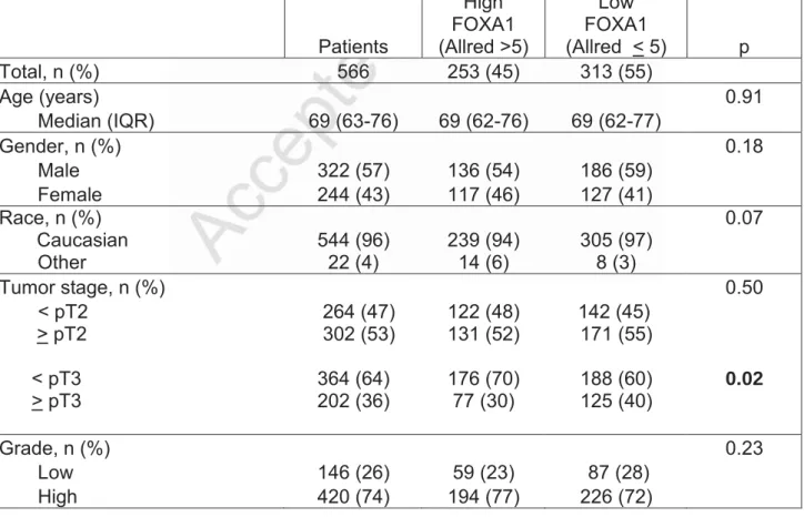
Table 1. Association of FOXA1 expression with clinical and pathologic characteristics in 566 patients managed by radical nephroureterectomy.
Patients High FOXA1 (Allred >5) Low FOXA1 (Allred < 5) p Total, n (%) 566 253 (45) 313 (55) Age (years) 0.91 Median (IQR) 69 (63-76) 69 (62-76) 69 (62-77) Gender, n (%) 0.18 Male 322 (57) 136 (54) 186 (59) Female 244 (43) 117 (46) 127 (41) Race, n (%) Caucasian Other 544 (96) 22 (4) 239 (94) 14 (6) 305 (97) 8 (3) 0.07 Tumor stage, n (%) 0.50 < pT2 264 (47) 122 (48) 142 (45) > pT2 < pT3 > pT3 302 (53) 364 (64) 202 (36) 131 (52) 176 (70) 77 (30) 171 (55) 188 (60) 125 (40) 0.02 Grade, n (%) 0.23 Low 146 (26) 59 (23) 87 (28)
Lymph node status, n (%) 0.60 pN0/pNx 522 (92) 235 (93) 287 (92) pN+ 44 (9) 18 (7) 26 (8) LVI, n (%) 0.74 Yes 120 (21) 52 (21) 68 (22) No 446 (79) 201 (79) 245 (78) Concomitant CIS, n (%) 0.006 Yes 104 (18) 35 (14) 69 (22) No 462 (82) 218 (86) 244 (78) Multifocality, n (%) 0.16 Yes 118 (21) 46 (18) 72 (23) No 448 (79) 207 (82) 241 (77) Necrosis, n (%) 0.33 Yes 74 (13) 37 (15) 37 (12) No 492 (87) 216 (85) 276 (88) Architecture, n (%) 0.86 Papillary 477 (84) 216 (85) 261 (83) Sessile 89 (16) 37 (15) 42 (17) Location, n (%) <0.001 Renal pelvis 418 (74) 164 (65) 254 (81)
Ureter & UEA 148 (26) 89 (35) 59 (19) Abbreviations
LVI = lymphovascular invasion CIS = carcinoma in situ
UEA = ureteroenteric anastomosis
Table 2. Univariate Cox regression analyses predicting disease recurrence and cancer-specific mortality in 566 patients undergoing radical nephroureterectomy.
Lower Recurrence-free survival Lower Cancer-specific survival HR 95 % CI p value HR 95 % CI p value Age 1.06 0.99 - 1.08 0.068 1.07 1.02 - 1.09 0.008 Male gender 0.74 0.53 - 1.05 0.081 0.81 0.62 - 1.04 0.14 Pathologic stage < pT3 (Reference) > pT3 9.78 4.36 - 36.74 <0.001 8.16 2.93 – 31.19 <0.001
Pathological high grade 1.56 0.88 – 1.99 0.37 1.61 0.74 - 2.12 0.18 LVI 1.28 0.91 - 1.62 0.52 1.23 0.81 - 1.84 0.22
Sessile Architecture 1.39 0.84 - 1.96 0.18 1.27 0.81 - 2.12 0.11
Necrosis (>10%) 0.98 0.92 – 1.17 0.46 1.03 0.60 – 1.15 0.36 Multifocality 1.49 1.02 - 2.15 0.04 1.83 1.31 - 2.88 0.003
Renal pelvis location 1.08 0.76 - 1.67 0.38 1.28 0.91 - 1.91 0.21 Lymph node metastasis 2.55 1.68 - 3.58 <0.001 2.83 1.41 – 4.08 <0.001
Lower FOXA1 Expression (< 5) 1.16 1.06 – 1.27 0.03 1.15 1.05 - 1.25 0.03 CI: confidence interval
LVI: lymphovascular invasion CIS: carcinoma in situ
HR: hazard ratio
Table 3. Multivariate Cox regression analyses predicting disease recurrence and cancer-specific mortality in 566 patients undergoing radical nephroureterectomy.
Lower
Recurrence-free survival Cancer-specific survival Lower
HR 95 % CI p value HR 95 % CI p value Age 1.03 0.81 - 1.17 0.124 1.05 1.01 - 1.16 0.074 Pathologic stage < pT3 (Reference) > pT3 12.48 5.11 - 42.24 <0.001 10.69 3.87 - 35.47 <0.001 Multifocality 1.38 0.96 – 1.88 0.047 1.92 1.41 - 2.98 0.028
Lymph node metastasis 5.15 2.71 - 7.62 0.008 4.54 2.31 – 7.28 0.009
Lower FOXA1 Expression (< 5) 1.11 1.05 - 1.62 0.044 1.17 1.03 - 1.92 0.039 HR: hazard ratio