HAL Id: in2p3-00448278
http://hal.in2p3.fr/in2p3-00448278
Submitted on 10 Feb 2010HAL is a multi-disciplinary open access
archive for the deposit and dissemination of sci-entific research documents, whether they are pub-lished or not. The documents may come from teaching and research institutions in France or abroad, or from public or private research centers.
L’archive ouverte pluridisciplinaire HAL, est destinée au dépôt et à la diffusion de documents scientifiques de niveau recherche, publiés ou non, émanant des établissements d’enseignement et de recherche français ou étrangers, des laboratoires publics ou privés.
ThomX - Conceptual Design Report
C. Bruni, R. Chiche, R. Cizeron, Y. Fedala, J. Haissinski, M. Jacquet, D.
Jehanno, M. Lacroix, L. Meignien, B. Mercier, et al.
To cite this version:
C. Bruni, R. Chiche, R. Cizeron, Y. Fedala, J. Haissinski, et al.. ThomX - Conceptual Design Report. 2009, pp.1-136. �in2p3-00448278�
LAL RT 09/28
SOLEIL/SOU-RA-2678
THOMX
Conceptual Design Report
Editors:
A.Variola
A.Loulergue
This Conceptual Design Report is dedicated to
Joseph Salomon
LIST OF CONTRIBUTORS
LAL CNRS, Université Paris-Sud 11, Batiment 200, 91898 Orsay, France:
C.Bruni, R.Chiche, R.Cizeron, Y.Fedala, J.Haissinski, M.Jacquet, D.Jehanno, M.Lacroix, L.Meignien, B.Mercier, B.Mouton, Y.Peinaud, C.Prevost, R.Roux, V.Soskov, A.Variola, G.Wormser, F.Zomer
Synchrotron-SOLEIL,Saint-Aubin, France :
P.Brunelle, M.E.Couprie, J.C.Denard, J.M.Filhol, N.Guillotin, P.Lebasque, A.Loulergue, P.Marchand, O.Marcouillé, F.Marteau, R.Nagaoka
Centre Lasers Intenses et Application, CNRS – Université de Bordeaux 1: P.Balcou, E. Cormier, M.C. Nadeau
C2RMF-UMR171 du CNRS/Ministère de la Culture : P.Walter
ILE, Ecole Polytechnique, CNRS, Palaiseau, France : N. Artemiev
L.M.A. CNRS, 7, Avenue Pierre de Coubertin VILLEURBANNE, France : R. Flaminio, C. Michel, L. Pinard, B. Sassolas.
THALES :
J.P. Brasile, Marie-Christine Nadeau
ESRF : European Synchrotron Radiation Facility, Grenoble 38403 France A.Bravin, G.LeDuc
THOMX ... 1
LIST OF CONTRIBUTORS ... 3
ACKNOWLEDGEMENTS ... 7
INTRODUCTION ... 8
- The ThomX working scheme ... 12
- X rays sources, Compton and alternative schemes. The ThomX positioning ... 13
- Compton ... 14
- Other sources ... 15
References ... 16
CHAPTER 1: Scientific Case ... 18
1.1] Cultural Heritage Applications ... 18
1.1.1] Introduction ... 18
1.1.2] Expected scientific impact ... 19
1.1.2.1] Analytical methods ... 19
1.1.2.2] X-ray imaging based on different physical properties ... 20
1.2] BioMedical Applications ... 22
1.2.1] Radiotherapy programs ... 22
1.2.2] Imaging ... 24
1.2.3] Conclusion for medical applications ... 27
1.3] X-ray crystallography ... 27
1.3.1] Protein structure determination ... 27
1.3.2] System dynamic studies ... 28
1.3.3] Compact Compton based source for protein study ... 28
References ... 28
CHAPTER 2. Thomson and Compton scattering ... 31
2.1] Photon scattering ... 31
2.2] High energy behaviour. Unpolarised and polarised Compton backscattering cross section ... 32
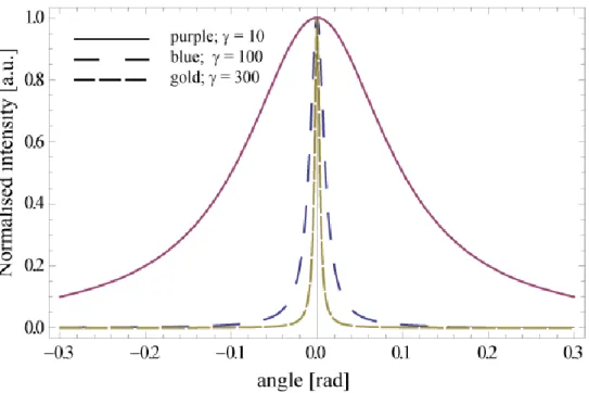
2.3] The frequency shift and the emission angular distribution ... 34
2.4] Parameterization of RadioThomX source. ... 38
2.4.1] Flux dependence on the beams waists sizes. ... 39
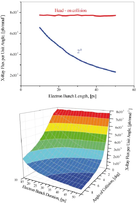
2.4.2] Flux dependence from the collision angle. ... 40
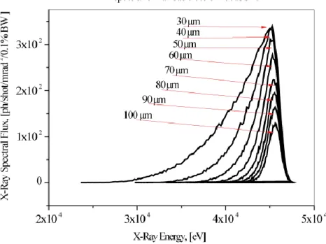
2.4.3] Brightness and monochromaticity. ... 43
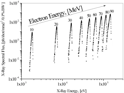
2.4.4] Flux dependence from the electron beam energy. ... 45
References ... 46
CHAPTER 3: Optical Systems ... 47
3.1] Laser and Fabry Perot resonator ... 47
3.1.2] Introduction ... 47
3.1.2] Laser ... 47
3.1.3] Frequency doubling ... 52
3.1.4] Laser cavity locking ... 52
3.1.5] Cavity design ... 54
3.1.6] Mechanical Solution ... 55
3.1.7] Cavity mirror coating ... 59
References ... 59
CHAPTER 4 : Accelerator ... 60
4.1] Injector ... 60
4.2]. RF simulations ... 60
4.3.1] Summary ... 65
4.4] Transfer line ... 65
4.5.1] Main requirements ... 68
4.5.2] Linear optics ... 69
4.5.3] Optics optimisation ... 70
4.5.4] Second order momentum compaction ... 73
4.5.5] Integration of the optical cavity ... 74
4.5.6] Orbits ... 75
4.5.7] Collective effects ... 75
4.5.8] Injection matching ... 77
Appendix 1 ... 82
Appendix 2 ... 82
4.5.9] Impact of Compton back scattering on the longitudinal and the transverse dynamics of the electron beam ... 83
4.5.10] Touschek effect ... 86 4.5.11] Intra-beam scattering ... 86 4.5.12] Vacuum effect ... 87 4.5.13] Ions instabilities ... 88 4.6] Equipments ... 89 4.6.1] Magnets ... 89 4.6.1.1] Power Supplies ... 92
4.6.2] Pulsed magnetic systems for the ring injection ... 93
4.6.3] Storage ring RF system ... 95
4.6.3.1] HOM impedance thresholds for coupled bunch mode instability (CBMI) ... 95
4.6.3.2] Cures to HOM driven CBMI ... 96
Conclusions ... 102
4.6.4] Diagnostics and beam dump ... 103
4.6.4.1] Charge, current and life time ... 103
4.6.4.2] Beam Position Monitors ... 103
4.6.4.3] Bunch length measurements ... 103
4.6.4.4] Transverse profile measurements ... 104
4.6.4.5] Beam dump ... 104
4.6.5] Vacuum ... 104
4.6.5.1] Pumping distribution in the ThomX ring ... 104
4.6.6] Synchronization ... 107
Summary of the synchronization constraints ... 109
4.6.7] X ray beam extraction and characterisation ... 110
References ... 111
CHAPTER 5: Radioprotection and Integration ... 113
5.1] Radiation Shielding Design ... 113
References ... 116
CHAPTER 6 : ERL and LINAC solutions. Feasibility considerations ... 117
6.1] Introduction ... 117
6.2] Normal conducting LINAC ... 117
6.3] Superconducting LINAC ... 117
6.4] Comparison of x rays fluxes ... 119
6.5] Energy Recovery Linac ... 120
6.6] Conclusion ... 121
References ... 121
7.1] Risk analysis ... 122
7.2] Planning ... 122
7.3] Budget ... 123
7.4] Industrialization ... 124
Annexe 1: Main source parameters ... 126
Annexe 2: Costing ... 129
CONCLUSIONS ... 132
ACKNOWLEDGEMENTS
We would like to thank S.Bielawski, P.Georges, V.Leflanchet, F.Meot, A.Mosnier, J.M Ortega, W.Scandale and M.Svanderlik for improving the text by providing valuable suggestions and remarks.
INTRODUCTION
Compton based photon sources generate a lot of interest since the rapid advancement in laser and accelerator technologies allows envisaging their utilisation for ultra-compact radiation sources. These should provide X-rays short pulses with a relatively high average flux. Moreover, the univocal dependence between the emitted photon energy and its scattering angle gives the possibility to obtain a quasi-monochromatic beam by a simple diaphragm system. For the most ambitious projects, such as the one presented here, the envisaged performances take into account a rate of 1012-1013 photons/s, an angular divergence of few mrad, an X ray energy cut-off of few tens of keV and a bandwidth E/E ~ 1-10%. Even if the integrated rate cannot compete with the synchrotron radiation sources, the cost and the compactness of these Compton based machines make them attractive for a wide spectrum of applications. For example the ThomX machine design footprint fits in a 70m2 surface (see fig.1).
Figure 1: Footprint of the ThomX machine.
Compton compact sources are extremely interesting as far as the chemistry component analysis field is concerned. One of the most promising applications is the cultural heritage preservation and associated domains. Tuneable and highly monochromatic hard X-rays can be obtained in Compton machines with the use of diaphragms (1-10% bandwidth) or monochromators (0.1% bandwidth). Diffraction techniques, laminography and painting components chemical analysis should take advantages of such radiation sources. A great advantage should be acquired by exploiting the complementarity between the analysis worked out by employing ion sources like elastic scattering (RBS, ERDA), the use of nuclear reactions for the detection of light elements, and the hard X-Ray techniques. Heavy elements analysis, absorption spectrometry, X-ray diffraction and diffusion, tomography and phase contrast imaging will allow an important widening of the application range in the masterpieces analysis domain. To detect the interesting elements associated to the cultural heritage preservation (Mn, Co, Fe, Cu, Zn, Zr, Ag, Cd, Sb, Sn, Ba, Hg, Pb), by XANES or
10m
analysis at the absorption edge, different photon energies are necessary. They correspond to the K edge of the overmentioned metals and to the Pb and Au LIII threshold. This means that a
photon energy ranging from 6.5 to 89 keV is necessary. The following table illustrates the required radiation performances for different analysis techniques:
XRF XRD XANES Tomography Edge enhancement Phase contrast Magnification Energy range [keV] 6.5-89 10-89 6.5-89 20-100 7-100 10-30 10-100 E/E 1-3% 3 – 10 % 5 – 10 % 3% bw 3 – 10 % 3% bw 3% bw
Source size 10-100 µm 10-100 µm Very small Very small
Size on the object 10-20 µm 10-20 µm 10-20 µm 10-50 cm 50 cm 50 cm 1-50 mm Flux on the object [ph/s] 109-1010 109 ph/s 107 ph/s 1011 109 1011 1011
Acquisition time 1s - 5 min 1s - 5 min 30 min
Coherence No No Yes Yes
Table 1: Requirements table for the analysis techniques used in heritage studies
So, by combining ion and photon measurement techniques on the same object, it is possible to assume that a complete material analysis will be worked out. These techniques provide precious information for the dating of the work of art, the employed techniques and the attribution [1]. Carbon detection can show the primitive sketch of the original drawing thus revealing the modifications during its realisation. Non destructive analysis of paintings permits also to reveal underlying drawings. An important synergy can be developed in integrated laboratories where physicists, chemists, art critics and historians collaborate in the same framework. Here the compactness of the Compton machine plays a fundamental role. The possibility to place such a source in an integrated laboratory gives to the expert direct access to the masterpieces in the Museum [2]. On the other side, an external facility, will involve extremely important insurance, security and transportation costs.
As far as the medical science applications are concerned an important benefit is, first of all given in the imaging field, by the development of the phase contrast method. Other applications are possible, for example, in static and dynamic imaging [3,4], 3D compressionless mammography [3,5], broncography, catheterless coronary arteries angiography [6] and in the K-edge radiography and therapy.
An important feature of Compton sources is the tunability at specific wavelength obtainable by varying the energy of the electron beam (quadratic dependence) or the wavelength of the impinging laser (linear dependence). This is attractive since a resonant reaction can be triggered by the interaction between the X rays with a determined energy, and an electronic shell of a contrast agent (like K-shell extraction with a subsequent energy release by Auger cascade). Contrast agents (like Gadolinium or Platinum) based cancer imaging and therapy [3] could represent the real breakthrough of the Compton machines in the medical field. The importance of the monochromaticity in biological tissue K-edge interactions is described in [3], where the imaging and therapy applications are illustrated.
Other interesting domains can be identified in the fields that usually operate synchrotron radiation sources but that do not need a very high average flux. A spin-off of the SLAC laboratory has created a commercial Compton source for X-ray diffraction protein
crystallography [7] where performances close to the synchrotron light sources ones are attained. This is the first example of a working mini-synchrotron based on the Compton effect. Moreover, due to the quadratic dependence of the Compton energy cut-off on the electron beam energy, it is easy to imagine harder photon production. This allows one to envisage different applications in the nuclear waste management and treatment industry [8] and in the field of nuclear isotope detection applied to infrastructures security. In this framework the atomic number identification by means of hard-X or gamma rays allows for nuclear application of the Compton scattering [9]. The high penetration power of these photons is extremely attractive when applied to security and to the radiography of shielded material. Also in this case resonant detection can be applied; one may take advantage from the relative monochromaticity degree of the photons pulses.
As one may expect, the required X-ray beam characteristics are different for different applications. Interesting rates range between 106 to 1013 photons/s. The Compton energy cut-off must vary from few keV for medical applications to the MeV range for nuclear applications. In this context one should point out that the Compton spectrum is continuous and quasi-constant (there is only a factor ~ 2 between its maximum and minimum) up to the energy cut-off (See fig. 2, Chapter 2). Therefore all the techniques requiring tunability can make use of monochromators at different wavelengths if X-ray optics is available in the explored range. Pulse length requirements can be very demanding in fast biochemistry studies (hundreds of fs range). Nevertheless other applications need to be exploited starting from the picosecond range. A general common requirement is the compactness: the main appeal of the Compton sources is to provide a high quality x ray beam from a source that can be easily hosted in a University laboratory, a Hospital, a Museum. The price is consequently strongly reduced with respect to the synchrotron sources.
Taking into account all these considerations several French laboratories [10] started to evaluate the feasibility of a compact Compton source: the project ThomX. This was the natural outcome of the different important technological results on Fabry-Perot optical resonators and fibre lasers obtained in these laboratories, and by their strong experience in design and building electron accelerators and storage rings. This allowed, as a first evaluation, to consider a prototype machine with performances at the top of the existing ones. In fact, an already financed ANR program aims to demonstrate the coupling of a high average power fibre laser with a very high finesse optical cavity. This will permit storing an incredible photon pulses average energy, in the order of 100kW-1MW. This system will be installed in the ATF ring (KEK – Tsukuba – Japan) as a demonstration of gamma factory for polarised positron production. The same system integrated in a low energy storage ring allows one to consider a very ambitious program, as far as the X Compton sources are concerned.
The ThomX French consortium is formed by different leading laboratories in the Compton associated technology. At present, accelerators systems, lasers and optical resonators are being developed by these teams reaching exceptional performances. This scenario ensures that the ThomX project should be implemented reaching the top of the actual performances in this domain.
Due to the wide spectrum of applications and requirements form different scientific communities, the ThomX project starts with a pre-defined performances range table permitting to summarize the needs in high average flux, photon energy (tens of keV), bandwidth, divergence etc etc. Moreover this prototype will allow acquiring an important
experience in operating such a machine and in understanding the subtleties of the beam dynamics under Compton collision regime. Only when the specific user community requirements will be definitively defined, it will be possible to finalise the operation mode and to fine tune this complex device required performances. Table 2 summarizes the design main parameters range of the ThomX project.
Source explored range
X energy 50-90 keV
Flux 1011 – 1013 ph/s
Bandwidth 10 %
Divergence < 2 mrad
Accelerator and laser
Ring and injector energies 50 MeV
Charge 1 nC
Emittance (normalised rms,) < 5 mm mrad
* 10 cm =>IP ~ 70m
Intracavity average power > 100 kW
Compton frep 50-200 MHz
Table 2 ThomX parameters ranges
It is important to stress that Compton sources are complementary to the third and fourth generation radiation facilities since they cannot compete in integrated emitted rates and in brightness. The performances of the most ambitious backscattering sources can be placed near the first generation synchrotron sources but with more attractive characteristics (flux, directivity, monochromaticity, tunability) than other X-ray sources like, for example, X-tubes. Classical bremsstrahlung X-ray tubes can deliver typical spectral rays with a large emission spectrum and different intensities (rotating anode or microfocus tubes with adaptive optics). At present the rotating anode X-ray tubes associated to a system for X Rays optics, are the most efficient sources if a laboratory integration is taken into account. The Rigaku FR-E model with the Vari-max HF optics (Osmic distr.) and a 2.475 kW tube, provide 7.109 ph/s maximum flux on a 200 µm diameter surface. Nevertheless these sources do not allow developing more ambitious techniques as far as diffraction, diffusion, absorption, imaging and spectroscopy are concerned. For this reasons these techniques are commonly used only at the synchrotron radiation facilities.
In fig.2 the general X-ray sources framework is illustrated by plotting the average brightness and average emitted flux of different facilities and devices. One notices that the proposed ThomX Compton sources, ranging in 1010 - 1011 brightness, can be compared with the old generation synchrotron sources, with the great advantages that the emitted photons energy cut-off allows one to provide for much harder X-ray beams.
Figure 2 : The performances of different generation synchrotron radiation sources are displayed. The Compton source is also visible in the figure to highlight the high energy cut-off. The considered photon emission is provided by an electron beam of 50 MeV, respectively a factor ten, sixteen and fifty-five less than the first, second and third generation facilities. Increasing the energy of the Compton source will reduce the beam emittance at the injection. The result is an increase of the brightness as the square root of the Lorentz gamma factor.
- The ThomX working scheme
To better introduce the ThomX project a first basic illustration of its working principle is presented. Actually, Compton sources devices are based on collisions between light pulses and electron beams. Nevertheless, diverse schemes are possible depending on the required performances. In fact, subject to the applications, average or peak brilliance, monochromaticity or peak energy cut off, beam source size or emission cone are preferred. In this framework different electron and laser systems can be chosen. Electron bunches should be provided by normal or super conductive linacs, recirculated in Energy Recovery Linacs (ERL) or accumulated in storage rings. On the other hand, light pulses can be delivered by laser systems after either active or passive amplification in optical cavities.
The ThomX machine is conceived to provide the maximum average flux in a fixed bandwidth. Consequently, the basic scheme takes into account a very important collision repetition frequency and therefore the possibility to have Compton interaction in a storage ring:
Electron bunches are injected and stored in the ring and discarded in a beam dump after 20 ms. To increase the pulse power of the light pulse the high average power laser is injected into a passive optical resonator (Fabry Perot cavity). Here the laser pulse is stacked on the pulse circulating in the cavity up to its limit given by the cavity finesse. The two systems are synchronised in a way that every turn the electron beam interacts with a laser pulse.
So, to better summarise the ThomX operation, the different working steps of the two systems are separately listed:
- Accelerator.
1) Electron bunches (~1nC) are produced in a RF gun at 50 Hz 2) A post-acceleration linac brings the electron energy to 50 MeV
3) The bunch is injected into the storage ring whose revolution frequency is 20.7 MHz 3) For 20 ms the bunch undergoes Compton collision in the storage ring
4) After 20 ms the bunch is discarded on a beam dump, and a new ―fresh‖ bunch is injected -Laser
1) Laser pulses are produced at 41.4 MHz
2) They are continuously injected into a four-mirror optical resonator
3) If the system is locked, the injected pulses are stacked in phase with the pulse circulating in the cavity
4) Energy amplification takes place up to the limit imposed by the mirrors losses 5) In stable regime the amplified pulse circulates in the cavity at 41.4 MHz
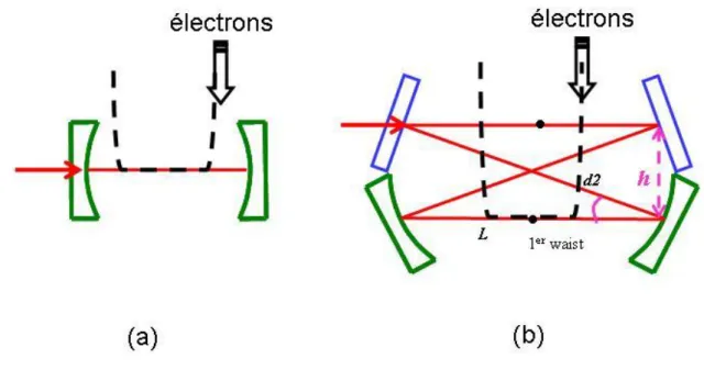
In a stable working condition, every turn the electron bunch impinges on the light pulse stored in the cavity with an angle of two degrees. The collision angle is imposed to allow the produced x ray beams to be extracted without damaging the high finesse cavity mirrors. Since the Fabry-Perot cavity operating frequency is a harmonic 2 of the storage ring, the operation with two stored bunches is suitable.
A sketch of the different ThomX components is shown in fig.3.
Laser beam Linac +RF gun
Electron beam pipe Electron Storage ring Collision point
Electron beam dump Four mirrors Fabry
Perot cavity
X rays
Laser beam Linac +RF gun
Electron beam pipe Electron Storage ring Collision point
Electron beam dump Four mirrors Fabry
Perot cavity
X rays
Laser beam Linac +RF gun
Electron beam pipe Electron Storage ring Collision point
Electron beam dump Four mirrors Fabry
Perot cavity
Laser beam Linac +RF gun
Electron beam pipe Electron Storage ring Collision point
Electron beam dump Four mirrors Fabry
Perot cavity
X rays
Figure 3 : ThomX different components. The electron beam is produced in the RF gun, accelerated in the linac and injected into the ring. Laser pulses are stored in the four-mirror optical resonator that is integrated in the collision region.
- X rays sources, Compton and alternative schemes. The ThomX positioning
As previously said Compton machines and other alternative compact x rays sources are becoming extremely attractive due to the technology advance allowing the increasing in the
sources brilliance. To have a better estimation of the ThomX performances in respect to the other projects, an overview of the principal other Compton backscattering based sources is provided followed by a short description of the other possible compact devices based on bremsstralhung or on alternative radiation mechanisms.
- Compton
As far as the Compton sources are concerned, currently the most ambitious projects aim to produce 1012-1013 ph/sec. The spectrum energy cut ranges in the few tens of KeV with a bandwidth of the order of 1-10%. In Table 3 an overview of the Compton compact sources projects and their main characteristics is illustrated.
Type Energy [KeV] Flux ( @ 10% bandwidth) Source size (m) *PLEIADES (LLNL) [11,12] Linac 10-100 107 (10 Hz) 18
*Vanderbilt [13,14] Linac 15-50 108 (few Hz) 30
*SLAC [15] Linac 20-85
*Waseda University [16,17] Linac 0.25-0.5 2.5 104 (5 Hz)
*AIST, Japan [18] Linac 10-40 106 30
*Tsinguha University [19] Linac 4.6 1.7 104
*LUCX (KEK) [20] Linac 33 5 104 (12.5 Hz) 80
+ UTNL, Japan [21,22] Linac 10-40 109
MIT project [23] Linac 3-30 3 1012 (100 MHz) 2
MXI systems [24] Linac 8-100 109 (10Hz)
SPARC –PLASMONX [25] Linac 20-380 2 108 -2 1010 0.5-13
Quantum Beam (KEK) [26,27] Linac 1013 3
*TERAS (AIST) [28] Storage ring 1-40 5 104 2
*Lyncean Tech [29,30,31] Storage ring 7-35 ~ 1012 30 Kharkov (SNC KIPT) [32] Storage ring 10-500 2.6 1013 (25 MHz) 35
TTX (THU China) [33,34] Storage ring 20-80 2 1012 35
ThomX France [35] Storage ring 50 1013 (25 MHz) 70
Table 3: Compact Compton X ray sources. Symbols * and + refers respectively to machines in operation and to machines in construction.
At present the experiments in operation [11,13,15,16,18,19,20,29] have already demonstrated the feasibility of X-rays production by inverse Compton scattering, but the delivered flux is not sufficient to exploit it for different applications where a higher brightness is required. The ThomX scheme, based on the multiple electron bunches-laser pulses collisions, is the one adopted for the already working Lyncean Tech machine [29,30,31] and for all the future sources which aim to produce an high average flux [23,32,33,34,35].
Although the MIT project [23] is not based on a storage ring it is able to foresee a very important average flux and brightness thanks to the use of a very low normalised emittance (< 1 mm mrad) superconductive linac at 100 MHz. Also the already funded Japanese project ―Quantum Beam‖ foresees a multi-bunch electron linac impinging on a pulsed amplified laser stacking device [26,27], producing an high quality and flux X ray beam.
It is important to stress that, the only machine in operation providing a flux comparable to the first generation synchrotron machines is the Lyncean Tech one. This project started in 2002 under the direction of R.Ruth. Today the machine provides a tuneable X ray beam of
1012ph/sec in 2% bandwidth [36] and the first phase contrast imaging has been recently obtained [37]
- Other sources
The X-ray technologies are driven by the imaging market with a trend towards high spectral brightness sources. A wide variety of technologies permit to produce X-rays but the potential alternative to Compton devices for quasi-monochromatic high flux are mainly represented by bremstralhung based sources. Therefore a lot of effort is performed to circumvent the inherent drawbacks of bremstrahlung sources, namely
The limited flux due to thermal constraints The finite size of the anode,
The angular and spectral of the X-ray beam
This has conducted to development of microfocus rotating anode with specific conditioning of the extracted beam. Rigaku [38], as an example, proposed an X-ray generator with 1.6 1011 photons/mm²/s (full spectrum) and possible dual wavelengths selection for phasing (chromium and copper K line selection at respectively 5.4 keV and 8 keV energies) also if the energy selection of the K line is not very efficient
The inherent thermal limitations of the interaction of the electron beam with matter have lead to two innovative designs, both connected to targets operating at higher temperature:
A metal-Jet-Anode Microfocus X-ray source with a potential 100 factor increase in brightness [39]. This technique remains at a research stage. It uses tin metal for the liquid anode, with a K line of 25.3 keV, below our requirements and with the same losses for the energy selection as mentioned above
A tungsten plasma X-ray generator [40] that provides a high instantaneous quasi-monochromatic flux at 59 keV (109 photons/cm² at 1 m from the source). This system is operating at present in single shot and evolution to repetitive operation remains problematic because of the plasma spread inside the tube during the shot.
MIRRORCLE [41]
Mirrorcle is based on the interaction of a high energy electron beam (up to 20 MeV), previously stored in a ring with a wire. The spectrum is therefore widespread.
The projected claimed brilliance is impressive (up to 1014 photons/s/mm²/mrad/0.1% BW for the 20 MeV system) but to date, the achieved level is lower (1011 photons/s/mm²/mrad²/0.1% BW - according to [42]. If monochromatic X-rays are requested, a monochromator can be used with the associated losses. We can assume that, at higher power, the inherent thermal limitation of the interaction between the beam and the wire target will appears
Alternative radiation mechanisms
Alternate exotic technologies, with their limitations to achieve high monochromatic flux, have been analyzed in [43]:
Channeling radiation, Coherent bremstrahlung, Microundulator radiation, Parametric-X-Radiation (PXR), Smith Purcell Radiation,
Among them, PXR can, achieve both monochromaticity and tunability, but the expected flux remains low, typically 6 105 photons/s within a 10-4 sr with a 6 MeV-100µA electron beam [44]
The following Conceptual Design Report is divided in seven chapters. The first chapter concerns the project scientific case. Among the different possible applications the cultural heritage and the medical applications are highlighted. In the second chapter an extensive introduction to the Compton backscattering physics is given. The first part covers the basic concepts taking into account the collision of an electron with a photon, whereas in the second part the parameterization of the collision between an electron bunch and a laser pulse is provided in the framework of the proposed source. The third chapter describes all the optical set-up sub-systems. It illustrate the laser and the optical cavity design and integration. Then, in the fourth chapter, the design of the electron injector and transfer line and the estimation of the emittance of the injected electron bunch are given. Successively the full description of the storage ring optics, beam dynamics, instabilities and collective effects is provided together with the general description of the hardware components like the magnets, the RF system, the injection septum and kicker, the diagnostics and the vacuum system. In the fifth chapter a short description of the radioprotection requirements, associated to the machine integration, are taken into account. Other intense X-ray source configurations are considered in the sixth chapter, where the electron drive beam is provided either by a Linac or an ERL. In the last chapter a first approximate costs evaluation of the various set-up items is given allowing an estimation of the required budget.
References
[1] M. Cotte et al., Anal. Chem., 79, 6988-6994 (2007). [2] See the Louvre laboratory web site: http://www.c2rmf.fr/ [3] Reviews of medical applications of Syncrotron radiation :
M.Ando and C Uyama, ―Medical Application of Syncrotron Radiation‖, Springer and Verlag (1998).
P. Suortti and W. Thomlinson, ―Medical applications of synchrotron radiation‖, Phys. Med. Biol. 48(2003)R1.
G. Margaritondo et al., ―Synchrotron in medical and materials science technology‖, Rev. Nuov. Cim. 7 (2004) 1.
[4] E.Rubistein et al. Proc SPIE 314,42,(1981).
[5] F.Carroll, Journal of Cellular Biochemistry 90 (2003)502. [6] E.G.Bessonov et al arXiv:physics/0405003 v1 (2004). [7] http://www.lynceantech.com/
[8] R.Hajima et al.,Proposal of Nondestructive Radionuclide Assay Using a High-Flux Gamma-Ray Source and Nuclear Resonance Fluorescence, Journal of Nuclear Science and Technology, Vol. 45 (2008), No. 5 p.441-451
[9] C.P.J Barthy and F.V Hartemann. UCRL-TR 206825, LLNL laboratory report [10] http://sera.lal.in2p3.fr/thomx/
[11] W.J Brown et al, Phys.Rev ST-AB 7, 060702 (2004) [12] D.J Gibson et al, Phys Plasmas 11 (2004), 2857-2864 [13] F.E. Carroll , AJR :179 (2002) 583-590
[14] F.E. Carroll et al, AJR :181 (2003) 1197-1202
[15] A.E Vlieks et al. Europen Particle Accelerator Conference EPAC04 Proceedings, Lucerne, Switzerland, (2004)
[17] K.Sakaue et al, Rad.Phys.Chem. 77 (2008), 1136-1141 [18] K.Yamada et al, Nucl.Inst. and Meth. A 608 (2009) S7-S10 [19] C.X.Tang et al CPC(HEP&NP), 33 (2009) 146-150
[20] K.Sakaue et al, Particle Accelrator Conference, PAC 07 Proceedings, Albuquerque, New Messico USA (2007)
[21] M. Uesaka et al, Nucl. Inst and Meth. B 261 (2007) 867-870
[22] F.Sakamoto et al, Nucl. Inst and Meth. A (2009), doi 10.1016/j.nima.2009.05.089 [23] W.S.Graves et al, Nucl. Inst and Meth. A (2009), doi 10.1016/j.nima.2009.05.042 [24] See http://www.mxisystems.com/specifications.html
[25] L.Serafini, Compton Sources for x/gamma rays Workshop, Alghero Italy, 7-12 September 2008
[26] K.Sakaue et al, LINAC08 Proceedings, Victoria BC Canada (2008)
[27] J.Urakawa, FJPPL/Compton meeting / LAL Orsay, France, 1-2 December 2008 [28] H Toyokawa et al, Nucl. Inst and Meth. A (2009), doi 10.1016/i.nima.2009.05.62 [29] Z.Huang and R .Ruth, Phys. Rev.Lett 80 (1998), 976-979
[30] R.Loewen, Thesis
[31] See http://www.lynceantech.com/sci_tech_cls.html
[32] E.Bulyak et al, Nucl.Inst. and Meth. A 487 (2002) 241-248 [33] C.X Tang et al, Nucl.Inst. and Meth. A 608 (2009) S70-S74 [34] P.Yu and W.Huang, Nucl.Inst. and Meth. A 592 (2008) 1-8
[35] C.Bruni et al, Europen Particle Accelerator Conference EPAC08 Proceedings Genova, Italy, (2008)
[36] J.Rifkin, POSIPOL workshop 2007, LAL Orsay France, 25-25 May 2007 [37] M Beck et al, J.Synchrotron Rad. 16 (2009) 43-47
[38] Rigatu FR-E+SuperBright ™
[39] A Compact High-Brightness Liquid-Metal-Jet X-Ray Source
[40] K-edge angiography utilizing a tungsten plasma X-ray generator in conjunction with gadolinium-based contrast media, E Sato & als, Radiation Physics and Chemistry 75 (2006) 1841-1849
[41] MIRRORCLE-6X - Photon Production Laboratory, Ltd.
[41] High Resolution X-Ray Imaging by Portable Synchrotron Radiation Source", MIRRORCLE - 6X, Yamada et al
[43] Exotic sources for iodine K-edge angiography - Roger Carr SLAC-PUB-634
[44] Tunable Source of Parametric X-Ray Radiation A.S Gogolev & A.P. Potylitsyn, ISSN 1063-7842, Technical Physics, 2008, Vol. 53, No 11
CHAPTER 1: Scientific Case
1.1] Cultural Heritage Applications 1.1.1] IntroductionNowadays the cultural heritage plays a crucial role, as far as the quality of the surrounding environment and the attractiveness of France are concerned, and has significant economic effects in many fields. The public at large takes great interest in it, as testified by the high number of visitors flocking to the many French monuments and museums. Consequently, knowing one‘s cultural heritage and preserving it in order to hand it over to future generations, is an increasingly important and shared concern in every country, especially those belonging to the European Union. At present in France all levels of society - government, regions, municipalities and companies - are involved in this effort.
Therefore the need was felt to carry out fundamental and applied research in order to best preserve this heritage and allow the public to access as many works of art as possible and learn about their history. This situation was described in the parliamentary report assessing the scientific and technological decisions on restoration techniques of works of art and the conservation of the national heritage [1]. On the one hand, in order to make progress in unveiling the history of works of art and in archeology it is necessary to identify the materials used; on the other hand, when conserving and restoring artifacts the effects of the aging of the materials cannot be neglected. To this end, the methods commonly used in materials science can prove extremely useful, especially those enabling researchers to perform a non-destructive analysis directly on the works of art or on tiny samples.
Research work on materials characterization is a big challenge from an analytical point of view, since there are many materials, they are often available in very small quantities, they have been processed or synthesized and then used by man, they have deteriorated in the natural environment or in the buildings in which they have been kept (museums, libraries, historical monuments, etc.). This is the case for paintings and graphical documents made up of various layers including mineral and organic ones (pigments, colorings, inks, binders, etc.): the chemical interaction between the different elements results in specific change or deterioration over a very long time. It is therefore necessary to develop and implement a number of innovative techniques to analyze works of arts.
A wide variety of instruments is commonly used. Most of them are classical laboratory techniques, whose set enables to probe samples at different levels (atomic, molecular, structural), at different scales (from millimeters to nanometers), and with different sensitivities (major to trace elements). Although lab instruments become more and more powerful and remain the prime equipment for the study of Cultural Heritage objects, two recent evolutions can be underlined. On the one hand, the necessity of reaching sites (excavation sites, museums, monuments…) fostered the development of portable instruments enabling in-situ analyses. On the other, specific studies require higher level of performance and are only possible on large-scale facilities (ion beam ―microprobe‖ accelerator such as AGLAE in the Louvre, synchrotron radiation, neutron sources) which provide brighter and smaller spots.
For this reason the setting-up of a ―micro-probe‖particle accelerator on the Louvre premises in late 1988 was a major step in the development of this research work. It made possible to investigate the composition of the museum‘s works of art directly, with a 20-30 µm spatial resolution which was ideal to study very complex and heterogeneous materials [2]. Today AGLAE is a unique and powerful analytical tool for the characterization of the materials of the museums‘ works of art and available to foreign researchers in the framework of a EU-financed program.
The AGLAE2 project aims at developing a new tool for the direct analysis and imaging of works of arts and archaeological items thanks to the co-localization of the present ion accelerator and a very intense X-ray source in a laboratory specialized in ―non-invasive‖ analysis of works of art to be set up in the Louvre security area.
These two sources will make it possible to develop additional analysis and imaging methods which would pave the way for the development of a new tridimensional analytical method of the museums‘ works of art to be combined with materials science methods. This could mark a radical change in what we know about works of art thanks to much more accurate scientific imaging. This device would be very useful for museums as it would be strategically positioned inside the Louvre. There would therefore be no cost constraints and no danger resulting from the need to move the museum‘s most precious works of art, namely Louvre‘s paintings, drawings and art objects.
1.1.2] Expected scientific impact 1.1.2.1] Analytical methods
Nowadays, the main research activities carried out in the laboratories working in the Cultural Heritage field focus on the identification of materials from a chemical and structural point of view (mineral, organic, hybrid, condense matter), the study of processes used for the elaboration of the works of art (origin of the materials, recipes of chemical synthesis, metallurgy, mechanical treatments and thermal annealing) and the study of alteration and ageing behavior, including the issues concerning the preventive conservation and the restoration. The ancient materials may be of natural origin, such as the gemstones, or stem from an artificial preparation at high temperature (glasses, ceramics and metals), or by wet chemistry, for the synthesis of pigments or pharmaceuticals for instance. Other materials have plant or animal origins, such as residues found in vases, sculptures made of wax, and human remains. Sometimes, it is the understanding of a diffusion mechanism, the observation of a patina on metals or the nature of nanocrystallized inclusions which are the purpose of the researches.
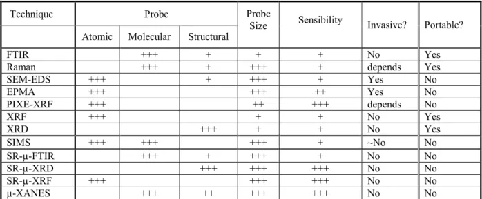
These activities require developing specific and appropriate physico-chemical analytical methods for the study of the cultural heritage masterpieces. The following table illustrates the techniques as far as painting is concerned [3]. It is possible to highlight the importance of applying a combination of different techniques to detect the underlying nature of the materials employed by the artists. Another important aspect of differentiation is that whereas in certain techniques a sample must be taken, the others allow for an in-situ analysis of the objects of art.
Technique Probe Probe
Size Sensibility Invasive? Portable? Atomic Molecular Structural
FTIR +++ + + + No Yes
Raman +++ + +++ + depends Yes
SEM-EDS +++ + +++ + Yes No EPMA +++ +++ ++ Yes No PIXE-XRF +++ ++ +++ depends No XRF +++ + + No Yes XRD +++ + + No Yes SIMS +++ +++ +++ + ~No No SR-µ-FTIR +++ + +++ + No No SR-µ-XRD +++ +++ +++ No No SR-µ-XRF +++ +++ +++ No No µ-XANES +++ ++ +++ +++ No No
Table 1: summary of classical and emerging techniques used for chemical analysis of ancient paintings.
The integration of a high flux monochromatic X-ray source, complementary to AGLAE, will provide a unique opportunity to have at one‘s disposal methods for non invasive elementary and structural analysis. A 20µm resolution, similar to that currently attained in AGLAE, is particularly well suited to the non-invasive study of ancient materials that often represent complex and heterogeneous samples.
Such an instrument should solve many problems in the context of different scientific cases. The ion beams are unique for the detection of light elements and the measurement of concentration gradients by nuclear reactions and also by elastic scattering (RBS and ERDA). Hard X-rays offer the opportunity to analyze heavy elements with high accuracy, to explore their environment by absorption spectrometry and to better understand the material organization by X-ray diffraction or small angle X-ray scattering (SAXS). By combining these two measurement categories on the same object, it is possible to assume that a ―total analysis of the material‖ will be worked out. This will remove any ambiguity in the interpretation of their nature in all the possible cases, independently from the metal, crystalline or amorphous nature of the sample.
1.1.2.2] X-ray imaging based on different physical properties
The use of radiography for the study of museum masterpieces has developed very rapidly after the discovery of X-rays by Röntgen in 1895. Today, C2RMF has 5 conventional radiography devices on the Louvre site. In the case of paintings, radiography provides information on the artist‘s techniques, the primitive draft variations, the paint overlays, and the conservation of the work. It reveals, for example, cracks in the paint layer. But the interpretation of the work of arts is much more than a mere conservation status analysis; it falls into the depths of the personality of the artist and sometimes shows the path of an artist toward its work creation. Radiography is very important to characterize objects, in particular in view of their restoration and to better understand the internal structure, i.e. the inhomogeneities (earth, plaster, bronze, inclusions, and bubbles), the formatting procedures (hammering, casting, lost wax or reversed ...), the assemblies, the alterations and the restorations.
An X-ray monochromatic intense source offers the opportunity to develop some techniques for three-dimensional imaging analysis of works of art aiming to a ―perfection level in direct imaging techniques‖: combining other imaging techniques in the visible, UV and near IR
ranges, it will allow providing a new scientific imaging report on each studied artefact, from the surface to the heart of the matter, with a spatial resolution of recording up to the micrometer scale. These scientific records will complement the documentation about its provenance and its historical and cultural environment.
The possibility of using a point source, monochromatic and coherent X ray beam makes it possible to consider different approaches:
(1) Absorption Tomography
This technique, commonly used in medicine, shows a fast development in the recent years. However, to provide the access to high quality of reconstruction and to quantitative data, an excellent signal to noise ratio is essential. It allows rapid data acquisition at very high spatial resolutions, resulting in precise mapping of the internal structures of the artefact. The use of an intense monochromatic beam is also preferable to conventional X-ray tubes because it avoids beam hardening effects that are frequently strong.
This technique is hardly used when dealing with objects almost flat, such as paintings: in this case the X-ray laminography technique should be used.
(2) Distribution of specific chemical elements analysis:
The edge-enhancement imaging technique is based on the same principle as the angiography of blood vessels in medicine. It is considered that each chemical element, with enough quantity in the work of art, may serve as a contrast agent when imaging with different X-ray energies. In medicine, ions are usually injected the vascular system. On the other side, as far as the paintings are concerned, lead, tin and mercury are characteristic of the different employed pigments. Therefore these images will map the spatial distribution of the chemical element. In practice, two images on each side of the K (or L) atomic absorption edge are measured. This technique and the resulting quantitative measurements can provide new information on the artists painting methodology: the substructure of a painting provides insight into the genesis of the object. Underlying layers may include the underdrawing, underpainting and modifications to the original sketch. In a growing number of cases conservators have discovered abandoned compositions on paintings, illustrating the artists‘ practice to re-use a canvas or panel and paint new compositions on top of existing ones. A painting from Vincent Van Gogh has been recently revealed by synchrotron-based XRF mapping [4].
(3) the phase contrast:
An inverse Compton scattering source should allow developing techniques of phase contrast imaging. These present the important advantage of the contrast enhancement in the case of low density materials. This is possible since the acquired image records the phase variations of the electromagnetic wave that occur when a variation of the refractive index occurs. The interest of this technique has been demonstrated on fossils in paleontology and on amber samples [5].
(4) magnification:
When the X-ray source sizes are very small and the emitted radiation sufficiently divergent (conical beam), it is possible to obtain a geometric magnification factor of the image by moving the detector away from the object. Thus, the projection of the object image is magnified, allowing a better accuracy in detecting details.
Other approaches could also be useful (Diffraction enhanced imaging, holo-tomography, X-ray grating interferometry, etc.).
We see therefore that the installation of a new X-rays source, simultaneously coupled with visible imagery, will provide useful data for the study of the paintings and the archaeological objects. Obtaining images of the surface and the core of the work of art, with spatial resolutions of the order of micrometers, is expected to provide to the researchers the access to digital reconstructions, with a much easier access than the original works. This will avoid to endanger the conservation of museum pieces particularly fragile: human rests and bones, wood or ivory masterpieces of the prehistoric era from the Musée de l'Homme, the National Museum of Prehistory and the National Archaeological Museum, the objects of ancient or medieval art from the Louvre Museum and the National Museum of the Middle Ages, etc. 1.2] BioMedical Applications
Biomedical applications of synchrotron radiation have been developed at Hasylab (Germany), Photon Factory (Japan), ELETTRA (Italia) and at the ESRF (France). Despite the fact that there are only a few dedicated beamlines in the world (two new ones are under construction at the Australian and Canadian synchrotrons), medical research is carried out in almost all synchrotron facilities through preclinical and clinical research protocol.
1.2.1] Radiotherapy programs
Gliomas are among the most frequent primary brain tumors in adults, with an incidence of approximately (5–11)/105 in industrial countries [6]. This pathology constitutes a major health problem. The treatment of high-grade gliomas is palliative rather than curative, despite a combination of surgery, chemotherapy and radiotherapy. Gliomas are extremely radioresistant, while surrounding normal tissues remain radiosensitive, especially in infants. The fundamental goal of radiation therapy is to deliver a high therapeutic dose of ionizing radiation to the tumor without exceeding normal tissue tolerance. This limitation is particularly severe in the case of brain tumors because of the high risk of adverse normal tissue morbidity. The needs, in particular for radioresistant tumours, are particularly high and may require very large facilities. For example, hadrontherapy [7] uses the enhanced energy deposition in the Bragg peak to achieve good conformity of the dose to the target, while sparing the healthy tissues. The main drawback of such large systems is the cost (in the range of 100 M€) and the accessibility.
At the ESRF, two radiotherapy techniques were developed, namely - the Microbeam Radiation Therapy (MRT) and
- the Stereotactic Synchrotron Radiation Therapy (SSRT) and clinical trials are foreseen to start in few years.
The Stereotactic Synchrotron Radiation Therapy (SSRT) consists in irradiating a tumour loaded with a contrast agent with quasi-monochromatic X-rays tuned at the energy above the K line of the contrast agent. The requested bandwidth is not a tight constraint and is naturally
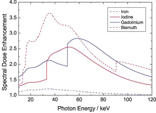
achieved with Inverse Compton Scattering without the need of a monochromator. The increase in the absorbing properties of the tumour relative to the surrounding tissue results in an internal dose enhancement based on the photoelectric effect as shown in fig.1. The photoelectric interactions generate photoelectrons, X-ray fluorescence and Auger electrons with the consequent cascade, which in turn increase the local radiation dose. In the energy
range of medical imaging (15–120 keV), the photoelectric effect plays a dominant role. As it displays a Z3-dependency, where Z is the atomic number, heavy elements absorb X-rays more strongly than the biological tissue light elements. This technique can be slightly optimized by loading the tumour with cis-platinum (cis-diamminedichloroplatinum or CDDP).The X-ray beam will induce a dose enhancement due to the high Z value of Pt in parallel with direct DNA damage as the chemotherapeutic effect of CisPt [8,9]
Figure 1: Dose enhancement versus X-ray energy for various contrast agents
In SSRT, the best survival curves were obtained by Biston et al. [8]. The brain tumor bearing rats were first inoculated with cisplatinum and then irradiated with X-rays of 78.8 keV. Median Survival Time (MeST) of untreated rats was 26 days. When cisplatin or SR alone was applied, the MeST was found to be 37 and 48 days, respectively, while when both treatments were combined, a very large increase in life span was obtained (MeST 206.5 days) compared to the controls. One year after treatment, 6 out of the 18 rats treated cisplatinum and radiation were still alive and cured. This is the largest life span increase obtained to date with this particular glioma model.
The Microbeam Radiation Therapy (MRT) [10] is an alternate approach for treating tumours and neurological disorders. It uses arrays of parallel, thin microplanar beams with two noticeable effects (fig. 2):
- single exposure MRT largely spare normal tissues including normal eyes, brain, spinal cord, and skin in spite of using an in-beam dose up to 20 times larger than those tolerated in broad beam radiotherapies,
- single exposure unidirectional MRT ablates intracerebral gliosarcomas in rats at an in-beam dose tolerated by contiguous normal tissues.
Experiments performed at the ESRF and BNL on the brains of adult rats, suckling rats, duck embryos and piglets have confirmed the sparing effect on normal tissues when using microbeams. In parallel, it was shown that MRT protocols can ablate highly aggressive
tumors like 9L brain gliomas, EMT-6 carcinoma and SCCVII carcinoma in animal models. Smilowitz et al. (2006) obtained 44% of longterm survivors (>300 days) in brain tumor bearing rats by combining MRT (entrance dose 625Gy, 50 microbeams, 25m wide and 200 m spaced) and gene mediated immunoprophylaxis. Furthermore, rats irradiated with MRT alone exhibited 20% long-term survivors [11].
This technique requests high flux density and high brightness beams that can only be provided by 3rd generation synchrotron to deliver very high doses (several hundreds of Gy) in an array of spatially fractionated quasi-parallel microbeams (25-75µm wide and spaced 100-400µm on-center). The 3rd generation sources are characterized by an adequate dose rate and energy spectrum which allows a minimal divergence of microscopic beams and consequently, the preservation of the steep gradients dose between parallel microbeams. ICS sources (as they are presently described) are below 3rd generation source flux by a factor of 100 approximatively. Present ICS source do not exhibit characteristics that are adequate for clinical trials, due to the fact the lack of flux will slow down the irradiation and increase the risk of blurring effects of the microbeams. However, ICS source could be used for performing MRT preclinical experiments on cells and animals as it is performed at the National Synchrotron Light Source (Brookhaven, National Laboratory, USA).
Figure 2: H&E staining of the horizontal section of piglet cerebellum, 15 months after irradiation (skin entrance dose: 300 Gy), 25 µm width. The microbeam paths are clearly visible although the piglets had a normal survival and no neurological signs or deficits.
1.2.2] Imaging
For imaging, it is necessary to avoid current radiograms that use 5-75 keV bremsstrahlung imaging, since the lowest energy part of the spectrum provides skin dose and no contrast while the highest part induces tissue dose and low contrast. Furthermore the traditional absorption imaging is very poor in differentiating soft tissues. Better in vivo systems need X-ray beams with the following characteristics (i) delivery of a very low integral dose and skin dose as small as possible (ii) high spatial resolution and in the same time high temporal resolution due to in vivo constraints (i) assess to quantitative information mimicking physiopathology or any functions of an organ. Among the techniques developed in synchrotron facilities, 2 of them have noticeably contributed to the biomedical field.
The K-edge digital subtraction imaging (KEDSI) method utilizes the sharp rise in the photoelectric component of the attenuation coefficient of a given element at the binding energy of the K-electron (e.g. 33.17 keV for I, 34.56 keV for Xe, 50.25 keV for Gd). Depending on the specific needs and constraints, experiments can be carried out either using two beams of energies bracketing the K-edge or by a single beam set at energy above the K edge. In the first case, the technique is indicated as ―energy subtraction‖ and the map of the contrast agent concentration is obtained by logarithmically subtracting the two energy images; In the second case, indicated as ―temporal subtraction mode,‖ the map is obtained by logarithmically subtracting the images taken at an energy above the K-edge, before and after the injection of the contrast agent. This latter modality is used when dealing with fixed samples and when the signal to noise ratio should be the best possible (brain imaging) [12]. Experiments have been performed in order to understand the uptake of Gd contrast agents in hepatitis-affected livers and in brain tumor-bearing animal models. In the frame of the radiotherapy programs, investigations have been carried out to optimize the delivery of drugs such as iodine, platinum, gold and/or gadolinium nanoparticles after intravenous of intracerebral delivery [13], in order to follow up their biodistribution and to study the tumor filling. From concentration maps of the contrast agent, brain perfusion parameters such as the cerebral blood volume and permeability can be derived using perfusion models [14](fig. 3).
Figure 3 : Microvascularization parametric maps, reliability maps (FMI maps) and H&E staining anatomic sections obtained in 5 rats brain bearing gliomas after iomeprol injection. The color scale for each parameter the red corresponds to high values and the blue to low values. In the FMI maps, a red pixel has a value of 1, attesting an excellent reliability of the model adjustment.
Left: physiological maps (blood flow FT, blood volume fraction VT, mean transit time MTT, permeability
surface product PS, and time delay D). Middle: reliability criteria FMI. Right: H&E staining (the labels on the right are the names of the rats).
To date, the KEDSI technique is the only one permitting the in vivo access of any drug concentration (if labeled with a high Z element such as Gd, I , Xe, Lu…) with such a spatial and temporal resolution.. This technique is of great interest for all laboratories (chemistry, pharmacy) who intend to characterize drugs and to study their bio-distribution.
Alternatively, the phase contrast imaging technique based on the recording of the phase variations occurring when X-rays pass through matter, has been demonstrated as an extremely powerful method since it permits contrast resolution of soft tissues (even if the elemental composition is almost uniform and the density variations are small) with far lower absorbed
dose levels than with conventional system as shown. This is in particular the case of breast, lung and articular cartilage tissues. Among the technique used on synchrotron beamlines, phase shifts are revealed by (i) using a crystal analyzer in order to depict the diffraction component (Analyser Based Imaging ie ABI)[15], (i) using the grating interferometry principle [16] (fig. 4) , as well as propagation phenomenon.
Figure 4 : Phase and attenuation-based tomography results. (a) Phase tomography slice through the rat‘s cerebellum showing a clear contrast between the white and gray brain matter. (b) Slice through a region of the brain containing a tumor (arrows indicate the tumor‘s ‗pushing front‘, the border between the tumor-invaded and healthy brain tissue). (c) and (d) Corresponding slices through the absorption-based reconstruction of the specimen. All images are displayed on a linear gray scale corresponding to ±2σ, where σ is the standard deviation of the pixel gray values in the image.
In the ABI technique, the X-rays transmitted through a sample are analyzed by a perfect crystal. The small angular acceptance of the analyzer permits the observation of the very small refraction angles (typically less than one microradian), to suppress scattered radiation and thereby increases the signal to noise ratio. The edge enhancement, which is characteristic of this method, occurs at the interfaces of regions with different refractive indices. The effects of refraction are converted to intensity variations by slightly detuning the analyzer away from the maximum of the reflectivity curve. The correlation of the radiographic findings with the morphologic changes in specimens analyzed in histo-pathological sections has been unequivocally confirmed. These advances in image quality make ABI a very promising candidate for clinical mammography at table top sources. In the case of osteoarthritis
joints of aging people, conventional radiography (including CT and MRI) is sensitive only in cases of advanced disease in which there has been a loss of cartilage. Measurements have been performed on human articular specimens. Cartilage defects, even at early stages of development, have been studied at 18 and 30 keV and compared with the absorption technique and show a clear, early visualization of the damage [17]. In addition, experiments were performed in rabbits and sheep samples with model implants to evaluate ABI as a tool in bone-implant research. AB images allow the identification of the quality of ingrowth of bone into the hydroxyapatite layer of the implant through the visualization of a highly refractive edge at the implant/bone border. Implants with bone fully grown onto the surface did not display a refractive signal.
1.2.3] Conclusion for medical applications
The SSRT technique seems to be fully compatible with ICS, since it can provide a flux and a brightness high enough together with a tunable quasi-monochromatic X-rays. The same prototype could serve both therapy and imaging since SSRT is derived from KEDSI method. These applications appeared to be the easiest ones to be developed in a first step. There techniques are also techniques where the synchrotron community has already a lot of knowledge and experience.
Regarding the MRT technique, although the flux is not sufficient enough for the realisation of clinical trials, an ICS source could be used anyway for preclinical studies.
Finally, the Inverse Compton Scattering process permits to provide high phase contrast and DEI imaging of soft tissues (adequate with its few percent bandwidth) on moving organs (because of its high available flux). The clinical interest of these techniques has to be however confirmed by in vivo measurement and several protocols are currently going on in other synchrotrons or with other synchrotron compact sources.
1.3] X-ray crystallography
The knowledge of the atomic structure of macromolecules such as proteins leads to a better understanding of the chemical reactions which take place in living organisms, how proteins are produced and how genetic information is forwarded.
1.3.1] Protein structure determination
Structure determination of proteins allowing a resolution at the level of distinguishing individual atoms is accomplished by two techniques: X-ray diffraction and Nuclear Magnetic Resonance (NMR) [18]. The NMR technique measures distances between atoms within a molecule in solution. Its principal advantage is to work on molecules in solution allowing a dynamic study to be performed. The complexity of the spectra for large molecules makes the analysis very difficult and protein size is the main limitation of the NMR technique. X-ray crystallography makes use of the diffraction pattern of X-rays that are shot through an object. The pattern is determined by the electron density within the crystal and this technique has no protein size limitation. It can solve structure of large and complex molecules but however requires proteins to be in an ordered crystal. Protein crystallization is inherently difficult due to the fragile nature of protein crystals and it is difficult to obtain a crystal with a suitable size (few hundred microns) for diffraction experiments [19]. For some proteins crystallisation, experiments will only ever produce extremely tiny crystals of the order of a few microns.
Since the average intensity of a diffraction peak for a crystal volume V and unit cell volume Vc in a beam of intensity I is approximately proportional to I(V/Vc), a thousand-fold increase in beam intensity is required to obtain the same strength diffraction from a 10 micron crystal compared to a 100 micron crystal. So, especially in case of complex structure determination, focused brightness X-ray beams are necessary to obtain sufficient resolutions [20]. The current intense synchrotron sources can provide the three-dimensional structure of small as well as large proteins with a precision of the order of the angstrom [21].
In a diffraction experiment, only intensities and diffraction angles of the diffracted beam are measured. To recover information about the phase of the diffracted beam which is essential for the solution of crystal structures, protein crystallography requires also a tuneable frequency X-ray source for the multi-wavelength anomalous dispersion (MAD) phasing technique [22]. High intensity polychromatic X-ray beams of the third generation synchrotron radiation sources are currently the most suited for protein structure determination.
1.3.2] System dynamic studies
A precise knowledge of a protein functioning requires the knowledge of its tree-dimensional structure but also its structural changes and its dynamic when the molecule is in activity. X-ray experiments make it possible to have access the timescale of molecular processes with a resolution in the picosecond range. Radiations from synchrotron sources have made it possible to conduct pump and probe experiments on chemical and biochemical systems down to a time-resolution of 50-200 ps. Pump and probe experiments consist to photolize a protein crystal sample with a femtosecond laser flash after which a probe time-delayed X-ray pulse provides diffraction data from excited molecules. The pump and probe sequence has to be repeated many time to build up sufficient signal to noise ratio for each diffraction pattern, to obtain patterns at different crystal orientations and at different pump-probe delays to access the time dimension of the process. The brightness and the pulse length of synchrotron source beams make possible the study of these time-dependant phenomena and allow to progress in the understanding of how proteins function [23, 24].
1.3.3] Compact Compton based source for protein study
A compact Compton based X-ray source would offer the possibility to perform protein studies mentioned above directly in a laboratory. With the envisaged performances (1012-1013 photons/s with an angular divergence of a few mrad and a bandwidth ΔE/E ~ 1-10%), a compact machine could carry out researches to progress in the understanding of protein structural properties and could also provided an X-ray beam having the proper pulse length required for system dynamic studies in the picosecond range.
References
[1] Rapport parlementaire sur « Les techniques de restauration des œuvres d'art et la protection du patrimoine face aux attaques du vieillissement et des pollutions », par M. Christian KERT, Office parlementaire d‘évaluation des choix scientifiques et technologiques, 15 juin 2006, rapport n°405.
[2] J. Salomon, J.C. Dran, T. Guillou, B. Moignard, L. Pichon, P. Walter, F. Mathis, ―Present and future role of ion beam analysis in the study of cultural heritage materials: The example of the AGLAE facility‖, Nuclear Instruments & Methods in Physics Research Section B-Beam Interactions with Materials and Atoms, 266 (10), 2273-2278 (2008).
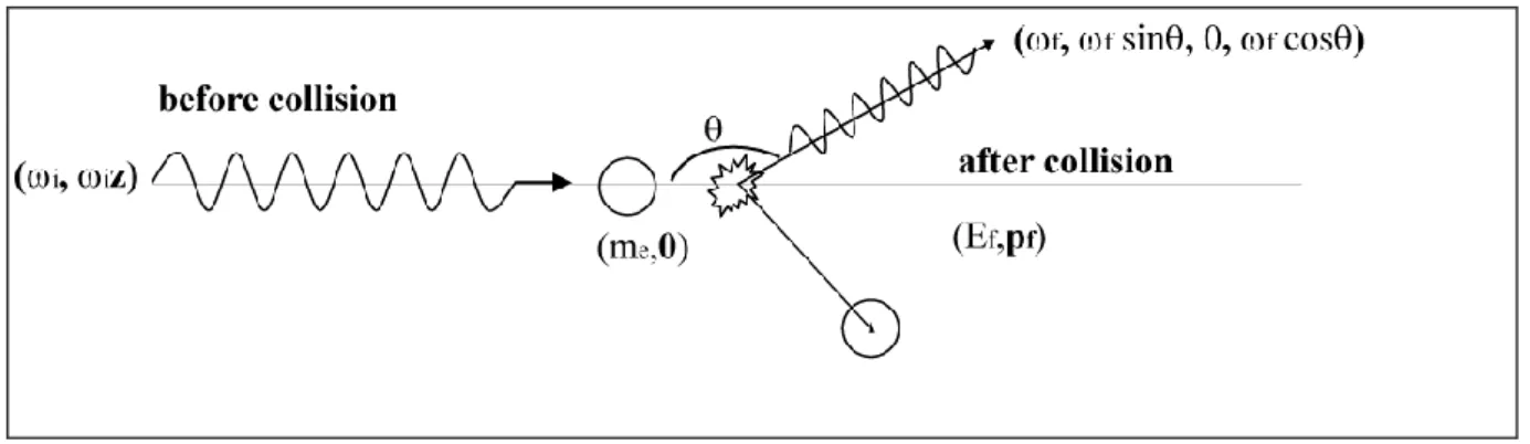
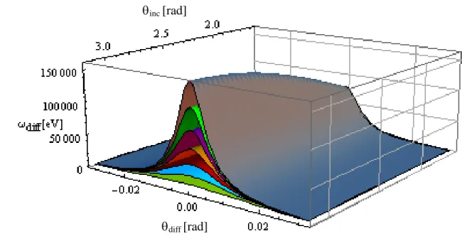
![Figure 5 : a] and b] panels: scattering energy dependence for head-on and orthogonal collision](https://thumb-eu.123doks.com/thumbv2/123doknet/12686794.354807/38.892.101.792.100.607/figure-panels-scattering-energy-dependence-head-orthogonal-collision.webp)