Conjugation of Extrachromosomal Replicons of
Rhodococcus erythropolis A N 12
Joyce Chun-Yi Yang
B.A., Biochemistry Rutgers University, 1 998
SUBMITTED TO THE DEPARTMENT OF BIOLOGY IN PARTIAL FULFILLMENT OF THE REQUIREMENTS FOR THE DEGREE OF
DOCTOR OF PHILOSOPHY
AT THE
MASSACHUETTS INSTITUTE OF TECHNOLOGY
MAY
2006
C a w
3oa
O 2006 Massachusetts Institute of Technology. All rights reserved.
Signature of Author: Department of Biology May 15,2006 Certified by:
-
- 8 I - l - --
I Anthony J. &inskey 'dProfessor of Microbiology Thesis Supervisor Accepted by: Stephen Bell Professor of Biology Chairman, Committee for Graduate Students1 MASSACHUSETTS N S ~ ~ ~ U T ' E
1Conjugation of Extrachromosomal Replicons of Rhodococcus erythropolis AN12
Joyce C. Yang
Submitted to the Department of Biology On May 15,2006 in Partial Fulfillment of the
Requirements for the Degree of Doctor of Philosophy in Biology
Abstract.
Bacteria belonging to the Gram-positive actinomycete species, Rhodococcus erythropolis, are diverse not only in terms of metabolic potentials but the plasmids they encode. Pulsed-field gel electrophoresis (PFGE) revealed three previously
uncharacterized megaplasmids in the genome of Rhodococcus erythropolis AN12. These megaplasmids, pREA400, pREA250 and pREA100, migrate at approximately 400 kb, 250 kb and 100 kb, respectively. Genetic screening of an AN1 2 transposon insertion library showed that two megaplasmids, pREA400 and pREA250, are conjugative. It is known for other bacterial systems that a relaxase encoded by the traA gene is required to initiate DNA transfer during plasmid conjugation. Sequences adjacent to the transposon insertion in megaplasmid pREA400 revealed a putative traA-like open reading frame. A novel site-specific gene disruption method was developed to generate a traA mutation in AN12, which allowed us to address the role of the traA gene for Rhodococcus
megaplasmid conjugation. We found that the AN12 traA mutant is no longer capable of transferring the pREA400 megaplasmid to Rhodococcus erythropolis SQ 1.
It was shown previously that the R. erythropolis AN12 genome harbors a 6.3 kb cryptic plasmid called pAN12. Here we show that pAN12 is conjugatively mobilizable into other rhodococcal strains. A series of plasmid deletion constructs were tested for loss of mobility to identify the pAN12 cis-acting conjugation requirement. In this way, an approximately 700 bp region was found to be required for plasmid transmission. A small 61 bp element within this region exhibited sequence similarity to the minimal 54 bp clt region known to be required for the conjugation of the streptomycete plasmid, pIJ101. The functionality of these cis-acting elements appears to be conserved, as the addition of this PAN 12 clt-like region confers mobility to an otherwise non-conjugative plasmid. However, unlike pJI 10 1 which encodes all necessary factors for transfer, pAN12 mobility is dependent on the presence of the AN12 megaplasmid, pREA400.
For my father (1 939- 1998)
from whom I've inherited a strict work ethic and a sometimes churlish stubborness,
which in its more noble form as persewerance
has sewed as my greatest asset on this journey
and
For my mother,
the bravest woman I know who taught me
(in additional to great phone manners)
ACKNOWLEDGEMENTS
First and foremost, this work would not have been possible without my thesis advisor, Dr. Sinskey. Certainly, had Tony not taken a chance on me about two and a half years ago, I would have written a much different thesis. Not only has Tony built a rich working environment, but his
encouragements and candid critiques both gave me faith in my own abilities and taught me how to be even better. The Sinskey lab's quest to understand basic biological phenomenons in the context of industrial metabolic engineering has rekindled in me the desire to focus on application-based research. I've also benefited greatly from Tony's unique perspective in his dual roles as professor and entrepreneur. Given the long history of MIT 1 W50K Entrepreneurship Competition winners from our lab, one can only conclude that others have felt that way too. Special thanks go to my MIT thesis committee members, Dr.
RajBhandary, Dr. Walker for their time and guidance, and most especially to Dr. Orr-Weaver for her unwavering support on my journey through three different laboratories. I am also indebted to Drs. Garrity and Solomon for their mentorship. Across the ocean at the other Cambridge, I am very grateful to my outside thesis examiner, Dr. Archer, who not only gave me insightfbl comments but went out of his way to attend my defense, well above and beyond the duties of being our collaborator.
I would not have pursued a scientific research path had it not been for the guidance of a Whippany Park High School physics and chemistry teacher, Mr. Altenderfer, and the endless patience of my
undergraduate advisor, Dr. Huibregtse, while attending Rutgers University. Thanks also to my collaborators at DuPont de Nemours, especially Drs. Nagarajan, Tomb, and Bramucci, who provided reagents, sequence and advice on working with rhodococci. I'm grateful for the financial support from the NIH Pre-Doctoral Training Grant, the MIT Dept. Biology, the Cancer Center Anna Fuller Fellowship, and the DuPont-MIT Alliance, throughout my graduate career.
I would like to thank the members of the Sinskey lab- especially Fen Tung, Dr. Vu Bui, Dr. Paolo Boccazzi, Dr. Jefferson Parker, Jes and Wil VanEssendelft, Joerg Schoenheit, Jae-Yeun Song, and Tim Ohlrich- for friendship, discussions, insights, and support. I've learned so much from all of you. Special thanks go to Xian OYBrien for optimizing pulsed-field gels, and undergraduates, Devin Currie, Neil Sengupta, and Steven Windsor for help in generating the transposon library. I am especially indebted to Dr. Philip Lessard for not only spearheading this and other Rhodococcus projects, but his mentorship, indomitable spirit, encyclopedic knowledge, and tireless reading of thesis chapters and manuscripts. Phil is also an excellent role model of how to juggle a family and a science career successfully. I don't know how he fits 48 hours into a day, but I'm not ruling out magic. Thanks also to lablteammates- Phil , Charles Budde and Nina Kshetry- for the unforgettable MIT 1K MicrobefuelMashville experience. At the Cancer Center, I thank Dr. Margaret Magendantz for teaching me how to do impeccable experiments, and Michael Moran, who rescued so many of my Macintosh computers from the brink of death. At the Garrity lab, I'd like to thank Monique Brouillette for help with the UAS-Ptpmeg constructs, and Dr. Jessica Whited, for being a vociferous ally, dear friend and terrific colleague.
I have been blessed by friendships with wonderful individuals, both at and outside of MIT. Some were members of the Class of 98 (Philina Lee, Arv Govindarajan, Suzanne Nguyen, Sandra Luikenhuis, Aron Eklund, John Doll, Jen Saionz, Brianna Burton, Tim Tayler, and Soni Lacefield). Others were found through WMBR (Rachel Grubb, Ted Young, Gene Fierro, Tani Chen, and Keith Sawyer). Yet more from the Rainbow Coffeehouse (Kevin Choi, Ajit Dash, Carlos Pacheco, Annie Frazer, An Nguyen, and Albert Chan), and through the Arts Scholars Program (Michele Oshima and Christa Starr). Also Elissa Lei, Stephanie Richard, Andrea Vala, Jared Nordman, Tim Montminy, and Mike Fisher at Harvard and Tufts for dating advice, friendships, and support. Thank you all for helping me cope with the challenging times, and sharing laughs through the good times.
Lastly, I'd like to acknowledge my family and extended family including; 1) my parents and my sister, Amy, whose unfailing love, faith and values are my ammunitions against any challenge, 2) my parents-in-law, Jan and Rich, for their unwavering support, generosity, and wisdom on life and
microbiology, 3) Ga and Steve, for stimulating conversations (scientific, literary, and otherwise) and for cultivating an unlikely appreciation for wine despite of my Asian Flush Syndrome, and finally, 4) my best friend, colleague, and wife Vicki, who has taught me everything else I needed to know.
TABLE OF CONTENTS Title page
...
1...
Abstract 2...
Dedication 3 Acknowledgements...
4 List of Tables...
8...
List of Figures 9 Chapter 1.
Introduction.
...
.
11...
1.
1. Background and motivation 12...
.
1.1 1. Introduction to the biology and versatility of Rhodococcus 12...
1.1.2. Importance of Rhodococcus in applied microbiology 13...
1 .1.2. 1. Rhodococcus and biodesulfurization 1 4 1.1.2.2. Use of Rhodococcus rhodochrous J1 in acrylamide production..
16...
1.1.3. Genetic analysis of Rhodococcus and tools development 18 1.2. Bacterial plasmids and conjugation...
201.2.1. Biology of Rhodococcus extrachromosomal replicons
...
201.2.2. The F-factor, a paradigm in bacterial conjugation
...
231.2.3. Important differences in Gram-positive plasmid conjugation: Actinomycetes appear to be exceptions to the rule
...
251.3. Thesis objectives and chapter outline
...
26...
1.4. References 27...
Chapter 2 . The conjugative megaplasmids of Rhodococcus erythropolis AN12 39 2.1.
Introduction...
-40...
...
2.2. Materials and methods.,.
-41 2.2.1. Bacterial strains and culturing conditions...
-412.2.2. DNA manipulation and plasmid construction
...
422.2.3. Preparation and standard transformation of electrocompetent
...
AN1 2 cells 43 2.2.4. Transposon mutagenesis...
432.2.5. Rhodococcus conjugation and megaplasmid mobilization frequency
...
432.2.6. Pulsed-field gel electrophoresis (PFGE)
...
-442.2.7. Southern blot analysis
...
-442.2.8. Rhodococcus erythropolis colony PCR
...
4 5 2.3. Results...
-452.3.1. Discovery of AN12 extrachromosomal elements
...
452.3.2. AN 1 2 transposon library construction and genetic screen for transmissible elements
...
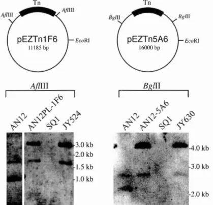
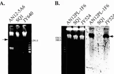
472.3.3. AN12PL- 1F6 and AN12-5A6 mutants bear transposon insertions in transmissible megaplasmids
...
-48...
2.3.4. Sequence analysis of the AN 12PL- 1F6 transposon insertion region 5 1
...
2.3.5. Sequence analysis of the AN 12-5F6 transposon insertion region 53
...
2.4. Discussion 53
...
2.5. References -56
Chapter 3
.
Development of a targeted gene disruption strategy for genetic and molecular analysis of traA required for plasmid conjugation in Rhodococcus erythropolis. and an analogous method utilized in the fruit fly. Drosophila...
melanogaster 59
...
3.1. Introduction -60
...
3.2. Materials and methods 6 2
...
3.2.1. Fly stocks. bacterial strains and culturing conditions 62
...
3.2.2. DNA manipulation and plasrnid construction 63
3.2.3. Preparation and standard transformation of electrocompetent
...
AN1 2 cells 65
3.2.4. Transformations of D
.
melanogaster with pUASt-Ptpmeg...
constructs and pTV2-MegISceI, and R . erythropolis AN12 with pJY37 66 3.2.5. Rhodococcus conjugation and megaplasmid mobilization...
frequency. 66
...
3.2.6. Pulsed-field gel electrophoresis (PFGE) 66
...
3.2.7. Southern blot analysis 66
3.2.8. Rhodococcus erythropolis colony PCR
...
66...
3.3. Results 67 3.3.1. Targeted gene disruption of the protein tyrosine phosphatase, Ptpmeg, in Drosophila melanogaster...
6 7 3.3.2. Sequence analysis of pREA400 encoded traA...
743.3.3. Development and deployment of a new targeted gene disruption method to study traA gene function
...
763.3.4. Phenotypic analysis of the traA mutant and genetic complementation of the traA defect
...
78...
3.4. Discussion 81 3.5. References...
-83Chapter 4
.
Characterization of the conjugation determinants of pAN12. a...
small replicon from Rhodococcus erythropolis AN12 90 4.1 Introduction...
91...
4.2 Materials and methods 9 3 4.2.1 Bacterial strains and culturing conditions...
934.2.2 Electrocompetent AN1 2 cells
...
9 3 4.2.3. Rhodococcus conjugation and plasmid conjugation efficiency...
934.2.4. DNA manipulation and plasmid construction
...
-944.2.5. Pulsed-field gel electrophoresis (PFGE)
...
-954.2.6. Southern blot analysis
...
954.2.7. Rhodococcus erythropolis colony PCR
...
954.3 Results
...
-96 4.3.1. Discovery of PAN 12 transmission, and definition of the cis-acting...
region required for conjugation 96
...
4.3.2. Identification and characterization of the pAN12 clt-like region 101 4.3.3. Co-Mobilization of AN 12 megaplasmids and pAN12 during
conjugation
...
-102...
4.3.4. Involvement of pREA4OO megaplasmid in pAL32 1 transfer 104 4.4 Discussion...
1 0 6 4.5. References...
1 0 8 Chapter 5.
Conclusions and recommendations for future work...
111...
5.1. Uncovering additional Rhodococcus megaplasmid conjugation determinants 112 5.1.1. Defining the pREA400 origin of transfer (orzT) region...
1125.1.2. Site-directed mutagenesis of TraA and purification of TraA proteins for enzymatic assays
...
1145.1.3. Isolation of additional pREA400 conjugation factors
...
1155.2. Spatial and temporal regulation of Rhodococcus erythropolis pAN12 transfer
...
1175.3. Outlook
...
1195.4. References
...
1 1 9 Appendix A. Colony morphology and solvent tolerance of Rhodococcus eryth ropolis AN1 2...
121A
.
1.
A correlation between Rhodococcus erythropolis AN1 2 morphology and the megaplasmid. pREA 1 00...
121A.2. Innate and gain of solvent tolerance in Rhodococcus erythropolis AN12
.
122 A.3. References...
126LIST OF TABLES
...
Table 1.1. Smaller ( ~ 2 0 kb) Rhodococcus plasmids and associated properties 21...
Table 1.2. Larger (>20 kb) Rhodococcur plasmids and associated properties 22...
Table 2.1. Bacterial strains. plasmids. and primers used in this study 42...
Table 2.2. Summary of plasmids used in this study 43
...
Table 3.1. Bacterial strains used in work described in this chapter 63Table 3.2. Summary of plasmids and cloning strategies used in this chapter
...
63Table 3.3. Summary of primers used in this chapter
...
65Table 3.4. Summary of mobilization fiequency of AN 12 megaplasmids to SQ 1
...
79Table 4.1. Summary of bacterial strains used in this study
...
94Table 4.2. Summary of plasmids and cloning strategies used in this study
...
95Table 4.3. Summary of primers used in this study
...
96LIST OF FIGURES
...
Figure 1.
1. Cell and colony morphologies of Rhodococcus bacteria 12Figure 1.2. The proposed Rhodococcus enzymatic pathway for
...
biodesulfurization of fossil fbels 16
...
Figure 1.3. The two microbial nitrile degradation pathways 17....
Figure 1.4. Metabolic engineering of rhodococci through megaplasmid conjugation 26...
Figure 2.1. Improvements in pulsed-field gel (PFG) electrophoresis 46...
Figure 2.2. Pulsed-field gel (PFG) profiles of wild type AN 12 and SQ 1 replicons 47Figure 2.3. Southern blot analysis of wild type, donors. recipient, and
...
transconjugants genomic DNA 49
...
Figure 2.4. Transposons-tagged megaplasmids can be transferred to SQ1 50Figure 2.5. Detailed schematic diagram of the plasmid rescue product fiom
...
recircularized genomic DNA isolated from AN 12PL- 1 F6 52
Figure 2.6. Sequence alignment of PemK toxin proteins
...
52Figure 2.7. The megaplasmid pREA250 transposon insertion region
...
53Figure 3.1. A schematic diagram of Drosophila Ptpmeg domains
...
68Figure 3.2. The splicing variants and end products of Ptpmeg
...
69Figure 3.3. In vivo targeting of the Drosophila Ptpmeg locus
...
70Figure 3.4. Genetics of generating a targetedptpmeg disruption
...
71Figure 3.5. Verification of the targeted ptpmeg locus
...
73Figure 3.6. Adult ptpmeg homozygous mutants exhibit duplicated scutellar bristles
....
73Figure 3.7. Alignment of four DNA relaxase domains
...
75Figure 3.8. The Rhodococcus targeted gene-disruption strategy
...
77Figure 3.9. PCR identification of the candidate traA disruption mutant strain
...
77Figure 3.10. Southern blot verification of the traA disruption mutant. JY825
...
78Figure 3.11. Genetic and phenotypic complementation of the traA mutant
...
79Figure 3.12. The integration of the complementation plasmid, pJY49B
...
80Figure 3.13. Southern blot confirmation
...
81Figure 4.1. PFG of Rhodococcus replicons following conjugation
...
97...
Figure 4.3. Southern blot analysis of pAL321 conjugation and maintenance 99...
Figure 4.4. PAN 12 deletion constructs. and summary of transmissibility to SQ 1 100...
Figure 4.5. Identification of the minimal PAN 12 clt-like region 101...
Figure 4.6. Addition of the clt-like region confers transmissibility to pAL28 1 102...
Figure 4.7. Colony PCR detection of the co-mobilization of pANl2 103Figure 4.8. Megaplasmid specific co-mobilization of PAN 1 2
...
103Figure 4.9. Involvement of pREA400 in pAL32 1 conjugation
...
105Figure 4.10. Megaplasmid encoded t r d function is not required for
pAN12 transfer
...
106Figure 5.1. Megaplasmids-specific libraries construction
...
114Figure 5.2. High-throughput screening strategy to identify Rhodococcus
conjugation mutants
...
117...
Figure 5.3. Fluorescence microscopy of rhodococci 118
Figure A.1. The absence of megaplasmid pREAlOO is correlated with dry colony
morphology
...
121...
Figure A.2. Inherent ethanol tolerance of Rhodococcus erythropolis AN 12 125...
. Figure A.3. Strategy for the genetic screening of a metagenome BAC library 126CHAPTER 1.
1.1. Background and motivation.
1.1.1. Introduction to the biology and versatility of Rhodococcus. Bacteria of
the genus Rhodococcus are Gram-positive, GC-rich, aerobic, non-motile members of the order Actinomycetales. As such, they are related to clinically and industrially important actinomycetes, such as Mycobacterium tuberculosis, the causative agent of tuberculosis, and various strains of Streptomyces, well known as producers of antibiotics.
Actinomyces literally means "ray-fungus" in Greek, as their sometime filamentous morphologies closely resemble small fungi. Indeed, the classification of rhodococci as a distinct genera historically has been difficult due to their pleomorphic nature, such that within a single isogenic strain, cells may appear as cocci, short rods, filaments, or take on elaborately branched morphotypes (Figure 1
-
1A). In addition, colony morphologies between different strains can vary; colonies may be smooth or rough edged, mucoid or dry, exhibiting colors from white to deep orange-red (Figure 1-
1 B).R. erqthmplis R. erythrupulis R. aetherivorans R. ruber
sQ1 AN1 2 I24 DD03 19
Figure 1.1. Cell and colony morphologies of Rhodococcus bacteria. (A) Morphologies that rhodococci cells can adopt during cycles of division and growth, adapted fi-om an insert on pg. 91 in the Biology of Actinomycetes (49). (B) Variance of colony
morphology between indicated strains of Rhodococcus. Photos taken (SQ1 through DD0319) after 1 month, 13 days, 1 month, and 8 days cultivation on LB agar at 30°C, respectively.
Rhodococci belong to the nocardioform family of bacteria, which are
differentiated from other actinomycetes based on cell wall composition. Specifically, these bacteria possess chemotype IV cell walls, consisting of tiered meshwork of
peptidoglycans, arabinogalactans, and lipoarabinomannans, atop basal lipid bilayers (92, 93, 125). Also found in these cell walls are mycolic acids, or large branched fatty acids between 20 and 90 carbons long, existing in both bound and free forms (15 1). Mycolic
acids are produced by other bacterial genera, including Corynebacteria, and
Mycobacteria; collectively, this subgroup of bacteria is referred to as the mycolata. It is thought that the perpendicular arrangement of mycolic acids with respect to the cell's lipid bilayer results in a chemically recalcitrant permeability barrier (1 12, 1 13).
Consistent with this theory, rhodococci as a group are especially noted for their solvent tolerance, being frequently isolated from contaminated soil and aquatic environments.
Though most Rhodococcus species are benign microbes, Rhodococcus equi has been found to be a pathogen of foals (1 02) and immune-compromised human (1 40). A few rhodococci have been found to be symbionts of termites (82, 101) and the insect, Rhodnius prolixus, associated with Chagas' disease (7,4 1). Strains of Rhodococcus fascians were discovered to cause tumor-like growths (leafy galls) near the base of the
sweet pea plants (10, 159). This pathogenistic process, known as fasciation, is due to the presence of three genetic loci- fas, att, and hyp on a 200 kb linear megaplasmid,
pFiD188 (1 8). Over 30 species of rhodococci have been taxonomically classified to date since the discovery of the type species, Rhodococcus rhodochrous, (aka Rhodococcus roseus) in 1891 (1 72).
1.1.2. Importance of Rhodococcus in applied microbiology. The veritable treasure trove of enzymatic pathways rhodococci encode, along with natural recalcitrance to organic solvents, makes this group of bacteria a particularly desirable resource for biotransformations, and leading candidate in considerations for industrial bioprocesses. Specifically, enzymes from rhodococci are capable of chiraVenantiomeric bioconversions of such exquisite specificity- such as the conversion of indene to cis-1,2-indiandiol(12, 162) and the resolution of racemic methyl nonactate to pure enantiomers (1 23)- that they could eventually augment or replace difficult chemical synthesis methods in the
production of pharmaceuticals. Rhodococcus species are also masters of biodegradation, acting upon substrates ranging from fuels (26,96) to halogenated organic compounds, in the form of chlorinated alkanes (20, 85, 141) or polycyclic aromatics (polychlorinated biphenyls or PCB) (5,9 1,107,121,146). As such, Rhodococcus bacteria have emerged as important microbes for bioremediation of oil spills and xenobiotic pollutants.
It is impossible to fully review the myriad of metabolic pathways documented for rhodococci; literally hundreds of scholarly articles have been published on this subject.
Instead, the discussion in this section will focus on the dominant roles that Rhodococcus plays in the biodesulfurization of fossil fuels, and the industrial production of acrylamide, as emerging and established bioprocesses, respectively. A more detailed list of plasmid- encoded enzymatic reactions in rhodococci and the possibility of using bacterial
conjugation as the basis of metabolic engineering- serving as the impetus for this thesis research- will also be covered in a later section of this chapter. Readers are referred to excellent reviews for detailed discussions of other metabolic pathways these microbes possess (4,27,44,90, 169).
1.1.2.1. Rhodococcus and biodesulfurization. This past decade bore witness to
a major surge in transportation fuel prices, from an average retail price of $1 .lO/gallon in May of 1996 to the current price of $2.60/gallon (www.eia.doe.gov), an increase well above the concurrent 27% inflation rate (www.bis.gov). Though this is largely due to the rising demand for petroleum, increased refinery costs to desulfurize fuel to the 30 ppm requirement mandated by the Clean Air Act of 1990, has also contributed to this price gain at the pump. Crude oil typically contains between 0.0 1 % to 5% sulfur (or 1000 to 30,000 ppm by weight), which largely exists in the form of an organic compound called dibenzothiophene, or DBT (53,72, 114). The release of sulfur from fuel combustion, and subsequent formation of sulphur oxides in the atmosphere results in smog and acid rain, the latter of which not only causes damage to fisheries, forests, and buildings/structures, but also negatively impacts human health (www.epa.gov). Removal of DBT and other sulfur compounds fiom crude oil is currently achieved by an energy intensive and costly process called hydrodesulfurization, or HDS (1 30).
According to a 2004 U.S. Department of Energy (DOE) fact sheet,
biodesulfurization (BDS) is a promising alternative to HDS which reduces capital and operating costs, as well as greenhouse gas emissions. BDS utilizes microbes to
metabolize the sulfur compounds in reactions that leave the hydrocarbons intact. Several strains, Rhodococcus erythropolis IGTS8 (3 1, 32,48, 5 1,99, 1 34), R. erythropolis KA2- 5- 1 (58, 78, 1 17, 120, 156), R. erythropolis D- 1 (64, 109, 128), Rhodococcus sp. ECRD- 1 (53, 54, 136), and Rhodococcus sp. KT462 (155) have been identified with the desirable properties for BDS.
Most of these bacteria possess enzymes in the so-called 4 s pathway (Figure 1.2A), in which a series of monoxygenases and a desulfinase act to transform DBT to 2- hydroxybiphenyl (HBP) and a sulfite ion. The genes encoding these activities are arranged in an operon. They were initially discovered by mutagenizing Rhodococcus sp. IGTS8 with shortwave W, then screening for mutants that no longer secreted HBP, a fluorescent compound whose presence can easily be detected visually following excitation with W (32). A mutant from this screen, W 1, was then transformed with a cosmid genomic library, and clones containing three open reading fiames (soxABC) spanning a 10 kb genomic region (Figure 1.2B) restored the ability to produce HBP (32). Interestingly, subsequent analysis of the UVl mutant by pulsed-field gel electrophoresis (PFGE) demonstrated the nature of the sox mutation was due to the curing of a 120 kb megaplasmid named pSOX (3 1). That the soxABC locus is plasmid-borne reflects a reccurring theme in a number of Rhodococc~ metabolic pathways to be described later in this chapter.
A paper which appeared a few months following Denome et al. (129) by an independent group described the cloning and characterization of the same operon in
Rhodococcus IGTS8, in which the sox genes were renamed dszABC (1 34). The second
nomenclature has since been adopted to avoid confusion with previously identified mammalian SOX genes. At about the same time, it was determined that molecular oxygen, the NADH reduced pyridine nucleotide, and the FMN flavin, along with the actions of a flavin oxidoreductase (DszD) are required in the reactions catalyzed by the DszC and DszA monoxygenases (5 1, 127, 129). Furthermore, an in vitro desulfurization system using purified enzymes allowed for detailed kinetic studies of this process, which showed that the reaction catalyzed by the desulfinase in the last step of this pathway is rate-limiting (5 1). Later studies which elucidated the gene regulatory region upstream of the dsz operon (99), and analyzed the desulfurization potential of modified strains that overexpressed dsz genes aimed at optimizing this reaction for commercial purposes (46).
DRT DBTO DBT02 HPBS HBY
Figure 1.2. The proposed Rhodococcus enzymatic pathway for biodesulfurization of fossil fuels. (A) A simplified schematic of the pathway for bioconversion of
dibenzothiophene (DBT) to 2-hydroxybiphenyl (HBP) adapted fiom Folsom et al. (1 999).
DBT
is first converted to intermediates, dibenzothiophene sulfoxide (DBTO) and dibenzothiophene sulfone (DBTO*) by the soxCldszC encoded monoxygenase. DBT02 is then converted by the soxAldsz4 monoxygenase to hydroxyphenyl sulfonate (HPBS), a reaction which opens the thiophenic ring. HPBS is then acted upon by the soxBldszB gene product, a desulfinase, to release 2-hydroxybiphenyl back into the oil, and sulfite ion for cellular metabolism. (B) Operon structure of the central Rhodococcusdesulfurization enzymes. Drawing represents the first cloned and sequenced desulfurization (sox) gene locus (Genbank No. U08850) found on a megaplasmid of Rhodococcus erythropolis IGTS8. s o d , soxB, and soxC are 1,362 bp, 1,098 bp, and
1,254 bp, respectively. These genes were subsequently renamed the dsz cluster.
The idea to use microbes for desulfurization reactions is not new. A study published in 1961 examined the use of Thiobacillus ferrooxidans to oxidize iron pyrite, FeS2, fiom coal (148). Despite this early discovery, it took roughly 40 years for the first microbially catalyzed biodesulfurization patent to be filed (24). Certain strains of Rhodococcus appear well-poised to be the most ideal biocatalysts in fossil fuel
biodesulfurization (1 14). It is anticipated that the significant challenges related to poor reaction kinetics and regeneration of the biocatalysts, also pointed out in this review, will be overcome in the near future to make Rhodococcus-mediated BDS platform a reality.
1.1.2.2. Use of Rhodococcus rhodochrous J1 in acrylamide production. Chemical production of the acrylamide and its versatile polymer, polyacrylamide, began in the 1950's, and now exceeds 400,000 metric tons per year (62). Because
polyacrylamides can be made cationic, anionic, or neutral in charge, they are central to many industrial applications, the largest uses of which include the potablehewage water treatments and the manufacturing of paper. Microbial synthesis of acrylamide was
prompted by the discovery of two different microbial pathways (Figure 1.3) to degrade nitriles (6, 13,62,76). The first pathway is mediated by a nihilase which directly breaks down nitriles (R-CN) to carboxylic acids (R-COOH) and ammonia (NH3). The second pathway is mediated stepwise by a nitrile hydratase (NHase), which converts R-CN to amides ( R - C o r n ) , followed by the action of an amidase, which converts arnides to R- COOH and NH3. The latter pathway has been investigated more thoroughly since nitrilases were found to be more labile.
2H20 R - C E N R-C, 0 nitrile H2° arnidase hydratase -C, NH2
Figure 1.3. The two microbial nitrile degradation pathways schematic, adapted fiom Warhurst and Fewson (1 994).
The Rhodococcus ( previously known as Brevibacteriurn) strain
R3
12 was initially considered for the microbial synthesis of acrylamide (17) since it yields 2.3M acrylamide every 30 minutes at room temperature. However, this enzymatic conversion was severely inhibited once the acrylamide concentration exceeded 2.8M. The nitrile hydratase of Rhodococcus rhodochrous J1 was found to be superior since it did not show significant inhibition by product concentration; indeed, it could produce up to 9.2M of acrylamide within 72 hours, and was thermostable up to 50°C (76). Furthermore, the arnidase activity was found to be very low, thus contaminating quantities of acrylic acid was not an issue for this bioprocess.The R. rhodochrous J1 nitrile hydratase activity can be attributed to two cobalt containing metalloenzymes; one of a lower molecular mass (L-NHase) (77), and one of a higher molecular mass (H-NHase) (1 15). Both enzymes are hetero-oligomers composed of two subunits named a and f3 (77), encoded by genes nhhA, nhhB, nhlA and nhlB. The crystal structure of a related Rhodococcus sp. R3 12 nitrile hydratase was later determined to 2.65
A
resolution (61). This study showed that the a and subunits of the R3 12activating metal ion bound by thiol groups of three cysteines found in the a subunit. In the case of R3 12 the metal ion is iron, but evidence suggests that cobalt is the equivalent metal in R. rhodochrous J1 (9). Huang et al. proposed that the metal ion acts as a Lewis acid, activating the carbon in nitrile to form a hydroxyl bond.
Because many Fe- and Co-type nitrile hydratase activities have been detected in diverse bacterial genera, a group of investigators recently completed a study examining the nitrile hydratase gene clusters in two different strains of Rhodococcus erythropolis (AJ270 and AJ300) and a strain of Microbacterium (AJ115) to address the possibility of horizontal gene transfer of these loci in the environment (124). They found that the nitrile hydratase gene clusters are indeed identical in these three bacteria. While linear and circular plasmid were detected, unlike the megaplasmid-encoded dsz genes discussed previously, the NHase genes in AJ270, AJ300 and AJ115 are chromosomally located. O'Mahony et al. did determine that a complete copy of transposable element, IS1 166, lies upstream of the NHase cluster, and concluded these genes most likely spread via
transposition. Ironically, the IS1 166 element was identified originally within the soxABC operon of desulfurizing bacteria, Rhodococcus erythropolis IGTS8 (33).
1.1.3. Genetic analysis of Rhodococcus and tools development. Bacterial genetics begins with mutagenesis and recombination. For rhodococci, this was initiated by Adams et al. in the 1960's when W-induced auxotrophic mutants- deficient for the production of various amino acids- of two different strains of Rhodococcus erythropolis (originally called Nocardia erythropolis and Nocardia canicruria) were mated to each other and prototrophic progenies were observed (1,2). Importantly, this series of work demonstrated both that genetic material can be transferred between Rhodococcus strains, and that irradiation can be used for mutagenesis. However, it was puzzling that unlike Escherichia coli in which homologous recombination occurs readily between cells of the same strain, recombination in rhodococci occurs readily only between different strains (1). It was suggested later that this phenomenon of self-incompatibility may be due to conjugation determinants located on plasmids (50,89). Complications in the genetic analysis of rhodococci have been compounded by the facts that: 1) no isogenic strain has been recognized as a type or reference strain; and 2) to date, there is no publicly available database with the complete and annotated genome sequence of any Rhodococcus, though
encouragingly, several groups are nearing this goal (J.-F. Tomb, J. Davies, and J. Archer, personal communication). Nevertheless, early genetic linkage maps of 65 traits were assembled for strains of R. erythropolis (1 1).
Besides UV mutagenesis, which was used to generate the auxotrophs above, and the dsz mutants mentioned previously, chemical mutagenesis has been successfully employed to study rhodococci (55,134, 168). However, by far the most popular way to generate Rhodococcus mutants for genetic analysis has been transposition. One of the first reports of transposon-based mutagenesis used an endogenous insertion sequence element, IS141 5, found proximal to the cobalamin synthesis genetic loci in R.
erythropolis N186/2 1 (1 16). Insertion sequence (IS) elements were initially identified in Rhodococcus fascians using an entrapment system (65), in which the Bacillus subtilis sacB gene encoding sucrose sensitivity can be inactivated via an IS transposition event.
Subsequently, they have been found in other Rhodococcus species either by accident through the study of nearby genes, as in the case of IS1 166 (33), or by intention via the
sacB method (97). The IS1415 or the resulting Tn5561, was found to have little, if any,
sequence bias; however, Tn5561 transposition appeared to be infiequent (1 event per pg vector DNA) and was not pursued further for mutagenesis. Since then, other transposon- based systems, commercial and otherwise, have been used successfully to generate mutants in a variety of Rhodococcus strains (3,43, 103,139, 156).
Study and utilization of bacterial plasmids, through the advent of recombinant DNA technology (14), have enabled many modern molecular biology techniques. Almost all strains of rhodococci harbor at least one plasmid (89), and study of their replication regions led to the development of shuttle vectors, generally chimeric plasmids that contain both an E. coli and a Rhodococcus replicon. The first of such, pMVS301,
was generated by Singer and Finnerty in 1988 (1 49), using a 3.8 kb region of the endogenous pMVS300 plasmid found in Rhodococcus sp. strain H13-A. It carries a thiostrepton resistance cassette cloned from Streptomycetes (1 57), and was found to be replicate stably as an independent replicon in Rhodococcus equi, Rhodococcus
globulerus, and Rhodococcus erythropolis. Since then, a dozen or so Rhodococcus
cryptic plasmids and derivative shuttle vectors have been constructed (21,29,30,39,56, 69, 83,98, 134, 147, 167), some of which include novel features such as; 1) the ability to
be mobilized directly from E. coli to Rhodococcus, bypassing often problematic rhodococci transformations (1 67); and 2) selective curing or integration of the plasmid based on replicon temperature sensitivity (98).
Post-genomic era tools for global Rhodococcus gene and protein expression analysis include DNA microarrays and proteomics (comparison of the 2-D mobility of expressed proteins). Studies using DNA microarrays to address questions in
Rhodococcus biology have not yet been published, though our lab is currently addressing aromatic compound metabolism of Rhodococcur aetherivorans I24 by this method (J.
Parker et al., unpublished). Reasons why Rhodococcus DNA microarray analyses have been so sparse include the lack of published genomic sequence, and difficulties in RNA
isolation from rhodococci (89). In contrast, protocols for the isolation of proteins- in particular the purification of desirable enzymes- are well established for this group of bacteria. One of the latest articles reports the purification of an alcohol dehydrogenase from Rhodococcus ruber that is stable in the presence of 50% acetone and 80%
isopropanol(8 1). A proteomics approach was taken very recently to identify genes specifically involved in phthalate metabolism in Rhodococcus sp. strain TFB (1 60). This method can prove to be a powerful analytic tool, when combined with genomic sequence information, to decipher the multitudes of seemingly redundant gene functions encoded
in some Rhodococcus genomes, as is the case with biphenyl metabolism in Rhodococcus
sp. RHA1 (132).
1.2. Bacterial plasmids and conjugation.
1.2.1. Biology of Rhodococcus extrachromosomal replicons. The term, plasmid, was coined by Joshua Lederberg in 1952 to describe an extrachromosomal genetic
element that can replicate autonomously (94). Plasmids can adopt either circular or linear topologies, in turn leading to differences in how they replicate and transfer. Plasmids can also vary in size. Miniplasmids can be engineered to carry only the essential replication region and selectible marker in less than 1,000 bp of DNA sequence. Naturally occuring megaplasmids can exceed 1 megabasepairs in size (16, loo), comparable to the gene coding capacity of a yeast chromosome. Plasmids profoundly influence the fitness of their host organisms, both negatively and positively. Uncontrolled plasmid proliferation
can be detrimental to the host, as plasmid replication is dependent on host factors (15, 165, 166). Conversely, the presence of plasmid-encoded enzymatic pathways often confer advantages to the host organisms, enabling their survival in otherwise inhospitable environments. In essence, bacteria that harbor plasmids exhibit dynamic genomes, which allows for rapid adaptation under selective pressures.
It would not be an overstatement to say that horizontal gene transfer
(HGT)
is a major facilitator of bacterial evolution. Nearly all bacterial species examined to date are capable of at least one of the following modes of exogenous DNA exchange, 1) uptake and direct transformation, 2) transposition via mobile genetic elements like transposons,3) transduction involving phage-intermediates, and 4) conjugation, referring to the exchange of genetic material most commonly in the form of plasmids. The exchange of plasmids through bacterial conjugation has been, and still is, one of the intensely studied forms of
HGT
with implications in human health, both negatively associated with plasmid-borne antibiotic resistances and virulence factors, and positively associated with plasmid-borne antibiotic production and pathways for bioremediation of xenobiotics.Rhodococci are hosts to one of the most diverse plasmid-based horizontal gene pool. Tables 1.1. and 1.2 summarize all plasmids and associated properties found in rhodococci; these tables have been modified fiom an earlier table fiom the Larkin review (1998) to include recent findings. Some of these plasmids have been found to be
conjugative, however, determinants governing their transmission are largely unknown. As this thesis directly addresses determinants of Rhodococcus plasmid conjugation, it will be prefaced here by what is currently known about the conjugation of a well- characterized Gram-negative bacterial plasmid, the F factor, and how Gram-positive conjugation, especially plasmids encoded by Actinomycetales, differ fiom this paradigm.
Table 1.1. Smaller (<20 kb) Rhodococcus plasmids and associated properties.
Strain Plasmida Size-kb Description Reference
R. opacus pHG3 1 -b 17 Cryptic circular plasmid (145) MRl 1
Rhodococcus pMVS200 20 Cryptic circular plasmid (1 49)
sp. H13-1A
Rhodococcus pMVS300 13 Cryptic circular plasmid (149) sp. H13-1A
R. erythropolis pFAJ2600 6 Cryptic circular plasmid; rolling-circle replicase; (29)
NI86/2 1 sequenced
R. rhodochrous pKA22 5 Cryptic circular plasmid; theta replicase; sequenced (84) NCIMB 13064 pKA4 9 (pKA22); cryptic circular plasmid (pKA4)
R. erythropolis pAN12 6 Cryptic circular plasmids; rolling-circle replicase; (83)
AN12 sequenced
R. rhodochrous pB264-1 5 Cryptic circular plasmid; theta replicase; highly (98) B264 similar to pKA22; sequenced; mobilizable
R. erythropolis pRE2895 z 1.9 Cryptic circular plasmid; theta replicase (1 19) JCM2895
R. erythropolis pRE8424 6 Cryptic circular plasmid; rolling-circle replicase; (1 18) DSM8424 9 1 % identical to PAN 12; sequenced
R. erythropolis pREC2 4 Cryptic circular plasmid; theta replicase; sequenced (144) PR4
a. Plasmids that can be mobilized are indicated (bold). Plasmids not indicated as such could be mobilizable though this property may not have been assayed.
Table 1.1. Larger (>20 kb) Rhodococcus plasmids and associated properties.
Strain plasm id(^)^ Size-kb Descri~tion Refer en ce
R. opacus MRl 1 R. opacus MR22 R hodococcus sp. TE1 R. fascians NCPPB10675 R. erythropolis ATCC12674 R. opacus MRl 1 Cryptic; circular Cryptic; circular
Atrazine and EPTC degradation; circular Chloramphenicol and cadmium heavy metal resistance; circular
Arsenic and cadmium resistance; linear Conjugative hydrogen autotrophy on pHG20 1 ; linear
R. opacus
MR22
Conjugative hydrogen autotrophy on pGH205; linear R. equi ATCC33701 R. equi L1 R. rhodochrous CTM R. rhodochrous CTM R. fascians Dl88
Virulence genes; circular Virulence genes; circular
2-methylaniline degradation; circular Cryptic; circular
Cadmium resistance (pD188); leafy gall formation and fasciation loci; linear (pFiD 1 8 8)
Biodesulfurization genes (dsdsox); circular R. erythropolis
IGTS8
R. erythropolis
p s o x
Isopropylbenzene metabolism; linear;
BD2 conjugative; completely sequenced
R. rhodochrous pRTL1, 100,80 Circular and chloroalkane degradation NCIMB 1 3064 pRTL2 (pRTL1); cryptic and circular (pRTL2)
R. equi 103 POTS 85 virulence genes; circular plasmid
Rhodococcus sp. RHAl R. erythropolis TA42 1 R. corallinus B-276 Rhodococcus sp. MP50 Rhodococcus sp. DK17 R. opacus 1 CP R. aetherivorans I24 R. erythropolis PR4
All linear plasmids; biphenyl metabolism (pRHA1 and pRHA2), cryptic and sequenced (pRHA3)
Biphenyl degradation; linear
All linear; pNC30 associated with propene metabolism
Aromatic nitrile degradation, circular Topology and conjugativity not determined; alkylbenzene metabolism (pDK2)
Linear; chlorocatechol catabolism
Circular and naphthalene degradation (pNid); linear and toluene metabolism (pI24) Linear and associated with alkane metabolism (pREL1); circular and cryptic (pREC1); pREC2 4 circular and cryptic (pREC2)
a. Plasmids determined to be conjugative have been highlighted (bold). Plasmids not indicated as such, may be conjugative, though this priperty may not have been assayed.
1.2.2. The F-factor, a paradigm in bacterial conjugation. Bacterial conjugation between cells of the Escherichia coli strain
K-
12 was discovered by J.Lederberg and E. Tatum in 1946 (95). It was shown a few years later by B. Davis that physical contact between the cells is necessary for bacterial mating, a core principle that extends to virtually all genera of bacteria (25). Significantly, in 1952, W. Hayes demonstrated another conjugation principle- that DNA transfer process proceeds unidirectionally from a donor to a recipient cells, and is mediated by a sex factor named F, for fertility (57). Lederberg then proposed that the F-sex factor might be a previously unrecognized form of extrachromosomal genetic material, which he termed a plasmid (94), and this was conclusively shown to be the case by Marmur et al. in 1961 (105).
Since these early discoveries, researchers have shown that one-third of the genes (the tra and trb loci) on the 99 kb F plasmid encode two types of transfer functions- those involved in initiating and stabilizing donor-recipient contact, or mating zair formation (Mpf), and those involved in DNA transfer and ~eplication (Dtr) (45). Mating begins when a thin flexible extracellular filament, called a pilus, fkom the (F+) donor cell makes contact with the (F-) recipient. The flexible nature of the F pilus allows for E. coli to
mate in liquid suspension. As part of the Mpf, the pilus is a polymer of homogenous subunits called pilins, which are encoded by the F-plasmid traA gene (1 11). A number of
the tra genes encode pilin processing, translocation, and assembly functions (47). Though it has been proposed in the past that the pilus is the physical bridge through which the plasmid DNA traverses, it is now thought that the pilus brings about the physical contact of the two cells by retraction, mediated by the depolymerization of its subunits. The actual DNA transfer may occur through electron-dense regions of the cell envelopes, known as conjugation junctions, established when the intimate contact of the donor and recipient cells is achieved (40). This mating pair stability is known to involve at least the traN gene product found at the outer membrane of the donor cell, and its association with recipient OmpA ( ~ u t e r ~ e m b r a n e rotei in) protein (75), as well as the inner membrane protein, TraG (1 04).
F is transferred as a single-stranded (ssDNA) molecule in the 5' to 3' orientation (63, 126), and is re-established as a double-stranded DNA (dsDNA) in the recipient cell following rolling-circle like DNA replication (88). The DNA processing is initiated at a region of the transferring strand called the origin of transfer, or orzT (1 58). The oriT sequence is bound by the relaxosome, a protein complex consisting of the plasmid- encoded tral relaxasehelicase (1 08, 137) and tray DNA binding protein (1 6 I), as well as a host-encoded histone-like protein called IHF, for integration host factor (164). It is the TraI relaxasehelicase which first recognizes a conserved 19 bp sequence
(TTTGCGTGGGGTGTGGTGC) called the nic site within oriT, then catalyses the scission reaction at the bolded thymine residue, the molecular mechanism of which will be discussed in greater detail in Chapter 3. Tray and IHF were found to stimulate the nicking activity of the TraI protein by creating DNA bending (122), and are preloaded onto the orzT before TraI (60). The TraD inner membrane gene product is a coupling protein and DNA transporter (13 I), linking the relaxosome-onT nucleoprotein complex (Dtr) to the Mpf via its interaction with the TraM DNA binding protein, which also binds to the orzT though it is not thought to be part of the relaxosome (35). Much is known about the how the replacement strand is synthesized in the recipient, as well as the organization and expression of the "early" genes located at the leader region (the first portion of the plasmid transferred to the recipient), which aid in plasmid establishment. For details on these processes, the readers are referred to the excellent review by Zechner et al. (1 71).
1.2.3. Important differences in Gram-positive plasmid conjugation: Actinomycetes appear to be exceptions to the rule. It is generally thought that the majority of Gram-positive plasmids transfer in a mechanistically conserved fashion as their Gram-negative counterpart with respect to the DNA processing and transport (Dtr) and the need for cell-to-cell contact (52). Indeed, plasmid-encoded components such as the relaxase and the oriT are found to be conserved not only in sequence but in function for Gram-positive plasmids from Streptococcus agalactiae pIP5O 1 (80,87) and
Enterococcus faecalis pRE25 (143). However, since no gene encoding for a pilus component has been found in Gram-positive bacteria, it is assumed that different
mechanisms than the Mpf exist to bring cells together. Consistent with the lack of pilus, most Gram-positive microorganisms require a solid surface for plasmid conjugation. At least two different methods to establish cell contact are beginning to be elucidated for Gram-positive bacteria; the pheromone-induced conjugation system exemplified by the enterococci plasmid, pADl (38), and the aggregation-based conjugation systems of Bacillus thuringienis pX0 16 (66) and Lactococcus lactis pRSO 1 (1 10).
For the Gram-positive bacteria of the genus Streptomyces, genetic recombination has been known for almost as long as that of E. coli (59). Conjugative transfer of various streptomycete plasmids, such as pIJ10 1, fiom isolated donors to a lawn of recipients, visibly results in the formation of pocks (73). Unlike other small mobilizable plasmids which depend on host or other plasmid encoded genes for transmission, pIJlOl does not appear to be dependent on the action of a relaxosome. In fact, only the function of one plasmid encoded gene called tra is required for pIJlO 1 conjugation (70,7 1, 133). The pIJl 01 Tra protein is a membrane protein not related to relaxases/helicases described earlier; rather, it resembles the DNA translocators FtsWSpoIIIE, which fbnction in chromosome segregation in bacilli and E. coli, respectively (42). Nor does the pIJlOl cis-acting site appear to be a substrate for nicking activity (36,37). Instead, it has been proposed that these Streptomyces plasmids are unique in the transfer of plasmid DNA to the recipients in a dsDNA form (52, 17 1).
1.3. Thesis objectives and chapter outline.
The existence of various rhodococci megaplasmids and associated properties suggests that it may be possible to engineer strains which carry combinatorial metabolic pathways through bacterial conjugation. This would aid in combining genetic traits associated with large or spatially separated loci that would otherwise be difficult to clone piecemeal. We have termed this concept "megapathway shuffling", illustrated below in Figure 1.4.
Recalcitrant
Strain
A
Recalcitrant
-
Strain
B
Process
Friendly
Strain
C
Figure 1.4. Metabolic engineering of rhodococci through megaplasmid conjugation.
With this ultimate objective in mind, we have examined the plasmid array and
conjugative properties of the Rhodococcus erythropolis strain AN1 2, originally isolated from industrial wastewater sludge. AN12 utilizes the aromatic compound aniline as its sole carbon source, and was found to encode a small cryptic plasmid called pAN12 (67). It was not known whether the R. erythropolis AN12 genome encodes other plasmids, nor whether any of its plasmids might be conjugative. Through characterization of AN12 plasmids, we endeavored also to understand how might the conjugation system of
rhodococci be similar or dissimilar to those defined for Gram-negative and positive plasmids, such as F and pIJ101, described earlier. The chapters of this thesis addresses the following specific aims: 1) Determining whether R. erythropolis AN12 encode other plasmids, and if so, 2) determining whether any AN1 2 plasmid is conjugative, and 3) defining cis- andlor trans-acting determinants for conjugative plasmids. Chapter 2 discusses the discovery of two conjugative AN12 megaplasmids, pREA400 and pREA250. Chapter 3 discusses generating a transfer-deficient mutant of AN12 using a novel site specific targeted gene disruption technique. Chapter 4 discusses the
mobilization of the small cryptic pAN12 and examines its dependence on one of the megaplasmids for its transfer. Chapter 5 outlines future experiments.
1.4. References.
Adams, J., and S. Bradley. 1963. Recombination events in the bacterial genus Nocardia. Science 140:1392-1394.
Adams, J.
N.
1964. Recombination between Nocardia Elythropolis and Nocardia Canicruria. J Bacteriol 88:865-76.Ashour, J., and M. KO Hondalus. 2003. Phenotypic mutants of the intracellular actinomycete Rhodococcus equi created by in vivo Himarl transposon
mutagenesis. J Bacteriol185:2644-52.
Ashraf, W., A. Mihdhir, and J. C. Murrell. 1994. Bacterial oxidation of propane. FEMS Microbiol Lett 122: 1-6.
Asturias, J. A., and K. N. Timmis. 1993. Three different 2,3- dihydroxybiphenyl- l,2-dioxygenase genes in the gram-positive
polychlorobiphenyl-degrading bacterium Rhodococcus globerulus P6. J Bacteriol 175463 1-40.
Banerjee, A., R. Sharma, and U. C. Banerjee. 2002. The nitrile-degrading enzymes: current status and future prospects. Appl Microbiol Biotechnol60:33- 44.
Beard, C. B., E. M. Dotson, P. M. Pennington, S. Eichler, C. Cordon-Rosales, and R V. Durvasula. 2001. Bacterial symbiosis and paratransgenic control of vector-borne Chagas disease. Int J Parasit0131 : 62 1 -7.
Behki, R., E. Topp, W. Dick, and P. Germon. 1993. Metabolism of the herbicide atrazine by Rhodococcus strains. Appl Environ Microbiol59: 1 95 5-9.
Brennan, B. A., J. G. Cummings, D. B. Chase, I. M. Turner, Jr., and M. J. Nelson. 1996. Resonance Raman spectroscopy of nitrile hydratase, a novel iron- sulfur enzyme. Biochemistry 35: 10068-77.
Brown, N. A. 1926. Sweet pea fasciation, a form of crowngall. Phytopathology 17~29-30.
bacteria., p. 201-228. In M. Goodfellow, M. Mordarski, and S. T. Williams (ed.), The biology of the actinomycetes. Academic Press, London.
Buckland, B. C., S. W. Drew, N. C. Connors, M. M. Chartrain, C. Lee, P. M. Salmon, K. Gbewonyo, W. Zhou, P. Gailliot, R Singhvi, R C. Olewinski, Jr., W. J. Sun, J. Reddy, J. Zhang, B. A. Jackey, C. Taylor, K. E. Goklen, B. Junker, and R L. Greasham. 1999. Microbial conversion of indene to
indandiol: a key intermediate in the synthesis of CRIXIVAN. Metab Eng 1:63-74.
Bunch, A. W. 1998. Biotransformation of nitriles by rhodococci. Antonie Van Leeuwenhoek 74: 89-97.
Chang, A C., and S. N. Cohen. 1974. Genome construction between bacterial species in vitro: replication and expression of Staphylococcus plasmid genes in
Escherichia coli. Proc Natl Acad Sci U S A 71: 1030-4.
Chittenden, T., A. Frey, and AD J. Levine. 1991. Regulated replication of an episomal simian virus 40 origin plasmid in COS7 cells. J Virol65:5944-5 1.
Coenye, T., and P. Vandamme. 2003. Simple sequence repeats and
compositional bias in the bipartite Ralstonia solanacearum GMI 1000 genome. BMC Genomics 4: 1 0.
Commeyras, A., A. Arnaud, P. Galzy, and J.-C. Jallageas. 1976. Process for the preparation of amides by biological hydrolysis. US patent 4,00 1,08 1.
Crespi, M., E. Messens, A. B. Caplan, M. van Montagu, and J. Desomer.
1992. Fasciation induction by the phytopathogen Rhodococcus fascians depends upon a linear plasmid encoding a cytokinin synthase gene. Embo J 11 :795-804.
Crespi, M., D. Vereecke, W. Temmerman, M. Van Montagu, and J. Desomer.
1994. The fas operon of Rhodococcus fascians encodes new genes required for efficient fasciation of host plants. J Bacteriol176:2492-50 1.
Curragh, H., 0. Flynn, MD J. Larkin, T. M. Stafford, J. T. Hamilton, and D. B. Harper. 1994. Haloalkane degradation and assimilation by Rhodococcus
rhodochrous NCIMB 13064. Microbiology 140 ( Pt 6): 1433-42.
Dabbs, E. R., B. Gowan, and S. J. Andersen. 1 990. Nocardioforrn arsenic resistance plasmids and construction of Rhodococcus cloning vectors. Plasmid 23:242-7.
Dabbs, E. R., and G. J. Sole. 1988. Plasmid-borne resistance to arsenate, arsenite, cadmium, and chloramphenicol in a Rhodococcus species. Mol Gen Genet 211: 148-54.
Dabrock, B., M. Kesseler, B. Averhoff, and G. Gottschalk. 1994. Identification and characterization of a transmissible linear plasmid from Rhodococcus
erythropolis BD2 that encodes isopropylbenzene and trichloroethene catabolism.
Appl Environ Microbiol 60:853-60.
Darzins, A., L. Xi, J. Childs, D. Monticello, and C. Squires. 1999. DSZ gene expression in Pseudomonas hosts. US patent 5952208.
Davis, B. D. 1950. Nonfiltrability of the agents of genetic recombination in
Escherichia coli. J Bacteriol60:507-8.
de Carvalho, C. C., and M. M. da Fonseca. 2005. Degradation of hydrocarbons and alcohols at different temperatures and salinities by Rhodococcus erythropolis
DCL14. FEMS Microbiol Ecol51:389-99.
erythropolis. Appl Microbiol Biotechnol67:7 15-26.
de la Pena-Moctezuma, A*, and J. F. Prescott. 1995. A physical map of the 85 kb virulence plasmid of Rhodococcus equi 103. Can J Vet Res 59:229-3 1.
De Mot, R, I. Nagy, A. De Schrijver, P. Pattanapipitpaisal, G. Schoofs, and J. Vanderleyden. 1997. Structural analysis of the 6 kb cryptic plasmid pFAJ2600 from Rhodococcus erythropolis NI86/2 1 and construction of Escherichia coli- Rhodococcus shuttle vectors. Microbiology 143 ( Pt 10):3 137-47.
Denis-Larose, C., H. Bergeron, D. Labbe, C. W. Greer, J. Hawari, M. J. Grossman, B. M. Sankey, and P. C. Lau. 1998. Characterization of the basic replicon of Rhodococcus plasmid pSOX and development of a Rhodococcus- Escherichia coli shuttle vector. Appl Environ Microbiol 64:4363 -7.
Denome, S. Am, C. Oldfield, L. J. Nash, and K. D. Young. 1994.
Characterization of the desulfurization genes from Rhodococcus sp. strain IGTS8. J Bacteriol176:6707- 16.
Denome, S. A.,
Em
S. Olson, and K. D. Young. 1993. Identification and Cloning of Genes Involved in Specific Desulfurization of Dibenzothiophene byRhodococcus sp. Strain IGTS8. Appl Environ Microbiol 59:2837-2843.
Denome, S. A., and K. D. Young. 1995. Identification and activity of two insertion sequence elements in Rhodococcus sp. strain IGTS8. Gene 161: 33-8.
Desomer, J., P. Dhaese, and M. Van Montagu 1988. Conjugative transfer of cadmium resistance plasmids in Rhodococcus fosciam strains. J Bacteriol 170:2401-5.
Disque-Kochem, C., and B. Dreiseikelmann. 1997. The cytoplasmic DNA- binding protein TraM binds to the inner membrane protein TraD in vitro. J
Bacteriol 179:6133-7.
Ducote, M. J., and G. S. Pettis. 2006. An in vivo assay for conjugation-mediated recombination yields novel results for Sheptomyces plasmid pIJlO1. Plasmid 55:
242-8.
Ducote, M. J., S. Prakash, and G. S. Pettis. 2000. Minimal and contributing sequence determinants of the cis-acting locus of transfer (clt) of streptomycete plasmid pIJ 10 1 occur within an intrinsically curved plasmid region. J Bacteriol 182:6834-41.
Dunny, G. M., B. A. Leonard, and P. J. Hedberg. 1995. Pheromone-inducible conjugation in Enterococcus faecalis: interbacterial and host-parasite chemical communication. J Bacteriol 177: 87 1-6.
Duran, R 1998. New shuttle vectors for Rhodococcus sp. R3 12 (formerly Brevibacterium sp. R3 12), a nitrile hydratase producing strain. J Basic Microbiol 38:lOl-6.
Durrenberger, M. B., W. Villiger, and T. Bachi. 199 1. Conjugational junctions: morphology of specific contacts in conjugating Escherichia coli bacteria. J Struct Biol107: 146-56.
Durvasula, R. V., A. Gumbs, A. Panackal, 0. Kruglov, S. Aksoy, R. B. Merrifield, F. F. Richards, and C. B. Beard. 1997. Prevention of insect-borne disease: an approach using transgenic symbiotic bacteria. Proc Natl Acad Sci U S A 94:3274-8.
Mol Cell Biol2:538-45.
Fernandes, P. J., J. A Powell, and J. A. Archer. 2001. Construction of
Rhodococcus random mutagenesis libraries using Tn5 transposition complexes.
Microbiology 147:2529-3 6.
Finnerty, W. R 1992. The biology and genetics of the genus Rhodococcus. Annu Rev Microbiol46: 193-2 1 8.
Firth, N., K. Ippen-Ihler, and R A. Skurray. 1996. Structure and h c t i o n of the F factor and mechanism of conjugation., p. 2377-2401. In F. C. Neidhard, R. Curtiss III., J. L. Ingraham, E. C. C. Lin,
K.
S. Low, B. Magasanik, W. S. Reznikoff, M. Schaechter, and H. E. Umbarger (ed.), Escherichia coli and Salmonella, vol. 2. ASM Press, Washington, D.C.Folsom, B. R, D. R Schieche, P. M. DiGrazia, J. Werner, and S. Palmer.
1999. Microbial desulfurization of alkylated dibenzothiophenes fiom a hydrodesulfurized middle distillate by Rhodococcur erythropolis I- 19. Appl Environ Microbiol65:4967-72.
Frost, L. S., K. Ippen-Ihler, and R A Skurray. 1994. Analysis of the sequence and gene products of the transfer region of the F sex factor. Microbiol Rev
58: 162-2 10.
Gallagher, J. R, E. S. Olson, and D. C. Stanley. 1993. Microbial
desulfurization of dibenzothiophene: a sulfur-specific pathway. FEMS Microbiol Lett 107:31-5.
Goodfellow, M., M. Mordarski, and S. T. Williams. 1984. The Biology of the actinomycetes. Academic Press, London ; Orlando.
Gowan, B., and E. R. Dabbs. 1994. Identification of DNA involved in
Rhodococcus chromosomal conjugation and self-incompatibility. FEMS
Microbiol Lett 115:45-50.
Gray, K. A , 0. S. Pogrebinsky, Go T. Mrachko, L. Xi, D. J. Monticello, and
C.
He
Squires. 1996. Molecular mechanisms of biocatalytic desulfurization of fossil fuels. Nat Biotechnol14: 1705-9.Grohmann, E , G. Muth, and M. Espinosa. 2003. Conjugative plasmid transfer in gram-positive bacteria. Microbiol Mol Biol Rev 67:277-301, table of contents.
Grossman, M. J., M. K. Lee, R C. Prince, K. K. Garrett, G. N. George, and I. J. Pickering. 1999. Microbial desulfurization of a crude oil middle-distillate fiaction: analysis of the extent of sulfur removal and the effect of removal on remaining sulfur. Appl Environ Microbiol65: 1 8 1 -8.
Grossman, M. J., M. K. Lee, R C. Prince, V. Minak-Bernero, G. N. George, and I. J. Pickering. 2001. Deep desulfurization of extensively hydrodesulfurized middle distillate oil by Rhodococcus sp. strain ECRD- 1. Appl Environ Microbiol
67: 1949-52.
Gutierrez, T., R. P. Learmonth, P. D. Nichols, and I. Couperwhite. 2003. Comparative benzene-induced fatty acid changes in a Rhodococcus species and its benzene-sensitive mutant: possible role of myristic and oleic acids in tolerance. J
Chem Ecol29:2369-78.
Hashimoto, Ye, M. Nishiyama, F. Yu, I. Watanabe, S. Horinouchi, and T. Beppu. 1992. Development of a host-vector system in a Rhodococcus strain and its use for expression of the cloned nitrile hydratase gene cluster. J Gen Microbiol
138: 1003-10.
Hayes, W. 1952. Recombination in Bact. coli K 12; unidirectional transfer of genetic material. Nature 169: 1 1 8-9.
Hirasawa, K., Y. Ishii, M. Kobayashi, K. Koizumi, and KO Maruhashi. 2001. Improvement of desulfurization activity in Rhodococcus erythropolis KA2-5- 1 by genetic engineering. Biosci Biotechnol Biochem 65239-46.
Hopwood, D. A 1959. Linkage and the mechanism of recombination in Streptomyces coelicolor. Ann N Y Acad Sci 81:887-98.
Howard, M. T., W. C. Nelson, and S. W. Matson. 1995. Stepwise assembly of a relaxosome at the F plasmid origin of transfer. J Biol Chem 270:28381-6.
Huang, W., J. Jia, J. Cummings, M. Nelson, Go Schneider, and Y. Lindqvist.
1997. Crystal structure of nitrile hydratase reveals a novel iron centre in a novel fold. Structure 5691 -9.
Hughes, J., Y. C. Armitage, and KO C. Symes. 1998. Application of whole cell rhodococcal biocatalysts in acrylic polymer manufacture. Antonie Van
Leeuwenhoek 74: 107- 1 8.
Ihler, G., and W. D. Rupp. 1969. Strand-specific transfer of donor DNA during conjugation in E. coli. Proc Natl Acad Sci U S A 63: 138-43.
Izumi, Y., T. Ohshiro, H. Ogino, Y. Hine, and M. Shimao. 1994. Selective Desulfurization of Dibenzothiophene by Rhodococcus erythropolis D- 1. Appl Environ Microbiol60:223-226.
Jager, W., A Schafer, J. Kalinowski, and A. Puhler. 1995. Isolation of insertion elements fiom gram-positive Brevibacterium, Corynebacterium and
Rhodococcus strains using the Bacillus subtilis sacB gene as a positive selection
marker. FEMS Microbiol Lett 126: 1-6.
Jensen, G. B., A. Wilcks, S. S. Petersen, J. Damgaard, J. A. Baum, and L. Andrup. 1995. The genetic basis of the aggregation system in Bacillus
thuringiensis subsp. israelensis is located on the large conjugative plasmid
pX016. J Bacteriol 177:2914-7.
Kalkus, J., C. Dorrie, D. Fischer, M. Reh, and H. G. Schlegel. 1993. The giant linear plasmid pHG207 fiom Rhodococcus sp. encoding hydrogen autotrophy: characterization of the plasmid and its termini. J Gen Microbiol 139:2055-65.
Kalkus, J., M. Reh, and H. G. Schlegel. 1990. Hydrogen autotrophy of
Nocardia opaca strains is encoded by linear megaplasmids. J Gen Microbiol 136:1145-51.
Kalscheuer, R., M. Arenskotter, and A. Steinbuchel. 1999. Establishment of a gene transfer system for Rhodococcus opacus PD630 based on electroporation and its application for recombinant biosynthesis of poly(3-hydroxyalkanoic acids). Appl Microbiol Biotechnol52: 508- 1 5.
Kendall, KO J., and S. N. Cohen. 1988. Complete nucleotide sequence of the
Streptomyces lividans plasmid pIJl 0 1 and correlation of the sequence with
genetic properties. J Bacteriol170:4634-5 1.
Kendall, KO J., and S. N. Cohen. 1987. Plasmid transfer in Streptomyces
lividans: identification of a kil-kor system associated with the transfer region of
pIJ101. J Bacteriol 169:4177-83.