HAL Id: tel-01171597
https://tel.archives-ouvertes.fr/tel-01171597
Submitted on 6 Jul 2015
HAL is a multi-disciplinary open access
archive for the deposit and dissemination of sci-entific research documents, whether they are pub-lished or not. The documents may come from teaching and research institutions in France or abroad, or from public or private research centers.
L’archive ouverte pluridisciplinaire HAL, est destinée au dépôt et à la diffusion de documents scientifiques de niveau recherche, publiés ou non, émanant des établissements d’enseignement et de recherche français ou étrangers, des laboratoires publics ou privés.
Cortical stiffness : a gatekeeper for spindle positioning in
mouse oocytes
Agathe Chaigne
To cite this version:
Agathe Chaigne. Cortical stiffness : a gatekeeper for spindle positioning in mouse oocytes. Cellular Biology. Université Pierre et Marie Curie - Paris VI, 2014. English. �NNT : 2014PA066288�. �tel-01171597�
Université Pierre et Marie Curie
Ecole doctorale Complexité du Vivant (ED515)
Centre Interdisciplinaire de Recherche en Biologie, Collège de France
Cortical stiffness : a gatekeeper for spindle positioning
in mouse oocytes
Présentée par Agathe Chaigne
Thèse de doctorat de Biologie Moléculaire et Cellulaire du Développement
Dirigée par Marie-Emilie Terret
Présentée et soutenue publiquement le 4 Juillet 2014
Devant un jury composé de :
Mme Sophie Louvet Présidente
Mr Guillaume Halet Rapporteur
Mr Péter Lénárt Rapporteur
Mr Laurent Blanchoin Examinateur
Mr Matthieu Piel Examinateur
Mme Marie-Emilie Terret Examinateur
Remerciements
Je voudrais d’abord remercier tous les gens de la Science (avec un S majuscule) :
Mon inestimable jury: Laurent Blanchoin, Guillaume Halet, Peter Lénart, Sophie Louvet et Matthieu Piel. Merci d’avoir accepté de juger mon travail.
Tous les gens qui m’ont aidé lors de cette thèse et en particulier mon comité de thèse, Arnaud Echard, Alexis Gautreau et Matthieu Piel, one more time, qui ont vraiment fait avancer mon travail d’une façon extrêmement productive.
Marie-Hélène alias The Big Boss pour m’avoir accueillie dans ton labo et donné les moyens
de faire plein de trucs marrants, d’aller tirer sur des ovocytes à Curie. Tu donnes à tous les gens du labo la possibilité de faire, d’essayer, de se tromper ; ce n’est pas donné à tout le monde et je t’en remercie. Merci aussi de m’avoir appris à dire JE, parfois.
Marie-Emilie alias The Boss pour m’avoir proposé un sujet cool, pour m’avoir super
encadrée, pour avoir discuté de Top Chef avec moi, pour m’avoir soutenue tout le temps et m’avoir remonté le moral quand j’ai fait la même manip pendant 6 mois en modifiant la concentration d’agarose 3 pM par 3 pM. J’ai vraiment passé une thèse formidable, à la fois passionnante et détendue et c’est en grande partie grâce à toi ! Ta façon d’encadrer tes étudiants, de leur manifester ton intérêt et ta patience comme tu le fais, tout en étant toujours disponible pour tout le monde te rend irremplaçable.
Isabelle à la fois pour ton aide précieuse, tes conseils et ta rigueur dans le travail, mais aussi
et surtout parce que je suis très contente de te connaître. Discuter avec toi m’aura souvent permis de comprendre les autres.
Maria pour tes innombrables conseils et anecdotes, et parce qu’on s’est bien marrées (comme
des vils petits scarabées). Ta confiance en toi m’a donné de la force pour défendre mes idées.
Agnieszka you gave me a great lesson of courage. You are a strong woman, the kind that
holds the whole world (and sometimes the whole lab!) on her shoulders, and you are still very missed in the lab.
Gosia , for a few fun parties at « La plage de Jérémie » with way too much vine (and
sometimes even vodka). Good luck for everything is the future !
Isma et Joanne on ne se connaît pas encore très bien mais je suis sûre que ça va très bien
marcher pour vous.
Hepburn dans « Vacances Romaines », et pour toutes tes idées en labmeeting.
Pascale on ne s’est pas beaucoup connu mais tu m’as accueillie très gentiment alors que
j’étais un peu stressée.
Jessica je te remercie surtout pour avoir démarré ce projet que j’ai adoré suivre, et parce que
tu t’es donné la peine de venir me voir juste pour m’expliquer comment analyser de la photoactivation.
Toute l’équipe de Cécile Sykes, en particulier Pierre Nassoy pour ses idées, Kévin et Joël pour le crawfish etouffé de New Orleans, et surtout mon inestimable collègue et ami Clément avec qui j’ai passé de longues heures enfermée dans une cave à critiquer ses goûts musicaux (pourris, il faut bien le reconnaître) en regardant mes ovocytes se déformer (ou pas). On se sera quand même bien marré dans cette cave.
Nir Gov and Raphaël Voituriez for thinking that modeling spindle migration in a mouse
oocyte is a good idea and for being able to say « is it quadratic? » in front of a biologist audience without blinking.
Tous les gens de l’étage: la Fekrije team et la Alexander team (en particulier Marion pour les descentes de pression).
Tous les gens qui m’ont aidée: Jean-Pierre, Frédéric, tous les animaliers (big up à Seb (malgré sa passion du PSG) pour m’avoir appris à buter les souris mos sans me faire mordre),
Claudette, Nicole, Sophie, Sidonie pour toutes les commandes, conventions, machins à
signer, Jérémie pour les appels et textos de type « y a un machin qui s’allume pas et le microscope fait un bruit chelou, je cours ? ».
Tous les gens qui m’ont donné des plasmides, inhibiteurs, machins, en particulier Sophie
Louvet, Franck Perez, Alexis Gautreau (one more time), Katja Wassmann. Je dois maintenant remercier tous mes amis (de la Science et d’ailleurs)
Claire, Hector, Florent, Imen, Filipe, Henri, Charlotte, Lauriane, Ludo, Rémy, Rémi, Morgan, Isabelle, Anne-So, Chou, Marianne, François, Claire B, Manu, Audrey, Lara, Alexis, Edouard (special thanks pour avoir répondu un long mail à mes questions pendant tes
vacances en t’excusant de ne pas pouvoir faire mieux !) Clémence, Alice, Benjamin et tous les autres. Je ne vais pas vous dire à tous pourquoi je suis contente de vous avoir parce que franchement, ça serait trop long, et puis vous le savez déjà, bande de moules.
dans mon embryon de carrière scientifique (ne nous emballons pas) :
Mr Le Nechet qui m’a donné envie de faire de la bio, tous mes profs de prépa de Nantes et de Paris, en particulier mes profs de bio Mr Auger et Périlleux (alias PP) et Mme Bonhoure, et aussi Toby sans qui je n’aurais JAMAIS eu 14 en maths à l’ENS et Dejan pour ses commentaires dithyrambiques sur mes copies de chimie orga, les gens que j’ai côtoyé à l’ENS, les profs de la prépa Agreg d’Orsay en particulier Patrick Pla, Hélène Vincent-Schneider et Samuel Rébulard.
Je remercie mes sponsors (non officiels)
Mariage Frères, La compagnie anglaise des thés, Kenzo, Labeyrie, Beillevaire, La Belle-iloise, Le Foie de Morue, Top Chef, HBO (Winter is coming), Mark Knopfler, Renaud, Puccini, Eminem, Scotland, l’Angélique Confite.
Enfin je dois remercier ma famille en général et la Famille en particulier (en mode mafia)
Tous les Lahouze pour la contrebande de confit et foie gras pour les soirs de manips ratées. Tous les Chaigne : Pierre, Brigitte, Caroline, Olivier, Antoine et Charlotte et en particulier
Benjamin qui trouve que la Science c’est marrant mais un peu bizarre et quand même galère.
Tous les Baron en particulier George, Eric, Nana, Julien (que j’ai copieusement bassiné avec mes histoires de graphiques), Olivier, Nadine, Max, Théo et Estelle qui doivent trouver ce que je fais très étrange aussi.
Une pensée pour tous ceux qui ne sont plus là.
Et surtout, merci à la Chaigne team : Daniel l’autre docteur es science qui sera toujours mon héros, Pascale la théoricienne de la lèvre supérieure rigide qui sera toujours mon pilier. Vous avez toujours été exigeants avec nous, ce n’est pas toujours facile (it is the less that we can say), mais je n’en serai pas ici sans vous et ne nous voilons pas la face, faire et défaire c’est toujours travailler. Merci aussi surtout à ma grande sœur Marion (ça n’a pas raté, patron !) et ma petite sœur Héloïse (spéciale dédicace pour les tutoriaux Adobe Illustrator personnalisés) pour avoir fait semblant de lire mon papier et commenté sur le joli machin qui s’épaissit, et m’avoir écouté râler. Et aussi Gibbon.
Table of content
Abbreviations list
Prologue
Introduction
A) Actin and cell division
1) Actin and its partners a) G-actin and F-actin
α. G-actin monomers β. F-actin filaments
Figure 1: Polarity of actin filaments χ. Actin polymerization mechanisms
b) Actin nucleators
α. Arp2/3, a branched actin filaments nucleator
Role
Structure and activity
Figure 2: Arp2/3 and branched actin nucleation
Activation
Figure 3: NPF type I structure
β. Formins, nucleator and/or elongator?
Role Structure
Figure 4: The Formin family
Activity
χ. Other types of nucleators
Spire JMY
Cordon-Bleu Leiomodin
c) Modulation of actin nucleation
α. Actin associated proteins
F-actin organizing proteins
Figure 6: Actin interacting proteins
F-actin Motors
Figure 7: Myosin-II and F-actin
β. Transduction pathways associated with actin polymerization
Rho GTPases
Figure 8: Actin and Rho-GTPases
MAPK pathway
Figure 9: The MAPK pathway
Figure 10: Transduction pathways associated with actin dynamics
2) The roles of actin in mitotic cell division a) Overview of mitotic cell division
α. The basic steps of mitotic cell division Figure 11: The steps of mitosis
Figure 12: Spindle organization during mitosis β. Asymmetric divisions
Figure 13: Simplified scheme of asymmetric divisions
b) The roles of actin in mitotic cell division
α. Cortical actin filaments β. Cytoplasmic actin filaments
χ. Actin flows
B) Meiotic maturation in mouse oocyte
1) Meiosis and the female gamete formation a) Overview on meiosis
Figure 14: Overview on Meiosis
b) A series of cellular arrests
Figure 15: Mouse meiotic maturation
2) Meiotic maturation in the mouse a) Cellular particularities of oocytes
b) Progression of mouse meiotic maturation
α. Prophase I and recombination β. Meiosis resumption
χ. Spindle formation
Figure 16: Spindle formation in mouse oocyte δ. Spindle migration
ε. Homologous chromosome segregation and polar body extrusion φ. Spindle reformation and anchoring
Figure 17: Actin flow and Metaphase II spindle γ. Later events
3) Breaking the symmetry in the oocyte a) Absence of polarity factors in Prophase I
α. None of the usual suspects β. A slightly off-centered nucleus
b) Spindle migration
α. Visualization of actin in cells
Lifeact Controversies
β. Role of F-actin
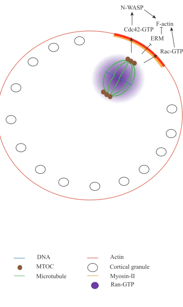
Figure 18: F-actin meshwork during Meiosis I in the mouse oocyte χ. Force generation
Figure 19: Force generator in mouse Meiosis I spindle migration
c) The role of Mos
Figure 20: Mos/MAPK during mouse meiotic maturation
d) Formation of the actin cap and polarization of the oocyte
Figure 21: Actin cap formation in Meiosis I
C) The physics of cell division
1) The physical parameters describing cell mechanics a) Tension
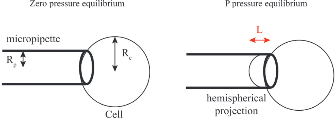
α. Micropipette aspiration
Figure 22: Micropipette aspiration and determination of cortical tension β. AFM
χ. Optical trap
b) Cytoplasmic viscosity
Figure 23: Micropipette aspiration and measure of the cell viscosity
c) Elasticity/Plasticity d) Models and scales
2) Physical constraints on a dividing cell a) From outside the cell
Figure 24: Mechanical constraints and spindle positioning in non-polarized cells
b) From inside the cell
Figure 25: Increased cortical tension allows proper spindle formation in mitosis β. Cortical tension and spindle motion in mouse oocyte
Figure 26: F-actin network and cortical tension in mouse oocytes, a contradiction? χ. Cell mechanics and cytokinesis
Figure 27: Shape instabilities, blebbing and pressure release during cytokinesis
D) Situation of the problem
Results
A soft cortex is essential for asymmetric spindle positioning in mouse oocytes
Cortical stiffness: a Gatekeeper for spindle positioning in mouse oocyte (in preparation)
Discussion and perspectives
A) Cooperation of two actin networks in positioning the Meiosis I spindle
1) Relationships between the two actin networks 2) Cortical tension: a catalyst for forces unbalance
B) Actin network architecture influences Myosin-II activity
C) Multiple roles for cortical tension?
1) A role for cortical tension in spindle formation?
Figure 28: Spindle defects in Meiosis I oocytes harboring a low cortical tension
2) A role for cortical tension in the switch between symmetric and asymmetric acentriolar division?
Figure 29: A role for cortical tension in mediating the switch between symmetric and asymmetric divisions in acentriolar cells?
References
Annexes
Abbreviations list
-/-: homozygous mutant allele ABP: Actin Binding Protein
ADF/Cofilin: Actin Depolimerization Factor/Cofilin ADP: Adenosine di Phosphate
AFM: Atomic Force Microscope AI: AutoInhibitory domain
APC-C: Adenomatous Polyposis Coli C terminal fragment APC/C: Anaphase Promoting Complex/Cyclosome
aPKC: atypical Protein Kinase C Arp2/3: Actin related protein 2/3
ARPC1-5: actin-related protein complex-1-5 ATP: Adenosine tri Phosphate
B: Basic binding domain
cAMP: Cyclic Adenosine 3’,5’-MonoPhosphate CapZ: Capping Protein Z
CC: Coil-Coiled
CDK1: Cyclin Dependent Kinase 1 COBL: Cordon-Bleu
CP: Capping Protein
CRIB: Cdc42-Rac Interactive Binding domain CSF: CytoStatic Factor
DAD: Diaphanous Autoinhibitory Domain DD: Dimerization Domain
Diaph1/2: Diaphanous 1 and 2 DID: Diaphanous Inhibitory Domain
DOC1R: Deleted in Oral Cancer 1 Related DRF: Diaphanous Related Formin
E3KARP: NHE3 kinase A regulatory protein
EBP50/NHE-RF: ERM Binding Protein 50 or Na+/H+ Exchanger Regulatory Factor Ect2: Epithelial Cell Transforming 2
ENA/VASP: Enabled/Vasodilator-Stimulated Phosphoprotein ERK: Extracellular signal-regulated kinases
ERK1/2: Extracellular signal-Regulated Kinase 1 and 2 ERM: Ezrin, Radixin, Moesin
F-actin: Filamentous actin FAK: Focal Adhesion Kinase
FERM domain: Four-point one, Ezrin, Radixin, Moesin domain FH1-FH2: Formin Homology domain 1 and 2
FMN1/2: Founding Mammalian Formin 1 or Formin-1 and Formin-2 FSI: Formin-Spire Interaction domain
G-actin: Globular actin
GAP: GTPase Activating Proteins GBD: GTPase Binding domain GDP: Guanosine Di Phosphate
GEF: Guanine nucleotide Exchange Factor GFP: Green Fluorescent Protein
GTP: Guanosine Tri Phosphate
HURP: Hepatoma Up-Regulated Protein JMY: Junction-Mediating regulatory protein kDa: kiloDalton
LH: Luteinizing Hormone
LMOD1-3: Leiomodin 1 , 2 and 3 MAP: Microtubule Associated Protein MAPK: Mitogen Activated Protein Kinase
MAPKAPK2: MAPK-Activated Protein Kinase 2 mDia: mammalian Diphanous
MEK: MAPK/ERK kinase Kinase
MISS: MAPK Interacting and Spindle Stabilizing MLCK: Myosin Light-Chain Kinase
Mos: Moloney murine Sarcoma virus MPF: M-phase Promoting Factor
MTOC: MicroTubule Organizing Center
N-WASP: Neural Wiskott-Aldrich Syndrome Protein Non-DRF: Non Diaphanous Related Formin
NPF: Nucleation Promoting Factor NuMA: Nuclear Mitotic Apparatus P: polyProline domain
PAK1-3: P21-Activated protein Kinase 1, 2 and 3 Par-3/6: Partitioning defective 3 and 6
PB1/2: Polar Body 1 and 2
PDZ domains: Postsynaptic-density protein of 95 kDa, Discs large and Zona occludens-1 domain
Pi: inorganic Phosphate
PICK1: Protein Interacting with C Kinase 1 PCM: PeriCentriolar Material
PRD: Prolin-Rich Domain
PtdIns(4,5)P2: PhosphatidylInositol 4,5-bisphosphate
Ran: Ras-related Nuclear protein RBD: Rho-Binding Domain
RCC1: Regulator of Chromosome Condensation 1 SAC: Spindle Assembly Checkpoint
SAF: Spindle Assembly Factor
SCAR: Suppressor of Cyclic AMP Repressor SHD: SCAR Homology Domain
SOP: Sensory Organ Precursors Spir: Spire
Spire1/2: Spire 1 and Spire 2 TBR: Tubulin-Binding Region
TIRF: Total Internal Reflection Fluorescence Tmod: Tropomoduline
TPX2: Targeting Protein for the Xenopus kinesin xklp2 WAHD1: WASH Homology Domain 1
WASH: WASP and Scar Homologue WASP: Wiskott-Aldrich Syndrome Protein
WAVE: WASP-family Verprolin-homologous protein
WCA domain: WASP-homology-2 (WH2 or W; also called verprolin-homology) domain; Central (also called cofilin-homology or connector) domain; Acidic domain
WHAMM: WASP Homologue associated with Actin, Membranes and Microtubules WMD: WHAMM Membrane-interaction Domain
Prologue
Asymmetric divisions are very important because they allow the formation of two different daughter cells thus generating cell diversity. Meiotic divisions during the formation of the female gamete, the oocyte, are examples of asymmetric divisions: in that case, the purpose of the divisions is to remove half of the DNA content to form an haploid cell, so that when it fuses with an haploid sperm it forms a diploid zygote. But, especially in species where eggs are laid such as Xenopus, asymmetric divisions are also very important to retain most of the cell cytoplasm in the oocyte, which contains maternal stores indispensable for further embryonic development. Hence, these divisions are highly asymmetric in size ensuring the segregation of chromosomes with minimum cytoplasmic loss.
The first female meiotic division (Meiosis I) in the mouse is peculiar because the asymmetry of the division depends on an off-center spindle positioning that is not microtubule dependent as in most mitotic cells but only actin dependent (Longo and Chen, 1985). Furthermore, it was shown recently that cortical tension, a parameter that accounts for cortex stiffness, decreases during Meiosis I (Larson et al., 2010), which is a very puzzling observation for two reasons. First, a very dense F-actin meshwork is formed during Meiosis I (Azoury et al., 2008; Schuh and Ellenberg, 2008) which we would expect to stiffen the cortex. Second, oocytes are round cells stable in shape, similar to mitotic cells that round up at the onset of mitosis because of an increase in cortical tension absolutely required for proper spindle function (Kunda et al., 2008; Lancaster et al., 2013).
During my PhD, I was interested in this drop in cortical tension and its role in spindle positioning. The goal of my PhD was first to understand where this drop was coming from and how it was related to F-actin present in the mouse oocyte, and second what was its role during Meiosis I. To this end, I also developed new physical approaches to study mechanical properties of the cytoskeleton in the oocytes that did not exist previously in the lab.
In the first part of the introduction, I will present F-actin and its partners playing a role in cell division in general and mouse meiotic divisions in particular. I will then present mouse meiotic divisions with a focus on spindle migration mechanisms during Meiosis I. Finally, I will discuss the mechanical properties of cells with an emphasis on cortical tension and their known roles in cell division.
Plus or barbed end
Fast polymerization
Minus or pointed end
Fast depolymerization
ADP-bound G-actin
Polymerization or
depolymerization
ATP-bound G-actin
ATP Cap
2
A) Actin and cell division
1) Actin and its partners
Actin is one of the most abundant proteins in cells; it has a major role in building the cytoskeleton, which confers to the cell its specific architecture. It is a very highly conserved protein among species and is involved in many cellular processes such as cell motility, cell division and intracellular trafficking.
a) G-actin and F-actin
Actin is present in almost all eukaryotic cell types. It is found in cells as monomers (globular actin or G-actin) or polymers (fibrillar actin, F-actin or microfilaments).
α. G-actin monomers
Actin monomers are 42 kDa proteins. In Vertebrates, three different isoforms have been identified: α-actin, β-actin and γ-actin. These isoforms are thought to sustain different functions in cells probably due to differences in their localization or their ability to bind proteins (for review see (Perrin and Ervasti, 2010)). Each monomer has a globular conformation and possesses a cleft that can bind Mg2+ and ATP (Adenosine Tri Phosphate), and hydrolyze ATP in ADP (Adenosine Di Phosphate) +Pi (inorganic Phosphate). In cells, G-actin is often associated with Profilin, a protein that has been shown to inhibit F-G-actin polymerization (Schutt et al., 1993; Perelroizen et al., 1994).
β. F-actin filaments
F-actin filaments consist of multiple actin monomers polymerized in an oriented fashion and forming an helix. The ATP-bound cleft in each monomer is oriented towards a particular end of the proto-filament. This extremity is able to polymerize faster than the other: it is called “plus-end” or “barbed-end” as opposed to “minus-end” or “pointed-end” (Figure 1).
When the actin monomer is added on a growing filament, it has a bound ATP molecule, which can then undergo hydrolysis. Therefore, because of the difference in polymerization rates between the two extremities, the plus-end of the actin filament is richer in ATP-bound monomers than the minus-end, thus forming an ATP-cap that might play a role in stabilizing the filament (Figure 1) (Blanchoin et al., 2000a).
70°
Nucleation
Elongation
Mother filament
Daughter
filament
G-actin
ARP2 and ARP3
3 χ. Actin polymerization mechanisms
F-actin filaments start to polymerize when G-actin reaches a threshold concentration called the critical concentration. De novo filament polymerization is achieved in two steps: the nucleation and the elongation. As mentioned before, the critical concentration is different at each end of the filament. In living cells, the critical concentration is reached and F-actin is at steady state.
The nucleation step, which is the formation of a seed of three monomers, is a slow process because the formation of an actin dimer is not thermodynamically favorable. However, the addition of the third monomer stabilizes the trimer and provides a starting point for elongation of the filament. The elongation step is a very favourable and quick reaction.
Actin filaments can undergo polymerization and depolymerization at both ends. But their rate is different leading to a process called treadmilling: even when there is actin assembly on the barbed end and disassembly on the pointed end the filament can harbor a constant length.
b) Actin nucleators
As mentioned before, the crucial event for filament polymerization is nucleation. Actin nucleators are a group of proteins that promote actin nucleation in cells (for review, see (Campellone and Welch, 2010)).
α. Arp2/3, a branched actin filaments nucleator
Role
The Arp2/3 complex is involved in many cellular processes, involving in particular a change in the architecture of the cell at its periphery. Indeed, Arp2/3 is localized and activated at the cortex of many cell types (see below). It has been involved for example in lamellipodia formation of migrating cells (Blanchoin et al., 2000b), during vesicular trafficking (Campellone et al., 2008), endocytosis (Kaksonen et al., 2006) and phagocytosis (May et al., 2000). It also plays a key role during mouse meiotic divisions that I will detail later on, promoting their asymmetry in size (Sun et al., 2011a; Chaigne et al., 2013).
Structure and activity
Arp2/3 is composed of seven subunits (ARP2, ARP3, ARPC1, ARPC2, ARPC3, ARPC4, ARPC5) forming a 220 kDa complex (for review, see (Goley and Welch, 2006; Rotty et al., 2013)), which is able to nucleate branches on a pre-existing actin filaments at a
WH1 B CRIB AI PRD V A
WASP
C WH1 B CRIB AI PRD V V CN-WASP
A SHD B PRD V C AWAVEs
WAHD1 TBR P V C AWASH
WHAMM
WMD CC P V V C A N CC P V VJMY
V L C ARegulatory motifs
Nucleation motifs
WH1: WASP Homology 1 domain
B: Basic binding
CRIB: Cdc42–Rac Interactive Binding
AI: AutoInhibitory domain
PRD: Prolin-Rich Domain
SHD: SCAR Homology Domain
WAHD1: WASH Homology Domain 1
TBR: Tubulin-Binding Region
P: Polyproline
WMD: WHAMM Membrane interaction Domain
CC: Coil-Coiled
N: N-term
V: WASP Homology domain 2
C: Connector
A: Acidic region
L: Linker
4 70° angle (Figure 2). The ability of this complex to nucleate actin filaments comes from the structural similarities between the ARP2 and ARP3 subunits and G-actin (Machesky et al., 1994; Kelleher et al., 1995). ARP2 and ARP3 bind to the pointed end of daughter filaments, thus probably providing a stable actin dimer-like structure allowing further elongation whereas ARPC2 and ARPC4 bind to the mother filament. The Arp2/3 complex seems to exist in two conformational states: opened and closed, the latter being the active conformation leading to creation of new branches (Smith et al., 2013).
Activation
For its nucleating activity, the Arp2/3 complex needs to be activated through a family of proteins called the NPF (Nucleation Promoting Factor) family (for review see (Campellone and Welch, 2010)).
Class I NPFs possess a characteristic VCA (or WCA) domain which is composed of a WH2 domain (also known as VH2 for Verprolin Homology domain 2), a connector domain (C), and an acidic domain (A). The VCA domain promotes the Arp2/3 complex-mediated nucleation by allowing the binding of G-actin (through the V motif) and Arp2/3 (through the CA motif) (Marchand et al., 2001) (Figure 3). Type I NPFs include WASP (Wiskott-Aldrich Syndrome Protein) which has also been shown recently to nucleate unbranched filaments in the absence of Arp2/3 (Urbanek et al., 2013), N-WASP (Neural WASP), three isoforms of SCAR/WAVE (Suppressor of Cyclic AMP Repressor/WASP-family Verprolin-homologous Protein) (Machesky et al., 1999), WASH (WASP and Scar Homologue) (Linardopoulou et al., 2007), WHAMM (WASP Homologue associated with Actin, Membranes and Microtubules) (Campellone et al., 2008) and JMY (Junction-Mediating regulatory protein) which can also nucleate unbranched actin filaments in the absence of Arp2/3 (Zuchero et al., 2009). Although the precise mechanism of Arp2/3 activation by the VCA is not completely elucidated, recent single molecule fluorescence colocalization techniques have further demonstrated that it stimulates both Arp2/3 binding to the mother filaments and Arp2/3 branching activity (Smith et al., 2013). Biochemical studies on WASP have shown that Arp2/3 is able to bind two VCA-containing molecules at once (Padrick et al., 2011), which could explain this dual function. Several type I NPFs have been shown to play a role during mouse meiotic divisions (Sun et al., 2011b, 2011a; Huang et al., 2012; Chaigne et al., 2013; Liu et al., 2012), that I will discuss later.
NPF can in turn be activated, for example Wave2 is phosphorylated in culture cells by ERK/MAPK (Extracellular signal-Regulated Kinases/Mitogen Activated Protein Kinase)
B: Nucleation and elongation by Formins
(adapted from Breitsprecher et al., 2012)
Formin recruting protein
G-actin-Profilin
Formin dimer
FH1 domains
FH2 domains
Regulatory domains
GBD DID DD CC FH1 DADDRF
mDia
FH2Non-DRF
Formin-2
Regulatory motifs
Nucleation motifs
GBD: GTPase Binding Domain
DID: Diaphanous autoInhibitory Domain
DD: Dimerization Domain
CC: Coil-Coiled
DAD: Diapnanous Autoinhibitory Domain
FH1: Formin Homology Domain 1
FH2: Formin Homology Domain 2
FH1 FH2 FSI
FSI: Formin-Spire Interaction Domain
A: Two examples of Mammalian Formins
5 (Nakanishi et al., 2006; Mendoza et al., 2011) adding another level of complexity (for review see (Mendoza, 2013)). Furthermore, they possess in their N-terminal parts motifs important for their regulation, for example some of them can be targeted to the membrane and activated via interaction between the Basic (B) domain (Figure 3) and membrane phospholipids such as PtdIns(4,5)P2 (PhosphatidylInositol 4,5-bisphosphate), explaining the many roles for Arp2/3
at the cell membrane.
Class II NPF such as Cortactin lack complete VCA domains, but they have acidic domains at their amino terminus that bind the Arp2/3 complex and tandem repeat domains that bind F-actin. They also activate Arp2/3 but in a less potent manner (Uruno et al., 2001; Weaver et al., 2001).
Arp2/3 activity is also dependent on ATP which binds to the subunits ARP2 and ARP3 in the same way that it binds to G-actin (Dayel et al., 2001). Its role is still controversial today, but it seems to promote a change in the conformation of the complex (Goley et al., 2004), a change in its affinity for the NPFs (Dayel et al., 2001), and its hydrolysis may also play a part in remodeling the F-actin network by promoting debranching (Martin et al., 2006).
Finally, several classes of negative regulators of the Arp2/3 complex have been identified in the past few years, including PICK1 (Protein Interacting with C Kinase 1) (Rocca et al., 2008) and Arpin (Dang et al., 2013). Very preliminary experiments conducted in the lab showed that overexpressing these proteins has no effect on mouse meiotic divisions (unpublished data).
β. Formins, nucleator and/or elongator?
Role
Like the Arp2/3 complex, Formins are also involved in cell migration, but also in cell polarity and cytokinesis. Furthermore, they seem to be able to stabilize microtubules and as such have recently been shown to play a role in cell division (for review see (Chesarone et al., 2010)).
Structure
Contrary to Arp2/3, the Formins are very potent nucleators of unbranched F-actin filaments (Figure 4). In all Formins, the C-terminal domain possesses the nucleating activity; it comprises the FH1 (Formin Homology 1) and FH2 (Formin Homology 2) domains (Figure
4A). The FH1 domain binds to G-actin-Profilin whereas the FH2 domain forms a donut-shaped dimer with an adjacent Formin around the barbed end of the growing F-actin filament (Figure 4B). The N-terminal part is variable according to the subfamilies and contains domains important for the control of the protein activity. The exact sequence of these domains can vary considerably from a subfamily to another and among species. There are two classes of Formins in Mammals (Figure 4A): the Diaphanous-Related Formins (DRFs), subdivided in three families, and the Non-Diaphanous-Related Formins (Non-DRFs), subdivided in four families. Among the Non-DRF class, the FMN family comprises two members, FMN1 (Founding Mammalian Formin 1 or Formin-1) and FMN2 (Founding Mammalian Formin 2 or Formin-2). Formin-2 in particular is expressed in mouse oocytes (Evsikov et al., 2006) and brain and plays a key role during mouse meiotic divisions, promoting their asymmetry in size (Leader et al., 2002; Dumont et al., 2007a; Azoury et al., 2008; Li et al., 2008; Schuh and Ellenberg, 2008). FMN1 and 2 both possess a Formin-Spire interaction domain (FSI) (see below).
Activity
The precise mechanism of actin nucleation by Formins is not yet understood. It seems that many Formins need another protein to start nucleating F-actin. For example, a recent study in vitro has shown by TIRF (Total Internal Reflection fluorescence) microscopy that the protein APC-C (Adenomatous Polyposis Coli C terminal fragment) would be one of these proteins: it recruits actin monomers which serve as a nucleation seed and mDia1-C (a DRF Formin), which is then propelled and acts as a processive elongator (Breitsprecher et al., 2012) (Figure 4B). It seems that similarly, other Formins would need the binding of another molecule to nucleate actin; for example, the C-terminal part of Formin-2 is able to bind via its FSI motif to Spire-1 (Figure 4A), an actin binding protein which can nucleate F-actin in vitro (Pechlivanis et al., 2009). In addition, the fly Formin Cappuccino, a non-DRF Formin, also interacts in vitro and in vivo in the oocyte with Spire (Rosales-Nieves et al., 2006; Quinlan et al., 2007). Thus, Formins should not be considered only as nucleators but also as elongators, as shown also in mouse oocytes (see part B)3)b)β) (Pfender et al., 2011; Montaville et al., 2014).
In contrast to the Arp2/3 complex, Formins are processive proteins that cap the barbed-end of the growing filaments (Pruyne et al., 2002; Sagot et al., 2002) (Figure 4B). The 2 FH2-domains bind transiently to the barbed end, somehow promoting a very potent filament elongation whereas the FH1 domains bind to G-actin-Profilin, probably then acting by
Figure 5: Structure of WH2-mediated actin nucleators
Regulatory motifs
Nucleation motifs
KIND: Kinase non-catalytic domain
FYVE: Fab1, YoTB, Vac1 and eeA1
domain
P: Polyproline
T: Tropomyosin- and actin-binding helices
LRR: Leucin-rich repeats
V: WASP Homology domain 2
h-b-h: helix-basic-helix
KIND L SPIREs P P V V P COBL V LRR T Leimodins V V V FYVE P h b h V VL: Linker
increasing the local concentration in G-actin (Vavylonis et al., 2006). χ. Other types of nucleators
Spire
Spire contains four WH2 domains required for actin nucleation that bind actin and in addition possesses a G-actin binding linker between the third and fourth WH2 domains (Quinlan et al., 2005) (Figure 5). The precise mechanism for F-actin polymerization is not yet completely understood, but evidence suggests that G-actin binds to the tandem WH2 domains and the actin-binding linker thus forming a linear core of three monomers (Quinlan et al., 2005). Spire can work on its own or cooperate with Formins. Mammalian Spire1/2 (Spire 1 and Spire 2) bind to FMN1 and FMN2 (Pechlivanis et al., 2009) and allow the polymerization of F-actin in the cytoplasm of mouse oocytes (Pfender et al., 2011; Montaville et al., 2014), a process that I will detail later.
JMY
As mentioned before, in addition to its role as a NPF for the branched actin nucleator Arp2/3, JMY is able to nucleate actin filaments by itself (Zuchero et al., 2009). It is composed of three WH2 domains, a C domain and an A domain like for all class I NPFs (see Figure 3). Similarly to Spire, JMY possesses an actin-binding linker between the second and third WH2 domains that are involved in the formation of a new actin seed (Zuchero et al., 2009).
Cordon-Bleu
COBL (Cordon-Bleu) has three WH2 domains that bind G-actin in vitro (Ahuja et al., 2007), but contrary to JMY and Spire no predicted actin-binding linker (Zuchero et al., 2009) (Figure 5). Although the precise mechanism has not been demonstrated, it is assumed that these domains provide a trimeric seed for further unbranched filament elongation (Ahuja et al., 2007).
Leiomodin
The Leimodin family comprises three members, LMOD1, LMOD2 and LMOD3 that are muscle proteins. They possess two actin-binding domains and a WH2 domain (Figure 5). These three actin-binding domains are predicted to allow further polymerization from this nucleated seed (Chereau et al., 2008).
extracellular
intracellular
ERM proteins
membrane protein
G-actin-Profilin
Figure 6: Actin-interacting proteins
Formin
ADF/Cofilin
ENA/VASP
Gelsolin
CapZ
c) Modulation of actin nucleation
Actin is a central protein in cells and as such interacts with many other proteins in order to regulate its function in time and space. We will discuss some proteins that have direct interactions with F-actin filaments, and some of the many transduction pathways that lead to modifications of the actin cytoskeleton.
α. Actin associated proteins
There are many actin-associated proteins, such as G-actin sequestering proteins, F-actin stabilizing or destabilizing proteins, F-F-actin organizing proteins and F-F-actin motors (Figure 6).
G-actin sequestering proteins
As mentioned before, G-actin in cells is often bound to Profilin (Schutt et al., 1993; Perelroizen et al., 1994). This binding promotes addition of actin monomers at barbed ends of filaments (Vavylonis et al., 2006).
Thymosine β4 is a small protein of the WH2 motif-containing protein family known for sequestering actin monomers (dos Remedios et al., 2003). It caps the actin monomers at both ends and competes with Profilin (Irobi et al., 2004), but can also bind to F-actin with a low affinity and destabilize the filaments (Carlier et al., 1996).
F-actin destabilizing proteins
ADF/Cofilin (Actin Depolimerization Factor/Cofilin) is a family of F-actin depolymerizing proteins (Bamburg et al., 1980), that comprises ADF and Cofilin in Vertebrates. These proteins possess binding sites for G-actin and F-actin and are responsible for the destabilization of F-actin in various ways, for example severing, increasing the off-rate at the pointed-end or direct depolymerization (dos Remedios et al., 2003).
Gelsolin is a superfamily comprising among others Gelsolin, Villin, and Severin (also called Fragmin); they all contain at least one gelsolin repeat (Kwiatkowski et al., 1986). Gelsolin binds to F-actin and upon Ca2+ fixation induces a change in conformation thats leads to the severing of the filament from their barbed ends (Robinson et al., 1999).
A less understood family is the ENA/VASP (Enabled/Vasodilator-Stimulated Phosphoprotein) family. ENA/VASP binds to G-actin, F-actin and Profilin and could act by preventing the capping of the filaments, inhibit Arp2/3 dependent nucleation or promote unbranching (for review see (Krause et al., 2003)).
Figure 7: Myosin-II and F-actin
G-actinMyosin-II heavy chain tail Myosin-II heavy chain head Myosin-II regulatory light chain Myosin-II essential light chain
Barbed-end
Barbed-end
F-actin relative movement Myosin-II movement
F-actin stabilizing proteins
The barbed-end of microfilaments can depolymerize very fast. However, in cells, they are stabilized by actin capping proteins proteins (for review see (dos Remedios et al., 2003) such as CapZ (Capping protein Z, also called CP for capping protein) (Casella et al., 1987). There are also proteins that stabilize the minus ends of the filaments, for example Tropomodulin (Tmod) that binds to pointed ends in presence of Tropomyosin.
F-actin organizing proteins
Many proteins such as Fimbrin, Actinin α, Villin, Spectrin organize actin filaments in higher order actin structure. Here, we will focus on another class of actin-organizing proteins, the ERM (Ezrin, Radixin, Moesin) proteins.
Ezrin, Radixin and Moesin are closely related proteins (Sato et al., 1992), they all possess a FERM (Four-point one, Ezrin, Radixin, Moesin) domain at their N-terminal parts. This domain is thought to have a clover leaf configuration such as it needs to bind to a ligand to unfold and thus allow the binding of F-actin to the C-terminal part of the protein (Bretscher et al., 2002). These proteins associate with the cytoplasmic tail of membrane protein such as the cell surface glycoprotein CD44 (Tsukita et al., 1994), but also with two scaffolding proteins EBP50/NHE-RF (ERM Binding Protein 50 or Na+/H+ Exchanger Regulatory Factor) and E3KARP (NHE3 kinase A regulatory protein)that themselves bind to a variety of membrane protein (Yun et al., 1998). Thus, ERM proteins are an interface between the plasma membrane and the actin cortex underneath (Charras et al., 2006) (Figure 6). Due to their close relation with the cell periphery, ERM proteins have been proposed to play various roles in cell cortex mechanics (for review see (Neisch and Fehon, 2011)) for example during blebbing (Charras et al., 2006) or during cell rounding at the onset of mitosis (Carreno et al., 2008; Kunda et al., 2008). In particular, it seems that upon activation, ERM proteins acting as crosslinkers between the actin cytoskeleton and the membrane force the F-actin filaments to become parallel to the cortex thus increasing the cortical tension of the cell and promoting cell rounding (Kunda et al., 2008). In mouse oocytes, cortical tension has also been shown to depend on ERM proteins (Larson et al., 2010), I will detail this process below (part C)2)b)β).
F-actin Motors
Myosins are motor proteins that can bind to actin; as such they form together a functional unit. Myosins possess a conserved motor domain on their “head” part, or N-terminal part and a variable “tail” at their C-N-terminal extremities. There are many different
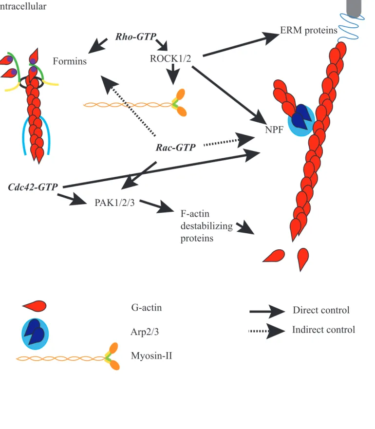
Figure 8: Actin and Rho GTPases
extracellular
intracellular
F-actin
destabilizing
proteins
ERM proteins
membrane protein
Formins
Rho-GTP
ROCK1/2
NPF
PAK1/2/3
Rac-GTP
Cdc42-GTP
G-actin
Direct control
Indirect control
Arp2/3
Myosins that are divided in thirty five families (Bloemink and Geeves, 2011). All but Myosin-VI are able to walk along the actin filaments towards the plus-end through ATP hydrolysis-mediated changes in Myosins’ “head” conformation. The tail domain contains motifs that can be important for the specific function of a specific Myosin, for example cargo transport or dimerization. Two Myosins play an important role during mouse meiotic divisions, Myosin-II and V; I will discuss it later. Myosin-X is also peculiar because it binds both to F-actin and microtubules in vitro and in Xenopus oocytes (Weber et al., 2004) and embryos (Woolner et al., 2008; Woolner and Papalopulu, 2012).
Myosin-II is one of the most studied Myosins. It is present in particular in the muscle and is named so because it has two “heads”, the very long tail of the two heavy chains supercoilling, thus forming a head-to-tail dimer (Figure 7). They also possess four light chains associated with each “head”. Myosin-II has a small processivity, which means that it can walk only on a short distance on the actin filament, but at a high speed. Myosin-II can be phosphorylated at several sites. Phosphorylation of the light chain by MLCK (Myosin Light-Chain Kinase) is of crucial importance because it unfolds the protein and allows F-actin-Myosin interactions (for review see (Hong et al., 2011)).
Myosin-Va and Myosin-Vb have also been shown to walk along actin filaments but moreover they can transport cargos (for review see (Hammer and Sellers, 2012)). This process seems to be used either for short distance transport or in cells lacking centrosomes for long distance transport for example in the mouse oocyte (Schuh, 2011), suggesting that this is an alternate pathway for the canonical microtubule/Dynein/Kinesin track (Hammer and Sellers, 2012).
Interestingly, Myosin activity depends on the architecture of the F-actin network. For example in vitro, Myosin-VI and Myosin-II induce a contraction followed by a disassembly of a branched actin network of antiparallel filaments, whereas parallel filaments with their barbed-end oriented towards the same direction would not contract or disassemble (Reymann et al., 2012). Moreover, it has also been shown in the amoeba Dictyostelium discoideum that Myosin-II binds preferentially to stretched F-actin filaments (Uyeda et al., 2011). Thus, the orientation, the architecture and tension of the F-actin filaments play a part in the activity of Myosins.
β. Transduction pathways associated with actin polymerization
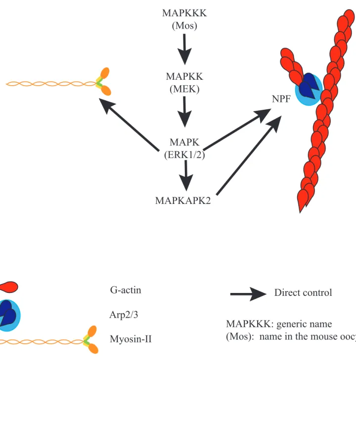
Figure 9: Actin and the MAPK pathway
G-actin
Direct control
Arp2/3
Myosin-II
MAPKKK: generic name
(Mos): name in the mouse oocyte
MAPKKK
(Mos)
MAPKK
(MEK)
MAPK
(ERK1/2)
MAPKAPK2
NPF
only present here the pathways that are relevant in mouse oocytes.
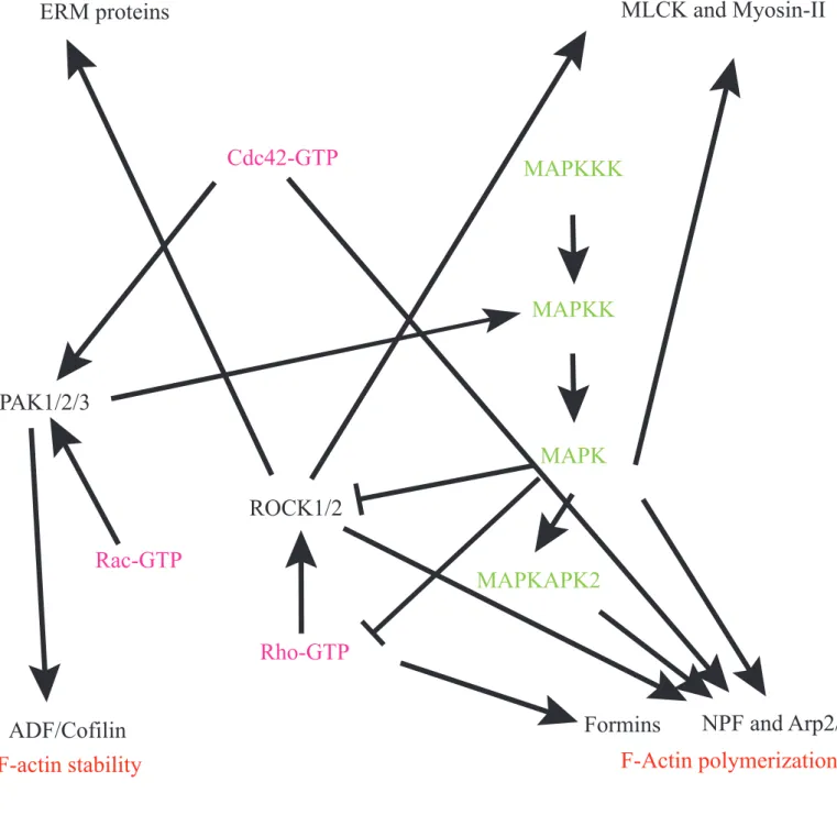
Rho GTPases
The Rho family of small GTPases includes Rho, Rac and Cdc42. These small molecules act as molecular switches; they exist bound to GTP (Guanosine Tri Phosphate), which is their active state, and GDP (Guanosine Di Phosphate). The switch between these states is mediated by GAPs (GTPase Activating Proteins) and GEFs (Guanine nucleotide Exchange Factor) (Jaffe and Hall, 2005). Rho has been shown very early on to have a major role in the control of the actin cytoskeleton and especially on stress fibers and focal adhesion (Paterson et al., 1990). Then Rac and Cdc42 were shown to control respectively the formation of lamellipodia, which are the leading edge of migrating cells, and filopodia, the cytoplasmic projections of migrating cells (for review see (Pullikuth and Catling, 2007)); these two structures rely on actin cytoskeleton.
The Rho GTPases have many downstream effectors (Figure 8) (for review see (Jaffe and Hall, 2005)). For example, Rho controls the Formins Diaph1 (Diaphanous 1) and Diaph2 (Diaphanous 2), Rock1 (Rho-associated, Coiled-coil-containing protein Kinase 1 or Rho Kinase 1) and Rock2 (Rho-associated, Coiled-coil-containing protein Kinase 2 or Rho Kinase 2) which in turn activate the phosphorylation and thus activation of Myosin-II light chain, activates WASH in Drosophila oocytes (Liu et al., 2009) and ERM proteins (Bretscher et al., 2002). Rac controls PAK1, PAK2 and PAK3 (P21-Activated protein Kinase 1, 2 and 3) which regulate Cofilin, an F-actin destabilizing protein, thus controlling actin depolymerization; it also interacts indirectly with a DRF Formin, mDia (mammalian Diaphanous) and Wave2 (Goh et al., 2012). Cdc42 controls WASP and N-WASP, which activates Arp2/3-dependent F-actin assembly, and the PAK family.
MAPK pathway
The MAPK (Mitogen-activated protein kinase) pathway is composed of a cascade of proteins phosphorylating each other, namely upstream a MAPKKK (MAP Kinase Kinase Kinase), then a MAPKK (MAP Kinase Kinase) and downstream a MAPK (MAP Kinase), which is a Serine/Threonine Kinase (Figure 9). In Mammalian oocytes, the ones expressed are respectively Mos (Moloney murine Sarcoma virus), MEK (MAPK/ERK Kinase) and ERK1/2 (Extracellular signal-Regulated Kinase 1 and 2). The MAPKs ERK1/2 have many substrates and are known for controlling the actin cytoskeleton in several ways. In cells, it has been shown to mediate Arp2/3 dependent nucleation via phosphorylation of the NPF Wave2
Figure 10: Transduction pathways associated
with actin dynamics
F-Actin polymerization
F-actin stability
F-Actin organization
F-Actin/motor interaction
Formins
NPF and Arp2/3
ERM proteins
MLCK and Myosin-II
ADF/Cofilin
MAPKKK
MAPKK
MAPK
MAPKAPK2
Rho-GTP
Rac-GTP
Cdc42-GTP
PAK1/2/3
ROCK1/2
Activation
Inhibition
(Nakanishi et al., 2006; Mendoza et al., 2011). In neutrophils, the MAPKAPK2 (MAPK-Activated Protein Kinase 2), a downstream effector of the MAPKKK has been shown to directly mediate Arp2/3 phosphorylation (Millard et al., 2003). Furthermore, ERK1/2 can phosphorylate MLCK and thus activate Myosin-II in vitro (Suizu et al., 2000).
In addition, the MAPK pathway and the Rho pathway are interconnected, bringing another level of complexity (Figure 10); it has been shown in various contexts that MEK can be activated by PAK (Pullikuth and Catling, 2007). Furthermore, MAPK can also modulate Rho signaling, for example focal adhesion remodeling during cell migration requires ERK activation of FAK (Focal Adhesion Kinase), which itself has been shown to inhibit Rho and ROCK (Pullikuth and Catling, 2007).
2) The roles of actin in mitotic cell division a) Overview of mitotic cell division
α. The basic steps of mitotic cell division
Cell division produces two daughter cells from a mother cell. During S phase, the DNA is replicated. Then for somatic cells mitosis follows, producing two daughter cells with the same amount of DNA as the mother cell. The challenge of cell division is to separate correctly the chromosomes and the cytoplasm between the two daughter cells, errors during cell division leading to aneuploidy and potential tumorigenic processes.
Mitosis is divided into four steps: prophase, metaphase, anaphase, telophase (Figure 11). During prophase, the replicated DNA is condensed into chromosomes composed of two sister chromatids, which progressively align on the metaphase plate during prometaphase. When the chromosomes are fully aligned and under equal tension on the microtubule spindle at metaphase, anaphase is triggered. The spindle is essential to align the chromosomes. It is composed of microtubules, very labile Tubulin α and Tubulin β polymers that can quickly polymerize and depolymerize. Microtubules are nucleated from MTOCs (MicroTubule Organizing Centers), which are usually centrosomes composed of two centrioles and pericentriolar material (PCM) comprising proteins such as γ-Tubulin and pericentrin. At mitosis onset, centrosomes duplicated during S phase separate and rapidly promote spindle bipolarization as they constitute the spindle poles. From these nucleating structures, microtubules organize as astral microtubules connecting the spindle to the cell cortex,
Figure 11: The steps of mitosis
DNA
Centrosome
Microtubules
Prophase
Metaphase
Anaphase
Telophase
Interphase
Nuclear Envelope
BreakDown (NEBD)
DNA condensation
Centrosomes separation
Formation of the spindle
Chromosomes align on
the metaphase plate
Separation of sister
chromatids
Elongation of the spindle
Decondensation of the
chromosomes
Cytokinesis
kinetochore microtubules connecting the kinetochores of the chromosomes to the spindle poles (referred also as K-fibers when stabilized), and polar microtubules that can slide with respect to each other via microtubule specialized motors and mediate spindle elongation at anaphase (Figure 12). Finally, cytokinesis is the separation of the two daughter cytoplasms after telophase. Cytokinesis always occurs in the middle of the microtubule spindle. Thus the division plane is specified by the position of the metaphase plate of the spindle.
β. Asymmetric divisions
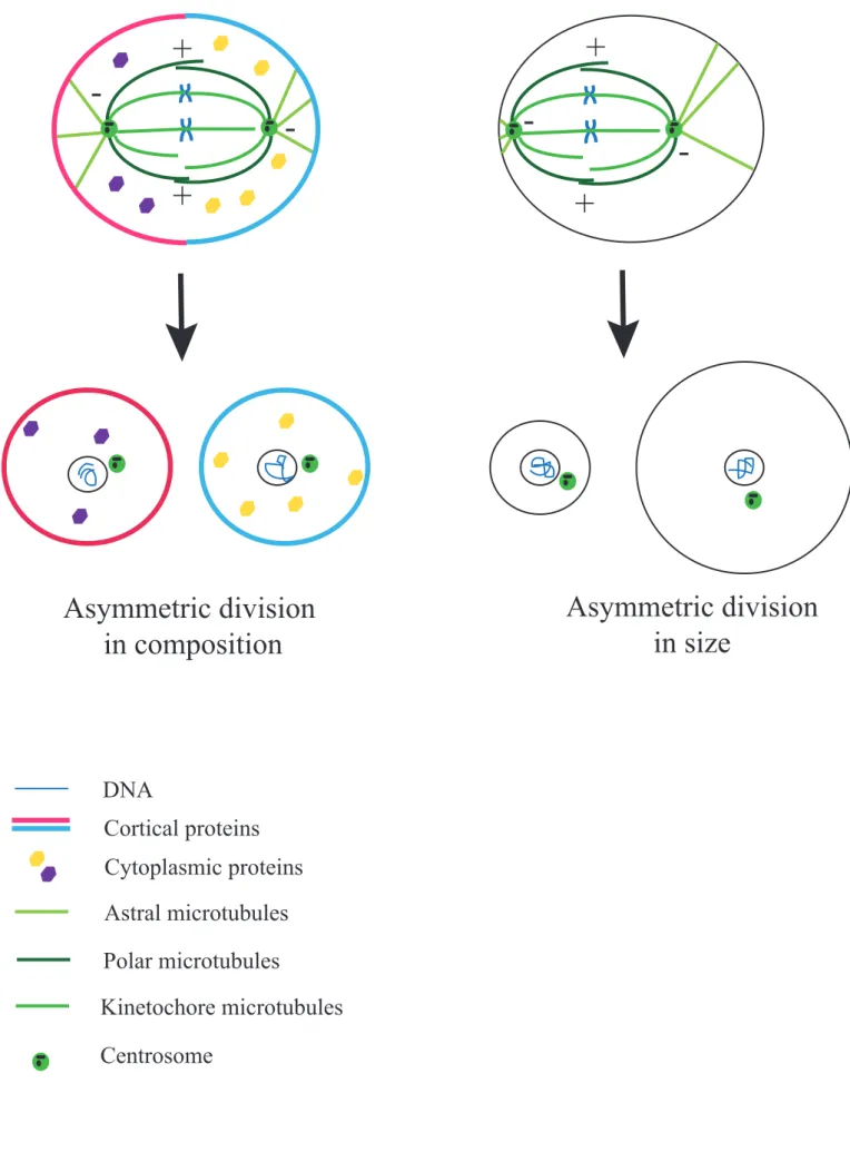
Asymmetric divisions produce two different daughter cells, allowing these two cells to have different fates. This is very important for example during development to promote cell diversity as stem cells self-renew and at the same time produce differentiated cells. It can also allow the partitioning of the genome with minimum cytoplasm loss, for example in female meiosis where half of the DNA needs to be extruded from the future oocyte but most of the cytoplasm containing maternal stores has to be retained. In addition to their role in development and adult life, defects in asymmetric cell divisions are associated with a variety of pathologies (for reviews see (Powell et al., 2010; Lu and Johnston, 2013; Terret et al., 2013)). The asymmetry of the division can be in composition, in size or both (Figure 13). We will focus here only on the asymmetry in size of the division, which occurs during female meiosis.
The asymmetry in size of the division is a consequence of the off-center positioning of the spindle in the cell, since the division plane is specified by the position of the metaphase plate of the spindle. In mitosis, the spindle is positioned through interaction between astral microtubules emanating from spindle poles and the cell cortex. The microtubule minus-end directed motor Dynein located at the cell cortex produces a pulling force on the spindle (Colombo et al., 2003) via astral microtubules (for review see (McNally, 2013; Moore and Cooper, 2010)). Although microtubules play a major part in cell division because they organize and position the spindle, growing evidence shows that actin is actively involved in cell division as well. We will focus here on the role of actin in mitosis and will discuss meiosis later.
b) The roles of actin in mitotic cell division
α. Cortical actin filaments
In mammalian cells, cortical actin is involved in spindle orientation. Indeed, stereotypical divisions become random when cells are treated with F-actin depolymerization
Figure 12: Spindle organization during mitosis
DNA
Astral microtubules
Polar microtubules
Kinetochore microtubules forming K-fibers
Centrosome with two centrioles and Pericentriolar Material
-+
-+
drugs (Théry et al., 2005; Toyoshima and Nishida, 2007). This is also the case in Xenopus embryos where disruption of F-actin leads to spindle mispositioning in the outer epithelial cells (Woolner et al., 2008). Cortical F-actin probably mediates spindle positioning through interactions between the actin cortex and astral microtubules (Toyoshima and Nishida, 2007). Indeed Myosin-X, a Myosin able to interact with microtubules and actin (Weber et al., 2004), is also required for spindle orientation (Toyoshima and Nishida, 2007; Woolner et al., 2008). In addition, it was recently shown that in mammalian cells a subcortical pool of actin is rotating around the cell and helps the cell to read external constraints coming from its environment in order to position its spindle accordingly (Fink et al., 2011). Thus, it is very possible that cortical F-actin, whose localization at the cortex could follow the external constraints coming from cell adhesions for example, mediates spindle positioning through interactions between Myosin-X and astral microtubules (for review see (Almonacid et al., 2014)).
In addition to its role on spindle orientation, cortical actin is essential for spindle morphogenesis by increasing cortical rigidity and thus contributing to cell rounding at mitosis onset in Drosophila cells (Carreno et al., 2008; Kunda et al., 2008). The increase of cortical rigidity is mediated by ERM-dependent actin localization in these cells (Carreno et al., 2008; Kunda et al., 2008). Cell rounding is crucial for proper spindle morphology since cells impaired of Moesin habor spindle defects (Kunda et al., 2008; Lancaster et al., 2013). Indeed, the spindle cannot form in a too confined environment: when the cell is flat, the microtubules are too small to reach and capture the chromosomes (Lancaster et al., 2013). In addition, without cell rounding, the spindle cannot position accurately. This could be because the cortex needs to be stiff in order for the microtubules to be properly anchored and thus properly pull on the spindle (Kunda et al., 2008). Thus, by mediating the cortex mechanical properties, cortical F-actin plays a crucial part in spindle morphogenesis and positioning (for review see (Kunda and Baum, 2009)).
β. Cytoplasmic actin filaments
Whereas the role of cortical F-actin has been investigated for a while, the role of cytoplasmic actin is still unclear, especially since it is hard to visualize cytoplasmic pools of actin (see part B)3)b)α). In yeast, actin cables have been shown to control spindle morphogenesis and function (for review see (Sandquist et al., 2011)). Although their precise role is not yet known, actin cables have also been detected during mitosis, for example in Xenopus embryos (Woolner et al., 2008). In plants where there are no centrioles, F-actin
Figure 13: Simplified scheme of
asymmetric divisions
Asymmetric division
in composition
Asymmetric division
in size
DNA
Cortical proteins
Astral microtubules
Polar microtubules
Kinetochore microtubules
Centrosome
Cytoplasmic proteins
+
+
-+
+
-
-cables running along the mitotic spindle have been reported (for review see (Sandquist et al., 2011)). Whether it is also the case in animal cells remains elusive, but it is possible since in many vertebrate embryos cytoplasmic actin seems to be up-regulated during M-phase (Field et al., 2011).
In Drosophila syncitial embryos, cytoplasmic actin seems to play a role in preventing the movement of the nuclei at the onset of mitosis, whereas nuclei are free to move in interphase (von Dassow and Schubiger, 1994). Similarly, in Drosophila oocytes, a F-actin meshwork is delaying a cytoplasmic streaming because when the F-actin meshwork is dismantled, the cytoplasmic streaming starts earlier (Dahlgaard et al., 2007). Thus, in that case, the F-actin acts as a “brake” to prevent cytoplasmic motion (Field and Lénárt, 2011). χ. Actin flows
Actin flows correspond to streaming movements of the actin cytoskeleton. They have first been described in lamellipodia of migrating cells. It has been shown in mitotic cells by monitoring the motion of beads in the cytoplasm that a cortical streaming due to contractions mediated by Myosin-II is responsible for spindle formation and in particular centrosome separation (Rosenblatt et al., 2004). Actin flows have also been implicated in the maintenance of the Metaphase II spindle at the cortex in mouse oocytes, that I will discuss later (Yi et al., 2011). A recent report also shows that in Xenopus oocyte extracts, actin flows develop spontaneously, under the dependence of F-actin nucleators (Pinot et al., 2012).
B) Meiotic maturation in mouse oocyte
Meiosis is the cell division leading to the formation of gametes, oocytes and spermatozoids. Generally speaking, it consists of two successive divisions without an intermediate DNA replication phase: the daughter cells thus contain half of the mother cell DNA such as upon fertilization the two haploid gametes form a diploid zygote. We will focus here on female meiosis only and on the formation of the female gamete, the oocyte.
1) Meiosis and the female gamete formation a) Overview on meiosis
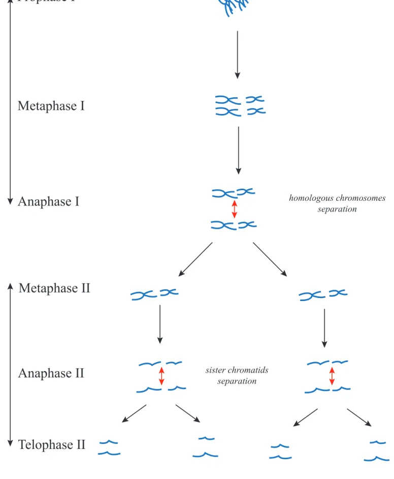
Meiosis is a series of two divisions (Figure 14). The first division is called Meiosis I and the second Meiosis II. Every step of the division is referred to as I or II, depending on
Figure 14: Overview on Meiosis
DNA
Prophase I
Metaphase I
Anaphase I
Metaphase II
Anaphase II
Telophase II
Meiosis I
Meiosis II
homologous chromosomes separation sister chromatids separation2N
N
N/2
whether it belongs to the first or second meiotic division. Meiosis I is a very atypical division, first because the homologous chromosomes are able to exchange parts of their DNA during Prophase I via the crossing overs, bringing genetic diversity, and second because at anaphase I homologous chromosomes segregate instead of sister chromatids. Meiosis II however resembles mitosis with separation of sister chromatids.
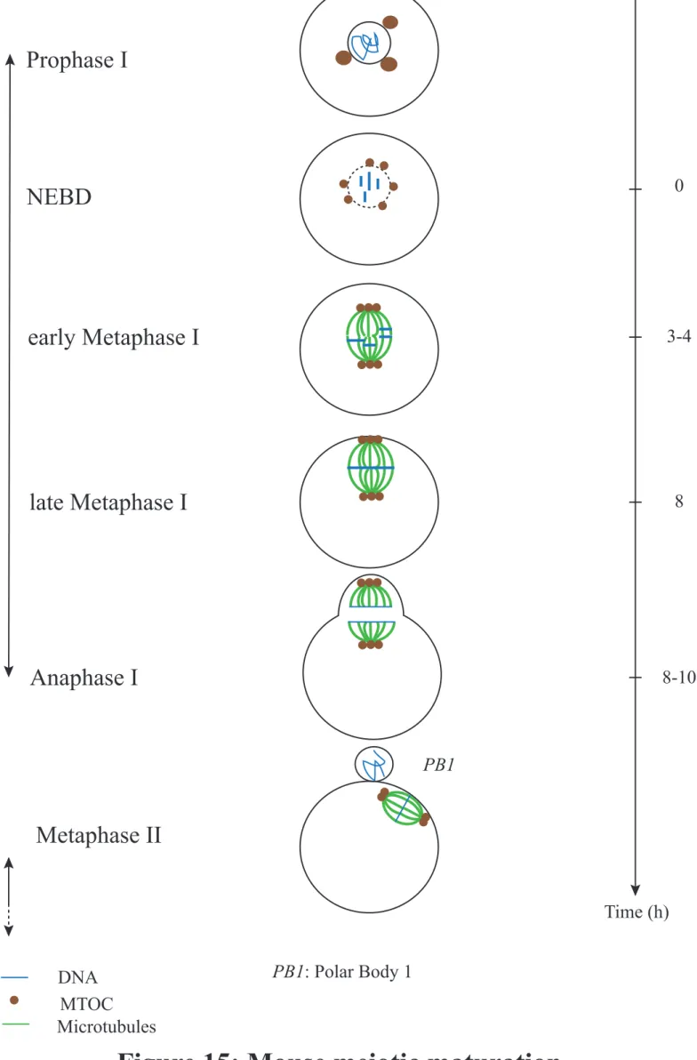
b) A series of cellular arrests
In the female vertebrate, the stock of incompetent oocytes is present at birth since oocytes enter meiosis during embryonic development and stay quiescent, arrested in Prophase I (Prophase of first meiotic division) until puberty. Then, upon periodic hormonal stimulation, a few oocytes depending on the species will enter a differentiation phase called growth phase. A peak of the sexual hormone LH (Luteinizing Hormone) will trigger meiosis resumption and the entry into the divisions per se, a phase known as meiotic maturation (Figure 15). The divisions will however stay incomplete because the oocyte experiences a second block in Metaphase II (Figure 15). This block will be overcome by fertilization that will trigger Meiosis II exit. If the block in Prophase I is universal, the second block depends on the species: in Metaphase II for vertebrates, in Metaphase I for most other species for example.
Meiosis is temporally regulated by the MPF (M-phase Promoting Factor) which is a complex promoting the entry to M phase of all Eukaryotic cells. It is composed of two subunits, CyclinB and CDK1 (Cyclin Dependent Kinase 1). CyclinB is the regulatory subunit; its synthesis and degradation are tightly controlled in all dividing cells. In oocytes arrested in Prophase I, MPF activity, sustained by the catalytic unit CDK1, is maintained low because cAMP (Cyclic Adenosine 3’,5’-MonoPhosphate) level is high. Indeed, cAMP indirectly regulates CDK1 activity by promoting its inhibitory phosphorylation (for review see (Holt et al., 2013)). cAMP is produced by the oocyte and surrounding follicular cells. Furthermore, CyclinB level is also maintained at a low level (Ledan et al., 2001) in Prophase I. When oocytes are removed from the ovaries, the level of cAMP drops allowing spontaneous exit from Prophase I and the initiation of meiotic maturation (Conti et al., 1998). This allows to artificially synchronize a pool of oocytes and have them complete Meiosis I at the same time. Alternatively, the addition of nucleotide derivatives in culture medium can mimic cAMP endogenous role and allow the maintenance of oocytes in Prophase I for several hours.
MPF activity increases slowly during Meiosis I (Verlhac et al., 1994) and diminishes abruptly when chromosomes are fully aligned on the metaphase plate, following CyclinB degradation and thus triggering Meiosis I exit (for review see (Holt et al., 2013)). Eventually,
Figure 15: Mouse meiotic maturation
DNA
MTOC
Microtubules
PB1: Polar Body 1
PB1
Prophase I
NEBD
late Metaphase I
Anaphase I
early Metaphase I
Metaphase II
Time (h)
8-10
8
3-4
0
Meiosis I
Meiosis II
Arrest
Arrest
MPF activity is rapidly restored by CyclinB resynthesis, allowing entry into Meiosis II and Metaphase II arrest. Fertilization will trigger CyclinB degradation, the drop in MPF activity and Meiosis II exit.
2) Meiotic maturation in the mouse a) Cellular particularities of oocytes
Oocytes are very peculiar cells. A first particularity of oocytes in general is that they are very big cells; for example, a fully-grown mouse oocyte has a diameter of 80 µm. A second particularity of most oocytes is that, as plant cells, they lack true centrosomes, centrioles being lost during the growth phase (Szollosi et al., 1972). Instead, their microtubules are nucleated by multiple MTOCs (MicroTubule Organizing Centers) devoid of centrioles. These MTOCs, that are cortical in small oocytes, become efficient microtubule nucleators and relocate to the nuclear envelope at the end of the growth phase (Luksza et al., 2013). The lack of true centrosomes in oocytes imposes alternative modes of spindle assembly and positioning that I will discuss in detail below.
b) Progression of mouse meiosis
In this part, the general progression of meiotic maturation in the mouse oocyte will be described (Figure 15). We will explain the symmetry breaking event and its consequences in more details in the last part.
α. Prophase I and recombination
Prophase I is divided into subphases called Leptotene, Zygotene, Pachytene and Diplotene. These stages correspond to the steps in DNA recombination (chromosome condensation, pairing, crossing-over, resolution). DNA recombination allows the exchange of DNA portions between homologous chromosomes, hence allowing the formation of new gene combinations that increase genetic diversity among the species. In mammals, Prophase I is a very long process that lasts for years because it starts in the embryo and finishes during adulthood where the oocytes stay blocked in Diplotene of Prophase I until meiosis resumes. Then, oocytes, surrounded by follicular cells, undergo a growth phase where they acquire the capacity to resume meiosis; it is notably characterized by an enormous volume increase. At that stage, the oocyte does not harbor any sign of polarization except a slightly off-centered nucleus that was actively centralized at the end the growth phase (Brunet and Maro, 2007;