HAL Id: hal-01394467
https://hal.sorbonne-universite.fr/hal-01394467
Submitted on 9 Nov 2016
HAL is a multi-disciplinary open access
archive for the deposit and dissemination of sci-entific research documents, whether they are pub-lished or not. The documents may come from teaching and research institutions in France or abroad, or from public or private research centers.
L’archive ouverte pluridisciplinaire HAL, est destinée au dépôt et à la diffusion de documents scientifiques de niveau recherche, publiés ou non, émanant des établissements d’enseignement et de recherche français ou étrangers, des laboratoires publics ou privés.
Mechanical Signals during Tendon Formation and
Healing
Ludovic Gaut, Nicolas Robert, Antony Delalande, Marie-Ange Bonnin,
Chantal Pichon, Delphine Duprez
To cite this version:
Ludovic Gaut, Nicolas Robert, Antony Delalande, Marie-Ange Bonnin, Chantal Pichon, et al.. EGR1 Regulates Transcription Downstream of Mechanical Signals during Tendon Formation and Healing. PLoS ONE, Public Library of Science, 2016, 11 (11), pp.e0166237. �10.1371/jour-nal.pone.0166237.t001�. �hal-01394467�
EGR1 Regulates Transcription Downstream
of Mechanical Signals during Tendon
Formation and Healing
Ludovic Gaut1☯
, Nicolas Robert1☯
, Antony Delalande2, Marie-Ange Bonnin1,
Chantal Pichon2, Delphine Duprez1*
1 Sorbonne Universite´s, UPMC Univ Paris 06, CNRS UMR7622, Inserm U1156, IBPS-Developmental Biology Laboratory, F-75005 Paris, France, 2 CNRS UPR4301-CBM, 45071 rue Charles Sadron, Orle´ans CEDEX2, France
☯These authors contributed equally to this work. *delphine.duprez@upmc.fr
Abstract
Background
Tendon is a mechanical tissue that transmits forces generated by muscle to bone in order to allow body motion. The molecular pathways that sense mechanical forces during tendon formation, homeostasis and repair are not known. EGR1 is a mechanosensitive transcrip-tion factor involved in tendon formatranscrip-tion, homeostasis and repair. We hypothesized that EGR1 senses mechanical signals to promote tendon gene expression.
Methodology/Principal findings
Using in vitro and in vivo models, we show that the expression of Egr1 and tendon genes is downregulated in 3D-engineered tendons made of mesenchymal stem cells when tension is released as well as in tendon homeostasis and healing when mechanical signals are reduced. We further demonstrate that EGR1 overexpression prevents tendon gene down-regulation in 3D-engineered tendons when tension is released. Lastly, ultrasound and microbubbles mediated EGR1 overexpression prevents the downregulation of tendon gene expression during tendon healing in reduced load conditions.
Conclusion/Significance
These results show that Egr1 expression is sensitive to mechanical signals in tendon cells. Moreover, EGR1 overexpression prevents the downregulation of tendon gene expression in the absence of mechanical signals in 3D-engineered tendons and tendon healing. These results show that EGR1 induces a transcriptional response downstream of mechanical sig-nals in tendon cells and open new avenues to use EGR1 to promote tendon healing in reduced load conditions.
a11111
OPEN ACCESS
Citation: Gaut L, Robert N, Delalande A, Bonnin M-A, Pichon C, Duprez D (2016) EGR1 Regulates Transcription Downstream of Mechanical Signals during Tendon Formation and Healing. PLoS ONE 11(11): e0166237. doi:10.1371/journal. pone.0166237
Editor: Chunfeng Zhao, Mayo Clinic Minnesota, UNITED STATES
Received: May 3, 2016 Accepted: October 25, 2016 Published: November 7, 2016
Copyright:© 2016 Gaut et al. This is an open access article distributed under the terms of the
Creative Commons Attribution License, which permits unrestricted use, distribution, and reproduction in any medium, provided the original author and source are credited.
Data Availability Statement: All relevant data are within the paper.
Funding: This work was supported by the Fondation pour la Recherche Me´dicale (FRM DEQ20140329500). The funders had no role in study design, data collection and analysis, decision to publish, or preparation of the manuscript. Competing Interests: The authors have declared that no competing interests exist.
Introduction
Tendon is a crucial component of the musculo-skeletal system, which transmits forces gener-ated by skeletal muscle to bone to allow body motion. Mechanical signals are known to be involved in tendon development, homeostasis and repair [1–4]. However, the mechanotrans-duction pathways involved in tendon cell differentiation in normal or pathological situations are not fully understood.
The functional component of tendon is type I collagen, which displays a tendon-specific spa-tial organization that confers tendon mechanical properties [5]. Type I collagen is composed of a triple helix of collagen chains (α1(2), α2(1)) coded by two different genes, Col1a1 and Col1a2. Unfortunately, neither of the Col1a genes is specific to tendons. They are also expressed in many other connective tissues, making it difficult to assess tenogenesis using Col1a gene expression. The bHLH transcription factor Scleraxis (Scx) is specifically expressed in embryonic, fetal and postnatal tendons [6–8]. From the 4thpostnatal month, Scx expression is restricted to the epite-non, but is reactivated in the tendon core by treadmill exercise [8]. Moreover, Scx expression is upregulated in tendons upon injury in animal models [9,10]. The type II transmembrane glyco-protein tenomodulin (Tnmd) is also considered as a relevant marker of tendon cell differentia-tion in mouse, rat and human [11–13]. During development, Scx is required and sufficient for
Tnmd expression in limb tendons [14,15]. Tnmd-/-mice show defective postnatal tendons [16] and reduced self-renewal of tendon stem cells in adult [17].
In addition to Scx, the Zinc finger transcription factor, Early Growth Response-1 (EGR1) has been identified as being involved in pre- and postnatal tendon formation [10,18]. EGR1 is not specific to tendons, since it is expressed in many other tissues; however, EGR1 has the remarkable ability to be sufficient for promoting tendon gene expression, including Scx and Col1a1, during development [18], in mouse mesenchymal stem cells [10] and in rabbit tendon stem cells [19]. The DNA-binding protein EGR1 is known to be a mechanosensitive gene in various cellular sys-tems. Mechanical stretch increases Egr1 transcription in endothelial cells [20], vascular smooth muscle cells [21] or skeletal muscle cells [22]. Dynamic compression also activates Egr1 transcrip-tion in 3-dimensional (3D) cultures of mouse primary chondrocytes [23]. EGR1 has been shown to be a mediator of mechanical input that contributes to vascular remodeling of vein grafts [24].
Since EGR1 is known to be a mechanosensitive gene and involved in tendon development, homeostasis and repair, we hypothesized that EGR1 transduces mechanical signals into transcrip-tional regulation to promote tendon cell differentiation during tendon formation, homeostasis and repair. We used in vitro 3-dimensional culture system and in vivo models to test this hypothesis.
Materials and Methods
Animals
The Egr1LacZ/+mice bred in a C57BL/6j background carry an insertion of a LacZ-neo cassette that inactivates the Egr1 gene [25] and allow the visualization of Egr1 expression with LacZ activity in a heterozygous context [10]. C57BLj wild-type mice were purchased from Janvier (France). All animal experiments were conducted in accordance with the guidelines of the french national ethic comity for animal experimentation N°05. The animal experiments shown in this study have been approved by the french national ethic committee for animal experimen-tation N°05 and are registered under the number 01789.02.
Engineered tendons made of mesenchymal stem cells
Mouse mesenchymal stem cells, C3H10T1/2 [26] were used to establish fibrin-based 3D con-structs. Tendon-like structures from mouse C3H10T1/2 cells or C3H10T1/2-EGR1 cells [10]
were performed as previously described [27]. For each construct, 400 μl of cell suspension (7.5 105cells) were mixed with 20 mg/ml fibrinogen (Sigma, St Louis, MO, USA) and 200 U/ml thrombin (Sigma, St Louis, MO, USA). The fibrin gels containing cells were seeded in already prepared SYLGARD-covered wells (Dow Chemical, Midland, MI, USA), in which two 8 mm-sutures (Ethican, Sommerville, NJ, USA) were pinned 10 mm apart. Culture medium contain-ing 200 μM of L-ascorbic acid 2-phosphate was added to the wells and gels were scored every day for a proper contraction into a linear construct. After 7 days, the C3H10T1/2 and C3H10T1/2-EGR1 cells formed continuous tendon-like constructs between the 2 anchors. Tension release was obtained by cutting one end of the construct as previously described in [28]. Gene expression was analyzed 24 hours after tension release. Each tendon construct made of C3H10T1/2 or C3H10T1/2-EGR1 cells under tension or after tension release was considered as a biological sample. We analyzed 12 constructs made of C3H10T1/2 cells, 7 constructs made of C3H10T1/2-EGR1 cells, 7 de-tensioned-constructs made of C3H10T1/2 cells and 5 de-ten-sioned-constructs made of C3H10T1/2-EGR1 cells. The mRNA levels of each construct were analyzed by q-RT-PCR.
Botox injection in muscles and Achilles tendon injury in adult mice
For the analysis of mechanical signal involvement in tendon homeostasis, 6 UI/kg of Botox preparation (Allergan) [29] and physiological saline solution were injected into the Gastrocne-mius muscles of right and left legs, respectively, of Egr1LacZ/+(N = 6) and wild-type (N = 12) adult mice (two- to four-month-old). Botox injection reduces muscle contraction and move-ments and consequently mechanical signals to tendons [30]. Experimental animals did not dis-play any obvious difficulty in moving, but limped on the Botox-injected legs. The Botox and physiological saline manipulated Egr1LacZ/+animals were kept for one week (N = 6) and ana-lyzed for LacZ staining. Tendons were fixed for 20 minutes in 4% paraformaldehyde and incu-bated in X-gal staining solution for 4 hours at 37°C. The Botox and physiological saline manipulated wild-type animals were kept for one (N = 8) or two (N = 11) weeks after injection (3 independent experiments) and then analyzed for gene expression.
For the analysis of mechanical signal involvement in tendon healing, Achilles tendon injury was performed on left legs of wild-type animals, as previously described in [10]. Briefly, adult mice were anesthetized by isoflurane inhalation. A 0.5-mm longitudinal full-thickness lesion parallel to the axis of the tendon was performed using a scalpel. In this type of injury, the ten-don tension was maintained. After injury, the skin was sutured using 2–0 Mersilk, and the ani-mals were kept for two weeks. The Botox or physiological saline was injected in the
Gastrocnemius muscles of the manipulated left legs. The mouse group, which had undergone Achilles tendon injury and Botox injection, constitute the experimental group (N = 11) that was compared to the control group with Achilles tendon injury and physiological saline injec-tion in muscles (N = 10). Experimental animals did not display any obvious difficulty in mov-ing. However, the animals in the group that had Achilles tendon injury and Botox injection were limping compared to those in the group with Achilles tendon injury and physiological saline injection. Achilles tendons, in the above experimental conditions, were processed for RT-q-PCR analyzes.
Ultrasound and microbubbles-mediated EGR1 gene delivery in Achilles
tendons followed by tendon injury and Botox injection into muscles
Ultrasound and microbubbles-mediated gene delivery is a technique also known as sonopora-tion, allowing for a high and sustained gene expression in mouse Achilles tendons [31]. 10 μg of EGR1 encoding plasmid DNA [18,32] and 3.75 μl of MicroMarkerTMmicrobubbles
(Bracco) in a total volume of 10 μl were injected into the Achilles tendon. Injection was immedi-ately followed by a 1-MHz ultrasound stimulation of 200 kPa (negative peak) during 10 minutes. Ultrasound was generated from a 0.5" diameter, IBMF-014 transducer with a frequency of 1 MHz (Sofranel, Sartrouville, France). A signal consisting of 40 cycles with a frequency of 1.0 MHz and a pulse repetition frequency of 10 kHz, a duty cycle of 40%, was generated by a 33220A arbitrary function generator (Agilent technologies, Les Ulis, France). The signal was amplified by a RF power amplifier (ADECE, Artannes, France) and used as the input for the transducer. The transducer was calibrated in a Perspex container using an HGL-200 PVDF bullet type hydro-phone (Onda, Sunnyvale, CA). EGR1 sonoporation experiments were performed one day before tendon injury and Botox treatment because intratendinous injection requires the integrity of ten-don sheath. Mice were euthanatized 15 days after tenten-don injury and Botox injection and tenten-dons were harvested in 500 μl of RNAlater solution (ThermoScientific) for RNA isolation and RT-q-PCR analyses. 10 mice displaying EGR1 overexpression, tendon injury and Botox injection and 6 mice displaying tendon injury and Botox injection were analyzed.
RNA isolation, reverse transcription and RT-q-PCR
Total RNAs were extracted from fibrin gel constructs made of C3H10T1/2 (N = 11) or C3H10T1/2-EGR1 cells (N = 7), tension-released fibrin gel constructs made of C3H10T1/2 cells (N = 7) or C3H10T1/2-EGR1 cells (N = 5). Total RNAs were extracted from adult mouse tendons under various experimental designs (following physiological saline or Botox injection in normal or injured conditions, after ultrasound-forced EGR1 expression followed by injury and Botox injection) according to the Qiagen RNeasy Minikit (QIAGEN, Germany). RNA (300 ng to 1 μg) was reverse transcribed using the High Capacity Retrotranscription Kit (Applied Biosystems). RT-q-PCR was performed using SYBR Green PCR Master Mix (Applied Biosystems). Relative mRNA levels were calculated using the 2−ΔΔCtmethod [33]. In all our experimental designs, the Cts of two housekeeping genes Gapdh and Hprt did not show any variations in control versus experimental conditions. The ΔCts were obtained from Cts nor-malized with Gapdh levels in each sample. RNA samples originating from 5 to 11 biological samples originating from 3 independent experiments were analyzed in duplicate. Primers used for RT-q-PCR are listed inTable 1.
Table 1. Primers used for quantitative RT-PCR.
Col1a1 Fwd 5’ CCAGCGAAGAACTCATACAGC Rev 5’ GGACACCCCTTCTACGTTGT Col1a2 Fwd 5’ CCAGCGAAGAACTCATACAGC Rev 5’ GGACACCCCTTCTACGTTGT Egr1 Fwd 50-CAGCGCCTTCAATCCTCAAG Rev 50-GCGATGTCAGAAAAGGACTCTGT Gapdh Fwd 5’ TTGTGGAAGGGCTCATGACC Rev 5’ TCTTCTGGGTGGCAGTGATG Hprt Fwd 5’AGGGCATATCCAACAACAAACTT Rev 5’GTTAAGCAGTACAGCCCCAAA Scx Fwd 5’ CCTTCTGCCTCAGCAACCAG Rev 5’ GGTCCAAAGTGGGGCTCTCCGTGACT Tgfb2 Fwd 5’ GAATAAAAGCGAAGAGCTCGAGG Rev 5’ GAGGTGCCATCAATACCTGCA Tnmd Fwd 5’ AACACTTCTGGCCCGAGGTAT Rev 5’ AAGTGTGCTCCATGTCATAGGTTTT doi:10.1371/journal.pone.0166237.t001
Statistical analysis
Sample sizes were based on historical experience of effect sizes for these types of experiments. Error bars in graphics represent the standard deviations or the standard errors of the mean, depending of the size of the samples. Statistics were calculated using non-parametric tests with the GraphPad Prism V6 software. The Mann-Whitney test was used for unpaired samples and the Wilcoxon test for paired samples.
Results
EGR1 overexpression prevents the decrease of tendon gene
expression in 3D tendon constructs after tension release
Fibrin-based 3D cell cultures recapitulate tendon formation based on tenogenic marker expres-sion and tendon-like collagen fibrillogenesis [27,34–37]. This in vitro engineered tendon sys-tem involves tension [34,35]. Forced expression of EGR1 in fibrin-based 3D C3H10T1/2 cell cultures increases Scx, Col1a1 and Col1a2 expression levels compared to control fibrin-based 3D C3H10T1/2 cell cultures [10]. We now show that the expression of the tendon differentia-tion marker Tnmd was also increased in the presence of EGR1 in 3D constructs (Fig 1A). The
Tgfb2 mRNA levels were also increased in EGR1-producing 3D constructs compared to 3D
constructs (Fig 1A). The tension release of the 3D constructs induces the appearance of imma-ture collagen fibrils with no preferred orientation in engineered chick tendons [27] and loss of
TNMD expression in engineered human tendons [28]. The tension was released by cutting one edge of the engineered mouse tendons made of C3H10T1/2 cells (Fig 1B). Tension release led to a decrease in the expression of Egr1 and tendon genes including Scx, Tnmd, Col1a1 and
Col1a2 (Fig 1B). The expression of Tgfb2 was also decreased in tension-released engineered tendons (Fig 1B). Tension release in Egr1-producing 3D-constructs did not trigger any signifi-cant changes in tendon gene expression compared to Egr1-producing 3D constructs under ten-sion (Fig 1C). This shows that EGR1 overexpresten-sion is able to activate tendon gene expresten-sion independently of tension in engineered tendons. We conclude that Egr1 expression is sensitive to tension in engineered mouse tendons and EGR1 forced expression prevents the downregula-tion of tendon gene expression in the absence of mechanical input.
The expression of tendon genes is downregulated in adult tendons
when movements are reduced
In order to assess the importance of mechanical signals for tendon gene expression in homeo-stasis, we developed a reduced load model in adult mice based on botulinium toxin A (Botox) injection into the Gastrocnemius muscle of hindlimbs (Fig 2A). One week after injection of Botox or physiological saline solution in muscles of Egr1LacZ/+mice, LacZ expression (reflecting Egr1 expression) appeared to be decreased in Achilles tendons (Fig 2B and 2C). Consistently, the Egr1 mRNA levels were decreased in Achilles tendons of Botox-injected legs compared to saline solution-injected legs, one and two weeks after injection in wild-type mice (Fig 2D). Scx expression was also decreased in tendons of Botox-injected legs compared to control legs, one and two weeks after Botox injection (Fig 2D), consistent with the decrease in Scx expression one week after Botox injection in Scx-GFP mice [30]. The tendon-associated collagen gene
Col1a2 also displayed decreased expression levels one and two weeks after Botox injection (Fig 2D). The expression of the terminal differentiation tendon marker, Tnmd did not significantly change (Fig 2D). Interestingly, Tgfb2 expression was also decreased in tendons, two weeks after Botox injection (Fig 2D). This showed that the expression of Egr1, Tgfb2 and tendon genes (Scx
and Col1a2) is sensitive to mechanical input in adult tendons. We conclude that mechanical signals are required for tendon gene expression in homeostasis.
Reduced mechanical signals decrease the transcriptional response
during tendon healing after injury
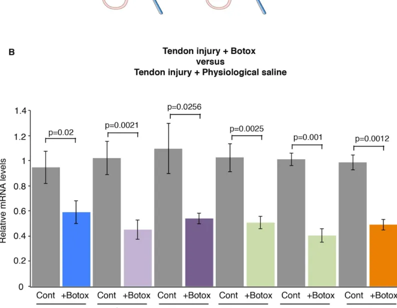
In order to analyze the effect of mechanical signals during healing, we used an Achilles tendon injury model in adult wild-type mice as previously described [10]. In this tendon injury model, a longitudinal incision was performed along the axis of the Achilles tendon, in which the ten-sion was maintained. One week after tendon injury, there is a dramatic increase in tendon gene expression, including Scx, Tnmd, Col1a1 and Col1a2 [10]. Egr1 expression is also increased by fold-3.6 in tendons, after injury and Egr1 is required for the normal tendon transcriptional response during the healing process [10]. In order to determine whether mechanical signals would influence the transcriptional response during tendon healing, we injected Botox or phys-iological saline solution in muscles in this mouse model of tendon injury (Fig 3A). We observed a significant decrease in the expression of Egr1, Scx, Tnmd, Col1a1, Col1a2 and Tgfb2 genes. after Botox injection compared to physiological saline solution injections, in injury conditions (Fig 3B). The decrease of mRNA levels of tendon genes ranged from 40% to 60% (Fig 3). This shows that a diminution of mechanical signals modifies the transcriptional response during the tendon healing process following injury. We conclude that mechanical signals are required for the full transcriptional response during tendon healing.
EGR1 forced expression in tendons prevents the diminution of tendon
gene response in reduced load conditions after injury
The expression of the mechanosensitive gene Egr1 was decreased in reduced load conditions in tendon homeostasis (Fig 2) and healing (Fig 3). To assess whether EGR1 acts downstream of mechanical signals during tendon healing, we performed in vivo EGR1 rescue experiments in reduced load conditions and after tendon injury. We took advantage of an ultrasound-based gene delivery method, which has been found to be efficient in tendons [31]. Plasmid DNA encoding for the Egr1 gene was delivered to the tendons with optimized parameters (1 MHz, 200 kPa, 10 min, 40% duty cycle and 10 kHz pulse repetition frequency). The following day, Botox was injected in muscle followed by tendon injury (Fig 4A). Two weeks after Botox injec-tion in muscle and tendon injury, we compared the expression of tendon genes in tendons overexpressing EGR1 or not during tendon healing and in reduced load conditions. In this experiment, Egr1 was increased by fold-2.6. EGR1 forced expression in tendons increased the expression of tendon-associated genes including Scx, Tnmd, Col1a1, Col1a2 and Tgfb2, when
Fig 1. EGR1 overexpression prevents the downregulation of tendon-associated gene expression in tension released engineered tendons. (A) Two week-old fibrin gel constructs made of mouse C3H10T1/2 cells or C3H10T1/2-EGR1 cells were analyzed for tendon gene expression by RT-q-PCR analyses. The mRNA levels of C3H10T1/2 constructs were normalized to 1. Errors bars represent standard errors of the mean of 5 C3HT101/2 constructs and 7 C3H10T1/2-EGR1 constructs. The p values were calculated using the Mann-Whitney test. The mRNA levels of Egr1, Scx, Tnmd and Tgfb2 genes were increased in C3H10T1/2-EGR1 constructs compared to those of C3H10T1/2 constructs. (B) Tension was released in C3H10T1/2 constructs by sectioning one end of the construct. Transcript levels were analyzed by RT-q-PCR analyses in tension-released C3H10T1/2 constructs and compared to C3H10T1/2 constructs. The mRNA levels of C3H10T1/2 constructs were normalized to 1. Errors bars represent standard errors of the mean of 7 C3H10T1/2 constructs and 7 tension-released C3H10T1/2 constructs. The p values were calculated using the Mann-Whitney test. We observed a decrease in the transcript levels of Egr1, Scx, Tnmd, Col1a1, Col1a2 and Tgfb2 genes in tension-released C3H10T1/2 constructs (TR) compared to tensioned C3H10T1/2 constructs (T). (C) The mRNA levels of tendon genes were analyzed in tension-released C3H10T1/2-EGR1 constructs and compared with tensioned C3H10T1/2-EGR1 constructs by RT-q-PCR analyses. The mRNA levels of C3H10T1/2-EGR1 constructs were normalized to 1. Errors bars represent standard errors of the mean of 7 C3HT101/2-EGR1 constructs and 5 tension-released C3H10T1/2-EGR1 constructs. The p values were calculated using the Mann-Whitney test. There was no significant change in tendon gene expression in tension-released C3H10T1/2-EGR1 constructs (TR) compared to tensioned C3H10T1/2-EGR1 constructs (T). T, Tension, TR, Tension release.
compared to control (Fig 4B). The levels of induction were similar to that of Egr1 overexpres-sion (Fig 4A). This shows that EGR1 is sufficient to prevent the diminution of tendon gene expression that is observed during healing in reduced load conditions.
Discussion
We show that reduced load conditions consistently lead to a decrease in expression of the tran-scription factor Egr1 and tendon genes in tendon homeostasis, tendon healing and in vitro engineered tendons. We also demonstrate that EGR1 forced expression prevents the decrease of tendon gene expression in reduced load conditions during tendon healing and in engineered tendons.
Egr1 is a mechanosensitive gene, which has been shown to act downstream of mechanical
signals in the vascular system [20,21,24]. Consistently, Egr1 expression is systematically downregulated in reduced load conditions in the in vivo and in vitro models of tendon biology. The decrease of Egr1 expression at the transcription level in reduced load conditions is consis-tent with the upregulation of Egr1 expression in overload conditions in adult tendons [38,39].
Egr1 expression is induced as early as 15 minutes after a loading episode in rat Achilles tendons
[40]. This indicates that Egr1 expression reflects a rapid transcriptional response following loading changes in tendons. In addition to being sensitive to mechanical signals, Egr1 appears to be sufficient to drive the tendon genetic program in the absence of mechanical signals. The presence of exogenous EGR1 abolishes the downregulation of Scx, Col1a1 and Tnmd in engi-neered tendons subjected to tension release and EGR1 also prevents the decrease of Scx, Col1a1 and Tnmd expression in Achilles tendons in reduced load conditions during tendon healing following injury. This shows that the Zinc-finger transcription factor Egr1 senses mechanical signals in tendons and modulates tendon gene expression upon loading changes. Although EGR1 has the ability to induce the expression of tendon genes in normal [10] or reduced load (Figs1and4) conditions, it remains unclear whether EGR1 directly regulates the transcription of tendon genes. Previous promoter and Chromatin Immuno-Precipitation (ChIP) analyses indicate that EGR1 trans-activates the tendon promoter of the mouse Col1a1 gene [18,32]. Moreover, EGR1 directly regulates Tgfb2 transcription in adult mouse tendons [10]. Tgfb2 expression is downregulated in reduced load conditions in tendon homeostasis or healing in adult mice (Figs2and3) and in engineered tendons made of mouse mesenchymal stem cells (Fig 1). EGR1 rescue experiments in conditions of reduced mechanical signals induced Tgfb2 expression in engineered tendons and during the tendon healing process (Figs1and4). These results suggest that Egr1 acts upstream of Tgfb2 to activate tendon gene expression upon load-ing. It should be noted that TGF-ß1 supplementation was not sufficient to prevent the decrease of tendon gene expression in de-tensioned 3D engineered tendons derived from human tendon cells [28]. It is not clear whether TGF-ß ligand is not sufficient to activate tendon gene expres-sion in reduced load conditions or TGF-ß1 activity differs from TGF-ß2 activity. Antagonist effects between TGF-ß1 and TGF-ß2 ligands have been reported on COL1A1 transcription in rat tendon fibroblasts [41]. TGF-ß supplementation activates Scx expression in various cellular
Fig 2. Reduced mechanical input induces a diminution of Egr1 and Scx expression in adult tendons. (A) Botox or physiological saline injections in Gastrocnemius muscles of adult mice. (B,C) LacZ staining (reflecting Egr1 expression) in tendons, 1 week following physiological saline or Botox injection in Egr1Lacz/+adult mice. (D) RT-q-PCR analyses of tendons, one or two weeks after Botox or physiological saline injections in muscles in adult mice. The mRNA levels of tendons following Botox injection were compared to those of tendons with physiological saline injection. Errors bars represent the standard deviations of 5 (one week) and 7 (two weeks) biological samples. The mRNA levels of Egr1, Scx, Col1a2, Tgfb2 genes were decreased one or two weeks after Botox injection compared to physiological saline injection. Tnmd mRNA levels were not significantly decreased after Botox injection. The p values were calculated using the Wilcoxon test.
Fig 3. Mechanical signals are required for normal tendon gene response following tendon injury. (A) Botox or physiological saline injections in Gastrocnemius muscles and tendon injury in adult mice. (B) RT-q-PCR analyses of tendons, 2 weeks after tendon injury and Botox injection in muscles. The mRNA levels of tendons following injury and physiological saline injection in muscles were normalized to 1. The errors bars represent the standard error of the means of 10 and 11 biological samples of injured tendons after physiological saline or Botox injections, respectively. The p values were calculated using the Mann-Whitney test. The mRNA levels of the Egr1, Scx, Tnmd, Col1a1, Col1a2 and Tgfb2 genes were all significantly decreased in reduced movement conditions compared to controls during the healing process, following tendon injury.
Fig 4. EGR1 forced expression in tendons prevents the diminution of tendon gene expression during tendon healing in reduced mechanical load. (A) Description of the experimental design for sonoporation. 10μg of EGR1 encoding plasmid and 3.75μl of MicroMarker microbubbles were injected in the Achilles tendon sheath. Tendons were then stimulated by ultrasound at 1 MHz during 10 minutes at 200 kPa, 40% duty cycle and 10 kHz pulse repeating frequency. The day after EGR1 sonoporation, a surgical lesion of the Achilles tendon was performed followed by a Botox or physiological saline solution injection in the muscle. Two weeks after treatment, tendons were harvested for analyses by RT-q-PCR. (B) RT-q-PCR analysis of tendon gene expression in EGR1-sonoporated tendons versus control-tendons, following tendon injury in immobilization conditions. The mRNA levels of control tendons following injury and Botox injection in muscles were normalized to 1. The error bars represent standard errors of the mean of 6 biological samples of injured tendons of Botox-injected legs in the absence of EGR1 and 10 biological samples of injured tendons of Botox-injected legs in presence of ectopic EGR1. The p values were calculated using the Mann-Whitney test. The mRNA levels of Egr1, Scx, Tnmd, Col1a2 and Tgfb2 were increased in Egr1-sonoporated tendons compared to control tendons, following tendon injury and Botox injection in muscles.
doi:10.1371/journal.pone.0166237.g004
Fig 5. Schematic representation of EGR1 regulation and function downstream of mechanical signals. Egr1 expression is regulated by mechanical signals in tendon cells. EGR1 positively regulates the transcription of tendon genes including Scx, Tnmd, Col1a1 and Co1a2. The transcription of Tgfb2 is also regulated by EGR1.
models and loss of TGF-ß leads to a decrease of Scx expression in developmental tendons [10, 42–44]. We failed to identify any EGR1 binding sites in mouse Scx promoter region and all our ChIP attempts to identify direct EGR1 recruitment to Scx promoter region were unsuccessful. Given the EGR1 direct binding to Tgfb2 promoter [10], it is tempting to suggest that TGF-ß2 mediates the Scx induction by EGR1 in tendons. Consistently, SCX and SMAD3 (an intracellu-lar component of the TGF-ß pathway) physically interact in 10T1/2 cells [45] and SCX is required for Smad3-mediated gene transcription in cardiac fibroblasts [46]. Whether EGR1 activates Tnmd via TGF-ß2 is less clear, since TGF-ß2 has been shown to downregulate Tnmd expression in mouse stem cells cultured in 2D [10,44,47] and do not have any positive effect on TNMD expression in human 3D tissue cultures [28]. In contrast, SCX gain- and loss-of-function experiments lead to upregulation and downregulation of Tnmd, respectively, during development [14,15]. This suggests that SCX could activate Tnmd expression during tendon differentiation.
In summary, we show that the expression of the zinc-finger transcription factor EGR1 is sensitive to mechanical signals and that EGR1 senses mechanical signals at the transcription level. Moreover, EGR1 is sufficient to promote tendon gene expression in the absence of mechanical forces during tendon formation and healing (Fig 5). This feature opens the possi-bility of exploiting EGR1 forced expression to accelerate tendon healing in reduced load conditions.
Acknowledgments
We thank laboratory members for comments on the manuscript and Sophie Gournet for illustrations.
Author Contributions
Conceptualization: DD CP. Formal analysis: LG. Funding acquisition: DD. Investigation: LG NR AD MAB. Methodology: LG NR AD MAB. Writing – original draft: DD.Writing – review & editing: DD LG AD CP.
References
1. Shwartz Y, Blitz E, Zelzer E. One load to rule them all: mechanical control of the musculoskeletal sys-tem in development and aging. Differentiation; research in biological diversity. 2013; 86(3):104–11. doi:10.1016/j.diff.2013.07.003PMID:23953954.
2. Wang JH, Guo Q, Li B. Tendon biomechanics and mechanobiology—a minireview of basic concepts and recent advancements. Journal of hand therapy: official journal of the American Society of Hand Therapists. 2012; 25(2):133–40; quiz 41. doi:10.1016/j.jht.2011.07.004PMID:21925835; PubMed Central PMCID: PMC3244520.
3. Heinemeier KM, Kjaer M. In vivo investigation of tendon responses to mechanical loading. Journal of musculoskeletal & neuronal interactions. 2011; 11(2):115–23. PMID:21625048.
4. Lavagnino M, Wall ME, Little D, Banes AJ, Guilak F, Arnoczky SP. Tendon mechanobiology: Current knowledge and future research opportunities. Journal of orthopaedic research: official publication of the Orthopaedic Research Society. 2015; 33(6):813–22. doi:10.1002/jor.22871PMID:25763779; PubMed Central PMCID: PMC4524513.
5. Nourissat G, Berenbaum F, Duprez D. Tendon injury: from biology to tendon repair. Nature reviews Rheumatology. 2015; 11(4):223–33. doi:10.1038/nrrheum.2015.26PMID:25734975.
6. Schweitzer R, Chyung JH, Murtaugh LC, Brent AE, Rosen V, Olson EN, et al. Analysis of the tendon cell fate using Scleraxis, a specific marker for tendons and ligaments. Development. 2001; 128 (19):3855–66. Epub 2001/10/05. PMID:11585810.
7. Pryce BA, Brent AE, Murchison ND, Tabin CJ, Schweitzer R. Generation of transgenic tendon report-ers, ScxGFP and ScxAP, using regulatory elements of the scleraxis gene. Developmental dynamics: an official publication of the American Association of Anatomists. 2007; 236(6):1677–82. doi:10.1002/ dvdy.21179PMID:17497702.
8. Mendias CL, Gumucio JP, Bakhurin KI, Lynch EB, Brooks SV. Physiological loading of tendons induces scleraxis expression in epitenon fibroblasts. Journal of orthopaedic research: official publica-tion of the Orthopaedic Research Society. 2012; 30(4):606–12. doi:10.1002/jor.21550PMID: 21913219; PubMed Central PMCID: PMC3245815.
9. Scott A, Sampaio A, Abraham T, Duronio C, Underhill TM. Scleraxis expression is coordinately regu-lated in a murine model of patellar tendon injury. Journal of orthopaedic research: official publication of the Orthopaedic Research Society. 2011; 29(2):289–96. doi:10.1002/jor.21220PMID:20740671; PubMed Central PMCID: PMC3951487.
10. Guerquin MJ, Charvet B, Nourissat G, Havis E, Ronsin O, Bonnin MA, et al. Transcription factor EGR1 directs tendon differentiation and promotes tendon repair. The Journal of clinical investigation. 2013; 123(8):3564–76. doi:10.1172/JCI67521PMID:23863709.
11. Jelinsky SA, Archambault J, Li L, Seeherman H. Tendon-selective genes identified from rat and human musculoskeletal tissues. Journal of orthopaedic research: official publication of the Orthopae-dic Research Society. 2010; 28(3):289–97. doi:10.1002/jor.20999PMID:19780194.
12. Sugimoto Y, Takimoto A, Akiyama H, Kist R, Scherer G, Nakamura T, et al. Scx+/Sox9+ progenitors contribute to the establishment of the junction between cartilage and tendon/ligament. Development. 2013; 140(11):2280–8. doi:10.1242/dev.096354PMID:23615282.
13. Havis E, Bonnin MA, Olivera-Martinez I, Nazaret N, Ruggiu M, Weibel J, et al. Transcriptomic analysis of mouse limb tendon cells during development. Development. 2014; 141(19):3683–96. doi:10.1242/ dev.108654PMID:25249460.
14. Shukunami C, Takimoto A, Oro M, Hiraki Y. Scleraxis positively regulates the expression of tenomodu-lin, a differentiation marker of tenocytes. Developmental biology. 2006; 298(1):234–47. doi:10.1016/j. ydbio.2006.06.036PMID:16876153.
15. Murchison ND, Price BA, Conner DA, Keene DR, Olson EN, Tabin CJ, et al. Regulation of tendon dif-ferentiation by scleraxis distinguishes force-transmitting tendons from muscle-anchoring tendons. Development. 2007; 134(14):2697–708. Epub 2007/06/15. dev.001933 [pii] doi:10.1242/dev.001933 PMID:17567668.
16. Docheva D, Hunziker EB, Fassler R, Brandau O. Tenomodulin is necessary for tenocyte proliferation and tendon maturation. Molecular and cellular biology. 2005; 25(2):699–705. doi:10.1128/MCB.25.2. 699–705.2005PMID:15632070; PubMed Central PMCID: PMC543433.
17. Alberton P, Dex S, Popov C, Shukunami C, Schieker M, Docheva D. Loss of tenomodulin results in reduced self-renewal and augmented senescence of tendon stem/progenitor cells. Stem cells and development. 2015; 24(5):597–609. doi:10.1089/scd.2014.0314PMID:25351164; PubMed Central PMCID: PMC4333258.
18. Lejard V, Blais F, Guerquin MJ, Bonnet A, Bonnin MA, Havis E, et al. EGR1 and EGR2 Involvement in Vertebrate Tendon Differentiation. J Biol Chem. 2011; 286(7):5855–67. Epub 2010/12/22.
M110.153106 [pii] doi:10.1074/jbc.M110.153106PMID:21173153; PubMed Central PMCID: PMC3037698.
19. Tao X, Liu J, Chen L, Zhou Y, Tang K. EGR1 induces tenogenic differentiation of tendon stem cells and promotes rabbit rotator cuff repair. Cellular physiology and biochemistry: international journal of experimental cellular physiology, biochemistry, and pharmacology. 2015; 35(2):699–709. doi:10. 1159/000369730PMID:25592085.
20. Stula M, Orzechowski HD, Gschwend S, Vetter R, von Harsdorf R, Dietz R, et al. Influence of sustained mechanical stress on Egr-1 mRNA expression in cultured human endothelial cells. Molecular and cel-lular biochemistry. 2000; 210(1–2):101–8. PMID:10976763.
21. Morawietz H, Ma YH, Vives F, Wilson E, Sukhatme VP, Holtz J, et al. Rapid induction and translocation of Egr-1 in response to mechanical strain in vascular smooth muscle cells. Circulation research. 1999; 84(6):678–87. PMID:10189355.
22. Pardo PS, Mohamed JS, Lopez MA, Boriek AM. Induction of Sirt1 by mechanical stretch of skeletal muscle through the early response factor EGR1 triggers an antioxidative response. J Biol Chem. 2011;
286(4):2559–66. doi:10.1074/jbc.M110.149153PMID:20971845; PubMed Central PMCID: PMC3024751.
23. Bougault C, Aubert-Foucher E, Paumier A, Perrier-Groult E, Huot L, Hot D, et al. Dynamic compres-sion of chondrocyte-agarose constructs reveals new candidate mechanosensitive genes. PloS one. 2012; 7(5):e36964. doi:10.1371/journal.pone.0036964PMID:22615857; PubMed Central PMCID: PMC3355169.
24. Wu X, Cheng J, Li P, Yang M, Qiu S, Liu P, et al. Mechano-sensitive transcriptional factor Egr-1 regu-lates insulin-like growth factor-1 receptor expression and contributes to neointima formation in vein grafts. Arteriosclerosis, thrombosis, and vascular biology. 2010; 30(3):471–6. doi:10.1161/ATVBAHA. 109.184259PMID:19965784.
25. Topilko P, Schneider-Maunoury S, Levi G, Trembleau A, Gourdji D, Driancourt MA, et al. Multiple pitui-tary and ovarian defects in Krox-24 (NGFI-A, Egr-1)-targeted mice. Molecular endocrinology. 1998; 12 (1):107–22. doi:10.1210/mend.12.1.0049PMID:9440815.
26. Reznikoff CA, Brankow DW, Heidelberger C. Establishment and characterization of a cloned line of C3H mouse embryo cells sensitive to postconfluence inhibition of division. Cancer research. 1973; 33 (12):3231–8. PMID:4357355.
27. Kapacee Z, Richardson SH, Lu Y, Starborg T, Holmes DF, Baar K, et al. Tension is required for fibripo-sitor formation. Matrix biology: journal of the International Society for Matrix Biology. 2008; 27(4):371– 5. doi:10.1016/j.matbio.2007.11.006PMID:18262777.
28. Bayer ML, Schjerling P, Herchenhan A, Zeltz C, Heinemeier KM, Christensen L, et al. Release of ten-sile strain on engineered human tendon tissue disturbs cell adhesions, changes matrix architecture, and induces an inflammatory phenotype. PloS one. 2014; 9(1):e86078. doi:10.1371/journal.pone. 0086078PMID:24465881; PubMed Central PMCID: PMC3897642.
29. Aoki KR. A comparison of the safety margins of botulinum neurotoxin serotypes A, B, and F in mice. Toxicon: official journal of the International Society on Toxinology. 2001; 39(12):1815–20. PMID: 11600142.
30. Maeda T, Sakabe T, Sunaga A, Sakai K, Rivera AL, Keene DR, et al. Conversion of mechanical force into TGF-beta-mediated biochemical signals. Curr Biol. 2011; 21(11):933–41. Epub 2011/05/24. S0960-9822(11)00423-4 [pii] doi:10.1016/j.cub.2011.04.007PMID:21600772; PubMed Central PMCID: PMC3118584.
31. Delalande A, Bouakaz A, Renault G, Tabareau F, Kotopoulis S, Midoux P, et al. Ultrasound and micro-bubble-assisted gene delivery in Achilles tendons: long lasting gene expression and restoration of fibromodulin KO phenotype. Journal of controlled release: official journal of the Controlled Release Society. 2011; 156(2):223–30. doi:10.1016/j.jconrel.2011.08.020PMID:21888933.
32. Lejard V, Brideau G, Blais F, Salingcarnboriboon R, Wagner G, Roehrl MH, et al. Scleraxis and NFATc regulate the expression of the pro-alpha1(I) collagen gene in tendon fibroblasts. J Biol Chem. 2007; 282(24):17665–75. doi:10.1074/jbc.M610113200PMID:17430895.
33. Livak KJ, Schmittgen TD. Analysis of relative gene expression data using real-time quantitative PCR and the 2(-Delta Delta C(T)) Method. Methods. 2001; 25(4):402–8. doi:10.1006/meth.2001.1262 PMID:11846609.
34. Kapacee Z, Yeung CY, Lu Y, Crabtree D, Holmes DF, Kadler KE. Synthesis of embryonic tendon-like tissue by human marrow stromal/mesenchymal stem cells requires a three-dimensional environment and transforming growth factor beta3. Matrix biology: journal of the International Society for Matrix Biol-ogy. 2010; 29(8):668–77. doi:10.1016/j.matbio.2010.08.005PMID:20736064; PubMed Central PMCID: PMC3611595.
35. Bayer ML, Yeung CY, Kadler KE, Qvortrup K, Baar K, Svensson RB, et al. The initiation of embryonic-like collagen fibrillogenesis by adult human tendon fibroblasts when cultured under tension. Biomateri-als. 2010; 31(18):4889–97. doi:10.1016/j.biomaterials.2010.02.062PMID:20356622; PubMed Cen-tral PMCID: PMC3485556.
36. Breidenbach AP, Dyment NA, Lu Y, Rao M, Shearn JT, Rowe DW, et al. Fibrin gels exhibit improved biological, structural, and mechanical properties compared with collagen gels in cell-based tendon tis-sue-engineered constructs. Tissue engineering Part A. 2015; 21(3–4):438–50. doi:10.1089/ten.TEA. 2013.0768PMID:25266738; PubMed Central PMCID: PMC4333253.
37. Yeung CY, Zeef LA, Lallyett C, Lu Y, Canty-Laird EG, Kadler KE. Chick tendon fibroblast transcriptome and shape depend on whether the cell has made its own collagen matrix. Scientific reports. 2015; 5:13555. doi:10.1038/srep13555PMID:26337655; PubMed Central PMCID: PMC4559659. 38. Eliasson P, Andersson T, Hammerman M, Aspenberg P. Primary gene response to mechanical
load-ing in healload-ing rat Achilles tendons. Journal of applied physiology. 2013; 114(11):1519–26. doi:10. 1152/japplphysiol.01500.2012PMID:23519232.
39. Hammerman M, Aspenberg P, Eliasson P. Microtrauma stimulates rat Achilles tendon healing via an early gene expression pattern similar to mechanical loading. Journal of applied physiology. 2014; 116 (1):54–60. doi:10.1152/japplphysiol.00741.2013PMID:24177691.
40. Eliasson P, Andersson T, Aspenberg P. Rat Achilles tendon healing: mechanical loading and gene expression. Journal of applied physiology. 2009; 107(2):399–407. doi:10.1152/japplphysiol.91563. 2008PMID:19541731.
41. Chan KM, Fu SC, Wong YP, Hui WC, Cheuk YC, Wong MW. Expression of transforming growth factor beta isoforms and their roles in tendon healing. Wound repair and regeneration: official publication of the Wound Healing Society [and] the European Tissue Repair Society. 2008; 16(3):399–407. doi:10. 1111/j.1524-475X.2008.00379.xPMID:18471258.
42. Pryce BA, Watson SS, Murchison ND, Staverosky JA, Dunker N, Schweitzer R. Recruitment and main-tenance of tendon progenitors by TGFbeta signaling are essential for tendon formation. Development. 2009; 136(8):1351–61. Epub 2009/03/24. 136/8/1351 [pii] doi:10.1242/dev.027342PMID:19304887; PubMed Central PMCID: PMC2687466.
43. Brown JP, Finley VG, Kuo CK. Embryonic mechanical and soluble cues regulate tendon progenitor cell gene expression as a function of developmental stage and anatomical origin. Journal of biomechanics. 2014; 47(1):214–22. doi:10.1016/j.jbiomech.2013.09.018PMID:24231248; PubMed Central PMCID: PMC4157071.
44. Brown JP, Galassi TV, Stoppato M, Schiele NR, Kuo CK. Comparative analysis of mesenchymal stem cell and embryonic tendon progenitor cell response to embryonic tendon biochemical and mechanical factors. Stem cell research & therapy. 2015; 6:89. doi:10.1186/s13287-015-0043-zPMID:25956970; PubMed Central PMCID: PMC4425922.
45. Berthet E, Chen C, Butcher K, Schneider RA, Alliston T, Amirtharajah M. Smad3 binds Scleraxis and Mohawk and regulates tendon matrix organization. Journal of orthopaedic research: official publication of the Orthopaedic Research Society. 2013; 31(9):1475–83. doi:10.1002/jor.22382PMID:23653374; PubMed Central PMCID: PMC3960924.
46. Bagchi RA, Roche P, Aroutiounova N, Espira L, Abrenica B, Schweitzer R, et al. The transcription fac-tor scleraxis is a critical regulafac-tor of cardiac fibroblast phenotype. BMC biology. 2016; 14(1):21. doi: 10.1186/s12915-016-0243-8PMID:26988708; PubMed Central PMCID: PMC4794909.
47. Liu H, Zhang C, Zhu S, Lu P, Zhu T, Gong X, et al. Mohawk promotes the tenogenesis of mesenchymal stem cells through activation of the TGFbeta signaling pathway. Stem cells. 2015; 33(2):443–55. doi: 10.1002/stem.1866PMID:25332192.