HAL Id: hal-01405171
https://hal.archives-ouvertes.fr/hal-01405171
Submitted on 10 Jan 2017HAL is a multi-disciplinary open access archive for the deposit and dissemination of sci-entific research documents, whether they are pub-lished or not. The documents may come from
L’archive ouverte pluridisciplinaire HAL, est destinée au dépôt et à la diffusion de documents scientifiques de niveau recherche, publiés ou non, émanant des établissements d’enseignement et de
model
Petru Bucur, Mohamed Bekheit, Chloe Audebert, Irene E. Vignon-Clementel,
Eric Vibert
To cite this version:
Petru Bucur, Mohamed Bekheit, Chloe Audebert, Irene E. Vignon-Clementel, Eric Vibert. Simplified technique for 75% and 90% hepatic resection with hemodynamic monitoring in large white swine model. Journal of Surgical Research, Elsevier, 2017, 209, pp.122-130. �10.1016/j.jss.2016.09.018�. �hal-01405171�
A final version of this manuscript can be found in Journal of Surgical Research, March 2017, Vol 209, pages 122-130, DOI: http://dx.doi.org/10.1016/j.jss.2016.09.018
Simplified technique for 75% & 90% hepatic resection with hemodynamic monitoring in a large white swine model
Petru Bucur Mohamed Bekheit Chloe Audebert Irene Vignon-Clementel Eric Vibert Authors` affiliations
1. AP-HP, Hôpital Paul Brousse, Centre Hépato-Biliaire, Villejuif, France 2. Inserm Unité 1193, Villejuif, France
3. Inria Paris-Rocquencourt, France
4. Sorbonne University, UPMC Univ. of Paris 6, Laboratoire Jacques-Louis Lions, Paris, France
Corresponding author:
EricVibert, MD, PhD
12 Avenue Paul VaillantCouturier 94804 Villejuif Cedex
Fax: 00 33 1 45 59 38 57 E-Mail: eric.vibert@pbr.aphp.fr
Authors` contribution:
Petru Bucur and Mohamed Bekheit equally contributed in this manuscript and are both first author.
Study concept and design: Petru Bucur and Mohamed Bekheit Surgical technique development: Petru Bucur.
Acquisition of data: Mohamed Bekheit, Petru Bucur, Chloé Audebert. Analysis and interpretation of data: Mohamed Bekheit, Petru Bucur, Chloé Audebert, Eric Vibert, Irene Vignon-Clementel
Drafting of the manuscript: Mohamed Bekheit, Petru Bucur, Chloé Audebert Critical revision of the manuscript: Eric Vibert, Irene Vignon-Clementel Final approval: Eric Vibert
Abstract Background
Accurate measuring of the hepatic hemodynamic parameters in humans is inconvenient. Swine has been a favourite surgical model for the study of liver conditions due to many similarities with human livers. However, pigs cannot tolerate pedicle clamping and to reduce bleeding during resection a simplified technique is required.
Aim:
The aim of this study is to present a simplified technique for different percentages of hepatic resection in a porcine model.
Methods:
Twenty two consecutive large white pigs were operated with 75% and 90% liver resection. CT liver volumetry is performed before and after surgery. In both types of surgery, hemodynamic monitoring was performed using a specialised apparatus. Results:
Resections were performed in both groups successfully. The residual volume in the planned 75% was 235 ±77 ml and 118 ±119 ml in the planned 90% resection. For 75% resection, the portal flow was reduced after resection by 8.13 ± 28%, which might be part of systemic circulatory depression. However, the portal pressure increased
by 20.1 ± 51 %. The hepatic artery flow decreased by 63.86 ± 26.3 % as well as the pressure by 5 ±28 %.
The central venous pressure at the start of surgery was 3.34 ± 1.9 mmHg and 2.8 ± 2.2 mmHg at the end of surgery. The portocaval pressure gradient was 4.4 ± 2.9 mmHg at the beginning of surgery and was 5.9 ± 2.8 mmHg at the end of surgery.
For 90% resection, the portal flow decreased by 33.6±12.6% and the pressure increased by 104±58%. The hepatic artery flow decreased by 88±7% and the pressure decreased by 5±14.8%. The central venous pressure was 3.5±1.7 mmHg before resection and 3±2.5 mmHg after resection. The portocaval pressure gradient was 3.8±1.1 mmHg before resection and 8±3.7 mmHg after resection.
The mean anaesthesia time was 6.6 ±1.05 hours and 6.9 ± 0.5 hours for 75% and 90% resection, respectively. The mean operative time was 4.6 ±0.9 hours and 5 ±0.7 hours for 75% and 90% resections, respectively. The mean time for hepatectomy was 1.23±0.76 hours and 2.4 ±0.1 hours for 75% and 90% resection, respectively. The mean time consumed in the measurements was 2.28±1.4 hours and 1.1 ±0.3 hours for 75% and 90% resections, respectively. The mean volume of aspirated fluid and blood in the 75% resection was 1062± 512 ml while it was 1050 ± 354 ml in 90% resections. Conclusions:
The hereby described technique is simple and easily applicable for major liver resection in a porcine model. . Portal flow decreases after 90% resection more than in 75% due to the relative reduction of remnant hepatic mass. There was a larger increase in portal
pressure following 90% compared to 75% resection. The hepatic artery flow decreases more in 90% than in 75% resections. .
Short title: Technique for 75 and 90 % porcine liver resection Key words:
Liver resection, hemodynamic monitoring, technique, major, pigs Background
The liver is one of the most heavily studied organs in modern medicine. Surgical parenchymal resection is a strong stimulating factor for regeneration (1). Among the various factors, the change in portal flow and pressure after resection were reported to be influential on regeneration (2).
Increased portal pressure after partial hepatic resection acts as a stimulus for liver
regeneration (3). Nonetheless, a limiting threshold has been identified after which further increase in portal pressure would lead to liver failure (4).Liver failure after extensive hepatectomy is a lethal complication (5).
Pressure and flow measurement of the splanchnic area in clinical settings is not feasible. For that, animal models were proposed as an alternative way to elaborate precise relations between the hemodynamic parameters and liver regeneration (6).The development of an animal model to study the relationship between the liver regeneration and changes in hepatic hemodynamics after major liver resection is necessary for better understanding and predicting the pathophysiological mechanisms related to this issue(7). We hereby describe our technique for 75% and 90% hepatectomy with mesenteric and hepatic
hemodynamic monitoring and to discuss some critical anatomical features and technique based on our experience.
Methods:
Ethical approval: The study was approved by the regional committee of ethics in animal research, and by the ministry of higher education and scientific research and ministry of agriculture and fishing, according to European Union directives.
Study setting Surgeries were performed at the CIRE plateform, INRA Centre Val de Loire, Nouzilly, France.
Animals: Twenty two large white female pigs were included in this study. The average age was 3 months +/- 10 days. The mean weight was 35.9 +/-7.5 kg. The pigs were kept under strict protocol. There was a period of conditioning before surgery varying from 4 to 6 days. The pigs were housed in individual pens with temperature regulated at 23 ± 1°C at ambient humidity. Lighting was natural through close to ceiling wide windows. Volumetric study:
Abdominal CT scan based volumetric analysis was conducted to every pig 7 days before surgery and immediately after the hepatic resection.
On average, an 80 ml (2 ml/kg) of iodinated contrast (Omnipaque TM, GE healthcare, Carrigtohil, Irland) was injected through an intravenous catheter with a rate of 3-4 ml/s. CT scans were performed with a Somatom (Definition AS, Siemens, Forchheim, Germany). Volume analysis was performed using the Myrian XP-Liver 1.14.1 software
(Intrasense, Montpellier, France). The resection is designed following anatomical landmarks to keep the residual segment/s with intact outflow and inflow (Figure 1a,b).
Figure 1: 3D reconstructed images for planned a) 75% and b) 90% resection. The images
demonstrate the planned resected lobes (S) and the residual lobes (R) with preserved portal inflow and hepatic venous outflow. PV=portal vein, IVC= inferior vena cava.
Preoperative preparation: Animals were left fasting the night before surgery. On the day of surgery, animals were given in their individualized cages as a pre-anaesthetic preparation 30 mg/kg ketamine (Ketamin, Panpharma) and 0.03 mg/kg acepromazine (Calmivet, Vetoquinol, France).
Anaesthesia and postoperative care:
Each pig received 100 mg of xylazine 2% (Rompun, Bayer Healthcare) with 750 mg ketamine (Ketamin Panpharma) for anaesthesia induction followed by tracheal intubation (6-7 mm in size, Portex, France). Subsequently, inhalational anaesthesia is started using a
60% FiO2 inhalational oxygen mixed with 2% isoflurane (Isoflurane, Belamont, France) at a rate of 2-3 ml mixed with 1.5-2 l/minute oxygen in 1.5 liters of air. Afterwards, 1 gram of amoxicillin (Amoxycilline, Mylan) was given i.v. Fentanyl (1 ml) was given subcutaneously after intubation and at the end of surgery and a Fentanyl patch was placed on the shaved skin of the right side of the thoracic cage after surgery. Twenty mg of atracurium (Tracrium, Glaxo Smith Klyne) muscle relaxant was given intramuscular. Crystalloids were infused at a rate of 2 ml/kg/h fasting in addition to 500-1000 ml. This was increased according to demands.
During surgery animals were covered with heat blankets and gastric aspiration through an oro-gastric tube was performed if gastric distension was observed at surgery. Surgery was conducted under sterile conditions. Animals were positioned in a dorsal decubitus and surgical site disinfection and draping were performed. Oxygen saturation, temperature, pulse rate, arterial pressure, and central venous pressure were monitored during surgery. Amoxycillin 1g (Amoxycilline, Mylan) b.i.d was administered intravenously for 5 days. In addition, animals received daily pantoprazole 40 mg i.v (Inipomp, Nycomed) and enoxaparine 0.2 ml s.c (Lovenox, Sanofi Aventis). Animals were givenfree access to water and food and if intake is not satisfactory fluid replacement was tailored.
Surgery: Longitudinal median neck incision was performed and canulation of the right carotid artery and the right internal jugular vein was performed with a 5 and 8 Fr vascular Desivalve (Vygon) cannula. A water column based barometer was connected to the side line of the arterial cannula in order to continuously measure the carotid pressure through
the anaesthesia monitor. Millar pressure probes (Millar, Texas, USA) were then inserted into the internal jugular vein (5 Fr, SPR 350) and carotid artery (4 Fr, SPR 340).
Subsequently a midline abdominal incision was performed. Table mounted costal retractors and self retaining abdominal retractors were installed.
Vascular cannulation:
Carotid artery cannulation was performed after dissecting a 4-5 cm segment. The artery was surrounded by a vascular tape through which traction was applied in a cranial direction. The caudal end of the dissected portion was gently grasped with a vascular forceps till the puncture was performed and the guide wire was introduced. The artery was grasped again over the guide wire to reduce bleeding from the puncture, and then the vascular cannula was introduced. The jugular vein was cannulated in a similar way except that the dissection around the vein was performed keeping a fine tissue pad around the vein to prevent its shearing. One more difference is that the caudal application of the forceps was not performed in order to keep the vein distended.
Hepatic artery cannulation was performed through the gastrodudenal artery, which was dissected above the head of pancreas to its confluence with the hepatic artery. Two absorbable 4/0 threads were passed under the vessel. The distal one (close to the head of pancreas) was ligated to direct all the flow that passes across the hepatic artery to the liver. The vessel was snipped with fine scissors, and then a 3Fr SPR-330 Millar pressure probe was introduced through this opening toward hepatic artery. Then the proximal ligature was tied over the probe.
The portal vein pressure probe (5 Fr, SPR-350 Millar) was inserted through the gastrodudenal vein in a similar way to the hepatic artery. Both probes were further secured in place with the proximal absorbable ties.
Probes positioning
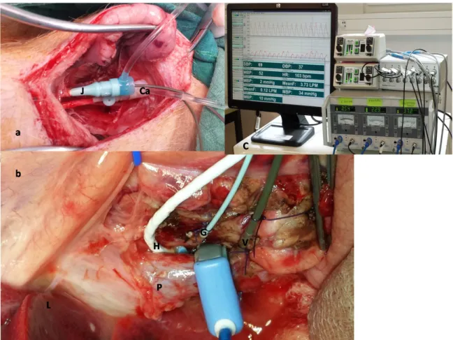
A 5 Fr and 4 Fr pressure probes were inserted into the right jugular vein (tip positioned in superior vena cava, verified by waveform) and carotid artery respectively (figure 2a); while a 3 Fr pressure probe was inserted into the hepatic artery through the ligated gastro duodenal artery and a 5 Fr pressure probe was inserted into the portal vein through a small medial gastro-duodenal tributary (figure 2b). For flow measurements, we used TranSonic transit time technology (TranSonic System Inc, Ithaca, USA). A 12 or 14 Fr flow probe (MA12 PAU or MA14 PAU) surrounded the portal vein and a 4 to 6 Fr flow meter probe (MA4PSS or MA 6PSS) was used around the hepatic artery, proximal to the gastroduodenal artery. A 10 to 14 Fr flow meter probe (MA 10/12/14 PAU) was
positioned around the supra-celiac aorta and a 8 or 10 Fr flow meter probe (MA 8PSS or MA 10PAU) surrounded the aorta between the mesenteric and the renal arteries Pressure probes were inserted to reside in the main stream of the designated vessel and were always of the same caliber, the flow meters size could be changed depending on the vessel size depicted from the preoperative CT scan.
Figure 2: a) Neck incision with cannulated jugular vein (J) and carotid artery (ca), b) probes installed for flow measurements around (p) portal vein, (H) hepatic artery and for pressure measurement inside (G) gastrodudenal artery, (V) gastrodudenal vein. C) Monitoring setup. The perivascular flow meters are connected to Transonic® T403 (Transonic Systems, Inc., Ithaca, NY, USA), and the Millar Mikro-Tip® Pressure Transducer Catheters are connected to pressure control unit PCU-2000 with two-channel connected to four Millar catheters (Millar Inc, Houston, Texas, USA).
Measurements are continuously recorded during surgery with a 16-channel amplifier connected to a computer running IOX2 acquisition software (Emka TECHNOLOGIES, Paris, FRANCE) (Figure 2c).
Resection of 75% of liver volume:
Based on the CT volumetric study, a 75% hepatectomy was planned for through resection of the left lateral, left medial and the right medial hepatic lobes leaving in place the right lateral lobe and the caudate lobe surrounding the inferior vena cava (IVC).
The liver was freed from its diaphragmatic attachments and from the lesser omentum (figure 3a). A capsular incision was performed all around the resected lobes at a 1 cm distance from the insertion of these lobes to the IVC on the dorsal aspect of the liver (figure 3b) while this incision is performed around the portal pedicles on the ventral aspect. A Kelly clamp was used for parenchymal fractionation of a 2 cm segment at the right border of the right medial lobe (figure 3c) to facilitate the placement of a clamping Rummel tourniquet (figure 3d), which was then placed to squeeze the parenchyma, inflow and outflow of the lobes to be resected in mass (figure 4a,b).Care must be taken to protect the right hepatic vein that passes from right lateral hepatic lobe to the IVC close to the fissure between the right medial and right lateral lobes. Each individual lobe pedicle is then ligated separately with a single absorbable 0 tie. The tie was passed around the pedicle with the aid of a right angled clamp that crosses posterior to the whole pedicle (figure 5 a,b). Subsequently, a common left hepatic trunk draining the left lateral and left medial lobes was ligated close to its confluence with the IVC. The hepatic vein draining the right medial lobe is then ligated and the remainder of the parenchyma is fractionated with a few Kelly crushings.
Figure 3: technical aspects during liver resection a) dissection of the medial attachment of the left lobe to the lesser omentum, b) capsular incision marking the line of resection, c) crushing of the parenchyma at the lateral border of the right medial lobe with Kelly clamp to facilicate the positioning of d) the tourniquet.
Figure 4: a) tightening of the clamping tourniquet around the 75% mass represented in the left lateral, left medial and right medial lobes. b) the efficacy of clamping is demonstrated by the
Figure 5: the passage of right angled clamp behind the first (a) and the second (b) portal pedicles supplying the resected lobes, and c) an after 75% resection view.
Gradual declamping takes place to complete the haemostasis. Haemostasis is completed using polypropylene 5/0 or bipolar electrocautery (figure 5c).
Technique for 90% hepatectomy
A more extended resection was performed in 6 out of the 22 animals. The surgery was performed in the same manner until completion of the 75% resection; thereafter a small parenchymal dissection was performed on the lateral border of the right lateral lobe using a bipolar electrocautery device. Then, using a monopolar device; the capsule of this lobe was incised all around its circumference close to the origin of its pedicle in order to
facilitate the use of a Rummel tourniquet, which is then tightened (figure 6 a,b). Subsequently, the liver parenchyma was dissected using a combination of Kelly clamp crushings and ties. Two large suprahepatic veins are generally encountered and tied with absorbable 0 sutures. Similarly, a single large portal pedicle was tied before its further subdivisions. Once resection was completed, gradual declamping and hemostais was performed in the same manner as in the 75% resection.
Figure 6: for 90% to be completed; a) passage and b) tightening of the clamping tourniquet around the right lateral lobe leaving just below it the caudate lobe.
After resection was completed, adapted pieces of gloves are left around the vessels as well as between the liver and the stomach to prevent adhesion formation in order to facilitate dissection at the time of sacrifice surgery.
75% resection:
The mean animals’ weight was 35.6 ±4.5 kg. Their mean whole liver volume based on CT estimation was 926 ±184 ml. The planned resection volume for the 75% hepatectomy was 558 ±84.5 ml with an estimated planned residual 365.6 ±113.5 ml. The planned resection volume based on CT volumetry was 60.9 ± 5.2 % of the whole liver.
The anatomic resection resulted in a real resected liver volume of 711 ± 175 ml (range 506-982 ml). The percentage of actual resection was 74.8±7.5%; based on the whole liver volume obtained from CT before and after resection. The residual volume was calculated from a CT scan performed immediately after the resection and it was 235 ±77 ml, while the planned residual volume was 365.6 ±113.5 ml. A discrepancy of 13.5 ±11.9 % between the planned and real resected volume was found. Likewise, a 31 ±23% difference was found between the planned residual and the actual residual volume. 90% resection:
The animals’ weight was 35 ±4 kg; their whole liver volume was 994 ±288 ml. We planned to resect 876 ±170 ml (89 ±8%) of the whole liver volume; leaving a residual volume 118 ±119 ml. The actual resected volume was 952 ± 267 ml; leaving a residual volume of 63 ± 21 ml. The percentage of resection based on the both pre and
postoperative volumetry was 94 ±.4% with an error in estimation of 7.3± 7.4%. The liver to body weight ratio was 2.8 ± .5 before surgery and 0.11 ±0.04 after surgery. Operative time
For 70% resection, the mean anaesthesia time was 6.6 ±1.05 (95% CI= 6-7.2) hours. The mean operative time was 4.6 ±0.9 (95% CI=4-5) hours. The mean time for hepatectomy was 1.23±0.76 (95% CI= .78-1.5) hours. The mean time consumed in measurements was 2.28±1.4 (95% CI=1.5-3) hours.
For 90% resection the mean anaesthesia time was 6.9 ± 0.5 hours. The mean operative time was 5 ±0.7 hours. The mean hepatectomy time was 2.4 ±0.1 hours. The mean time consumed for measurement was 1.1 ±0.3 hours.
Blood loss
The blood loss could not be accurately estimated due to its mix with aspirated fluid, which is mainly lymphatic. The mean volume of aspirated fluid and blood in the 75% resection was 1062± 512 ml while it was 1050 ± 354 ml in 90% resections.
Hemodynamic assessment 75% resection
At the start of surgery; the mean portal flow was 0.775 ±0.198 l/m, while at the end of surgery there was a reduction in the portal flow to 0.689 ±0.19 L/m. The percent in reduction was 8.13 ± 28% (t=1.1, p=0.27). The portal pressure at the start of surgery was 7.8 ±2 mmHg and 8.79 ±2 mmHg at the end of surgery. The increase in the portal
pressure was equivalent to 20.1 ± 51 % (t=-1.2, p=.24).
The hepatic artery flow decreased by 63.86 ± 26.3 % from 0.18 ± 0.071 l/m to 0.052 ± 0.024 l/m (t=6.2, p<0.001) and the hepatic artery pressure decreased by 5 ±28 % from
The Aortic flow measured above the celiac trunk was 2.3 ±.78 l/m, which insignificantly decreased to 2.1 ± 0.59 l/m (t=0.93, p=0.36) at the end of surgery. The aortic flow above the origin of the renal arteries was 0.83 ± 0.39 l/m at the start of surgery and was 0.74 ± 0.4 l/m at the end of surgery (t=0.54, p=0.59) at the end of surgery. The carotid artery pressure was 52.5 ± 7.6 mmHg at the beginning of surgery and was 46.8 ± 13.6 at the end of surgery (t=1.3, p=0.2).
The central venous pressure at the start of surgery was 3.34 ± 1.9 mmHg and 2.8 ± 2.2 mmHg at the end of surgery (t=0.6, p= 0.54). The portocaval pressure gradient was 4.4 ± 2.9 mmHg at the beginning of surgery and was 5.9 ± 2.8 mmHg at the end of surgery (t=-1.3, p=0.2).
90% resection:
The mean portal flow at the start of surgery was 0.8±0.08 l/min and 0.48±0.025 l/min the end of surgery (p=0.09). The percent in reduction was 33.6±12.6%. The portal pressure at the start of surgery was 6.4±2.3 mmHg and 11.1±1.2 mmHg at the end of surgery with a 104±58% increase after resection (p=0.6).
The hepatic artery flow decreased by 88±7% from 0.16±0.06 l/min to 0.04±0.02 l/min (p=0.007) and the hepatic artery pressure decreased from 34.8±10.1 mmHg to 22±14 mmHg (p=0.8).
The Aortic flow measured above the celiac trunk was 2±1.2 l/min before resection and 1.2±0.76 l/min after resection. The carotid artery pressure was 43±11 mmHg before resection and 37 mmHg after resection.
The central venous pressure was 3.5±1.7 mmHg before resection and 3±2.5 mmHg after resection (p=0.06). The portocaval pressure gradient was 3.8±1.1 mmHg before resection and 8±3.7 mmHg after resection (p=0.02).
Discussion:
The hereby described technique to perform 75 and 90% liver resection in a swine model as well as our technique for invasive continuous monitoring of hemodynamics during liver resection is simple. There are certain precautions that should be kept in mind while proceeding with this particular technique in a swine model.
Anatomical consideration
The porcine liver is divided into the left lateral and medial lobes, the right medial and lateral lobes and the caudate lobe (8). The left lateral lobe represents around 25% of the total liver volume (9) and is consistently the largest of all lobes (10).
Hepatic hilum
The hepatic artery runs off the celiac artery on the posterior aspect to the postero medial aspect of the portal vein, where it gives off the gastro duodenal artery just above or behind the head of pancreas. Nearly at the same level in a more superficial plane; the gastrodudenalvein joins the portal vein. The bile duct is situated on the antero-medial side of the portal vein.
The portal vein and the hepatic artery are dissected carefully throughout their extra-hepatic course, and the lymph nodes on the postrolateral and medial sides of the portal
Aorta
While dissecting the supra celiac part of the aorta, the plane is accessed through an opening in between the oesophageal and aortic crura. The pleura insert low into the coverings of the aorta on the anterior and the left aspects of the vessel, around one to two centimetres above the origin of the celiac trunk and the landmark to its insertion is where the dorsal sling converges with the ventral sling of the right crus. Trying to access the plane around the aorta below the insertion of the pleura might expose the suprarenal gland to manipulations to which the pig is highly sensitive. Manipulation around the suprarenal area usually caused the pigs to manifest hemodynamic instability. Having that observed during the first few pigs; we therefore preferred to access the plane a little bit higher, taking the risk of opening the pleura. Opening of the pleura could be nevertheless, avoided if the plane was accessed between the two crural slings and below the white fibers of the diaphragm. At the end of the intervention, the pleura, if opened; is drained using a suction drain that is activated after the abdominal closure and left in place for around 30 minutes (ie: time for pig to recover from anaesthesia).
On the right side of the aorta, a large lymphatic vessel will be consistently found. This lymphatic channel is sometimes found creeping on the anterior aspect of the aorta and becomes difficult to avoid injuring.
At the origin of the renal arteries portion of the aorta, the large lymphatic vessels surround the aorta from many directions and were breached nearly constantly while positioning the supra-renal flow meter. The injury to the lymphatic channels at that level
is responsible for loss of significant amounts of fluids which increases the risk of mortality after surgery.
RESECTION SURGERY
One of the advantages offered by the illustrated clamping technique is that it avoids pedicle dissection. In that way the time required for resection is theoretically shorter. Moreover, the intraparenchymal individual pedicle ligation avoids injury to the right lateral bile duct that commonly inserts into the left main duct (10).
We planned the resection based on the CT volumetry. Nonetheless, resections were performed based on the anatomic segmentation leading to a discrepancy of 13.5 ±11.9 % between the planned and real resected volume. The error in estimating the resected liver volume before surgery could be related to the difficulty in appreciation of the anatomic lobes in pigs despite the prominent fissures between lobes that might be difficult to visualize in the CT scan. However, the anatomic resection resulted in a resection that corresponds to the planned resection in the 90% group, since the delineation of the caudate lobe was precise in the preoperative CT.
An estimation error is systematically reported different between the CT measured volume and the actual liver volume. This difference is attributed to the intrahepatic blood volume that is included in the CT measurement but not during the operative measurement (11), which could be adjusted for using a factor of 0.85 in the calculation of volume based on CT (12).
that the initially planned resected volume was around 60%. Despite that error; the correlation between the resected and the estimated volumes was excellent (r=0.8, p=0.003). Attributing the intrahepatic blood volume to the difference between the CT measured volume and the ex-vivo volume was reported in one study (13).
The variation range in the real resection volume percent indicates that anatomic
resections might not always result in the desired percent of resection; similar to what was found in humans (14). This is particularly evident in the estimation of 75% resection as opposed to the lower error in the estimation of 90%, which suggests that the contribution of the four lobes to the whole liver volume could be slightly different from animal to animal. Nevertheless, the relatively narrow range of standard deviation suggests its usefulness as a preoperative planning tool.
For partial hepatectomy pig model, resection usually starts from the left lateral lobe then further resections are performed in an anti-clock wise manner. The caudate lobe has a peculiar position, together with the uncountable small veins that drains the segment directly in the IVC which resides inside the parenchyma (15); make the resection of this segment is very difficult to pursue.
We used to measure the hepatic hemodynamics in humans using a small needle
connected to a water column barometer. This technique although widely adapted yet it conveys some difficulties given the sensitivity of the measurement set to calibration and positioning. We find that the hereby described measurement setting is much more reproducible and robust. Despite that it is technically invasive, the electromagnet flow meters require the vessel of interest to be librated all around and the pressure probe to be
introduced through a small vessel that could be sacrificed or introduced directly into a vessel that will be repaired after extraction of the barometer probe.
Conclusions:
The hereby described technique is simple and reproducible. It avoids the pedicle clamping, which is intolerable in large white pigs. The anatomical considerations
described are important to consider to perform a successful surgery. Portal flow decreases after 90% resection more than in 75% due to the reduction of remnant hepatic mass, which lead to increase in tissue resistance. There was a larger increase in portal pressure following 90% compared to 75% resection. The hepatic artery flow decreases more in 90% than in 75% resections.
Conflicts of interest: none
Source of funding: this study was funded mainly by the “Agence de la Biomedecine” through its program of Research (AOR 2009). Eric Vibert, Petru O. Bucur, Mohamed Bekheit acknowledge funding by project ANR-13-TECS-0006 (IFlow).
Acknowledgement:
The authors would like to acknowledge the contribution of Hans Adriansen, Fracois Le Compte at the INRA, Tours, France, in data acquisition.
Figures legends:
Figure 2: a) Neck incision with cannulated jugular vein (J) and carotid artery (ca), b) probes installed for flow measurements around (p) portal vein, (H) hepatic artery and for pressure measurement inside (G) gastrodudenal artery, (V) gastrodudenal vein. C) Monitoring setup.
Figure 3: technical aspects during liver resection a) dissection of the medial attachment of the left lobe to the lesser omentum, b) capsular incision marking the line of resection, c) crushing of the parenchyma at the lateral border of the right medial lobe with Kelly clamp to facilicate the positioning of d) the tourniquet
Figure 4: a) tightening of the clamping tourniquet around the 75% mass represented in the left lateral, left medial and right medial lobes. b) the efficacy of clamping is
demonstrated by the colour difference between the clamped and unclamped lobes. Figure 5: the passage of right angled clamp behind the first (a) and the second (b) portal pedicles supplying the resected lobes, and c) an after 75% resection view.
Figure 6: for 90% to be completed; a) passage and b) tightening of the clamping tourniquet around the right lateral lobe leaving just below it the caudate lobe. References:
1. Chen M-F, Hwang T-L, Hung C-F. Human liver regeneration after major
hepatectomy. A study of liver volume by computed tomography. Annals of surgery 1991;213:227.
2. Niiya T, Murakami M, Aoki T, Murai N, Shimizu Y, Kusano M. Immediate increase of portal pressure, reflecting sinusoidal shear stress, induced liver regeneration after partial hepatectomy. Journal of hepato-biliary-pancreatic surgery 1999;6:275–280. 3. Park M, Lee Y, Rha S, Oh S, Byun J, Kim D. Correlation of portal venous velocity
donor liver transplants. In: Transplantation proceedings. 2008: 1488–1491. 4. Allard M-A, Adam R, Bucur P-O, Termos S, Cunha AS, Bismuth H et al.
Posthepatectomy portal vein pressure predicts liver failure and mortality after major liver resection on noncirrhotic liver. Annals of surgery 2013;258:822–830.
5. Lin X-J, Yang J, Chen X-B, Zhang M, Xu M-Q. The critical value of remnant liver volume-to-body weight ratio to estimate posthepatectomy liver failure in cirrhotic patients. Journal of Surgical Research 2014;188:489–495.
6. Pouyet M, Méchet I, Paquet C, Scoazec J-Y. Liver regeneration and hemodynamics in pigs with mesocaval shunt. Journal of Surgical Research 2007;138:128–134. 7. Arkadopoulos N, Defterevos G, Nastos C, Papalois A, Kalimeris K, Papoutsidakis N
et al. Development of a porcine model of post-hepatectomy liver failure. Journal of
Surgical Research 2011;170:e233–e242.
8. Gravante G, Ong SL, Metcalfe MS, Lloyd DM, Dennison AR. The porcine hepatic arterial supply, its variations and their influence on the extracorporeal perfusion of the liver. Journal of Surgical Research 2011;168:56–61.
9. Huisman F, van Lienden KP, Damude S, Hoekstra LT, van Gulik TM. A review of animal models for portal vein embolization. Journal of Surgical Research 2014. 10. Court F, Wemyss-Holden S, Morrison C, Teague B, Laws P, Kew J et al. Segmental
nature of the porcine liver and its potential as a model for experimental partial hepatectomy. British journal of surgery 2003;90:440–444.
11. Niehues S, Unger J, Malinowski M, Neymeyer J, Hamm B, Stockmann M. Liver volume measurement: reason of the difference between in vivo CT-volumetry and intraoperative ex vivo determination and how to cope it. European journal of medical
research 2010;15:345.
12. Karlo C, Reiner C, Stolzmann P, Breitenstein S, Marincek B, Weishaupt D et al. CT-and MRI-based volumetry of resected liver specimen: comparison to intraoperative volume and weight measurements and calculation of conversion factors. European
journal of radiology 2010;75:e107–e111.
13. Müller SA, Pianka F, Schӧbinger M, Mehrabi A, Fonouni H, Radeleff B et al. Computer-based liver volumetry in the liver perfusion simulator. Journal of Surgical
Research 2011;171:87–93.
14. Abdalla EK, Denys A, Chevalier P, Nemr RA, Vauthey J-N. Total and segmental liver volume variations: implications for liver surgery. Surgery 2004;135:404–410.
15. Court FG, Laws PE, Morrison CP, Teague BD, Metcalfe MS, Wemyss-Holden SA et al. Subtotal hepatectomy: a porcine model for the study of liver regeneration. Journal
of Surgical Research 2004;116:181–186.