JJBS
Volume 13, Number 3, September 2020 ISSN 1995-6673 Pages 403 - 412Jordan Journal of Biological Sciences
Pomegranate Peel Extract Activities as Antioxidant and
Antibiofilm against Bacteria Isolated from Caries and
Supragingival Plaque
Sabria Benslimane, Ouafa Rebai
*, Rachid Djibaoui, and Abed Arabi
Laboratory of Microbiology and Vegetal Biology, Faculty of Natural Sciences and Life, University of Mostaganem, Algeria Received: October 5, 2019; Revised: November 20, 2019; Accepted: November 22, 2019
Abstract
The present study aimed to extract polyphenols from pomegranate (Punica granatum L.) peel extract (PPE) by maceration using three different solvants: acetone 70%, ethanol 70% and methanol 70% (v/v). The antioxidant capacity potential was determined by scavenging activity of free radicals (DPPH) and ferric reducing power (FRAP) assays. The antimicrobial activity of PPE was evaluated against six oral pathogens isolated from dental caries and supragingival plaque (Streptococcus mutans, Enterococcus faecalis, Gemella morbillorum, Staphylococcus epidermis, Enterococcus bugandensis and Klebsiella oxytoca). The highest total phenolic and flavonoid contents were obtained with ethanolic PPE (204.67 ± 15.26 26 mg gallic acid equivalents (GAE)/g dry weight (DW), 67.67 ± 1.53 mg quercetin equivalent (QE)/g DW respectively). The highest proanthocyanidin content was observed with acetonic extract (220 ± 17.32 mg catechin equivalent (CE)/g DW). The phenolic profile of ethanolic PPE was determined by HPLC analysis; peduncalagin, punigluconin and punicalagin as a predominant ellagitannin have been identified. The highest scavenging activity (87.37 ± 1.36%) was exhibited by ethanolic PPE with the lowest IC50 value (220 ± 14µg/ml) for DPPH, whereas the highest reducing power assay was observed with acetonic PPE with a value of 1.48 at 700 nm. The antibacterial activity was investigated by microdilution method, all bacteria were sensitive to the extract with MIC (minimum inhibitory concentration) ranging from 0.0125 to 100 mg/ml, the Gram-positive bacteria are the most sensitive. Antibiofilm activity of ethanolic PPE was tested by crystal violet. The maximum biofilm inhibition was observed at the highest concentration of the extract (MIC) with E. faecalis (91.95%) and S. epidermis (90.7%). Results indicate the potential application of PPE as antioxidant and antibacterial agent against oral pathogens and that it has great potential for prevention and treatment of dental caries.
Keywords: Punica granatum peel, polyphenols, antioxidant, oral biofilm, minimum inhibitory concentration (MIC).
*
Corresponding author e-mail: : rebaiouafa@yahoo.fr.
1. Introduction
For a long period in history, a large number of aromatic, spicy, medicinal, and other plants have been used for their medicinal or aromatic properties. The plant derived extracts and essential oils are a potential source of natural and safer antibacterial, antioxidant, anticarcinogenic, antifungal, analgesic, insecticidal, anticoccidial and hypoglycemic agents (Hussain et al., 2008 ; Dadashi et al., 2016). During the growth, Plants generate a variety of secondary metabolites for their defense against negative biotic and abiotic environmental factors. Polyphenols, one of the main bioactive secondary metabolites, are natural antioxidant agents which have an important role in human health because of their ability to scavenge free radicals (Saeed et al., 2012) which have been implicated in the development of a number of disorders, including cancer, neurodegeneration and inflammation (Halliwell, 2007; Ferguson, 2010), giving rise to studies of antioxidants for the prevention and treatment of diseases.
Punica granatum Linn is a plant of punicaceae family; the tree may grow up to five meters in height. It has
glossy, leathery leaves and bears red flowers at the branch tips (Khalil, 2004). The pomegranate is fruit of this plant, locally known as romane variety safferi. The tree is native in Asian countries; it has been cultivated and naturalized over the whole Mediterranean region since ancient times (Ahangari et al., 2012). Pomegranates have prominent medical history, and possess remarkable medicinal properties (Longtin, 2003) such as inflammation, anti-diabetes, anti diarrhea, treat dental plaque and aphtae and to combat intestinal infections and malarial parasites (Ismail et al., 2012). Recent studies also revealed the efficacy of the pomegranate fruit against cancer, atherosclerosis, infectious and coronary heart diseases (Lansky et al., 2007; Fischer et al., 2011).The pomegranate peels represent 50% of total weight of fruit, they are an important source of bioactive compounds as phenolics, flavonoids, ellagitannins, and proanthocyanidin compounds (Li et al., 2006), minerals, mainly potassium, nitrogen, phosphorus, magnesium, sodium, and calcium (Mirdehghan and Rahemi, 2007), and complex polysaccharides (Jahfar et al., 2003).
The oral cavity harbors diverse and complex microbial community, more than 700 species have been detected in human oral cavity, and they are composed by both
commensal and pathogenic species (Paster et al., 2001; Dewhirst et al., 2010). Some of these adhere to the teeth and initiate formation of a dental biofilm, which is the major cause of dental caries and periodontal disease (Hardie, 1992; Aas et al., 2005; Do et al., 2013). Oral pathologies such as cavities and periodontal disease are among the most prevalent worldwide (Petersen et al., 2005). The poor oral health has a significant impact on quality of life, as well as implications for systemic health; there is a strong association between severe periodontal diseases and diabetes, cardiovascular diseases, rheumatoid arthritis, osteoporosis, stomach ulcers and gastric cancer and the risk of pregnancy complications, such as preterm low-birth weight (Watabe et al., 1998; Petersen et al., 2005; Yeo et al., 2005; Rautemaa et al., 2007).
The aim of this research is to investigate the effects of different solvents on the extraction of polyphenol from pomegranate peels extract. We also investigate and compare antioxidant, antimicrobial and antibiofilm activity of acetone, ethanol and methanol pomegranate peel extract. This study hints for a plausible economic exploitation of an agroindustrial wastes in pharmaceutical and food industries.
2. 2. Materials and Methods
2.1. Preparation of the Samples
The pomegranate fruits were collected from the region of Hassi Mameche (Mostaganem, Algeria), during the months of September and October, and identified by Microbiology and vegetal biology laboratory at Mostaganem University. Fresh pomegranate fruits were peeled manually and collected peels were then rinsed with distilled water. Peels were further cut into small pieces and dried in an incubator at 30 °C for several days (6-7 days). Dried pieces were ground into a fine powder by a blender and stored away from light for further studies. The solvent extraction procedure was carried out according to the method described by Soares et al. (2009). The plant extracts were prepared by mixing 5 g of grounded material in 100 mL of different solvents with increasing polarity: acetone 70%, ethanol 70% and methanol 70% (v/v). The mixture was then shaken for 24 hours at ambient temperature prior to filtration. The filtrates were concentrated under reduced pressure with a rotary evaporator (Heidolph Laborota 4000) at 40°C. The resulting crude extracts were stored at 4°C for further analysis.
2.2. Phytochemical Analysis of Plant Extracts 2.2.1. Total Phenolic Content
The total phenolic content of all extracts was measured using the Folin-Ciocalteu method described by the International Organization for Standardization (ISO 14502-1:2005(E)). Aliquots of 1 mL of diluted extracts (2 mg/mL) were mixed with 5 mL Folin-Ciocalteu reagent at 10% (v/v). After 5 minutes, 4 mL of 7.5% sodium carbonate solution was added to the mixture and incubated for 60 minutes at room temperature in the dark. The absorbance was measured at 765 nm against a blank. The total phenolic content was calculated by the regression equation of the calibration curve of gallic acid (ranging from 10 to 50 µg/mL), and the results were expressed as
mg of gallic acid equivalents per gram of dry weight (mg GAE/g of DW).
2.2.2. Total Flavonoid Content
Total flavonoids of the samples were measured by the aluminum chloride spectrophotometric assay according to Kumazawa et al. (2004). Briefly, 0.5 mL of trichloridric aluminium (AlCl3) at 2% (ethanolic solution) was added to
0.5 mL of appropriately diluted extract. After 10 minutes of incubation at room temperature, absorbance was measured at 420 nm. Quercetin was used as a standard for the construction of the calibration curve (ranging from 0.125 to 40μg/mL). The results were expressed in mg equivalent of quercetin per g dry weight (mg QE/g DW). 2.2.3. Proanthocyanidin Content
The proanthocyanidin content was carried out by the modified vanillin assay (Sun et al., 1998b). 2.5 mL of each methanolic solutions of a 1:3 (v/v) sulfuric acid and 1% (w/v) of a vanillin solution were mixed with 1mL of appropriately diluted extract in distilled water (1mg/mL). The tubes were incubated at 30°C for 15 minutes, the absorbance was measured using a spectrophotometer (6715 UV / VIS, Jenway) at a wavelength of 500 nm. The tannin content is estimated in mg equivalent of catechin per gram of dry weight (CE/ g) from the calibration curve (ranging from 50 to 400μg/mL).
2.3. High Performance Liquid Chromatography Analysis
HPLC analyses are performed using a Shimadzu instrument equipped with a high pressure liquid chromatography pump (LC-2030C) with a deuterium UV detector (LC-2030/2040 PDA). Phenolic compounds were separated on Restek IBD ultra C8 reversed-phase column (250mm x4.6mm ,5µm), using water/acetic acid (0.075%) (Solvent A) and methanol/ acetic acid (0.075%) (Solvent B) at pH 3. Flow rate for each sample was 0.8 mL/min, column temperature was 35ºC and the detection was monitored at 280 nm. The phenolic compounds were identified by comparing their retention times with those of pure standards.
2.4. Antioxidant Activity 2.4.1. DPPH Scavenging Assay
The antioxidant activity of the PPE was measured in terms of radical scavenging ability, using the DPPH modified method of Sanchez-Moreno et al. (1998). 50 µl of each plant extracts at different concentrations from 0.025 to 5 mg/mL was added to 1950 µl of methanolic solution of DPPH (0.025 g/l). The mixture was shaken vigorously and left to stand for 30 minutes in the dark, at room temperature. The absorbance was recorded at 515 nm on spectrophotometer. The control was prepared as above without any extract and methanol was used as blank. The scavenging ability (SA) was calculated as follows:
DPPH scavenging activity (%) = (Ac –As /Ac) x 100.
Where, Ac is the absorbance of the control reaction and As
is the absorbance of the test compound.
The EC50 value is the concentration of the sample necessary to decrease initial concentration of DPPH by 50%; it was calculated from the nonlinear graph of scavenging activity (%) versus concentration of samples. Lower absorbance of the sample indicated the higher free
© 2020 Jordan Journal of Biological Sciences. All rights reserved - Volume 13, Number 3 405
radical scavenging activity but higher IC 50 value indicates a weaker capacity to scavenge DPPH radicals. 2.4.2. Ferric Reducing Power Assay
The reducing power of the PPE was carried out according to the method of Oyaizu (1986). 2.5 mL of phosphate buffer solution (0.2 M, pH 6.6) and 2.5 mL of 1% potassium ferricyanide solution (K3 [Fe(CN)6]), were mixed with 1 mL of various concentrations of the extracts (0.1, 0.2, 0.3, 0.4 and 0.5 mg/mL) diluted in distilled water. After incubation for 20 minutes at 50°C, 2.5 mL of 10% trichloracetic acid (w/v) was added to the mixture. After the centrifugation (Rotofix 32A) at 3000 rpm for 10 minutes, 2.5 mL of the supernatant was mixed with 2.5 mL of distilled water and 0.5 ml of 0.1% ferric chloride solution. The absorbance of the mixture was measured at 700 nm. Ascorbic acid was used as a positive control.
2.5. Antibacterial Activity
2.5.1. Sample Collection and Culturing Conditions Samples were taken from the patients who exhibited the sign and symptoms of dental caries and periodontal diseases (gingivitis, periodontitis). Sterile cotton swaps were used to collect microbial samples (from caries and supragingival plaque) appropriately under the assistance of dentist. Sample collection was performed at a community health center in Mostaganem Algeria, on a total of 37 patients (male and female, age ranging from 4 to 61 year). Informed consent was obtained from all patients in accord with the protocol approved by the local ethics committee. The samples were taken to the laboratory of microbiology and vegetal biology at Mostaganem. The sticks were taken out and put in nutrient broth and incubated at 37°C for 24 hours. For the isolation of etiological agents involved in causing dental caries and teeth plaques, several media are used to inoculating all samples; Trypticase soy agar (TSA) + 5% blood for the isolation of a facultative anaerobic bacteria, Mac Conckey’s medium for the isolation of aerobic Gram-negative bacteria, Chapman agar for isolation of staphylococcus strains. Then the inoculated plates are incubated at 37°C for 24 or 48 hours in aerobic or anaerobic conditions. After the purification of the isolates, a bacterial identification is carried out by the conventional methods of microbiology (morphology of colony, gram stain reaction, and biochemical tests including catalase, oxidase, Voges-Proskauer and methyl red), hemolytic test and the utilization of API 20 strep, API staph and API 20E (Biomérieux, France). Isolated cultures were identified and screened for biofilm formation. 2.5.2. Determination of Minimum Inhibitory Concentration (MIC) and Minimum Bactericidal Concentration (MBC)
MIC of ethanolic, methanolic and acetonic crude extract of pomegranate peels was evaluated by the microdilution method by using 96-well microplates (Gulluce et al., 2007). The stock solution of each extract (200 mg/mL) was prepared in 10% dimethylsulfoxide (DMSO), from this solution twofold serial dilutions were made in a concentration range from 200 mg/mL to 0.0125 mg/mL. 100 μl of stock solution, is added to the wells of the first line of the microplate, then 100 μl of the different dilutions are added successively in the following lines. 90 µl of nutrient broth and 10µl of inoculums (adjusted at 0.5
Mc Farland) were introduced in each well. The microplates were incubated at 37°C for 24 hours. The MIC was determined as the lowest extracts concentration of the permitting no visible growth of microorganism (no turbidity).
The minimum bactericidal concentration (MBC) is the lowest concentration of the extract that inhibited visible growth of the microorganism. The MBC were determined by subculturing 10 µL of the culture from each negative well on Mueller Hinton Agar, and incubated at 37°C for 24 hours. All these analyses were performed in triplicate.
2.6. Determination of Antibiofilm Activity 2.6.1. Biofilm Formation Assay
The ability of the isolates to adhere to the abiotic surface in vitro was tested by the adopted tube method (Lim et al., 2008) with some modifications. The bacterial strains were inoculated in TSB at 37°C with stirring overnight, then the density was adjusted to 108CFU/mL at 600 nm. Isolates were inoculated (200µl) in 2 mL trypticase soy broth (TSB) with 5% sucrose and incubated for 2, 4, 6, 24 and 48 hours at 37°C at static condition. The quantitative analysis of biofilm formation was performed using crystal violet staining of the attached cells (Djordjovic et al., 2002). The planktonic-phase cells were gently removed and the glass tubes were washed three times with PBS, allowed to air dry for 20 minutes, then stained for 20 minutes with 0.1 % crystal violet (w/v) and rinsed thoroughly to remove excess stain and dried for 20 minutes. 2 mL of 95% ethanol was added into each tube and incubated for 30 minutes. The optical density of 2 mL distained solution was examined at 595 nm using a spectrophotometer. All tests were performed in triplicate. 2.6.2. . Quantitative Assay of Biofilm Inhibition
In this study, the ethanolic PPE was chosen because of its efficacy. The ability of the extract to inhibit biofilm formation of oral pathogen was tested by tube method, by the adopted method from Cramton et al., (1999) with little modifications. Briefly 1mL of inoculated fresh TSB+5% sucrose (adjusted to 108 CFU/mL at 600 nm) was
dispensed into each test tube in presence of 1 mL of different concentrations of PPE (ranging from final concentration MIC to 6.25% MIC). The tubes with bacterial suspension and D.W are considered as negative control. All the tubes are incubated at 37°C for 48 hours. After incubation, the biofilm was assayed using the crystal violet staining method (Djordjovic et al., 2002).
The results are expressed as percentage inhibition: [(OD growth control – OD sample) / OD growth control] × 100.
2.7. Statistical Analysis
All analyses were carried out in triplicate; the values were then presented as average values along with their standard derivations. Data were statistically analyzed by One-way analysis of variance (ANOVA) complemented with Tukey’s test with SPSS version 25. Results were considered significant if p <0.05.
3. Results
3.1. Determination of Polyphenol, Flavonoid and Proanthocyanidin Contents
Data in table 1 summarizes the contents of total phenolic, flavonoid and proanthocyanidin of the 70% acetonic, 70% ethanolic and 70% methanolic extracts of pomegranate peels. The total polyphenol contents of PPE are in the range of 80.67 ± 1.15 to 204.67 ± 15.26 mg gallic acid equivalents (GAE)/g dry weight (DW). The highest TPC was obtained with 70% ethanol. With respect to TPC extraction yield, solvents used in the present study could be classified in the following decreasing order: ethanol 70% (204.67 ± 15.26 mg GAE/g DW), acetone 70% (176.67 ± 3.06 mg GAE/g DW) and methanol 70% (80.67 ± 1.15 mg GAE/g DW). The total flavonoids contents in PPE ranged from 54.67 ± 4.62 to 67.67 ± 1.53 mg quercetin equivalent (QE) /g DW.
The solvent classification with respect to their extraction efficiency was similar for polyphenols and flavonoids. Ethanolic extract showed the highest value of the flavonoids contents, while the lowest value was obtained by methanolic extract.
Based on the results, it is evident that the PPE is rich in proanthocyanidin and ranged from 115 ± 13.23 to 220 ± 17.32 mg CE/g; the highest level of condensed tannins was observed with acetonic extract, followed by ethanolic extract.
Table 1. The total phenolic, flavonoid and tannin contents of
pomegranate peel extracts Pomegranate peel extract Total phenolic mg GAE/g Total flavonoid mg QE/g Proanthocyanidin mg CE/g Acetonic PPE 176.67 ± 3.06 60 ± 20 220 ± 17.32 Ethanolic PPE 204.67 ± 15.26 67.67 ± 1.53 145 ± 50 Methanolic PPE 80.67 ± 1.15 54.67 ± 4.62 115 ± 13.23
The data are displayed with mean standard deviation of twice replications.
3.2. HPLC Analysis
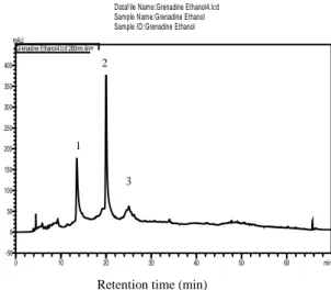
In order to identify components from PPE, HPLC analysis was performed on ethanolic PPE (Figure 1), which has shown the highest level of polyphenols. The results revealed the presence of tannins as peduncalagin, punicalagin and punigluconin respectively in ethanolic PPE by comparisons to the retention time and UV spectra of authentic standards. In the present study, the punicalagin was determined as predominant in the extract; it constituted of 36.451% of the total extracted compounds followed by peduncalagin (23.430%) and punigluconin (9.486%).
Dataf ile Name:Grenadine Ethanol4.lcd Sample Name:Grenadine Ethanol Sample ID:Grenadine Ethanol
0 10 20 30 40 50 60 min -50 0 50 100 150 200 250 300 350 400 mAU Grenadine Ethanol4.lcd 280nm,4nm
Figure 1. HPLC chromatogram of ethanolic pomegranate peels
extract recorded at 280 nm. Peak 1 is peduncalagin; peak 2 is punicalagin; peak 3 is punigluconin.
3.3. DPPH Assay
In the present study, we have evaluated the free radical scavenger activity of ethanol 70%, acetone 70% and methanol 70% PPE; the scavenging activity of DPPH in PPE solution was determined in percentage and compared with a standard ascorbic acid. The decrease in absorbance of DPPH radical is caused by the reaction between antioxidant found in a mixture solution of the PPE and DPPH, which induces the change in color of the mixture from purple to yellow. The hydroalcoholic extracts showed concentration dependant on antiradical activity. Antioxidant activity of phenolic compounds varied greatly according to tested solvent as given in Table 2. The highest scavenging activity (87.37 ± 1.36%) was exhibited by ethanolic PPE followed by acetonic PPE (57.84 ± 4.85%), their DPPH radical scavenging activity are not significantly different, whereas methanolic PPE had the lowest antioxidant property (22.23 ± 0.76%) and is significantly different from ethanolic PPE (p = 0.043). When compared to a standard ascorbic acid, the DPPH radical scavenging activity of ethanolic PPE was higher at concentrations ranging from 25 µg/mL to 50 µg/mL; however, its antiradical activity is lower than ascorbic acid at concentrations ranging from 100 µg/mL to 1000 µg/ mL. The EC 50 values obtained for the investigated plant extracts were in the range from 220 ± 0.02 µg/mL to 700 ± 37 µg/mL. The EC50 of ethanolic PPE had the lower value (220 ± 0.02 mg/mL) in comparison with the different extracts (Table 3).
In this present study, a positive relationship exists between the total phenolic content, flavonoid content and the antioxidant activity of the PPE. This is because the highest antioxidant capacity was exhibited by 70% ethanolic extract which also showed the highest concentrations of phenolics and flavonoids. Likewise, methanolic PPE extract exhibited the lowest concentrations of phenolics, flavonoids and antioxidant activity.
3 1
2
© 2020 Jordan Journal of Biological Sciences. All rights reserved - Volume 13, Number 3 407
Table 2. DPPH radical scavenging activities (%) of 70% acetonic,
70% ethanolic and 70% methanolic PPE. Ascorbic acid was used as positive control Concentration (µg/mL) % of inhibition Acetonic PPE Ethanolic PPE Methanolic PPE Ascorbic acid 25 20.27± 0.88 44.74± 1.05 4.57± 2.16 5.41± 2.19 50 34.81± 0.54 46.79± 0.68 13.09± 0.82 21.04± 3.92 100 44.53± 1.97 50.96± 0.84 17.03± 2.28 63.61± 6.61 500 46.01±1.84 70.54± 0.63 22.01± 0.55 90.24± 1.78 1000 57.84±4.85 87.37± 1.36 22.23± 0.76 97.24± 0.90
The data are displayed with mean ± standard deviation of twice replications.
Table 3. EC50 concentrations of DPPH scavenging activity from
bioactive compounds.
Bioactive compounds (PPE) EC50 (µg/mL)
Acetonic PPE 510± 20
Ethanolic PPE 220± 14
Methanolic PPE 700± 37
Ascorbic acid 200± 26
Values are expressed as mean of three replicates. EC50: the sample concentration required to scavenge 50% free radicals. Ascorbic acid was used as positive control.
3.4. FRAP Assay
The reducing power was used to investigate the ability of the antioxidants found in the extract to donate an electron and reduce Fe+3 to Fe+2. In this experiment,
depending on the reducing power of the sample, the yellow color of the test solution changes to pale green and blue color. As shown in Figure 2, it was found that the increase in extract concentration increases the reducing power activity. The reducing power of acetonic, ethanolic and methanolic PPE at different concentrations showed a range of absorbance values from 0.13 to 1.48 at 700 nm; the activity of the three extracts was not significantly different (p= 0.657). These values were lower than those obtained with ascorbic acid with absorbance values from 0.71 to 2.28; the reducing power of the reference compound is significantly higher (p <0.05) than that of methanolic and ethanolic PPE. The lowest reducing power was found in the methanolic PPE; this result confirmed that obtained by DPPH assay.
Figure 2. Reducing power of PPE. Eth extr = ethanol extract;
Meth extr = methanol extract; Acet extr = acetone extract; Ascorbic acid was used as positive control.
3.5. Identification and Selection of Oral Pathogen In this study, samples were collected from 37 patients, males and females, of different age groups; all samples were cultured on different media, and conventional methods were used for isolation and identification. Strains were identified as Streptococcus mutans, Streptococcus salivarius, Streptococcus constellatus, Enterococcus faecalis, Enterococcus faecium, staphylococcus epidermis, staphylococcus aureus, Lactobacillus sp, Klebsiella oxytoca, Enterobacter bugandensis. Six strains exhibiting significant biofilm formation were selected for our study, for which four were selected for antibiofilm activity and subjected to molecular identification (data not shown).
Table 4. Gram staining and cell morphology of isolated bacteria.
Strains Gram staining Morphology
Streptococcus mutans + Cocci
Gemella morbillorum + Cocci
Enterococcus faecalis + Cocci
Staphylococcus epidermis + Cocci
Klebsiella oxytoca - Rod
Enterobacter bugandensis - Rod
3.6. Antibacterial Effect
Antibacterial activity was evaluated by determining the MIC and MBC of the PPE against oral pathogen by microdilution method, according to Gulluce et al. (2008).
The three PPE, screened for antimicrobial activity, showed antibacterial activity against all strains. As evident from the Table 5, the intensity of antibacterial activity of the extracts varied depending on the species of bacteria. Statistical analysis reveals that extracts from pomegranates peels exhibited significantly different antimicrobial potential against tested strains (p= 0.026). Gram-negative bacteria are more resistant to the extracts. The highest MIC values were observed with E. bugandensis (from 3.15 mg/mL tо 100 mg/mL), followed by K. oxytoca with action interval of PPE from 12.5 to 25 mg/mL. PP extracts are the most effective against S. mutans and E. faecalis (MIC values from 0.0125 to 0.025 mg/mL). In comparison between the effects of the ethanolic, methanolic and acetonic PPE on each strain, there is not statistically significant difference in activity (p < 0.05). Action interval of acetonic, ethanolic and methanolic extract was from 0.0125 mg/mL to 12.5 mg/mL, from 0.025 mg/mL to 100 mg/mL and from 0.0125 mg/mL to 100 mg/mL respectively.
On the other hand, results of minimum bactericidal concentrations (MBC) are listed in Table 5, where the ethanolic and acetonic PPE have the lowest MBC values and have approximately the same values against oral pathogens. They were in the range from 6.25 mg/mL to ˃200 mg/mL. CMBs of methanolic PPE were in the range from 25 mg/mL to 200 mg/ML; there are no significantly differences between the MBC values of the three extracts. MBC results show that S. mutans and E. faecalis were the most sensitive, E. bugandensis and K. oxytoca were the most resistant microorganisms and have significantly different values of MBC than the other strains, which confirm the results of the MIC. A bacteriostatic activity of the PPE was observed in all the strains.
Table 5. MIC and MBC of acetonic, ethanolic and methanolic
PPE.
Species Acetone Ethanol Methanol Acetone Ethanol Methanol MIC (mg/mL) MBC (mg/mL) S. mutans 0.0125 0.025 0.025 6.25 6.25 100 G.morbillorum 12.5 12.5 25 12.5 12.5 25 E. faecalis 0 .0125 0.025 0.0125 6.25 12.5 25 S. epidermis 0.4 0.05 0.05 25 12.5 12.5 K. oxytoca 12.5 12.5 25 ˃200 ˃200 200 E.bugandensis 3.15 100 100 100 200 200
MIC: minimum inhibitory concentration; MBC: minimum bactericidal concentration.
3.7. Antibiofilm Activity
In this study, the in vitro antibiofilm effect of the ethanolic PPE was evaluated against four selected oral pathogens by tube method. The effect of PPE on biofilm formation was evaluated by staining with crystal violet as previously reported (Djordjevic et al., 2002). The adherence capabilities of the strains were classified into four categories, no/low (OD595 < 0.2), moderate (0.2 < OD595 < 0.4), high (0.6 < OD595 < 1), and very high (OD595 > 1) biofilm producers (Russo et al., 2018). All the strains showed positive result on their capability of biofilm formation (Figure 3). G. morbillorum and E. bugandensis formed the thickest biofilm (very high producer), E. faecalis, S. epidermis, K. oxytoca were classified as high biofilm producers and S. mutans as moderate biofilm producer. There are no significant differences in biofilm formation between strains.
Susceptibility of biofilms against plant extract showed that the effect of tested ethanolic PPE inhibited the biofilm formation of all the strains (Figure 4), and the obtained effect was dose dependent. The extract showed good antibiofilm activity ˃50% against all tested bacteria. Significant differences (p< 0.05) were observed between different extract concentrations in biofilm inhibition values. Doses of MIC, 50% MIC and 25% MIC had a greater influence in biofilm eradication than doses of 12.25% MIC and 6.25% MIC. The maximum biofilm inhibition was observed at the highest concentration of the extract (MIC) with E. faecalis (91.95%) and S. epidermis (90.7%), and the lowest biofilm reduction was observed with E. bugandensis (62.66%).
Figure 3. Quantification of biofilm formation by tube method
after 2, 4, 6, 24 and 48 hours of incubation. Values represent means ± standard errors for three replicates. Data with different letters indicate significant difference at p< 0.05.
Figure 4. The effect of ethanolic PPE on the formation of
bacterial biofilms. Values represent means ± standard errors for three replicates. Different letters denote significant difference from one another (p < 0.05).
4. Discussion
Recently, the use of medicinal plants as antibacterial agent has increased due to opposing effects of antibiotics and the increasing resistant pattern of bacteria which made that antibiotics lose their efficacy (Davies, 1994; Service, 1995; Ahmad et al., 1998). For this, it is necessary to find an alternative of antibiotics for the treatment of infectious diseases. Among these, Punica granatum, known for its antioxidant and antimicrobial activities, these properties could be attributed to the production of several secondary metabolites by the plants (Khan et al., 2017). Therefore, in the current study we have evaluated different classes of phenolic compounds. The results indicated that the pomegranate peel extract is rich in polyphenol and proanthocyanidin contents. Our results are in agreement with Elfalleh et al. (2012) and Shinde et al. (2015) who reported that total phenolic content of pomegranate peels is 85.60 ± 4.87 and 212 ± 20.55 mg GAE/g DW respectively. The ethanolic PPE has the highest polyphenol and flavonoid contents. Ethanol has been known as a good solvent for polyphenol extraction and is safe for human consumption (Dai and Mumper, 2010). Malviya et al. (2014) and Wang et al. (2011) also reported that content of polyphenols and flavonoids in ethanol extract of pomegranate peels are higher than water, methanol and acetone extracts. There is a different factor that influenced the variation in the recovery of phenolics and flavonoids from natural products, the type of plant, the nature of compounds to extract and the efficiency of extraction solvents to dissolve such compounds (Shabir et al., 2011; Roudsari et al., 2007). Also, the use of mixed solvents (solvent+ DW) results in an enrichment of the extracts in polyphenols, which is due to the increase in the solubility of the phenolic compounds in the extracts (Mohammedi and Atik, 2011; Trabelsi et al., 2010). The results indicated that PPE contained higher level of tannins compounds, which is in compliance with the literature (Machado et al., 2002; Voravuthikunchai et al., 2005; Al zoreky, 2009). Wang et al. (2011) found that the proanthocyanidins content of ethanol and acetone extracts were higher than the methanol extract. Also, Vijayalakshmi et al. (2017) revealed the presence of highest total tannin content in the ethanolic PPE.
HPLC analysis was performed to obtain more information about the nature of the phenolic compounds present in the ethanolic PPE. The results showed the
© 2020 Jordan Journal of Biological Sciences. All rights reserved - Volume 13, Number 3 409
presence of three ellagitannins with punicalagin as major components. The pomegranate peel is a rich source of ellagitannins, especially punicalagin, gallic acid, ellagic acid (Gil et al., 2000; Cerdà et al., 2003). Punicalagin, which belongs to the class of hydrolisable tannins, is the most abundant component and is characteristic compounds in pomegranate peel (Seeram et al., 2006; Bakkalbaşi et al., 2009). Our findings are in correlation with other studies, Middha et al. (2013) and Kwak et al. (2005) revealed the presence of punicalagin as a major ellagitannin in PPE. Other authors (Choi et al., 2011; Çam and Hişil, 2010) found in addition to punicalagin other compounds such as ellagic acid and gallic acid. Furthermore, HPLC analysis of methanolic PPE carried out by Ali et al. (2014) revealed the presence of pyrogallol, chlorogenic acid, coumaric acid, ferulic acid, cinnamic acid, benzoic acid, catechin, rutin, acacetin, genistein and kaempferol. These variations in results are probably due to the used solvent, which have an impact on the composition of PPE (El-Falleh et al., 2012). Also several factors (environmental, processing and post harvesting) influenced the composition of the peel (Houston, 2005).
In vitro radical scavenging and antioxidant capacity of the PPE were studied by using DPPH free radical scavenging assay and Ferric reducing power assay. The DPPH test is used to measure the capability of antioxidants in plants extracts to scavenge free radicals from the DPPH solution by donating hydrogen atoms or electrons, which converts free radicals into more stable products (Roudsari., 2007; Sajid et al., 2012).
In the DPPH test, the scavenging activity of the extract varies according to the extraction methodology and the polarity of solvent used for extraction; this could be due to the solubility or insolubility of different antioxidant compounds found in the extract with different chemical characteristics and polarities in particular solvent (Bushra et al., 2009; Boeing et al., 2014). In the present studies, ethanol is a better solvent than acetone and methanol. Results were consistent with the findings of Malviya et al. (2014), who reported such higher antioxidant activity was found in 100 % water and 70 % ethanol: 30 % water.
The FRAP assay is a simple method frequently used in the evaluation of antioxidant compounds of fruits, vegetables and some biological samples (Wong et al., 2006). In this method, acetonic extract gave the highest reducing power followed by ethanolic extract. Similar results were reported by Thitipramote et al. (2019) where acetonic and ethanolic PPE exhibited the highest bioactive compound and antioxidant activity in ferric reducing power assay. Other authors found the best results of antioxidant activity in methanolic PPE (Li et al., 2006; Pagliarulo et al., 2016). The differences in results may be attributed to the type of pomegranate variety, storage time, harvesting date, climate conditions and method of extraction, as well as solvent concentration (Moure et al., 2001; Robards , 2003; Pinelo et al., 2004).
In the current study, PPE showed antibacterial activity against all tested bacteria. The intensity of antibacterial activity varied depending on the tested bacteria. positive strains are more sensitive to PPE than Gram-negative bacteria. This variation in sensitivity is ascribed to the differences in cell wall composition. The cell wall of Gram-positive bacteria contains a thick peptidoglycan
layer, while the cell wall of Gram-negative bacteria contains a thin peptidoglycan layer that is surrounded by a thick cytoplasmic membrane and perisplastic space, which prevents the penetration of antimicrobial substances (Staszewski et al., 2011; Oliveira et al., 2013). Alvarez-Ordonez et al. (2014) reported that the plant extracts were able to disrupt the molecular structure of the bacteria cell wall and caused leakage of intracellular content, causing increasing in membrane permeability. Several authors have also observed low MIC values for plant extracts for Gram-positive bacteria and high values was reported for Gram-negative bacteria (Burt, 2004; Al-Zoreky, 2009; Hayrapetyan et al., 2012; Tayel et al., 2012). The present research was in line with other studies demonstrating antibacterial effect of punica granatum L. peel extracts against S. aureus, Salmonella enterica, Shigella sonnei, E. coli, Bacillus subtilis and Enterococcus faecalis (Machado et al., 2002; Voravuthikunchai et al., 2005; Pagliarulo et al., 2016; Rosas-Burgos et al., 2017). In the work carried by Malviya et al. (2014) the antibacterial activity of ethanolic PPE of Ganesh variety was demonstrated on Staphylococcus aureus, Enterobacter aerogenes, Salmonella typhi and Klebsiella pneumonia. In the study by Jaisinghani et al. (2018), the inhibitory effect of ethanolic PPE cultivars against both Gram-negative and positive bacteria strains have been reported. The ellagitannins are the major components of pomegranate peel polyphenols, which have been implicated in antimicrobial, anti-inflammatory and anti-cancer activities (Adams et al., 2006; Glazer et al., 2012). Bialonska et al. (2009) has reported effect of pomegranate tannin constituents on the growth of various species of human gut bacteria in vitro. The tannins increase bacteriolysis, interfere with mechanisms of bacterial adhesion onto the tooth surfaces (Pereira et al., 2006).
The six oral isolates were screen for the biofilm formation by tube method. All the strains showed adherence of the stain in test tube. The antibiofilm activity was tested at concentrations ≤ CMI of ethanolic PPE against four selected oral pathogens. Results showed that the tested extract could inhibit the biofilm formation for all tested bacteria in a dose-dependent manner, the inhibitory effect increased with increasing concentration. Vasconcelos et al. (2006) demonstrate the potential of PPE to significantly reduce the adherence of three standard streptococci strains (mutans ATCC 25175, sanguis ATCC 10577, and mitis ATCC 9811) of dental plaque and the high efficiency of pomegranate gel in inhibition of bacterial adherence. Menezes et al. (2006) observed the effect of the pomegranate hydroalcoholic extract to significantly reduce the bacteria of dental plaque and concluded that this extract could be helpful in the prevention of diseases caused by plaque bacteria. These results are supported by an in vitro study of several authors (Kakiuchi et al., 1986, Pereira et al., 2006) who demonstrated that the antimicrobial action of Punica granatum Linn on the inhibition of adherence of dental biofilm bacteria is the result of disturbance of glucan synthesis, thus preventing the adhesion of these bacteria to dental surface.
5. Conclusion
This study showed that pomegranate peels are a potential resource for phenolics, flavonoids and proanthocyanidins. The phytochemistry of ethanolic extract was the highest followed by acetonic extract.
This research revealed the highest antioxidant activities of pomegranate peels which may be attributed to ellagitannins identified by HPLC analysis. All extracts of pomegranate peels have inhibitory effect against both Gram-negative and Gram-positive bacteria. Also, the ethanolic PPE are able to inhibit biofilm formation of E. faecalis, S. epidermis, K. oxytoca and E. bugandensis. So, the use of pomegranate peels, which is the by product, could be interesting for its antioxidant and antimicrobial activities against oral pathogen by its incorporation in gum, mouthwash, toothpaste and in the elaboration of products to reduce caries and dental plaque.
Acknowledgment
The authors would like to thank laboratory of Microbiology and Vegetal Biology at University of Mostaganem (Algeria) for their financial support, Ms. Katarina Kvoriaková, Dr. Dahloum Lahouari and Mr. Benchehida Abdelkader for their help with the article.
References
Aas JA, Paster BJ, Stokes LN, Olsen I and Dewhirst FE. 2005. Defining the normal bacterial flora of the oral cavity. J Clin Microbiol, 43: 5721-5732.
Adams LS, Seeram NP, Aggarwal BB, Takada Y, Sand D and Heber D. 2006. Pomegranate juice, total pomegranate ellagitannins, and punicalagin suppress inflammatory cell signaling in colon cancer cells. J Agric Food Chem, 54: 980-985. Ahangari B and Sargolzaei J. 2012. Extraction of pomegranate seed oil using subcritical propane and supercritical carbon dioxide. Theor Found Chemen, 46:258-265.
Ahmad I, Mehmood Z and Mohammad F. 1998. Screening of some Indian medicinal plants for their antimicrobial properties. Journal of Ethnopharmacology, 62: 183-193.
Ali SL, El-Baz FK, El-Emary GAE, Khan EA and Mohamed AA. 2014. HPLC analysis of poly phenolic compounds and free radical scavenger activity of pomegranate. International journal of pharmaceutical and clinical research, 6: 348-355.
Alvarez-Ordonez A, Begley M, Clifford T, Deasy T, Considine K, O’Connor P, Ross RP and Hill C. 2014. Investigation of the Antimicrobial Activity of Bacillus licheniformis Strains Isolated from Retail Powdered Infant Milk Formulae. Probiotics and Antimicrobial Proteins, 6: 32-40.
Al-Zoreky NS. 2009. Antimicrobial activity of pomegranate (Punica granatum L.) fruit peels. Int. J. Food Microbiol, 134: 244-248.
Bakkalbaşi E, Mentes O and Artik N. 2009. Food Ellagitannins- Occurrence, Effects of Processing and Storage, Crit. Rev. Food Sci. nut, 49: 283- 298.
Bialonska D, Kasimsetty SG, Schrader KK and Ferreira D. 2009. The effect of pomegranate (Punica granatum L.) by products and ellagitannins on the growth of human gut bacteria. Journal of agricultural and food chemistry, 57: 8344-8349.
Boeing JS, Barizão ÉO, E Silva BC, Montanher PF, Cinque Almeida V and Visentainer JV. 2014. Evaluation of solvent effect on the extraction of phenolic compounds and antioxidant capacities from the berries: application of principal component analysis. Chem Cent J, 8:48.
Burt S. 2004. Essential oils: their antibacterial properties and potential applications in foods – a review. International Journal of Food Microbiology, 94: 223-253.
Bushra S, Farooq A and Muhammad A. 2009. Effect of Extraction Solvent/Technique on the Antioxidant Activity of Selected Medicinal Plant Extracts. Molecules, 14: 2167-2180.
Çam M and Hişil Y. 2010. Pressurized water extraction of polyphenols from pomegranate peels. Food Chem, 123: 878-885. Cerda B, Llorach R, Ceròn JJ, Espìn JC and Tomàs-Barberàn FA. 2003. Evaluation of the bioavailability and metabolism in the rat of punicalagin, an antioxidant polyphenol from pomegranate juice. Eur J Nutr, 42:18-28.
Choi JG, Kang OH, Chae YS, Oh YC, Brice OO, Kim MS, Sohn DH, Kim HS, Park H, Shin DW, Rho JR and Kwon DY. 2011. In Vitro and in Vivo Antibacterial Activity of Punica granatum Peel
Ethanol Extract against Salmonella. Evidence- Based
complementary and Alternative Medicine, 5: 690518.
Cramton SE, Gerke C, Schnell NF, Nichols WW and Götz F. 1999. The intercellular adhesion (ica) locus is present in Staphylococcus aureus and is required for biofilm formation. Infect Immun, 67: 5427-33.
Dadashi M, Hashemi A, Eslami G, Fallah F, Goudarzi H, Erfanimanesh S and Taherpour A. 2016. Evaluation of antibacterial effects of Zataria multiflora Boiss extracts against ESBL-producing Klebsiella pneumoniae strains. Avicenna Journal of Phytomedicine, 6:336-343.
Dai J and Mumper RJ. 2010. Plant Phenolics: Extraction, Analysis and Their Antioxidant and Anticancer Properties. Molecules, 15: 7313-7352.
Davies J. 1994. Inactivation of antibiotics and the dissemination of resistance genes. Science, 264: 375-382.
Dewhirst FE, Chen T, Izard J, Paster BJ, Tanner AC, Yu WH, Lakshmanan A and Wade WG. 2010. The human oral microbiome. J Bacteriol, 192: 5002-5017.
Djordjevic D, Wiedmann M and Mc Landsborough LA. 2002. Microtiter plate assay for assessment of Listeria monocytogenes biofilm formation. Applied and Environ- mental Microbiology,
68: 2950-2958.
Do T, Devine DA and Marsh PD. 2013. Oral biofilms: molecular analysis, challenges, and future prospects in dental diagnostics. Clin Cosmet Investig Dent, 5: 11-19.
Elfalleh W, Hannachi H, Tlili N, Yahia Y, Nasri N and Ferchichi A. 2012. Total phenolic contents and antioxidant activities of pomegranate peel, seed, leaf and flower. J Med Plants Res, 6: 4724-4730.
Ferguson LR. 2010. Chronic inflammation and mutagenesis. Mutat. Res. Fund. Mol, 690: 3-11.
Fischer UA, Carle R and Kammerer DR. 2011. Identification and Quantification of Phenolic Compounds from Pomegranate (Punica granatum L.) Peel, Mesocarp, Aril and Differently Produced Juices by HPLC-DAD– ESI/Msn. Food Chemistry, 127: 807-821. Gil MI, Tomàs- Barberàn FA, Hess-Pierce B, Holcroft DM and Kader AA. 2000. Antioxidant activity of pomegranate juice and its relationship with phenolic composition and processing,
48:4581-4589.
Glazer I, Masaphy S, Marciano P, Bar-Ilan I, Holland D, Kerem Z and Amir R. 2012. Partial identification of antifungal compounds from Punica granatum peel extracts. J Agric Food Chem, 60: 4841-4848.
Gulluce M, Sahin F, Sokmen M, Ozer H, Daferara D, Sokmen A, Polissiou M, Adiguzel A and Ozkan H. 2007. Antimicrobial and antioxidant properties of the essential oils and methanol extract from Mentha longifolia L. ssp. longifolia. Food Chem, 104: 1449-1456.
© 2020 Jordan Journal of Biological Sciences. All rights reserved - Volume 13, Number 3 411
Halliwell B. 2007. Oxidative stress and cancer: have we moved forward? Biochem. J, 401: 1-11.
Hardie JM. 1992. Oral microbiology: Current concepts in the microbiology of dental caries and periodontal disease. Br Dent J,
172:271.
Hayrapetyan H, Hazeleger WC and Beumer RR. 2012. Inhibition of Listeria monocytogenes by pomegranate (Punica granatum) peel extract in meat pate at different temperatures. Food Control,
23: 66-72.
Houston MC. 2005. Nutraceutical, vitamins, antioxidants and minerals in the prevention and treatment of hypertension. Progress in Cardiovascular Diseases, 47: 396-449.
Hussain AI, Anwar F, Hussain ST and Przybylski R. 2008. Chemical composition, antioxidant and antimicrobial activities of basil (Ocimum basilicum) essential oils depends on seasonal variations. Food chemistry, 108: 986-995.
Ismail T, Sestili P and Akhtar S. 2 0 1 2 . Pomegranate peel and fruit extracts. A review o f p ot en t i a l a n t i -inflammatory and anti-infective effects. Journal of Ethnopharmacology, 143: 397-405.
ISO 14502-1: 2005. Determination of substances characteristic of green and black tea. Part 1: C/ontent of total polyphenols in tea. Colorimetric method using Folin-Ciocalteu reagent.
Jahfar M, Vijayan KK and Azadi P. 2003. Studies on a polysaccharide from the fruit rind of Punica granatum. Res. J. Chem. Environ, 7: 43-50.
Jaisinghani RN, Makhwana S and Khanojia A. 2018. Study on antibacterial and flavonoid content of ethanolic extract of punica granatum (pomegranate) peel. Microbiology research, 9:7480. Kakiuchi N, Hattori M and Nishizawa M. 1986. Studies on dental caries prevention by traditional medicines. Inhibitory effect of various tannins on glucan synthesis by glycosyltransferase from Streptococcus mutans. Chem Pharm Bull, 34:720-725.
Khalil EA. 2004. Antidiabetic effect of an aqueous extract of pomegranate (Punica granatum L.) peels in normal and alloxan diabetic rats. Egy. J. Hosp. Med, 16: 92-99.
Khan NH, Ying ALT, Tian CGZ, Yi OW and Vijayabalan S. 2017. Screening of Punica granatum seeds for antibacterial and antioxidant activity with various extracts. J Gastroenterol Dig Dis,
1:1-7.
Kumazawa S, Hamasaka T and Nakayama T. 2004. Antioxidant activity of propolis of various geographic origins. Food Chemistry, 84: 329-339.
Kwak HM, Jeong HH, Sohng BH et al. 2005. Quantitative analysis of antioxdants in Korea pomegranate Husk (Granati pericarpium) cultivated in different site. Journal of the Korean Society for Applied Biological Chemistry, 48: 431-434.
Lansky EP and Newman RA. 2007. Punica granatum (Pomegranate) and Its Potential for Prevention and Treatment of Inflammation and Cancer. Journal of Ethnopharmacology, 109: 177-206.
Li Y, Guo C, Yang J, Wei J, Xu J and Cheng S. 2006. Evaluation of antioxidant properties of pomegranate peel extract in comparison. Food Chemistry, 96: 254-260.
Lim J, Lee KM, Kim SH, Nam SW, Oh YJ, Yun HS, Jo W, Oh S, Kim SH and Park S. 2008. Nanoscale characterization of Escherichia coli biofilm formed under laminar flow using atomic force microscopy (AFM) and scanning electron microscopy (SEM). Bull. Kor. Chem. Soc, 29: 2114-2118.
Longtin R. 2003. The pomegranate: nature’s power fruit? JNCI J Natl Cancer Inst, 95: 346-348.
Machado TDB, Leal ICR, Amaral ACF, Dos Santos KRN, Da Silva MG, and Kuster RM. 2002. Antimicrobial ellagitannin of Punica granatum fruits. Journal of the Brazilian Chemical Society,
13: 606-610.
Malviya S, Jha A and Hettiarachchy N. 2014. Antioxidant and antibacterial potential of pomegranate peel extracts. Journal of food science and technology, 51: 4132-4137.
Menezes SMS, Cordeiro LN and Viana GSB . 2006. Punica granatum (pomegranate) extract is active against dental plaque. Journal of Herbal Pharmacotherapy, 6: 79-92.
Middha SK, Talambedu U and Pande V. 2013. HPLC evaluation of phenolic profile, nutritive content, and antioxidant capacity of extracts obtained from Punica granatum fruit peel. Advances in Pharmacological Sciences, 2013: 1-6.
Mirdehghan SH and Rahemi M. 2007. Seasonal changes of mineral nutrients and phenolics in pomegranate (Punica granatum L.) fruit. Scientia Horticulturae, 111: 120-127.
Mohammedi Z and Atik F. 2011. Impact of solvent extraction type on total polyphenols content and biological activity from Tamarix aphylla (L.) karst. Inter J Pharma Bio Sci, 2: 609-615. Moure A, Franco D, Sineiro J, Domĭngguez H, Nūňez MJ and Lema JM. 2001. Antioxidant activity of extracts from Gevuina avellana and Rosa rubiginosa defatted seeds. Food Research International, 34: 103-109.
Oliveira DA, Salvador AA, Smânia A, Smânia EF, Maraschin M, Ferreira SR. 2013. Antimicrobial activity and composition profile of grape (Vitis vinifera) pomace extracts obtained by supercritical fluids. J Biotechnol, 164: 423-432.
Oyaizu M. 1986. Studies on products of browning reactions: Antioxidant Activities of Products of browning reaction prepared from glucose amine. Jpn J Nutr, 44: 307-315.
Pagliarulo C, De Vito V, Picariello G, Colicchio R, Pastore G, Salvatore P and Volpe MG. 2016. Inhibitory effect of pomegranate (Punica granatum L.) polyphenol extracts on the bacterial growth and survival of clinical isolates of pathogenic Staphylococcus aureus and Escherichia coli. Food Chemistry,
190: 824-831.
Paster BJ, Boches SK, Galvin JL, Ericson RE, Lau CN, Levanos VA, Sahasrabudhe A and Dewhirst FE. 2001. Bacterial diversity in human subgingival plaque. J Bacteriol, 183: 3770-3783. Pereira JV, Pereira MSV, Sampaio FC, Sampaio MCC, Alves PM, Araújo CRF and Higino JS. 2006. In vitro antibacterial and antiadherence effect of Punica granatum Linn extract upon dental biofilm microorganisms. Braz J Pharmacogn, 16: 88-93.
Petersen PE, Bourgeois D, Ogawa H, Estupinan-Day S and Ndiaye C. 2005. The global burden of oral diseases and risks to oral health. Bulletin of theWorld Health Organization, 83: 661-669.
Pinelo M, Manzocco L, Nunez MJ and Nicoli MC. 2004. Interaction among phenols in food fortification: negative synergism on antioxidant capacity. J Agric Food Chem, 52: 1177-1180.
Rautemaa R, Lauhio A, Cullinan MP and Seymour GJ. 2007. Oral infections and systemic disease—an emerging problem in medicine. Clinical Microbiology and Infection, 13: 1041-1047. Robards K. 2003. Strategies for the determination of bioactive phenols in plants, fruit and vegetables. J Chromatogr A, 1000: 657-691.
Rosas-Burgos EC, Burgos-Hernández A, Noguera-Artiaga L, Kacániová M, Hernández-García F, Cárdenas-López JL and Carbonell-Barrachinaa ÁA. 2017. Antimicrobial activity of
pomegranate peel extracts as affected by cultivar. J Sci Food
Agric, 97: 802-810.
Roudsari MH. 2007. Subcritical water extraction of antioxidant compounds from canola meal, M.Sc. Thesis, University of Saskatchewan, Saskatoon.
Russo P, Hadjilouka A, Beneduce L, Capozzi V, Paramithiotis S, Drosinos EH and Spano G. 2018. Effect of different conditions on Listeria monocytogenes biofilm formation and removal. Czech J. Food Sci, 36: 208-214.
Saeed N, Khan MR and Shabbir M. 2012. Antioxidant activity, total phenolic and total flavonoid contents of whole plant extracts
Torilis leptophylla L. BMC Complement. Altern. Med, 12: 221.
Sajid ZI, Anwar F, Shabir G, Rasul G, Alkharfy KM and Gilani AH. 2012. Antioxidant, antimicrobial properties and phenolics of different solvent extracts from bark, leaves and seeds of
Pongamia pinnata (L.) pierre. Molecules, 17: 3917-3932.
Sanchez-Moreno C, Larrauri JA and Saura-Calixto F. 1998. A procedure to measure the antiradical efficiency of polyphenols. J. Sci. Food Agric, 76: 270-276.
Seeram Np, Zhang Y, Reed JD, Krueger CG and Vaya J. 2006. Pomegranates: Ancient Roots to Modern Medicine, CRC press, Taylor and Francis Group, Boca Raton, FL.
Service RF. 1995. Antibiotics that resist resistance. Science, 270: 724-727.
Shabir G, Anwar F, Sultana B, Khalid ZM, Afzal M, Khan QM, and Ashrafuzzaman M. 2011. Antioxidant and antimicrobial attributes and phenolics of different solvent extracts from leaves, flowers and bark of gold mohar [Delonix regia (Bojer ex Hook.) Raf.]. Molecules, 16: 7302-7319.
Shinde PA, Reddy VKS and Patange SB. 2015. Quality of Indian mackerel as affected by pomegranate peel and tea leaf extracts during ice storage. SAARC J. Agr, 13: 109-122.
Soares AA, Marques de Souza CG, Daniel FM, Ferrari GP, Gomes da Costa SM and Peralta RM. 2009. Antioxidant activity and total phenolic content of Agaricus brasiliensis (Agaricus blazei Murril) in two stages of maturity. Food Chemistry, 112: 775-781.
Sun B, Ricardo da Silva J and Spranger I. 1998b. Critical factors of the vanillin assay for catechins and proanthocyanidins. Journal of Agricultural and Food Chemistry, 46: 4267-4274.
Tayel AA, El-Tras WF, Moussa SH and El-Sabbagh SM. 2012. Surface decontamination and quality enhancement in meat steaks using plant extracts as natural biopreservatives. Foodborne Pathog. Dis, 9: 755-761.
Thitipramote N, Maisakun T, Chomchuen C, Pradmeeteekul P, Nimkamnerd J, Vongnititorn P, Chaiwut P, Thitilertdecha N and Pintathong P. 2019. Bioactive Compounds and Antioxidant Activities from Pomegranate Peel and Seed Extracts. Food and Applied Bioscience Journal, 7: 152-161.
Trabelsi N, Megdiche W, Ksouri R, Falleh H, Oueslati S, Bourgou S, Hajlaoui H and Abdelly C. 2010. Solvent effects on phenolic contents and biological activities of the halophyte Limoniastrum monopetalum leaves. Food Sci Tech, 43: 632-639.
Vasconcelos LCDS, Sampaio FC, Sampaio MCC, Pereira MDSV, Higino JS, and Peixoto MHP. 2006. Minimum inhibitory concentration of adherence of Punica granatum Linn (pomegranate) gel against S. mutans, S. mitis and C. albicans. Brazilian Dental Journal, 17: 223-227.
Vijayalakshmi K, Sreedevi1 P and Venkateswari R. 2017. Phytochemical evaluation of Punica granatum L. leaf extract. Int J Curr Pharm Res, 9: 14-18.
Von Staszewski M, Pilosof AM and Jagus RJ. 2011. Antioxidant and antimicrobial performance of different Argentinean green tea varieties as affected by whey proteins. Food Chem, 125: 186-192. Voravuthikunchai SP, Sririrak T, Limsuwan S, Supawita T, Iida
T, and Honda T. 2005. Inhibitory effects of active compounds
from Punica granatum pericarp on verocytotoxin production by enterohemorrhagic Escherichia coli O157:H7. Journal of Health Science, 51: 590-596.
Wang Z, Pan Z, Ma H and Atungulu GG. 2011. Extract of phenolics from pomegranate peels. The Open Food Sci. J, 5:17-25.
Watabe K, Nishi M, Miyake H and Hirata K. 1998. Lifestyle and gastric cancer: a case-control study. Oncol Rep, 5: 1191-1194. Wong CC, Li HB, Cheng KW and Chen F. 2006. A systematic survey of antioxidant activity of 30 Chinese medicinal plants using the ferric reducing antioxidant power assay. Food Chem, 97: 705-711.
Yeo BK, Lim LP, Paquette DW and Williams RC. 2005. Periodontal disease—the emergence of a risk for systemic conditions: pre-term low birth weight. Annals of the Academy of Medicine Singapore, 34: 111-116.