HAL Id: hal-01608337
https://hal.archives-ouvertes.fr/hal-01608337
Submitted on 25 May 2020HAL is a multi-disciplinary open access archive for the deposit and dissemination of sci-entific research documents, whether they are pub-lished or not. The documents may come from teaching and research institutions in France or abroad, or from public or private research centers.
L’archive ouverte pluridisciplinaire HAL, est destinée au dépôt et à la diffusion de documents scientifiques de niveau recherche, publiés ou non, émanant des établissements d’enseignement et de recherche français ou étrangers, des laboratoires publics ou privés.
Distributed under a Creative Commons Attribution| 4.0 International License
SOLEIL shining on the solution-state structure of
biomacromolecules by synchrotron X-ray footprinting at
the metrology beamline
Anna Baud, Laure Aymé, Florence Gonnet, Isabelle Salard, Yann Gohon,
Pascale Jolivet, Konstantin Brodolin, P. da Silva, Alexandre Giuliani, B.
Sclavi, et al.
To cite this version:
Anna Baud, Laure Aymé, Florence Gonnet, Isabelle Salard, Yann Gohon, et al.. SOLEIL shining on the solution-state structure of biomacromolecules by synchrotron X-ray footprinting at the metrology beamline. Journal of Synchrotron Radiation, International Union of Crystallography, 2017, 24, pp.576-585. �10.1107/S1600577517002478�. �hal-01608337�
J. Synchrotron Rad. (2017). 24, doi:10.1107/S1600577517002478 Supporting information
Volume 24 (2017)
Supporting information for article:
SOLEIL SHINING ON THE SOLUTION-STATE STRUCTURE OF
BIOMACROMOLECULES BY SYNCHROTRON X-RAY
FOOTPRINTING AT THE METROLOGY BEAMLINE
A. Baud, L. Ayme, F. Gonnet, I. Salard, Y. Gohon, P. Jolivet, K. Brodolin, Paulo Da Silva, A. Giuliani, B. Sclavi, T. Chardot, P. Mercère, P. Roblin and R. Daniel
J. Synchrotron Rad. (2017). 24, doi:10.1107/S1600577517002478 Supporting information, sup-1
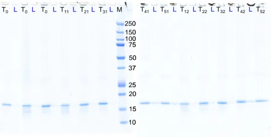
Figure S1 SDS-PAGE of oil bodies purified from S. cerevisiae expressing S3 oleosin and irradiated by X-ray beam from 0 to 100 ms. Three non-irradiated samples (T0) were injected, followed by two series of irradiated samples (series Tx1 and Tx2) submitted to increasing irradiation
time (2.5 ms [x = 1]; 10 ms [x = 2]; 25 ms [x = 3]; 50 ms [x = 4]; 100 ms [x = 5]). The intermediate washing fractions (L) were also analyzed in order to check all collected fractions.
250 150 100 75 50 37 25 20 15 10 T0 L T0 L T0 L T11 L T21 L T31L M T41 L T51 L T12 LT22 LT32 L T42 L T52 250 150 100 75 50 37 25 20 15 10 T0 L T0 L T0 L T11 L T21 L T31L M T41 L T51 L T12 LT22 LT32 L T42 L T52
J. Synchrotron Rad. (2017). 24, doi:10.1107/S1600577517002478 Supporting information, sup-2
Figure S2 Mass spectrometry control of synchrotron-irradiated FH. Positive MALDI-TOF mass spectrum in linear mode of Factor H (2 pmol spotted) before (A) and after (B) synchrotron irradiation for 30 ms. Please note that no mass shift of FH molecular weight was observed after the irradiation, confirming the integrity of the protein. Irradiation-specific mass shift could not be observed due to the low resolution of the MALDI-TOF MS. Matrix is 2,4,6-trihydroxyacetophenone (THAP) at 10 mg/mL in acetontrile/water (50:50 v/v) 0.1% trifluoroacetic acid. MALDI parameters: Voltage: 25 kV, grid %: 70%, extraction delay: 1000 ns, laser attenuation (λ=337 nm): 3400 arbitrary units and 500 laser shots were averaged.
Table S1 Quantification of S3 oleosin bands from fig. S1 gel by using gel densitometry
(MultiGauge software). Determined optical densities after background subtraction where normalized by the average intensity of the non-irradiated sample.
30000 68000 106000 144000 182000 220000 Mass (m/z) 0 10 20 30 40 50 60 70 80 90 100 % Intensi ty 1590005 79519 53005 39755 [Factor H + 2H]2+ [Factor H + H]+ [Factor H + 3H]3+ [Factor H + 4H]4+ 30000 68000 106000 144000 182000 220000 Mass (m/z) 0 10 20 30 40 50 60 70 80 90 100 % In ten si ty 158989 79494 52990 39741 [Factor H + 2H]2+ [Factor H + H]+ [Factor H + 3H]3+ [Factor H + 4H]4+