HAL Id: hal-02958993
https://hal.archives-ouvertes.fr/hal-02958993
Submitted on 6 Oct 2020HAL is a multi-disciplinary open access archive for the deposit and dissemination of sci-entific research documents, whether they are pub-lished or not. The documents may come from teaching and research institutions in France or abroad, or from public or private research centers.
L’archive ouverte pluridisciplinaire HAL, est destinée au dépôt et à la diffusion de documents scientifiques de niveau recherche, publiés ou non, émanant des établissements d’enseignement et de recherche français ou étrangers, des laboratoires publics ou privés.
Fluorocarbon Gas Exposure Induces Disaggregation of
Nanodiamond Clusters and Enhanced Adsorption,
Enabling Medical Microbubble Formation
Estefania Mendoza-Ortega, Marc Dubois, Marie Pierre Krafft
To cite this version:
Estefania Mendoza-Ortega, Marc Dubois, Marie Pierre Krafft. Fluorocarbon Gas Exposure Induces Disaggregation of Nanodiamond Clusters and Enhanced Adsorption, Enabling Medical Microbubble Formation. ACS Applied Nano Materials, American Chemical Society, 2020, 3 (9), pp.8897-8905. �10.1021/acsanm.0c01651�. �hal-02958993�
Fluorocarbon Gas Exposure Induces Disaggregation
1
of Nanodiamond Clusters and Enhanced Adsorption,
2
Enabling Medical Microbubble Formation
3
Estefania E. Mendoza-Ortega
1, Marc Dubois
2, and Marie Pierre Krafft
1*
4
1University of Strasbourg. Institut Charles Sadron (CNRS). 23 rue du Loess. 67034 Strasbourg,
5
France. 6
2Université Clermont Auvergne (UCA), CNRS, Sigma Clermont, ICCE, UMR 6296, 63178
7
Aubière, France 8
9
Corresponding Author* E-mail: krafft@unistra.fr 10
ORCID: ID: orcid.org/0000-0002-3379-2783 11
KEYWORDS. nanoparticle, adsorption, fluid interface, contrast agent, diagnostic, theranostic. 12
ABSTRACT
1
Introducing fluorocarbon vapor in the air above an aqueous dispersion of clusters of 2
nanodiamonds induces their disaggregation, a prerequisite for most of their biomedical uses. 3
Furthermore, the fluorocarbon gas promotes the adsorption of nanodiamonds at the gas/water 4
interface. As an example of the benefits that can be gained from our findings relative to the 5
implementation of nanomaterials for practical uses, we investigated the role that a fluorocarbon 6
gas may play in the generation of microbubbles, which are currently actively investigated in 7
ultrasound-mediated diagnosis and therapy. Remarkably, the fluorocarbon gas enables the 8
production of microbubbles shelled only with nanodiamonds, in the absence of any other 9
surfactant, which could not be achieved without the fluorocarbon being present. This demonstrates 10
that a supernatant gas can decisively affect the adsorption of nanoparticles from an aqueous phase 11
to an air/water interface, likely through physical adsorption at the nanodiamond surface. The 12
investigations involved solid-state NMR and FTIR, microbubble generation experiments with 13
acoustic attenuation monitoring, and bubble profile analysis tensiometry on spontaneously 14
adsorbed Gibbs films. Perspectives include control of aggregation of nanodiamonds and their 15
retrieval from aqueous dispersions, and applications in multimodal diagnostic imaging, 16
bioimaging and therapeutic cell tracking. It is proposed that the disaggregating potency of 17
fluorocarbons can be applied to other nanomaterials, providing a simple and effective means of 18
alleviating aggregation. 19
KEYWORDS 20
nanoparticle, nanomaterial, adsorption, fluid interface, contrast agent, diagnostic, theranostic. 21
GRAPHICAL TABLE OF CONTENT 1 2 3 4 5
Exposing aqueous dispersion of nanodiamonds to fluorocarbon vapor disaggregates clusters and 6
allows recruitment of the nanoparticles at the air/gas interface. This effect is stronger with 7
fluorinated nanodiamonds. This new gas-driven phenomenon allowed preparation of 8
nanodiamond-shelled microbubbles for potential use in bioimaging, cell tracking and ultrasound-9
mediated diagnostic and therapy. 10
11 12 13
INTRODUCTION
1
Nanodiamonds (NDs) capture considerable attention in various fields, from electronics and 2
optics to biomedicine, due to a range of specific properties, such as high refractive index and 3
scattering efficiency, outstanding mechanical performance (e.g. super-hardness), and versatile 4
surface chemistry and biocompatibility.1-4 In biology and medicine, they are actively investigated
5
for sensing, bioimaging and drug delivery.5-7 In particular, fluorescent NDs are being investigated 6
as a less-toxic alternative to semiconductor quantum dots (QDs).8 NDs can be surface-conjugated
7
with many biologically active molecules and targeting ligands using covalent or non-covalent (e.g. 8
electrostatic or hydrophobic) interactions.4 However, most biomedical applications of NDs are still 9
impeded due to their strong tendency for aggregation when dispersed in an aqueous phase. Various 10
techniques of ND disaggregation, crushing and fractionation have been reviewed,9 including 11
zirconia microbead-assisted wet ball milling,10 salt (or sugar)-assisted dry attrition and ball
12
milling,11 and bead-assisted sonic disintegration12-14 Although these techniques proved efficient, 13
and produced colloidal dispersions of nanodiamonds with particles lower than 10 nm,11 the risk of 14
contamination, either by metal ions or ceramic debris, remained, and made mandatory additional 15
purification steps, reducing the overall efficiency and increasing the cost. A salt-assisted ultrasonic 16
disaggregation process that is based on sonication of aqueous NaCl/ND slurries followed by 17
washing/centrifugation steps was also effective.15 Although the authors claimed the absence of 18
contamination, a long sonication time (over 1 h) using a titanium tip is well-known to produce 19
metal contamination by erosion of the probe due to the high intensity cavitation energy.16 20
Functionalization of NDs with zwitterionic moieties,17 or adsorption of polyelectrolytes,18 or 21
amino acids19 improved the dispersibility of NDs. Various methods of ND encapsulation, as in
phospholipid liposomes,20 or in silica21 or dopamine22 shells have also been reported. Control of 1
ND aggregation in aqueous solutions through simple approaches is therefore sought for. 2
We propose here a simple mean to induce dispersion of NDs in aqueous solutions that does not 3
involve chemical modification of the NDs, which consists in exposing ND dispersions to vapor of 4
a fluorocarbon (FC). We show that the capacity for a FC gas to recruit NDs to a gas/water interface 5
also unlocks the possibility to prepare microbubbles (MBs), which, to our knowledge, is 6
impossible in the absence of the FC. Microbubbles are used in the clinics as ultrasound contrast 7
agents, and their efficacy as mediators in ultrasound therapy is actively investigated.23-28 Medical 8
microbubbles, especially those stabilized by a phospholipid shell, are often stabilized by a 9
fluorocarbon gas. It is noteworthy that fluorocarbons are chemically and biologically inert, due to 10
the strength of the C-F bond. A variety of nanoparticles (NPs), including gold, magnetic iron oxide 11
or CdTe QDs, can be attached to MBs, or form the bubble shell, in order to extend the range of 12
their uses, as for combining multimodal imaging procedures, targeted drug delivery or therapeutic 13
energy deposition.29-30 Despite increasing interest for NP-coated microbubbles, MBs with a shell 14
of nanodiamonds have never been reported. Controlling nanoparticle adsorption and behavior at 15
interfaces is key to achieving versatile ultrasound imaging and theranostic agents. 16
We have previously shown that perfluorohexane (F-hexane) vapor substantially accelerates the 17
adsorption at a gas/water interface of a variety of molecules, including fluorinated biomarkers,31 18
proteins32 and block copolymers33, helps control their self-assembly at the interface and promote 19
efficient stabilization of MBs. Also relevant to this work, we found in a previous report that 20
exposure to F-hexane can disaggregate protein Hydrophobin HFBII.32 We thus questioned whether 21
FC-induced adsorption would occur with nanoparticles tens of nanometers in size, and whether
22
such enhanced adsorption could ensure stabilization of MBs that would be shelled solely by these 23
NPs. It should be noted that, although the parameters that condition the adsorption of NPs at fluid 1
interfaces have been investigated,34 the potential effect of the nature of the supernatant gas phase
2
has, to our knowledge, never been considered. Here, we report the capacity for FC gases to 3
disaggregate ND clusters, recruit NDs at the air/water interface, and form MB shells. This 4
investigation was achieved with fluorinated and non-fluorinated NDs in the presence, or absence, 5
of FC gases (Scheme 1). 6
Scheme 1. Schematic representation of aqueous dispersions of nanodiamonds (ND25 or F-ND25)
7
exposed to air saturated with vapors of perfluoropentane or perfluorohexane. 8
9 10 11 12
We found that 1) exposure to F-hexane or perfluoropentane (F-pentane) vapor readily induces 13
significant disaggregation of the clusters formed by both types of nanodiamonds in aqueous 14
dispersions; 2) the adsorption kinetics of both types of NDs at a gas/water interface is markedly 15
impacted by exposure to the FC, both in terms of rate and interfacial tension lowering, and 3) 16
microbubbles (2-4 m in average diameter) can be produced from both types of NDs as sole shell 17
components when, and only when, F-hexane is introduced in the gas phase. 18
EXPERIMENTAL SECTION
19
Materials and Methods. Nanodiamonds (ND25, 25 nm in diameter, Tomei Diamond Co,
20
Tokyo, Japan) were prepared by size separation of powdered bulk diamond synthesized using a 21
static high-pressure high-temperature (HPHT) method. They were subsequently treated with heated 22
solutions of acids to remove impurities and characterized following previous reports.35-36
Fluorinated analogs of ND25 (F-ND25) were obtained from the same commercial batch by direct 1
fluorination of ND25 with difluorine (F2, 1 atm, 500°C, 12 h).35-36 Perfluorohexane (F-hexane, 98%)
2
and perfluoropentane (F-pentane, 98%) were purchased from Fluorochem (UK). Water was 3
obtained from a Milli-Q (Millipore) system (surface tension: 71.7 ± 0.2 mN m−1 at 20 °C; resistivity: 4
18.2 MΩ cm). FTIR experiments were performed on a Vertex 70 (Bruker) spectrometer. Solid state 5
NMR experiments were performed on a Tecmag spectrometer (Tecmag Inc., Houston, TX) with 6
operating frequencies of 500.33, 470.74 and 125.81 MHz for 1H, 19F and 13C, respectively. A simple
7
sequence (-acquisition) was used with a single π/2 pulse length of 3.5 μs for 1H and 13C nuclei and
8
a recycle time of 3 and 5 s. For 19F MAS spectra, the π/2 pulse duration was 5.5 μs and the recycle 9
time was 5 s. 19F chemical shifts were referenced to CFCl3, and tetramethylsilane (TMS) was the
10
reference for both 1H and 13C chemical shifts. The contact angles of water droplets (3-10 µL) 11
deposited on the surface of pellets of nanodiamonds (obtained under a pressure of 0.2 ton; 5 mm in 12
diameter) were measured using an Attension Theta Lite Optical Tensiometer with an imaging source 13
camera. The measurements were repeated 3 times at different pellet locations in a temperature-14
controlled room (19°C) with constant relative humidity (33%). 15
Preparation of Aqueous Dispersions of Nanodiamonds (NDs) Exposed to Air or to a
16
Fluorocarbon gas. ND25 and F-ND 25 were dispersed in MilliQ water (2 mL) at room temperature
17
and sonicated in an ultrasound bath (UCS 300T, 45 kHz, 80 W, 2.8 L, VWR) for 10 min at 25°C. 18
Concentrations were 0.05 g L-1 for the DLS measurements (2 mL) and 0.1 g L-1 for the tensiometry 19
(6 mL), cryogenic transmission electron microscopy (cryo-TEM) and microbubble experiments (1 20
mL). The pH of the ND aqueous dispersions was adjusted to 12 using a NaOH solution (1 M) in 21
order to allow ionization of the acid functions present on the ND surface, after which the dispersions 22
were sonicated in the sonication bath for another 20 min. Aliquots for analysis by DLS and 23
tensiometry, and for the preparation and characterization of microbubbles, were placed into the 1
appropriate measuring cells. For the experiments conducted under fluorocarbon exposure (F-hexane 2
or F-pentane), N2 was allowed to bubble through three successive vials containing the fluorocarbon
3
(more details are provided in 37) prior to being flushed for 10 min into the measuring cells used in 4
DLS, tensiometry and microbubble preparation. For comparison, sonication was also achieved using 5
a tip sonicator (Vibracell, Bioblock Scientific, Illkirch, France) equipped with a 3 mm titanium 6
probe (setting 5, duty cycle 40%) and operated at 20 kHz. 7
Dynamic Light Scattering and Zeta Potential. The ND aqueous dispersions (0.05 g L-1) were 8
prepared as described above and filtered on 0.22 µm filters (Millex-GP PES 33 mm Millipore). 9
They were either exposed to air or to F-hexane- (or F-pentane)-saturated air in capped cylindrical 10
glass cuvettes (outer diam.: 10 mm, l: 75 mm, Hellma Analytics). The size distributions of the 11
nanodiamond clusters in aqueous dispersions were measured using an experimental set-up (ALV 12
GmbH, Langen, Germany) equipped with a He-Ne laser (22 mW, 0 = 632.8 nm), a compact ALV
13
CGS-8 goniometer and an ALV-7002 autocorrelator. The samples were analyzed at 90°, after 14
preparation, after treatment in the sonication bath and after 2 days at room temperature. The 15
autocorrelation functions were based on 60 runs with 60 s counting time. The data were analyzed 16
using the CONTIN algorithm,38 which allows determination of the mean hydrodynamic radius rh
17
and polydispersity index (PDI). PDI values varied from 13 to 22%. Zeta potential was measured 18
after preparation with a Zetasizer Nano ZS (Malvern Panalytical, Grovewood Road, Malvern, 19
UK). It was deduced from the electrophoresis mobility using the Henry equation: UE =
20
2f(Ka)/3, UE being the electrophoretic mobility, the dielectric constant, the zeta potential,
21
f(Ka) the Henry function and η the viscosity of the medium. The results are the average of 3 runs
22
with 100 s counting time and they were repeated 3 times (error was ± 1 mV). 23
Cryogenic Transmission Electron Microscopy of Aqueous Dispersions of Nanodiamonds.
1
Cryo-TEM experiments were performed with a FEI Tecnai G2 electron microscope (operating at 2
200 kV) under low dose conditions with an Eagle slow scan CCD camera. A laboratory-built 3
humidity and temperature-controlled vitrification system was used to prepare the samples. 4
Humidity was kept close to 80% for all experiments and the temperature was set at 22°C. 5 μL 5
aliquots of the ND dispersions (0.1 g L-1) were placed onto a grid covered by a lacey carbon film 6
(Ted Pella, Redding, United States) that serves as an electron transparent support. This grid was 7
rendered hydrophilic using a Glow 2 discharge system (Elmo, Cordouan Technologies, Bordeaux, 8
France). Excess solution was removed by blotting with filter paper, and the sample grid was 9
vitrified by rapid plunging into liquid ethane. The grids were kept in liquid nitrogen before being 10
transferred into a Gatan 626 cryo-holder. 11
Preparation and Observation of Nanodiamond-Shelled Microbubbles by Optical
12
Microscopy. A 1 mL aliquot of the ND dispersions (0.1 g L-1) was transferred to a cylindrical 13
glass tube (outer diameter: 10 mm, length: 25 mm) and subjected to agitation/amalgamation using 14
a Vialmix® (2 cycles of 45 s, Lantheus Medical Imaging N. Billerica, MA) at room temperature. 15
The samples were observed using a Nikon Eclipse 90i microscope (Nikon Instruments Europe, 16
Amsterdam, The Netherlands). One 8 µl droplet was placed into a concave slide and covered with 17
a glass slide. Bubble mean diameter and distribution width were determined on 5−10 slides using 18
Fiji (an open-source image processing package39) and the standard deviations using Origin9 19
(OriginLab Corp. Northampton, MA, USA). 20
Echogenicity and Acoustical Determination of the Nanodiamond-Shelled Microbubble
21
Size Distribution and Half-Life. Our method is based on the attenuation, that is, the reduction in
22
amplitude, of an acoustical wave that propagates through an aqueous dispersion of bubbles. A 23
method was developed that fits standard simple-harmonic resonator curves to measured 1
attenuations to infer the size of the bubbles.40 A low-power emitter was used to avoid altering
2
bubble characteristics and stability (acoustical power: 0.1 W cm−2; peak-to-peak acoustical 3
pressure <3 × 10⁴ Pa). More experimental details can be found in37. An aliquot of the ND-shelled 4
microbubbles (1 mL, ND concentration: 0.1 g L-1) was injected in the ultrasonic measuring cell 5
(cell volume 140 mL) and thermoregulated at 25° C. The mean diameter of the microbubbles was 6
measured immediately after preparation and over time. 7
Adsorption kinetics of nanodiamonds at the air/water interface (Bubble profile analysis
8
tensiometry). Axisymmetric bubble shape analysis was applied to a rising gas bubble (7 µL)
9
formed in aqueous dispersions of nanodiamonds. Time dependence of during adsorption at the 10
gas/liquid interface was measured using a Tracker® tensiometer (Teclis Scientific, Lyon, France). 11
For the experiments achieved in the presence of F-hexane or F-pentane, a 100 µL syringe was 12
purged 10 times with the fluorocarbon-saturated air that surmounts liquid fluorocarbon according 13
to the protocol provided in 39. At 25°C, F-hexane saturated vapor pressure and concentration at 14
25°C are 2.9 104 Pa and 11.66 mol m-3, respectively; water solubility is 2.7 10-4 mol m-3; F-pentane
15
saturated vapor pressure and concentration are 8.5 104 Pa and 34.14 mol m-3, respectively; water 16
solubility is 4 10-3 mol m-3).41 The bubbles were formed in rectangular cuvettes (40 mm x 23.6 17
mm x 15 mm, Hellma Analytics) containing the ND aqueous dispersions (0.1 g L-1). All 18
measurements were achieved at 24 ± 1 °C and repeated three to five times. The characteristic time 19
of adsorption was determined by fitting the adsorption profiles with an exponential decay 20
function. 21
RESULTS AND DISCUSSION
Characterization of Nanodiamonds. Commercial nanodiamonds (ND25, 25 nm in diameter),
1
prepared by size separation of powdered bulk diamond synthesized using a static high-pressure 2
high-temperature (HPHT) method were treated with heated solutions of acids to remove impurities 3
and were characterized according to35-36. Advantages of HPHT NDs over detonation NDs include 4
a substantially lower nitrogen content, which makes them better suited for fluorescent imaging 5
applications.3 ND25 have a crystalline sp3 carbon core and their surface is covered with an 6
amorphous sp2 carbon shell displaying functional surface groups, mainly CH, COH, CO and
7
COOH.35-36 Fluorinated analogs of ND25 (F-ND25) were obtained from the same commercial 8
batch by direct fluorination of ND25 with difluorine (F2, 1 atm, 500°C, 12 h).36 Fluorination
9
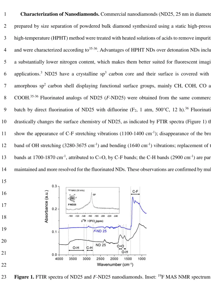
drastically changes the surface chemistry of ND25, as indicated by FTIR spectra (Figure 1) that 10
show the appearance of C-F stretching vibrations (1100-1400 cm-1); disappearance of the broad 11
band of OH stretching (3280-3675 cm-1) and bending (1640 cm-1) vibrations; replacement of the
12
bands at 1700-1870 cm-1, attributed to C=O, by C-F bands; the C-H bands (2900 cm-1) are partly 13
maintained and more resolved for the fluorinated NDs. These observations are confirmed by multi- 14 15 16 17 18 19 20 21 22
Figure 1. FTIR spectra of ND25 and F-ND25 nanodiamonds. Inset: 19F MAS NMR spectrum of 23
nuclear solid-state NMR. The 1H13C CP-MAS spectrum confirms the presence of COOH (160 1
ppm), COH (75 ppm), CH (45 ppm) groups, of sp3 C (35 ppm) and of a low content of sp2 C
2
(amorphous C shell on ND surface, 120 ppm) (SI, Figure S1a). In the 1H MAS spectrum recorded 3
after fluorination, only C-H band (at 1.5 ppm) is observed and no more C-OH (SI, Figure S1b). 4
The 19F MAS spectrum reveals the presence of covalent C-F bonds with a chemical shift of -170 5
ppm (inset in Figure 1). 6
The contact angles of a droplet of water deposited on the surface of pellets formed by the NDs 7
under pressure were measured. These contact angles are only “apparent”, because of the roughness 8
of the pellets. Nevertheless, the method provided evidence of significant differences in surface 9
chemistries between non-fluorinated and fluorinated nanodiamonds. The contact angle was 130° in 10
the case of F-ND25, reflecting the hydrophobic character of the F-ND25 pellet surface. By contrast, 11
the water droplets were readily absorbed into the pellets of ND25, preventing any measurement but 12
confirming the hydrophilic character of the ND25 surface. 13
Effect of Fluorocarbon Gases on Nanodiamond Disaggregation in Aqueous Solutions. We
14
studied the effect of exposing aqueous dispersions of ND25 and ND25 to hexane and F-15
pentane vapors. An essential requirement for the development of any biological application is 16
indeed to be able to obtain stable, minimally aggregated, reproducible aqueous ND dispersions, 17
which depends on various parameters, including the surface potential of the particles, pH, 18
temperature and concentration.42 Aqueous dispersions of ND25 and F-ND25 (0.05 g L-1) were 19
prepared in Milli-Q water at room temperature and pH was adjusted to 12.0 in order to allow 20
ionization of the carboxylic acid functions in the case of ND25, and provide similar ionic 21
environment in the case of F-ND25. The mean hydrodynamic radius rh of the ND clusters was
22
determined by dynamic light scattering (DLS) after filtration of the dispersions on 0.22 µm filters. 23
The zeta potential of the dispersions was measured under air and under F-hexane- (or F-pentane) 1
saturated air after preparation. We achieved a study to compare the effects of bath sonication versus 2
tip sonication, as an attempt to disaggregate the ND clusters in aqueous dispersions. We 3
systematically found larger clusters in the case of tip sonication. For example, under air, ND25 4
clusters with rh of 69 2 nm were obtained after 2 min of tip sonication. Increasing sonication
5
time only resulted in an increase of rh to 112 24 nm and 157 31 nm after 5 and 10 min of
6
sonication, respectively. After 5 days of storage at 25°C, the cluster sizes reached 220 nm. By 7
contrast, bath sonication led to smaller ND25 clusters (58 ± 4 nm) after preparation that grew up 8
to 151 ± 4 nm after 2-week storage at room temperature. Bath sonication was thus used in all the 9
experiments. The data are collected in Table 1 and Figure 2. For ND25, rh was 58 ± 4 nm when
10
measured under air immediately after preparation (i.e. the clusters contain 9 primary particles). 11
This value did not change significantly when subjecting the dispersions to ultrasound bath 12
treatment (25°C, 20 min). After 2 weeks of storage at room temperature, significant aggregation 13
occurred in the aqueous dispersions, leading to larger clusters with a mean radius of 151 ± 4 nm 14
(163 primary particles). By contrast, exposing the ND25 dispersions to F-hexane, or F-pentane, 15
vapors drastically changed their aggregation behavior. After ultrasound bath treatment, rh
16
decreased significantly from 62 ± 3 nm (11 primary particles) to 35 ± 1 nm (2 primary particles),
17
when exposed to F-hexane, and from 65 ± 2 nm (13 primary particles) to 29 ± 1 nm (1 primary 18
particles) when exposed to F-pentane (SI Figure S2). decreased significantly from -58.2 mV (air) 19
to -61.1 mV, and to -62.3 mV for F-hexane and F-pentane, respectively. After two-week storage, 20
the NDs exposed to the FC gases had maintained their size (37 and 32 nm, respectively). Zeta 21
potentials were found unchanged after one week at 25°C. These results indicate that exposure to 22
FCs promotes disaggregation of the ND25 clusters and prevents their re-aggregation of the NDs
1
over time. 2
Table 1. Mean hydrodynamic radius rh and zeta potential of dispersions of nanodiamonds 3
exposed to air, F-hexane or F-pentane. In parentheses, the number of primary particles contained 4
in the nanodiamond clusters. 5 6 ND Gas phase rh Prep. (nm) rh Sonic bath (nm) rh Storage* (nm) Prep. (± 1 mV) ND25 Air 58 4 (9) 57 2 (8) 151 4 (163) -58.2 F-hexane 62 3 (11) 35 1 (2) 37 1 (2) -61.1 F-pentane 65 2 (13) 29 1 (1) 32 2 (1) -62.3 F-ND25 Air 81 3 (25) 83 3 (26) 146 4 (147) -48.1 F-hexane 87 2 (31) 89 2 (32) 75 1 (19) -50.4 F-pentane 88 4 (32) 88 3 (32) 74 3 (19) -51.2 *Two weeks at room temperature.
7
Where F-ND25 NDs are concerned, rh was 81 ± 3 nm (25 primary particles) under air after
8
preparation. This larger size relative to that of ND25 reflects the higher hydrophobicity of the 9
fluorinated NDs at the surface of which the polar functions have been replaced by C-F bonds, and 10
their higher tendency for aggregation in aqueous media. The less negative zeta potential of the F-11
ND25 dispersions exposed to air (-48.8 mV vs. -58.2 mV for ND25) also reflects the substitution 12
of polar functions by fluorine atoms. When exposed to air for 2 weeks at room temperature, 13
pronounced aggregation was observed, leading to F-ND25 clusters reaching 146 ± 4 nm in size 14
(147 primary particles). By contrast, exposure to F-hexane or F-pentane led to significant 15
disaggregation of the F-ND25 clusters (~75 nm, ~19 primary particles). Disaggregation was 16
noticeable after 2 days at 25°C, which means that the process is slow for F-ND25. Importantly, 17
ND aggregation did not recommence over time when the FC gas was present. Exposure to 18
fluorocarbon gases slightly decreases the zeta potential of F-NDs (from -48.1 to -50.4 and -51.2 19
for F-hexane and F-pentane, respectively). These values did not change after one week at 25°C. 20
1 2 3 4 5 6 7 8 9
Figure 2. DLS profiles of ND25 (left panel) and F-ND25 (right panel) in aqueous dispersions
10
(0.05 g L-1) under air (a, b) and F-hexane-saturated air (c, d) showing the variation of intensity as 11
a function of the hydrodynamic radius after preparation (black), after ultrasound bath treatment 12
(red), and after two-week storage at room temperature (blue). In d), the difference between the 13
values measured after preparation (87 ± 2 nm) and after storage (75 ± 1 nm) is significant. Arrows 14
indicate the direction of peak radii variation. 15
The disaggregation effect of F-hexane on aqueous dispersions of NDs was also investigated by 16
cryogenic transmission electron microscopy (cryo-TEM). Figure 3 shows that significant 17
aggregation occurs in the aqueous dispersions of ND25 under air (3.a), while the aggregation is 18
limited under F-hexane (3.b). More isolated primary particles that look transparent, as well as 19
small aggregates can be observed (3.c-d), while only large aggregates are seen when the NDs are 20
exposed to air. 21
22 23
1 2 3 4 5 6 7 8 9 10 11 12
Figure 3. Cryo-TEM images of aqueous dispersions of ND25 (0.1 g L-1) exposed a) under air and 13
b) under F-hexane-saturated air (b-d). b-d) reveal the presence of relatively small isolated clusters 14
that cannot be seen under air. Scale bar:100 nm. 15
One hypothesis for the mechanism of disaggregation and for the FC-induced improved ND 16
dispersion stability is that, although the solubility of the FCs in water is extremely low (2.7 x 10⁻⁴
17
mol m-3 and 4.0 x 10⁻³ mol m-3 for F-hexane and F-pentane, respectively, at 25°C)41, it is not null. 18
It can therefore be envisioned that FC molecules present in the aqueous phase interact with the 19
clusters of NDs through hydrophobic interactions, which may break some of the interactions that 20
ensure ND cluster cohesion, and induce their progressive disaggregation (Scheme 2). The more 21
negative values measured when both ND25 and F-ND25 are exposed to the FC gases support 22
this view. Addition of surfactants was indeed shown to increase the colloidal stability of ND 23
aqueous dispersions,43 and the surface activity of F-hexane at interfaces has been established.39 24
1 2 3 4 5 6 7
Scheme 2. Schematic representation of the aggregation of ND clusters that occurs under air over
8
time (a), whilst disaggregation occurs under F-hexane (b) (green entities: F-hexane; not on scale). 9
The fact that the more water-soluble F-pentane is more efficient than F-hexane for ND25 cluster 10
disaggregation also supports this view. The stronger disaggregating effect of the FC gas on non-11
fluorinated ND25 was unexpected. It may be possible that disaggregating F-ND25 clusters, which 12
are larger than ND25 ones (they contain 25 primary particles versus 9), may require more 13
fluorocarbon molecules, which is limited by the low water solubility of F-hexane and F-pentane. 14
It may also be possible that F-NDs25 are more tightly packed. 15
Adsorption of Nanodiamonds at the Air/Water Interface. In order to shed light on the effect
16
of the FC on MB formation, we determined the kinetics of the F-hexane-driven adsorption of 17
ND25 and F-ND25 at the gas/water interface at 25°C using bubble profile analysis tensiometry. A 18
syringe filled with F-hexane-saturated air was fitted on the measuring cell of the tensiometer.39 19
The adsorption profiles of dispersions of ND25 and F-ND25 (0.1 g L-1) exposed – or not – to F-20
hexane gas are provided in Figure 5. The characteristic time of adsorption was determined by 21
fitting the adsorption profiles with an exponential decay function. Under air, ND25 adsorb only 22
slightly and slowly ( 10.3 min) at the interface. The equilibrium value of the interfacial tension 23
was reached after 30 min (eq 66.4 mN m-1). The weak adsorption of ND25 particles is explained
1
by their significant hydrophilic character, as measured by contact angle, due to the polar functions 2
present on the ND25 surface, and low affinity for the interface. Exposure of ND25 to F-hexane 3
results in a much faster ( 0.86 min) and more pronounced adsorption (eq 59.5 mN m-1, i.e. a
4
lowering = 6.9 mN m-1). It should be reminded that F-hexane alone induces a lowering of
5
the tension of water of only 4.5 mN m-1 (Figure 5). This indicates a synergistic effect between F-6
hexane and the NDs at the interface. By contrast with ND25, F-ND25 do not adsorb at all at the 7
interface under air, as assessed by a constant value of 71.5 mN m-1, which is probably due by
8
the large size of the clusters. By contrast, exposure to F-hexane leads to a pronounced adsorption, 9
causing a tension lowering by 15.8 mN m-1 with respect to the value measured in air. These
10
results suggest that fluorous interactions develop bet- 11 12 13 14 15 16 17
Figure 5. a) Schematic representation of the adsorption of nanodiamonds at the air/water interface
18
using bubble profile analysis tensiometry. b) Variation of the gas/water interfacial tension over 19
time for aqueous dispersions of ND25 (black) and F-ND25 (blue) under air and under F-hexane-20
exposure (0.1 g L-1, pH 12). Adsorption profile of F-hexane in the absence of nanodiamonds 21
(green). Concentration: 0.1 g L-1; pH 12.0; temperature: 25°C. 22
-ween F-hexane and the fluorinated NDs, enabling the recruitment of the latter to the interface, 1
and their stabilization at this interface. Although the interfacial behavior of spread films of 2
nanodiamonds functionalized by alkyl (and fluoroalkyl) chains has been described,44 our data are 3
the first report of spontaneously adsorbed films of nanodiamonds at the gas/water interface. 4
To sum-up, the disaggregating effect exerted by the fluorocarbon both facilitates the diffusion 5
of the nanodiamond particles to, and their organization at the interface, likely by filling the 6
interparticle gaps. For F-NDs, these effects are enhanced, likely by fluorine-fluorine hydrophobic 7
interactions that develop between the C-F groups and F-hexane, as was recently reported for 8
magnetic nanoparticles fitted with dendrons terminated with fluorinated groups.45 9
Microbubbles with Nanodiamond Shells. In order to exploit the observed dispersive effect of
10
FC gases on ND in aqueous dispersions, and influence of their adsorption at an air/water interface,
11
we investigated the possibility of preparing microbubbles that would be shelled by NDs only, that 12
is, without recourse to any conventional surfactant or foaming agent. Essentially all the 13
commercially available medical microbubbles use FC gases for stabilization.25-26 The latter acts as 14
an osmotic agent and as a co-surfactant to the bubble shell-forming material (usually 15
phospholipids).23, 46 Preparation of MBs was attempted by bubbling either N2 or
F-hexane-16
saturated N2 into aqueous dispersions of ND25 or F-ND25 (0.1 g L-1). The MBs were prepared
17
using a Vialmix® shaker in the presence, or absence, of F-hexane. Their size and stability 18
characteristics were measured by optical microscopy and by an acoustical attenuation method.40 19
We found that, for both ND25 and F-ND25, only the dispersions that were exposed to F-hexane 20
did produce MBs stable enough for characterization. All our attempts to produce MBs in the 21
absence of FC failed. The impossibility to generate MBs in the absence of F-hexane is strongly 22
related to the interfacial behavior of nanodiamonds, as observed in tensiometry. For generating 23
MBs, it is crucial that the shell components access and stabilize the interface rapidly.47 The 1
tensiometry experiments demonstrate that, under air, neither of the NDs are adsorbing at the 2
air/water interface in a significant manner. Under F-hexane exposure both types of nanodiamonds 3
are readily adsorbing at the interface, likely due to the fact that F-hexane fills in gaps left between 4
the nanodiamond particles, thus facilitating their organization in a compact film. 5
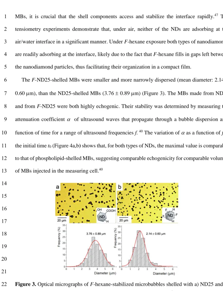
The F-ND25-shelled MBs were smaller and more narrowly dispersed (mean diameter: 2.14 6
0.60 m), than the ND25-shelled MBs (3.76 0.89 m) (Figure 3). The MBs made from ND25 7
and from F-ND25 were both highly echogenic. Their stability was determined by measuring the 8
attenuation coefficient of ultrasound waves that propagate through a bubble dispersion as a 9
function of time for a range of ultrasound frequencies f. 40 The variation of as a function of f at 10
the initial time t0 (Figure 4a,b) shows that, for both types of NDs, the maximal value is comparable
11
to that of phospholipid-shelled MBs, suggesting comparable echogenicity for comparable volumes 12
of MBs injected in the measuring cell.40 13 14 15 16 17 18 19 20 21
Figure 3. Optical micrographs of F-hexane-stabilized microbubbles shelled with a) ND25 and b)
22
F-ND25, with corresponding size distributions. Concentration: 0.1 g L-1; pH 12.0.
1 2 3 4 5 6 7 8 9
Figure 4. Variation of the attenuation coefficient as a function of ultrasound frequency f for
10
microbubbles prepared with dispersions of a) ND25 and b) F-ND25 under F-hexane. Size 11
distributions of MBs shelled with c) ND25 and d) F-ND25 as measured acoustically after 5 (black), 12
10 (red) and 15 (blue) min. 13
The dependence of the MB volume fraction with size is plotted in Figure 4c,d for different time 14
points. The distribution of ND25-shelled MBs is shifted toward larger radius values over time. By 15
contrast, the mean radius of the fluorinated F-ND25-shelled MBs remains constant over time for 16
at least 15 min. A hypothesis for the difference in the size distribution evolution is that F-ND25 17
are attracted to, and immobilized at the air/water interface through fluorine-fluorine interactions 18
with supernatant F-hexane molecules. This is supported by the increased adsorption of F-ND25 19
upon F-hexane exposure, as monitored by tensiometry. It is also in line with previous data showing 20
that iron oxide nanoparticles fitted with fluorinated group-terminated dendrons provided efficient 21
stabilization of F-hexane-saturated air MBs.45 The variation of the acoustically determined MB 22
volume fraction plotted as a function of time allowed determination of the MB half-lives, which 23
were ~15 min (SI Figure S3). 24
Altogether, these data show that ND-shelled MBs with small mean size can be generated owing 1
to the presence of the FC gas. These MBs present echogenic properties that qualify them as a 2
contrast agent for ultrasound diagnostics. More generally, the unique potential of NDs for sensing, 3
bioimaging and drug delivery can be combined to the portfolio of applications of MBs in medicine 4
and biology. 5
CONCLUSIONS AND PERSPECTIVES
6
In summary, exposure to a supernatant fluorocarbon gas strongly impacts, and in multiple ways, 7
the behavior of aqueous dispersions of two types of nanodiamonds with different surface 8
chemistry, nanodiamonds with a relatively polar surface, and more hydrophobic ones with a 9
fluorinated surface. First, the FC effectively disaggregates the clusters that form spontaneously for 10
both types of NDs. This helps surmount a critical, recurrent obstacle in nanodiamond 11
implementation. As compared to other techniques of ND disaggregation, our method does not 12
require dry (or wet) milling or prolonged tip sonication, processes that often produce contaminants 13
that need to be removed by additional purification step. Second, the introduction of a supernatant 14
FC gas strongly promotes the adsorption/recruitment of both types of NDs to the gas/water
15
interface. Third, the fluorinated NDs experience increased adsorption relative to the non-16
fluorinated ones, revealing hydrophobic interactions with the FC. Fourth, ND-shelled 17
microbubbles can be generated only when the FC is present, and this for both types of NDs, the 18
fluorinated NDs leading to smaller MBs. Generation of microbubbles shelled with nanodiamonds 19
is enabled by the FC gas that allows disaggregation of ND clusters in aqueous solution, and thus 20
facilitates their diffusion to, and organization at the interface. Shorter, more water-soluble FCs 21
deserve investigation in order to enhance the disaggregating effect vis-à-vis other types of NDs 22
(e.g. detonation NDs). Our data support a novel means of controlling nanodiamond aggregation, 23
as well as a novel approach to stabilize nanodiamond-shelled microbubbles, allowing investigation 1
of their potential as agents for bimodal contrast for ultrasound/fluorescence imaging, and drug 2
delivery. We believe that these findings open new perspectives and trigger new research, both 3
fundamental and mechanistic, and promote new applications in bioimaging and cell tracking. 4
ASSOCIATED CONTENT
5
Supporting Information. multinuclear solid-state NMR spectra, disaggregation induced by
6
perfluoropentane as monitored by DLS, and nanodiamond-shelled microbubble half-life (PDF). 7
AUTHOR INFORMATION 8
Corresponding Author* E-mail: krafft@unistra.fr,ORCID: ID: orcid.org/0000-0002-3379-2783 9
Author Contributions. The manuscript was written through contributions of all authors. All authors 10
have given approval to the final version of the manuscript. 11
Funding Sources. Ph.D grant for E.E.M.O. from CONACYT (grant #459199). 12
The authors declare no competing financial interest. 13
ACKNOWLEDGMENTS 14
We are grateful to Dr. F. Schosseler (ICS, Strasbourg) for help in DLS experiments. We also 15
acknowledge CONACYT (Mexico) for a Ph.D fellowship (E.E.M.O., grant #459199). We thank 16
Dr. M. Schmutz (electron microscopy platform, Institut Charles Sadron ICS) for help in electron 17
microscopy, and M. Legros (ICS characterization platform) for support with DLS experiments. 18
REFERENCES
19
(1) Krueger, A. Diamond nanoparticles: Jewels for chemistry and physics. Adv. Mater. 2008, 20, 20
2445–2449. 21
(2) Mochalin, V. N.; Shenderova, O.; Ho, D.; Gogotsi, Y. The properties and applications of 1
nanodiamonds. Nat. Nanotechnol. 2011 7,11-23. 2
(3) Nunn, N.; Torelli, M.; McGuire, G.; Shenderova, O. Nanodiamond: a high impact 3
nanomaterial. Curr. Opin. Solid State Mater. Sci. 2017, 21, 1-9. 4
(4) Reina, G.; Zhao, L.; Bianco, A.; Komatsu, N. Chemical functionalization of nanodiamonds: 5
opportunities and challenges ahead. Angew. Chem. Int. Ed. 2019, 58, 17918-17929. 6
(5) McGuinness, L. P.; Yan, Y.; Stacey, A.; Simpson, D. A.; Hall, L. T.; Maclaurin, D.; Prawer, 7
S.; Mulvaney, P.; Wrachtrup, J.; Caruso, F.; Scholten, R. E.; Hollenberg, L. C. L. Quantum 8
measurement and orientation tracking of fluorescent nanodiamonds inside living cells. Nat. 9
Nanotechnol. 2011, 6, 358-363.
10
(6) Zhu, Y.; Li, J.; Li, W.; Zhang, Y.; Yang, X.; Chen, N.; Sun, Y.; Zhao, Y.; Fan, C.; Huang, Q. 11
The biocompatibility of nanodiamonds and their application in drug delivery systems. 12
Theranostics 2012, 2, 302.
13
(7) Happel, P.; Waag, T.; Schimke, M.; Schweeberg, S.; Muzha, A.; Fortak, K.; Heesch, D.; Klask, 14
L.; Pilscheur, M.; Hoppe, F.; Lenders, T.; Meijer, J.; Lepperdinger, G.; Krueger, A. Intrinsically 15
32P‐labeled diamond nanoparticles for in vivo imaging and quantification of their biodistribution 16
in chicken embryos. Adv. Funct. Mater. 2018, 28, 1802873. 17
(8) Montalti, M.; Cantelli, A.; Battistelli, G. Nanodiamonds and silicon quantum dots: ultrastable 18
and biocompatible luminescent nanoprobes for long-term bioimaging. Chem. Soc. Rev. 2015, 44, 19
4853-921. 20
(9) Shenderova, O.; Nunn, N. Production and purification of nanodiamonds. In Nanodiamonds: 21
Advanced Materials, Analysis, Properties and Applications; Arnault, J. C., Ed.; Elsevier:
22
Amsterdam, 2017; Chapter 2, pp 25-56. 23
(10) Krüger, A.; Kataoka, F.; Ozawa, M.; Fujino, T.; Suzuki, Y.; Aleksenskii, A. E.; Vul, A. Y.; 1
Osawa, E. Unusually tight aggregation in detonation nanodiamond: identification and 2
disintegration. Carbon 2005, 43, 1722-1730. 3
(11) Pentecost, A.; Gour, S.; Mochalin, V.; Knoke, I.; Gogotsi, Y. Deaggregation of nanodiamond 4
powders using salt- and sugar-assisted milling. ACS Appl. Mater. Interfaces 2010, 2, 3289–3294. 5
(12) Liang, Y.; Ozawa, M.; Krueger, A. A general procedure to functionalize agglomerating 6
nanoparticles demonstrated on nanodiamond. ACS Nano 2009, 3, 2288–2296. 7
(13) Ozawa, M.; Inaguma, M.; M.Takahashi; Kataoka, F.; Krüger, A.; Osawa, E. Preparation and 8
behavior of brownish, clear nanodiamond colloids. Adv. Mater. 2007, 19, 1201–1206. 9
(14) Pedroso-Santana, S.; Sarabia-Saínz, A.; Fleitas-Salazar, N.; Santacruz-Gómez, K.; Acosta-10
Elías, M.; Pedroza-Montero, M.; Riera, R. Deagglomeration and characterization of detonation 11
nanodiamonds for biomedical applications. J. App. Biomed. 2017, 15, 15-21. 12
(15) Turcheniuk, K.; Trecazzi, C.; Deeleepojananan, C.; Mochalin, V. N. Salt-assisted ultrasonic 13
deaggregation of nanodiamond. ACS Appl. Mater. Interfaces 2016, 8, 25461−25468. 14
(16) Betts, J. N.; Johnson, M. G.; Rygiewics, P. T.; King, G. A.; Andersen, C. P. Potential for 15
metal contamination by direct sonication of nanoparticle suspension. Environ. Toxicol. Chem. 16
2013, 32, 889–893.
17
(17) Merz, V.; Lenhart, J.; Vonhausen, Y.; Ortiz-Soto, M. E.; Seibel, J.; Krueger, A. Zwitterion-18
functionalized detonation nanodiamond with superior protein repulsion and colloidal stability in 19
physiological media. Small 2019, 15, 1901551. 20
(18) Tiainen, T.; Myllymäki, T. T. T.; Hatanpää, T.; Tenhu, H.; Hietala, S. Polyelectrolyte 21
stabilized nanodiamond dispersions. Diam. Relat. Mater. 2019, 95, 185-194. 22
(19) Wang, T.; Handschuh-Wang, S.; Qin, P.; Yang, Y.; Zhou, X.; Tang, Y. Enhancing the 1
colloidal stability of detonation synthesized diamond particles in aqueous solutions by adsorbing 2
organic mono-, bi- and tridentate molecules. J. Colloid Interface Sci. 2017, 499, 102-109. 3
(20) Sotoma, S.; Hsieh, F. J.; Chen, Y.-W.; Tsai, P.-C.; Chang, H.-C. Highly stable lipid-4
encapsulation of fluorescent nanodiamonds for bioimaging applications. Chem. Commun. 2018, 5
54, 1000-1003.
6
(21) Bumb, A.; Sarkar, S. K.; Billington, N.; Brechbiel, M. W.; Neuman, K. C. Silica encapsulation 7
of fluorescent nanodiamonds for colloidal stability and facile surface functionalization. J. Am. 8
Chem. Soc. 2017, 135, 7815−7818.
9
(22) Jung, H.-S.; Cho, K.-J.; Seol, Y.; Takagi, Y.; Dittmore, A.; Roche, P. A.; Neuman, K. C. 10
Polydopamine encapsulation of fluorescent nanodiamonds for biomedical applications. Adv. 11
Funct. Mater. 2018, 28, 1801252.
12
(23) Schutt, E. S.; Klein, D. H.; Mattrey, R. M.; Riess, J. G. Injectable microbubbles as contrast 13
agents for diagnostic ultrasound imaging: The key role of perfluorochemicals. Angew. Chem. Int. 14
Ed. 2003, 42, 3218-3235.
15
(24) Kooiman, K.; Vos, H. J.; Versluis, M.; de Jong, N. Acoustic behavior of microbubbles and 16
implications for drug delivery. Adv. Drug Deliv. Rev. 2014, 72, 28-48. 17
(25) Wang, S.; Hossack, J.; Klibanov, A. L. Targeting of microbubbles: contrast agents for 18
ultrasound molecular imaging. J. Drug Target. 2018, 26, 420-434. 19
(26) Chong, W. K.; Papadopoulou, V.; Dayton, P. A. Imaging with ultrasound contrast agents: 20
current status and future. Abdom. Radiol. 2018, 43, 762–772. 21
(27) Stride, E.; Segers, T.; Lajoinie, G.; Cherkaoui, S.; Bettinger, T.; Versluis, M.; Borden, M. 22
Microbubble agents: New directions. Ultrasound Med. Biol. 2020, 46, 1326-1343. 23
(28) Kooiman, K.; Roovers, S.; Langeveld, S. A. G.; Kleven, R. T.; Dewitte, H.; O’Reilly, M. A.; 1
Escoffre, J.-M.; Bouakaz, A.; Verweij, M. D.; Hynynen, K.; Lentacker, I.; Stride, E.; Holland, C. 2
K. Ultrasound-responsive cavitation nuclei for therapy and drug delivery. Ultrasound Med. Biol. 3
2020, 46, 1296-1325.
4
(29) Fu, L.; Ke, H.-T. Nanomaterials incorporated ultrasound contrast agents for cancer 5
theranostics. Cancer Biol. Med. 2016, 13, 313-324. 6
(30) Jamburidze, A.; Huerre, A.; Baresch, D.; Poulichet, V.; Corato, M. d.; Garbin, V. 7
Nanoparticle-coated microbubbles for combined ultrasound imaging and drug delivery. Langmuir 8
2019, 35, 10087−10096.
9
(31) Yang, G.; O'Duill, M.; Gouverneur, V.; Krafft, M. P. Recruitment and immobilization of a 10
fluorinated biomarker across an interfacial phospholipid film using a fluorocarbon gas. Angew. 11
Chem. Int. Ed. 2015, 54, 8402-8406.
12
(32) Gazzera, L.; Milani, R.; Pirrie, L.; Schmutz, M.; Blanck, C.; Resnati, G.; Metrangolo, P.; 13
Krafft, M. P. Design of highly stable echogenic microbubbles through controlled assembly of their 14
hydrophobin shell. Angew. Chem. Int. Ed. 2016, 55, 10263-10267. 15
(33) Ando, Y.; H. Tabata; Sanchez, M.; Cagna, A.; Koyama, D.; Krafft, M. P. Microbubbles with 16
a self-assembled poloxamer shell and a fluorocarbon inner gas. Langmuir 2016, 32, 12461-12467. 17
(34) Garbin, V.; Crocker, J. C.; Stebe, K. J. Nanoparticles at fluid interfaces: Exploiting capping 18
ligands to control adsorption, stability and dynamics. J. Colloid Interface Sci. 2012, 387, 1-11. 19
(35) Dubois, M.; Guérin, K.; Batisse, N.; Petit, E.; Hamwi, A.; Komatsu, N.; Kharbache, H.; 20
Pirotte, P.; Masin, F. Solid state NMR study of nanodiamond surface chemistry. Solid State Nucl. 21
Magn. Reson. 2011, 40, 144-154.
(36) Zagrebina, E. M.; Generalov, A. V.; Klyushin, A. Y.; Simonov, K. A.; Vinogradov, N. A.; 1
Dubois, M.; Frezet, L.; Mårtensson, N.; Preobrajenski, A. B.; Vinogradov, A. S. Comparative 2
NEXAFS, NMR, and FTIR study of various-sized nanodiamonds: As-prepared and fluorinated. J. 3
Phys. Chem. C 2015, 119, 835−844.
4
(37) Szijjarto, C.; Rossi, S.; Waton, G.; Krafft, M. P. Effects of perfluorocarbon gases on the size 5
and stability characteristics of phospholipid-coated microbubbles - Osmotic effect versus 6
interfacial film stabilization. Langmuir 2012, 28, 1182-1189. 7
(38) Hassan, P. A.; Rana, S.; Verma, G. Making sense of Brownian motion: Colloid 8
characterization by dynamic light scattering. Langmuir 2014, 31, 3−12. 9
(39) Nguyen, P. N.; Veschgini, M.; Tanaka, M.; Waton, G.; Vandamme, T.; Krafft, M. P. 10
Counteracting the inhibitory effect of proteins towards lung surfactant substitutes: a fluorocarbon 11
gas helps displace albumin at the air/water interface. Chem. Commun. 2014, 50, 11576-11579. 12
(40) Rossi, S.; Waton, G.; Krafft, M. P. Phospholipid-coated gas bubble engineering - Key 13
parameters for size and stability control as determined by an acoustic method. Langmuir 2010, 26, 14
1649-1655. 15
(41) Kabalnov, A.; Klein, D.; Pelura, T.; Schutt, E.; Weers, J. Dissolution of multicomponent 16
microbubbles in the blood stream: 1. Theory. Ultrasound Med. Biol. 1998, 24, 739-749. 17
(42) Gibson, N.; Shenderova, O.; Luo, T. J. M.; Moseenkov, S.; Bondar, V.; Puzyr, A.; Purtov, K.; 18
Fitzgerald, Z.; Brenner, D. W. Colloidal stability of modified nanodiamond particles. Diam. Relat. 19
Mater. 2009, 18, 620–626.
20
(43) Xu, X.; Yu, Z.; Zhu, Y.; Wang, B. Effect of sodium oleate adsorption on the colloidal stability 21
and zeta potential of detonation synthesized diamond particles in aqueous solutions. Diamond 22
Relat. Mater. 2005, 14, 206-212.
(44) Machida, H.; Ohashi, T.; Akasaka, S.; Fujimori, A. Formation of organized films with 1
fluorocarbon-modified inorganic nanoparticles and their nanodispersion behavior in solvent. J. 2
Fluorine Chem. 2020, 230, 109433.
3
(45) Shi, D.; Wallyn, J.; Nguyen, D.-V.; Perton, F.; Felder-Flesch, D.; Bégin-Colin, S.; Maaloum, 4
M.; Krafft, M. P. Microbubbles decorated with dendronized magnetic nanoparticles for biomedical 5
imaging. Effective stabilization via fluorous interactions. Beilstein J. Nanotechnol. 2019, 10, 2103-6
2115. 7
(46) Shi, D.; Liu, X.; Counil, C.; Krafft, M. P. Fluorocarbon exposure mode markedly affects 8
phospholipid monolayer behavior at the gas/liquid interface: Impact on size and stability of 9
microbubbles. Langmuir 2019, 35, 10025-10033.. 10
(47) Kwan, J. J.; Borden, M. A. Lipid monolayer collapse and microbubble stability. Adv. Coll. 11
Interface Sci. 2012, 183-184, 82-99.
12 13


![[PDF] Support de formation Perfectionnement Word 2007 | Cours informatique](data:image/gif;base64,R0lGODlhAQABAIAAAP///wAAACH5BAEAAAAALAAAAAABAAEAAAICRAEAOw==)