Plant, Cell and Environment (2004) 27, 219–228
© 2004 Blackwell Publishing Ltd 219
Blackwell Science, LtdOxford, UKPCEPlant, Cell and Environment0016-8025Blackwell Science Ltd 20032004 272219228
Original Article
Aquaporins and hydraulic conductance of agave roots G. B. North
et al.
Correspondence: Gretchen B. North. Fax: 323-341-4974; e-mail: gnorth@oxy.edu
Aquaporins account for variations in hydraulic conductance
for metabolically active root regions of
Agave deserti
in wet,
dry, and rewetted soil
G. B. NORTH1
, P. MARTRE2,3
& P. S. NOBEL2
1Department of Biology, Occidental College, Los Angeles, California 90041, USA, 2Department of Organismic Biology,
Ecology, and Evolution, University of California, Los Angeles, California 90095–1606, USA and 3Unite d’Agronomie, INRA
– Site de Crouel, 234 Avenue du Brezet F-63039 Clermont-Ferrand, Cedex 2, France
ABSTRACT
The importance of aquaporins for root hydraulic conduc-tance (LP) was investigated along roots of the desert
succu-lent Agave deserti in wet, dry and rewetted soil. Water channel activity was inferred from HgCl2-induced
reduc-tions of LP that were reversible by 2-mercaptoethanol.
Under wet conditions, HgCl2 reduced LP for the distal root
region by 50% and for the root region near the shoot base by 36% but did not affect LP for the mid-root region. For
all root regions, LP decreased by 30–60% during 10 d in
drying soil and was not further reduced by HgCl2. After soil
rewetting, LP increased to pre-drying values and was again
reduced by HgCl2 for the distal and the basal root regions
but not the mid-root region. For the distal region, water channels in the epidermis/exodermis made a disproportion-ately large contribution to radial hydraulic conductance of the intact segment; for the basal region, water channel activity was highest in the cortex and endodermis. The role of water channels was greatest in tissues in which cells were metabolically active both in the distal root region, where new apical growth occurs in wet soil, and in the basal region, which is the most likely root region to intercept light rainfall.
Key-words: endodermis; exodermis; hydraulic conductivity;
mercury sensitivity; root water uptake; water channels; water transport.
Abbreviations: JV, volume flux density (m s-1); Kh, axial
hydraulic conductivity of the xylem (m4 s-1 MPa-1); L
P, root
hydraulic conductance (all conductances based on the
outer surface area of intact root segments; m s-1 MPa-1);
LR, radial hydraulic conductance; LR,EpEx, radial hydraulic
conductance of the epidermis plus exodermis; LR,-EpEx,
radial hydraulic conductance of root segments with the
epi-dermis and exoepi-dermis removed; LR,CoEn, radial hydraulic
conductance of the cortex plus endodermis; LR,S, radial
hydraulic conductance of the stele; QV, volumetric flow of
water through a root segment (m3 s-1).
INTRODUCTION
The relative permeability of roots to water, known as the
root hydraulic conductance (LP, also called the root
hydraulic conductivity), tends to vary directly with water availability in the soil (Huang & Nobel 1994). For a number of species, including many desert succulents and rainforest
cacti, decreases in LP due to soil drying can be quickly
reversed when the soil is rewetted (e.g. North & Nobel
1991, 1992, 1994). Drying-induced changes in LP that are
reversed upon rewetting and that are not accompanied by changes in root morphology or anatomy occur for a number
of species, including the desert cactus Opuntia
acantho-carpa (Martre, North & Nobel 2001) and the more
meso-phytic Mediterranean species Olea oleaster (Lo Gullo et al.
1998). Such flexible, short-term regulation of root water uptake appears to involve aquaporins, a family of trans-membrane proteins that occur in the plasma trans-membrane, tonoplast and other intracellular membranes and that are abundantly expressed in roots (Javot & Maurel 2002; Tyer-man, Niemitz & Bramley 2002).
Water channel activity can vary with time of day
(Hen-zler et al. 1999; Tsuda & Tyree 2000), with root
develop-ment (Barrowclough, Peterson & Steudle 2000; North &
Nobel 2000; Martre et al. 2001; Hukin et al. 2002), and in
response to various stresses such as salinity (Carvajal, Mar-tinez & Alcaraz 1999; MarMar-tinez-Ballesta, MarMar-tinez & Car-vajal 2000), low nutrients (CarCar-vajal, Cooke & Clarkson
1996; Clarkson et al. 2000), and drought (Martre et al. 2001,
2002; Siefritz et al. 2002). This variability is not surprising,
given the diversity of aquaporins and the variety of their locations within cells and tissues. As the plasma membrane is generally much less permeable to water than is the tono-plast (Javot & Maurel 2002), the aquaporins that are more important for regulating root water uptake are likely to be proteins integral to the plasma membrane (PIPs). The expression of a variety of PIPs is generally reduced in roots
during drought or osmotic stress (Smart et al. 2001;
220 G. B. North et al.
although some studies indicate no change (Morillon &
Las-salles 2002) or an increase in PIP expression (Kirch et al.
2000). Water channel activity can also be regulated by post-translational mechanisms, such as phosphorylation, as shown for aquaporins in the plasma membrane of leaf
mes-ophyll cells for Spinacia oleracea (Johansson, Larsson &
Kjellbom 1996, Johansson et al. 1998). Whether water
chan-nel opening is inhibited or stimulated by water stress may depend on the adaptive value of decreasing or increasing root permeability to water. For desert plants, the closure of water channels during drought would help prevent root water loss to a soil that generally has a lower water poten-tial than does the plant.
The desert plant examined in the present study, Agave
deserti, is a monocotyledonous, stemless rosette with
essen-tially unbranched roots that are chiefly adventitious and nearly constant in diameter along their length. The roots arise from the base of the succulent shoot, which maintains the basal root region in a relatively well-hydrated condition even during prolonged drought. Even though the basal root region is the oldest root region, its anatomy is similar to that of the much younger, actively growing distal root
region (North & Nobel 1998). LP for the basal root region
is also relatively high even after soil drying, which may be critical in the field, where light rainfall often rewets only the uppermost few centimetres of soil. Rainfall is also likely
to be channelled toward the basal root region by A. deserti’s
concave, upward-angled leaves. The ability of the roots of
desert plants in general and of A. deserti in particular to
respond to such temporal and spatial heterogeneity in soil moisture may depend on the possible closing of water chan-nels during drying and their reopening upon rewetting.
For young roots of A. deserti, comparable with the distal
root segments examined in the present study, LP is reduced
by treatment with HgCl2, a known inhibitor of water
chan-nel activity (Javot & Maurel 2002), under wet conditions but not after 45 d of drought (North & Nobel 2000). During
a shorter period of drying, such as the 10 d imposed on A.
deserti in the present study, few anatomical changes in roots
are expected, allowing a sharper focus on the role of
aqua-porins in regulating LP. Specifically, three questions with
respect to possible regulation of LP in roots of A. deserti
are addressed: (1) how do water channels respond to short-term soil drying and rewetting; (2) are water channels active along the entire length of a root, specifically, in the distal region (including the tip), at mid-root and at the base of the shoot; and (3) in what root tissues or cell layers (epi-dermis/exodermis, cortex/endodermis and stele) is water channel activity most important to hydraulic conductance? To assess the presence of aquaporins as well as their
importance in root water transport, LP was measured for
root segments first in water and then in HgCl2. The use of
HgCl2 to close water channels raises a number of concerns,
such as the possibility that reduction in water transport may be due to membrane damage or metabolic poisoning (Zhang & Tyerman 1999). However, preliminary
experi-ments confirmed that an inhibitory effect on LP for roots of
A. deserti could be achieved with a low concentration of
HgCl2 (25 mM) and that such inhibition was reversible by
10 mM 2-mercaptoethanol. A further concern with this
technique is that the relative impermeability of outer root
tissues may preclude entry of HgCl2 (Barrowclough et al.
2000). For a number of species, including A. deserti, the
exodermis in older root regions tends to be more heavily suberized than in younger regions, thus possibly acting as a
barrier to substances such as Hg2+ (Barrowclough et al.
2000). Removal of this layer allowed HgCl2 to penetrate,
and hence the mercury-sensitivity of water channels in internal root tissues could be determined. In addition,
sequential tissue removal followed by measurements of LP
allowed the effect of HgCl2, and, by extension, the
impor-tance of aquaporins in water transport, to be assessed radi-ally tissue by tissue from the epidermis/exodermis to the stele at three locations along a root.
MATERIALS AND METHODS
Plant material
Four-year-old-plants of Agave deserti Engelm.
(Aga-vaceae), approximately 18 cm tall and 25 cm in diameter with an average of eight unfolded leaves, were obtained from a commercial nursery. They were grown in a green-house at the University of California, Los Angeles in plastic
trays 52 cm long ¥ 26 cm wide ¥ 6 cm deep that were filled
with vermiculite. The plants received a mean total daily
photosynthetic photon flux of 38 mol m-2 d-1 (80% of
ambi-ent solar radiation), with daily maximum/minimum air
tem-peratures averaging 28/16 ∞C and daily minimum/maximum
relative humidities averaging 40/70%. The soil water
poten-tial (Ysoil; MPa) in the rooting zone was maintained above
-0.1 MPa by watering every other day with 0.1-strength
Hoagland’s solution no. 2 supplemented with micronutri-ents. Twenty-four plants were maintained under wet condi-tions for at least 45 d in the greenhouse before water was withheld to initiate soil drying for half of the plants.
The water content of the vermiculite was determined by weighing 7–10 g of soil before and after drying for 48 h in
a forced-draught oven at 105 ∞C, and the vermiculite water
potential in the rooting zone was calculated using a mois-ture-release curve for vermiculite (Dubrovsky, North &
Nobel 1998). Under wet conditions, Ysoil was >-0.2 MPa,
and at 10 d of drying it was -1.6 MPa. At 10 d of soil drying,
six plants were watered so that Ysoil was increased to
-0.1 MPa and then maintained at that value by watering every other day. After 45 d in wet soil, main roots arising from the stem were 300–350 mm long and averaged 3.6 mm in diameter. Three regions of main roots were examined: (1) distal (the youngest region), from the tip to 80 mm back; (2) mid-root, from 130 to 210 mm back from the tip; and (3) basal (the oldest region), 10–90 mm from the base of the succulent shoot.
Root hydraulic properties
Root hydraulic conductance (LP, m s-1 MPa-1) was
Aquaporins and hydraulic conductance of agave roots 221
about 75 mm in final exposed length (Nobel, Schulte & North 1990). Root segments about 150 mm long were gently excavated and trimmed by 50 mm from the proxi-mal end (distal end for basal root segments) under dis-tilled water with a razor blade. All tissues external to the stele were removed from a 15-mm length at the proximal end of the segment, and the exposed stele was then trimmed 5 mm under water and inserted into a 10-mm-length of Tygon tubing affixed to a glass capillary (inside diameter, 1.6 mm) that was half-filled with distilled water. A watertight seal between the stele and the tubing was made by inserting the tubing through a silicone gasket in a brass compression fitting. The junction between the tub-ing and the stele as well as the distal cut end of the mid-root and basal segments (about 5 mm) were sealed with hydrophilic vinyl polysiloxane (Reprosil; Dentsply Inter-national, Milford, DE, USA) and coated with clear nail polish. The root segment was then suspended in 200 mL of distilled water.
Water flow through the root segment was induced by applying a partial vacuum to the open end of the capillary
at pressures of -40, -30, -20 and -10 kPa, regulated with a
needle valve and monitored with a digital manometer (PS309; Validyne Engineering, Northridge, CA, USA). The
flow rate (QV, m3 s-1) was recorded at each pressure after
the rate stabilized (in less than 10 min). LP was calculated
as the slope of the relationship between the volumetric flux
density (JV = QV per unit root surface area, m3 m-2 s-1, or
m s-1) and the vacuum pressure. The root surface area was
calculated from root length and mean diameter.
Before measuring axial (xylem) hydraulic conductivity
(Kh,(m3 s-1)/(MPa m-1), or m4 s-1 MPa-1), root segments
were first trimmed under water at about 10 mm from the distal seal. About 1 mm at the cut end of the segment was
then immersed in distilled water. QV was used to calculate
Kh:
Kh = QV/DP/Dx) (1)
where the pressure difference DP (10 kPa) was applied along the length Dx (m) of the root segment. Measurements
of QV were made during the first 10 s after immersion of
the segment to avoid blockage of vessels by debris.
The radial hydraulic conductance (LR, m s-1 MPa-1,
cal-culated based on the outer root surface area) along a root
equals JV at the root surface divided by the difference in
water potential from the root surface to the root xylem. LR
averaged over the entire root segment was calculated by
incorporating measured values of LP and Kh together with
the length (l, m) and the radius (rroot, m) of the root segment
into a model of Landsberg & Fowkes (1978) based on leaky cable theory:
LR = LPa/tanh(al) (2)
where a (m-1) equals (2pr
rootLR/Kh)1/2 which represents the
length along the root xylem across which the pressure
decreases by half (Landsberg & Fowkes 1978). LR was
ini-tially set equal to LP and gradually increased to solve Eqn
2 by iteration.
Radial hydraulic conductance of concentric root
tissues
After LP was measured for an intact root segment, the
epidermis and the exodermis were removed using fine
for-ceps under a stereomicroscope, and LP was then measured
on the stripped segment (North & Nobel 1995; Martre et al.
2001). Measured values of LP and mean values of Kh for
the appropriate root region were then used to calculate LR
for the stripped segment (LR,-EpEx) using Eqn 2 and the
surface area of the root segment. For another set of seg-ments, a similar procedure was followed except that all cell layers outside the stele were removed (epidermis, exoder-mis, cortex and endodermis). Radial conductances are in series and are based on the outer surface area of the intact
root segment in all cases (note that the same QV occurs
across each of the concentric tissue layers). Thus, the
recip-rocal of LR for an intact root segment equals the sum of the
reciprocals of LR for the epidermis plus exodermis (LR,EpEx),
the cortex plus endodermis (LR,CoEn), and the stele (LR, S), so
LR,EpEx = 1/(1/LR - 1/LR,-EpEx) (3)
For the cortex plus endodermis,
LR,CoEn = 1/(1/LR - 1/LR,EpEx - 1/LR, S) (4)
Assessment of water channels
After measurement of LP in water, the root segments were
transferred to 25 mM HgCl2 for 10 min at a pressure of
-30 kPa. The pressure was then decreased to -40 kPa and LP
was measured as before. The root segments were then
briefly rinsed in water, immersed in 10 mM
2-mercaptoeth-anol under the same conditions (10 min, -30 kPa), and LP
was measured again. To check for artifacts due to repeated
measurements of LP on the same root segment, comparable
segments were repeatedly measured in water with the same
protocol; LP varied less than 3% among three such
sequen-tial measurements on four representative root segments.
Anatomy
To investigate anatomical features, root segments were sec-tioned with a razor blade and stained with 0.05% (w/w) toluidine blue O in water for 30 s. Sections were mounted in water and then examined with an Olympus BH2 micro-scope (Lake Success, NY, USA) at magnifications of 100– 1000¥. Sections were examined for lignin by staining with 0.5% (w/w) phloroglucinol in water followed by 20% HCl (Jensen 1962). The presence of suberin in cell walls was investigated by staining with 0.1% (w/w) Sudan red 7B in 70% ethanol; suberin lamellae appeared red, and Caspar-ian bands were not stained. Suberin and lignin were also located in untreated sections by their autofluorescence under violet and ultraviolet light.
Statistics
Statistical analysis was done using SigmaStat 4.0 (SPSS Inc.,
treat-222 G. B. North et al.
ments were analysed using one-way ANOVA (a = 0.05)
fol-lowed by a Tukey’s test, after verifying that the treatment effects were normally distributed with equal variance.
Dif-ferences in LP due to HgCl2 treatment were analysed using
paired t-tests or Student’s t-test. Data are presented as means ±1 SE (n = number of measurements).
RESULTS
Root hydraulic conductance and HgCl
2sensitivity of intact (non-dissected)
root segments
The volume flux density (JV) of the distal root region of
Agave deserti for intact segments immersed in water, in
25 mM HgCl2, or in 10 mM 2-mercaptoethanol (ME)
follow-ing the HgCl2 treatment had a linear relationship with
applied pressure differences (Fig. 1). Linear relationships
between JV and pressure were also obtained for mid-root
and basal root regions (r2
= 0.978 ± 0.005). Although the
responses to HgCl2 differed, JV for root segments in 10 mM
ME ranged from 78 to 100% of JV in water for all root
regions, averaging 83 ± 9%. The slope of these relationships
was used to calculate the root hydraulic conductance (LP).
For the distal region under wet conditions, LP for root
segments in HgCl2 was 50% lower than LP for root
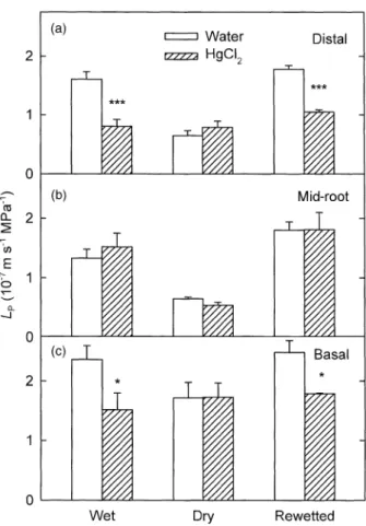
seg-ments in water (P < 0.001; Fig. 2a). After 10 d of soil drying
(Ysoil = -1.6 MPa), LP for the distal region was 59% lower
than under wet conditions and did not decrease further in
HgCl2. After 3 d of rewetting (Ysoil = -0.1 MPa), LP for the
distal region was restored to its value under wet conditions
and was again reduced by HgCl2 (P < 0.001).
For the mid-root region, changes in LP measured in water
in response to soil drying and rewetting were similar to the changes in the distal region (Fig. 2b). However, unlike the
case for the distal region, HgCl2 did not affect LP under any
of the soil moisture conditions.
Under wet, dry and rewetted conditions, LP for the basal
root region was higher than for distal and mid-root regions (P < 0.05; Fig. 2c). Similar to the case for the distal region,
LP measured in HgCl2 for the basal region was lower than
in water under wet soil conditions, although the reduction
was only 36%. After 10 d of soil drying, LP for the basal
region was 27% lower than under wet conditions (P < 0.05)
and did not decrease further in HgCl2. After 3 d of
rewet-ting, LP was restored to its value under wet conditions and
was reduced 28% in response to treatment with HgCl2.
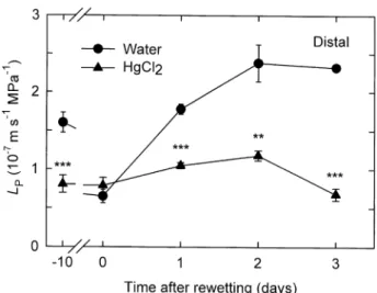
Changes in LP and root growth for the distal root region
in response to rewetting after soil drying were examined
daily for 3 d. At 1 d of rewetting, LP was 2.7 times higher
than its value under dry conditions (day 0, Fig. 3) and was
41% lower when measured in HgCl2 (P < 0.001). No new
apical growth (as detected by root color) was apparent at
1 d. At 2 d of rewetting, LP was 3.7 times higher than its
value under dry conditions, and the reduction due to HgCl2
was 50% (P < 0.01). New apical growth at 2 d averaged
3.8 ± 0.2 mm (n = 4 roots). No further increase in LP
mea-sured in water occurred at 3 d of rewetting, although the
Figure 1. Relationship between JV and the applied pressure
dif-ference for a distal root segment of Agave deserti under wet soil (vermiculite) conditions. JV was measured sequentially in water,
HgCl2 (25 mM) and 2-mercaptoethanol (10 mM).
Figure 2. LP for intact segments from the distal (a), mid-root (b)
and basal (c) regions of roots of A. deserti measured in water (open bars) and in 25 mM HgCl2 (hatched bars) in wet, dry and rewetted
soil (vermiculite). Soil water potential was -0.1 MPa under wet conditions, -1.6 MPa at 10 d of soil drying and -0.1 MPa during 3 d of rewetting. Data are means +1 SE (n = 5 roots from different plants). Asterisks indicate significant differences due to the HgCl2
Aquaporins and hydraulic conductance of agave roots 223
reduction due to HgCl2 was greater, 71% (P < 0.001), and
new apical growth averaged 9.8 ± 0.9 mm (n = 4).
Root hydraulic conductance and HgCl
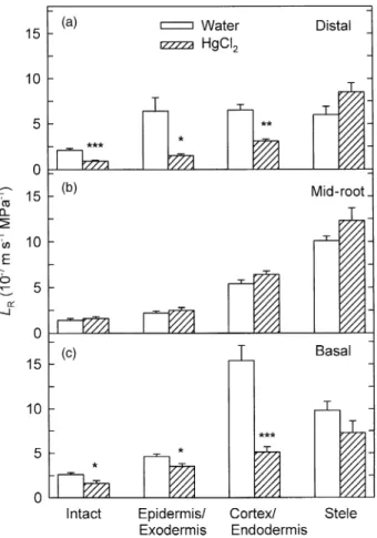
2sensitivity of concentric root tissues
For distal root segments with the epidermis and exodermis
removed, LP in water was 37% higher than LP for intact
distal root segments (Fig. 4a versus Fig. 2a). For both the
mid-root and basal regions without these layers, LP was
about twice as high as for the intact segments (Fig. 4a versus Fig. 2b & c). For distal segments with the epidermis and
exodermis removed, LP in HgCl2 was 23% lower than in
water (Fig. 4a; P < 0.01). For mid-root segments with these
layers removed, LP in HgCl2 was slightly but not
signifi-cantly higher than in water. For basal segments with the
epidermis and exodermis removed, LP was 45% lower in
HgCl2 than in water (P < 0.05).
For distal, mid-root and basal root regions, LP in water
for the stele (the epidermis, exodermis, cortex and endo-dermis having been removed) was 2.1, 6.6 and 2.7 times higher, respectively, than for the intact segments (Fig. 4b
versus Fig. 2). For the stele of the three root regions, HgCl2
had no significant effect on LP.
Axial hydraulic conductance (Kh) was 1.2 ± 0.5 ¥ 10-11
m4 s-1 MPa-1 for the distal root region, 9.8 ± 2.6 ¥ 10-11 m4
s-1 MPa-1 for the mid-root region and 5.4 ± 2.3 ¥ 10-11 m4
s-1 MPa-1 for the basal region (n = 5 plants). Root radial
hydraulic conductance (LR) calculated from LP and Kh was
2.1 ¥ 10-7 m s-1 MPa-1 for intact segments from the distal
region, which was 50% higher than for the mid-root region
and similar to LR for the basal region (Fig. 5). For LR of
intact segments, the effect of HgCl2 was similar to its effect
on LP, with a reduction of 57% for the distal region, no
reduction for the mid-root region and a reduction of 38% for the basal region.
The radial hydraulic conductance of the epidermis and
exodermis (LR,EpEx) for the distal region was about three
times greater than LR,EpEx for the mid-root region and
slightly but not significantly higher than LR,EpEx for the basal
region (Fig. 5). HgCl2 reduced LR,EpEx for the distal region
by 77%, had no effect on LR,EpEx for the mid-root region
and reduced LR,EpEx for the basal region by 24% (Fig. 5).
The radial hydraulic conductance of the cortex and
endo-dermis (LR,CoEn) for the distal region was slightly but not
significantly higher than for the mid-root region and about
60% lower than for the basal region. HgCl2 reduced LR,CoEn
by 72% for the distal region, had no effect on LR,CoEn for
the mid-root region and reduced LR,CoEn by 67% for the
basal region. For the stele, the radial hydraulic conductance
(LR,S) was about 40% lower for the distal region than for
the mid-root and basal regions. HgCl2 had no significant
effect on LR,S for the three root regions (Fig. 5).
Root anatomy
Distal, mid-root and basal regions of roots of A. deserti differed in the extent of suberization and lignification of various cell layers (Fig. 6). For the distal region under wet
conditions, 1.4 ± 0.2 cell layers directly inside the
epider-mis had suberin lamellae and were thus considered exo-dermis. After 10 d of soil drying, epidermal cells in the
Figure 3. LP for intact root segments from the distal region of
roots of A. deserti measured in water and in 25 mM HgCl2 under
wet conditions, at 10 d of soil drying (day 0) and at 1, 2 and 3 d after rewetting. Data are means ±1 SE (n = 4 plants). Asterisks indicate significant differences due to the HgCl2 treatment (see
Fig. 2).
Figure 4. LP for distal, mid-root and basal root segments of A.
deserti (a) from which the epidermis and exodermis had been removed and (b) from which the epidermis, exodermis, cortex and endodermis had been removed, leaving the vascular cylinder (= stele), measured in water (open bars) and 25 mM HgCl2 (hatched
bars) under wet conditions. Data are means +1 SE (n = 4–6 plants). Asterisks indicate significant differences due to the HgCl2
224 G. B. North et al.
distal region had lost turgor and the exodermis was 2.0 ± 0.3 layers (Fig. 6a), an increase that was not signifi-cant (Mann–Whitney rank sum test, P = 0.056; n = 5 roots from different plants). Nuclei were present in exodermal layers in the distal region under both wet and drying con-ditions. In the inner cortex adjacent to the endodermis, 3.5 ± 0.8 cortical cell layers were lignified, although the walls remained relatively thin (Fig. 6b). The endodermis of the distal region lacked suberin lamellae (in sections taken 30–40 mm from the root tip) under both wet (Fig. 6b) and drying conditions, and lignified Casparian bands were apparent.
For the mid-root region, the exodermis consisted of 3.6 ± 0.2 suberized cell layers under both wet (Fig. 6c) and drying conditions. Nuclei were not present in the outermost layer of the exodermis, but occasionally were apparent in the inner exodermal layers. Under both wet and drying conditions, the outermost layers of the cortex at mid-root consisted of irregularly shaped, collapsed cells that were
apparently dead. Outside the endodermis, 3.8 ± 0.4 cortical cell layers were lignified and had thickened cell walls under both wet (Fig. 6d) and drying conditions. The endodermis consisted primarily of cells with suberin lamellae and thick-ened inner tangential walls, although about 10% of endo-dermal cells were thin-walled and lacked suberin lamellae (i.e. were passage cells; Fig. 6d).
The basal region was anatomically more heterogeneous than the other two root regions. At 60–70 mm from the shoot base, the anatomy was similar to that of the mid-root region. At 15–40 mm from the shoot base, the root diame-ter increased, and cell layers tended to be less lignified and suberized than in the region further from the shoot. Specif-ically, the exodermis was 2.6 ± 0.2 layers at 30 mm from the base under both wet (Fig. 6e) and drying conditions, and nuclei were apparent in the outer exodermal layers. Although the outer cortical cells were collapsed, as at mid-root, most of the cortical cells just outside the endodermis were unlignified, and nuclei were often apparent under both wet and drying conditions (Fig. 6f). The percentage of endodermal cells lacking suberin lamellae (passage cells) ranged from 22 to 100% (Fig. 6f).
DISCUSSION
Except under dry conditions, treatment with 25 mM HgCl2
inhibited water transport for the distal (youngest) and the basal (oldest) root regions of Agave deserti, suggesting the involvement of aquaporins (Chaumont et al. 2000; Javot & Maurel 2002). The reversibility of inhibition by
2-mercap-toethanol further suggests that the primary effect of HgCl2
was to close water channels. In addition, root segments left
in water for 12 h after treatment with HgCl2 and
2-mercap-toethanol exhibited apical growth and gravitropism,
indi-cating a lack of residual toxicity. Using HgCl2 allowed water
channel activity to be assessed on a tissue-by-tissue basis, complementing research that demonstrates gene expres-sion and aquaporin presence in individual root cell layers (Eckert et al. 1999; Baiges et al. 2002; Javot & Maurel 2002; Tyerman et al. 2002).
Location of water channel activity under wet
soil conditions
Under wet soil conditions, HgCl2 reduced hydraulic
con-ductance (LP) for the distal root region of A. deserti by
about 50%, comparable to previous results for this species (North & Nobel 2000) and within the range reported for roots of comparable age from several other species, includ-ing Opuntia acanthocarpa (Martre et al. 2001). As was the
case for O. acanthocarpa, treatment with HgCl2 did not
affect water transport in the older mid-root region. Aqua-porins in both the tonoplast (TIPs) and in the plasma membrane (PIPs) are more abundant in younger than in older root tissues of Mesembryanthemum crystallinum (Yamada et al. 1995), Picea abies (Oliviusson, Salaj & Hakman 2001) and hybrid Vitis (Baiges et al. 2002). The
reduction of LP by HgCl2 for the basal root region of A.
Figure 5. LR for distal, mid-root and basal regions of roots of A.
deserti under wet conditions. LR for intact segments, the epidermis
plus exodermis (LR,EpEx), the cortex plus endodermis (LR,CoEn) and
the stele (LR,S) was calculated from LP measured in water (open
bars) and in 25 mM HgCl2 (hatched bars), using mean values of Kh
(text data, assumed not to change in HgCl2) for each root region;
Eqn 2 was used for intact segments and Eqns 3 and 4 for sequential cell layers. Data are means ±1 SE (n = 5–6 plants). Asterisks indi-cate significant differences due to the HgCl2 treatment (Student’s
Aquaporins and hydraulic conductance of agave roots 225
deserti, which was similar to that for the distal region, at first appears anomalous, yet this region near the shoot base is anatomically more similar to the distal than to the mid-root region. In addition, both the distal and the basal regions had a higher proportion of living cells in all tissues than did the mid-root region. Radial hydraulic
conduc-tance (LR) was similar for the distal and the basal regions
and 50–85% higher than at mid-root. Thus, greater abun-dance and/or greater opening of water channels may help explain the greater hydraulic conductance for the distal and basal regions than for the mid-root region. Such a pattern may be adaptive for A. deserti in the desert, where new apical growth in the distal root region occurs in wet soil and where the basal root region is positioned to inter-cept light rainfall that may wet only the top few centime-tres of soil and is channelled by the leaves toward the plant base.
For the distal region of roots of A. deserti, LR,EpEx (radial
hydraulic conductance for the epidermis and exodermis),
LR,CoEn (for the cortex and endodermis) and LR,S (for the
stele) were all similar. In terms of resistance, each of these sequential layers thus represented about one-third of the resistance of the intact root segment. Similarly, the hydrau-lic resistance for young roots of Zea mays is relatively evenly distributed across the root (Peterson, Murrmann &
Steudle 1993). For distal root segments treated with HgCl2,
the resistance of the epidermis and exodermis increased four-fold and became 60% of the total radial resistance, suggesting that closure of water channels in these outer cell layers had a disproportionate effect on transport across the
root. Treatment of onion roots with HgCl2 also indicated
that closure of water channels in the epidermis and exoder-mis greatly increases radial hydraulic resistance (Barrow-clough et al. 2000). In the distal region of roots of A. deserti, the exodermis generally consisted of one layer of cells with suberin lamellae, although this layer is actually dimorphic, with unsuberized, densely cytoplasmic cells alternating irregularly with suberized cells (North & Nobel 1991). As
suberized cell walls can block the passage of HgCl2
(Bar-rowclough et al. 2000), a likely site for the inhibitory effect
of HgCl2 on water transport is the plasma membrane of
these unsuberized cells.
For the distal root region, treatment with HgCl2
decreased the radial conductance of the cortex and
endodermis (LR,CoEn) by about 50% but did not change
the relative contribution of these layers to the radial resistance, due in part to the decreased proportional resistance of the stele. Thus, although water transport through the cortex and endodermis appeared to involve aquaporins, their role in these layers in the distal region was not as important as in the epidermis and exodermis. Some water flow may have occurred in the apoplastic pathway through these tissues, as has been shown previ-ously for the distal 20 mm of roots of A. deserti under wet conditions (North & Nobel 1995). As the endoder-mis lacked suberin lamellae in most of the distal region, apoplastic flow was more likely than in the mid-root and the basal regions.
For the mid-root region, treatment with HgCl2 had no
effect on LR for any of the three tissue components
consid-ered. The suberized cell layers of the exodermis and endo-dermis as well as the lignified cortical cell layers outside the
Figure 6. Photomicrographs of cross-sections of roots of A. deserti. Sections were made at 35 mm back from the root tip, showing the outer (a) and the inner (b) tissues of the distal region; at 170 mm from the root tip, showing the outer (c) and the inner (d) tissues of the mid-root region; and at 30 mm from the shoot base, showing the outer (e) and the inner (f) tissues of the basal region. Cross-sections in (a) and (f) were made at 10 d of soil drying and all others were made under wet conditions. All cross-sections were stained with toluidine blue O; unlignified parenchyma is purple, lignified cell walls are blue-green or blue and suberin lamellae in endodermis are unstained. Black arrows indicate nuclei; white arrows indicate passage cells in the endodermis; en, endodermis; ep, epidermis; ex, exodermis; c, cortex; s, stele. Scale bars = 50 mm.
226 G. B. North et al.
endodermis could pose substantial barriers to the
penetra-tion of HgCl2 to the plasma membrane of cells in these
layers, possibly rendering the effect of HgCl2 on aquaporins
inconclusive (Barrowclough et al. 2000). However, removal of the epidermis and fully suberized exodermis did not
increase sensitivity to HgCl2 for the mid-root region.
Pas-sage cells lacking suberin lamellae were few in the endo-dermis, indicating a more limited apoplastic pathway than in the distal region. Thus, water flow in the mid-root region would be expected to occur primarily through the cell-to-cell pathway (Steudle & Peterson 1998), yet insensitivity to
HgCl2 indicates that aquaporin involvement was minimal.
With the exception of LR,S, radial conductance for the
tis-sues of the mid-root region was lower than for tistis-sues of the distal region, consistent with lower aquaporin activity and/or lower apoplastic flow.
For the basal region, LR,EpEx was one-third of LR,CoEn
and one-half of LR,S, perhaps reflecting the relatively
extensive suberization of the exodermis. The epidermis and exodermis represented 56% of the total radial resis-tance for the intact segment in water and 46% of the
total resistance in HgCl2. In contrast to the case for the
distal region, treatment with HgCl2 increased the relative
contribution of the endodermis and cortex to the total resistance, indicating greater aquaporin involvement in flow through these tissues than through the epidermis and exodermis in the basal region. Similar to the case at mid-root, a possible explanation is the relatively extensive suberization of the exodermis in comparison with the endodermis in this region. At 15–40 mm from the shoot base, many passage cells were present in the endodermis, suggesting that the 67% loss of conductance due to
treat-ment with HgCl2 could reflect closure of aquaporins in
these cells. Analogously, mRNA distribution indicates abundant plasma membrane aquaporins in the endoder-mis of roots of M. crystallinum (Yamada et al. 1995), as does GUS expression in the endodermis and stele of roots of Arabidopsis thaliana (Javot et al. 2003). Aquapor-ins in cortical cells of A. deserti may also have been affected, although the outer cells of the cortex in the basal region were collapsed and apparently dead under both wet and drying conditions.
Treatment with HgCl2 had no significant effect on water
transport through the stele in any of the three root regions, in contrast to results for roots of O. acanthocarpa (Martre et al. 2001). Thus, aquaporins do not appear to have been important in radial conductance through the stele of A. deserti. With no suberized cell layers present (the endoder-mis having been removed), and under the conditions of vacuum-induced water uptake in the current study, apo-plastic flow may have predominated in the radial path to the xylem. Aquaporins in stelar parenchyma, as indicated for a number of species including M. crystallinum (Yamada et al. 1995) and Zea mays (Barrieu, Chaumont & Chrispeels 1998), could be more important for osmotically induced water uptake than for hydrostatically induced flow, accord-ing to the composite model of root hydraulic conductivity (Steudle & Peterson 1998).
Changes in root hydraulic conductance and
water channel activity during soil drying
and rewetting
Soil drying reduced LP by 50–60% for the distal and the
mid-root regions and by about 30% for the basal region of
roots of A. deserti, and treatment with HgCl2 had no
addi-tional inhibitory effect on LP. Interestingly, for both the
distal and the basal root regions, LP after 10 d of soil drying
did not differ from LP after treatment with HgCl2. In most
other plant species, stress similarly reduces the effect of mercurial compounds on water transport. For example, N-and P-deprived roots of Lotus japonicus lose their
sensitiv-ity to Hg2+ (Clarkson et al. 2000), and salinity stress reduces
not only LP for roots of Cucumis melo (Martinez-Ballesta
et al. 2000), Triticum aestivum (Carvajal et al. 1996) and Capsicum annuum (Martinez-Ballesta, Martinez & Carva-jal 2003) but also their sensitivity to further inhibition by
HgCl2. In such cases, the stress may down-regulate
aqua-porin expression, close or narrow the opening of existing
water channels, or render aquaporins insensitive to HgCl2
in a manner unrelated to water transport. For the roots of a number of relatively stress-tolerant species, including M. crystallinum (Kirch et al. 2000), Nicotiana glauca (Smart et al. 2001) and Hordeum vulgare (Katsuhara et al. 2002), the expression of PIPs is moderately to strongly down-regulated during water or salinity stress.
A possible mechanism for the regulation of water chan-nel activity in roots of A. deserti as soil moisture varied is by the phosphorylation and dephosphorylation of aquapor-ins. According to a model based on work with leaves of Spinacia oleracea, high apoplastic water potential leads to
full cell turgor and the opening of stretch-activated Ca2+
channels in the plasma membrane; high levels of Ca2+
stim-ulate aquaporin phosphorylation, and water channels open (Johansson et al. 1998). When cell turgor decreases during
drying, Ca2+ channels close, aquaporins are
dephosphory-lated, and water channels close. Young roots of A. deserti shrink during drying and quickly regain turgor upon rewet-ting (Nobel & Cui 1992), changes that were paralleled in the current study by the loss and re-acquisition of
sensitiv-ity to HgCl2. In particular, the complete recovery of
hydraulic conductance for distal root segments of A. deserti after 1 d of rewetting occurred in the absence of new apical
growth and was accompanied by a 41% reduction of LP due
to HgCl2, similar to the 49% reduction caused by HgCl2
under wet conditions. The reopening of water channels was apparently central to the recovery of root water uptake upon rewetting, corroborating results for Arabidopsis thaliana showing that plant recovery after soil drying and rewetting was faster and more complete for wild-type plants than for PIP1/PIP2 antisense plants (Martre et al. 2002).
CONCLUSIONS
The variable contribution of aquaporins to water transport
Aquaporins and hydraulic conductance of agave roots 227
along the length and across the tissue layers of roots of A.
deserti under wet, dry and rewetted soil conditions. HgCl2
reduced hydraulic conductance for the distal and the basal root regions but not at mid-root, suggesting a positive asso-ciation between water channel opening and the frequency of living, metabolically active cells. Water channel activity was thus greatest in the root regions most likely to encoun-ter wet or rewetted soil in the desert. In the distal root region, the epidermis and exodermis were the site of the major contribution of aquaporins to root hydraulic conduc-tance, whereas water channel activity in the basal region was greatest in the cortex and endodermis. Soil drying led to lower root hydraulic conductance, which was not
reduced further by HgCl2, yet rewetting restored both
hydraulic conductance and its sensitivity to HgCl2 for the
distal and the basal regions. The percentages of LP
inhibi-tion due to treatment with HgCl2 indicate that most of the
quantitative variations in LP for the distal and the basal root
regions were due to changes in water channel activity in response to variations in soil moisture. The apparent lack of anatomical changes and the rapid recovery of water uptake after rewetting both point to a central role for aqua-porins in regulating root responses to changes in water availability. Thus, aquaporins may represent an efficient way for A. deserti to modify root hydraulic conductance in a habitat where sporadic light rainfall must be utilized quickly.
ACKNOWLEDGMENTS
This work was supported by the National Science Founda-tion (Grant no. 9975163).
REFERENCES
Baiges I., Schäffner A.R., Affenzeller M.J. & Mas A. (2002) Plant aquaporins. Physiologia Plantarum 115, 175–182.
Barrieu F., Chaumont F. & Chrispeels M.J. (1998) High expression of the tonoplast aquaporin ZMTIP1 in epidermal and conduct-ing tissues of maize. Plant Physiology 117, 1153–1163. Barrowclough D.E., Peterson C.A. & Steudle E. (2000) Radial
hydraulic conductivity along developing onion roots. Journal of Experimental Botany 51, 547–557.
Carvajal M., Cooke D.T. & Clarkson D.T. (1996) Responses of wheat plants to nutrient deprivation may involve the regulation of water-channel function. Planta 199, 372–381.
Carvajal M., Martinez V. & Alcaraz C.F. (1999) Physiological function of water channels as affected by salinity in roots of paprika pepper. Physiologia Plantarum 105, 95–101.
Chaumont F., Barrieu F., Jung R. & Chrispeels M.J. (2000) Aqua-porins constitute a large and highly divergent protein family in maize. Plant Physiology 125, 1206–1215.
Clarkson D.T., Carvajal M., Henzler T., Waterhouse R.N., Smyth A.J., Cooke D.T. & Steudle E. (2000) Root hydraulic conduc-tance: diurnal aquaporin expression and the effects of nutrient stress. Journal of Experimental Botany 51, 61–70.
Dubrovsky J.G., North G.B. & Nobel P.S. (1998) Root growth, developmental changes in the apex, and hydraulic conductivity for Opuntia ficus-indica during drought. New Phytologist 138, 75–82.
Eckert M., Biela A., Siefritz F. & Kaldenhoff R. (1999) New
aspects of plant aquaporin regulation and specificity. Journal of Experimental Botany 50, 1541–1545.
Henzler T., Waterhouse R.N., Smyth A.J., Carvajal M., Cooke D.T., Schäffner A.R., Steudle E. & Clarkson D.T. (1999) Diur-nal variations in hydraulic conductivity and root pressure can be correlated with the expression of putative aquaporins in the roots of Lotus japonicus. Planta 210, 50–60.
Huang B. & Nobel P.S. (1994) Root hydraulic conductivity and its components, with emphasis on desert succulents. Agronomy Journal 86, 767–774.
Hukin D., Doering-Saad C., Thomas C.R. & Pritchard J. (2002) Sensitivity of cell hydraulic conductivity to mercury is coincident with symplasmic isolation and expression of plasmalemma aqua-porin genes in growing maize roots. Planta 215, 1047–1056. Javot H. & Maurel C. (2002) The role of aquaporins in root water
uptake. Annals of Botany 90, 301–313.
Javot H., Lauvergeat V., Santoni V., et al. (2003) Role of a single aquaporin isoform in root water uptake. Plant Cell 15, 509–522. Jensen W.A. (1962) Botanical Histochemistry: Principles and
Prac-tice. W.H. Freeman, San Francisco, CA, USA.
Johansson I., Karlsson M., Shukla V.K., Chrispeels M.J., Larsson C. & Kjellbom P. (1998) Water transport activity of the plasma membrane aquaporin PM28A is regulated by phosphorylation. Plant Cell 10, 451–459.
Johansson I., Larsson C. & Kjellbom P. (1996) The integral pro-teins of spinach leaf plasma membranes are putative aquaporins and are phosphorylated in response to Ca2+ and apoplastic water
potential. Plant Cell 8, 1181–1191.
Katsuhara M., Akiyama Y., Koshio K., Shibasaka M. & Kasamo K. (2002) Functional analysis of water channels in barley roots. Plant Cell Physiology 43, 885–893.
Kirch H.H., Vera-Estrella R., Golldack D., Quigley F., Micha-lowski C.B., Barkla B.J. & Bohnert H.J. (2000) Expression of water channel proteins in Mesembryanthemum crystallinum. Plant Physiology 123, 111–124.
Landsberg J.J. & Fowkes N.D. (1978) Water movement through plant roots. Annals of Botany 42, 493–508.
LoGullo M.A., Nardini A., Salleo S. & Tyree M.T. (1998) Changes in root hydraulic conductance (KR) of Olea oleaster seedlings
following drought stress and irrigation. New Phytologist 108, 25– 31.
Martinez-Ballesta M.C., Martinez V. & Carvajal M. (2000) Regu-lation of water channel activity in whole roots and in protoplasts from roots of melon plants grown under saline conditions. Aus-tralian Journal of Plant Physiology 27, 685–691.
Martinez-Ballesta M.C., Martinez V. & Carvajal M. (2003) Aqua-porin functionality in relation to H+-ATPase activity in root cells
of Capsicum annuum grown under salinity. Physiologia Plan-tarum 117, 413–420.
Martre P., Morillon R., Barrieu F., North G.B., Nobel P.S. & Chrispeels M.J. (2002) Plasma membrane aquaporins play a significant role during recovery from water deficit. Plant Physi-ology 130, 2101–2110.
Martre P., North G.B. & Nobel P.S. (2001) Hydraulic conductance and mercury-sensitive water transport for roots of Opuntia acan-thocarpa in relation to soil drying and rewetting. Plant Physiol-ogy 126, 352–362.
Morillon R. & Lassalles J.P. (2002) Water deficit during root devel-opment: effects on the growth of roots and osmotic water per-meability of isolated root protoplasts. Planta 214, 392–399. Nobel P.S. & Cui M. (1992) Hydraulic conductances of the soil,
the root-soil air gap, and the root: changes for desert succulents in drying soil. Journal of Experimental Botany 43, 319–326. Nobel P.S., Schulte P.J. & North G.B. (1990) Water influx
charac-teristics and hydraulic conductivity for roots of Agave deserti Engelm. Journal of Experimental Botany 41, 409–415.
228 G. B. North et al.
North G.B. & Nobel P.S. (1991) Changes in hydraulic conductiv-ity and anatomy caused by drying and rewetting roots of Agave deserti (Agavaceae). American Journal of Botany 78, 906–915.
North G.B. & Nobel P.S. (1992) Drought-induced changes in hydraulic conductivity and structure in roots of Ferocactus acan-thodes and Opuntia ficus-indica. New Phytologist 120, 9–19. North G.B. & Nobel P.S. (1994) Changes in root hydraulic
con-ductivity for two tropical epiphytic cacti as soil moisture varies. American Journal of Botany 81, 46–53.
North G.B. & Nobel P.S. (1995) Hydraulic conductivity of concen-tric root tissues of Agave deserti Engelm. under wet and drying conditions. New Phytologist 130, 47–57.
North G.B. & Nobel P.S. (1998) Water movement and structural plasticity along roots of a desert monocot during and after pro-longed drought. Plant, Cell and Environment 21, 705–713. North G.B. & Nobel P.S. (2000) Heterogeneity in water
availabil-ity alters cellular development and hydraulic conductivavailabil-ity along roots of a desert succulent. Annals of Botany 85, 247–255. Oliviusson P., Salaj J. & Hakman I. (2001) Expression pattern of
transcripts encoding water channel-like proteins in Norway spruce (Picea abies). Plant Molecular Biology 46, 289–299. Peterson C.A., Murrmann M. & Steudle E. (1993) Location of the
major barriers to water and ion movement in young roots of Zea mays L. Planta 189, 288–297.
Siefritz F., Tyree M.T., Lovisolo C., Schubert A. & Kaldenhoff R.
(2002) PIP1 plasma membrane aquaporins in tobacco: from cel-lular effects to function in plants. Plant Cell 14, 869–876. Smart L.B., Moskal W.A., Cameron K.D. & Bennett A.B. (2001)
MIP genes are down-regulated under drought stress in Nicotiana glauca. Plant Cell Physiology 42, 686–693.
Steudle E. & Peterson C.A. (1998) How does water get through roots? Journal of Experimental Botany 49, 775–788.
Suga S., Komatsu S. & Maeshima M. (2002) Aquaporin isoforms responsive to salt and water stresses and phytohormones in radish seedlings. Plant Cell Physiology 43, 1229–1237.
Tsuda M. & Tyree M.T. (2000) Plant hydraulic conductance mea-sured by the high pressure flow meter in crop plants. Journal of Experimental Botany 51, 823–828.
Tyerman S.D., Niemietz C.M. & Bramley H. (2002) Plant aqua-porins: multifunctional water and solute channels with expand-ing roles. Plant, Cell and Environment 25, 173–194.
Yamada S., Katsuhara M., Kelly W.B., Michalowski C.B. & Bohnert H.J. (1995) A family of transcripts encoding water channel proteins: tissue-specific expression in the common ice plant. Plant Cell 7, 1129–1142.
Zhang W.H. & Tyerman S.D. (1999) Inhibition of water channels by HgCl2 in intact wheat root cells. Plant Physiology 120, 849–
857.
Received 1 August 2003; received in revised form 6 October 2003; accepted for publication 7 October 2003