HAL Id: hal-02483977
https://hal.archives-ouvertes.fr/hal-02483977
Submitted on 18 Feb 2020
HAL is a multi-disciplinary open access
archive for the deposit and dissemination of
sci-entific research documents, whether they are
pub-lished or not. The documents may come from
teaching and research institutions in France or
abroad, or from public or private research centers.
L’archive ouverte pluridisciplinaire HAL, est
destinée au dépôt et à la diffusion de documents
scientifiques de niveau recherche, publiés ou non,
émanant des établissements d’enseignement et de
recherche français ou étrangers, des laboratoires
publics ou privés.
Cybergology and bioprinting: The biotechnological
future of maxillofacial rehabilitation
Adrien Naveau, Rawen Smirani, Murielle Rémy, Philippe Pomar, Florent
Destruhaut
To cite this version:
Adrien Naveau, Rawen Smirani, Murielle Rémy, Philippe Pomar, Florent Destruhaut. Cybergology
and bioprinting: The biotechnological future of maxillofacial rehabilitation. International Journal of
Maxillofacial Prosthetics, Tokyo: [International Journal of Maxillofacial Prosthetics], 2019, 1,
pp.20-26. �10.26629/ijmp.2019.05�. �hal-02483977�
Keywords:
Maxillofacial prosthetics, bibliometrics, trends.
Corresponding Author:
Dr Adrien Naveau, Unité de réhabilitation dentaire et parodontale, Hôpital Saint André, 1 Rue Jean Burguet, 33000 Bordeaux, France.
Phone :+33 5 57 62 34 34 email: adrien.naveau@u-bordeaux.fr Received: 02 October 2019 Received in revised form: 30 October 2019
Accepted: 02 November 2019
Maxillofacial prostheses now benefit from the growing advances in converging technologies, such as nanotechnologies, biotechnologies, informatics and cognitivism (NBIC). Instead of being laid and passive, prostheses can now ensure true neurophysiological interactions with their wearers through complex phenomena of hybridization and vicariance. These new devices get closer to “maxillofacial amplification prostheses”, by improving the world perception through restored sensory properties and new extra-sensory properties. These technological devices also benefit from the bioengineering revolution that will soon allow the bioprinting of graft prostheses with an intimate integration to the organic maxillofacial support. In this article, the authors would like to present some major advances in cybergology and bio-printing in maxillofacial hybridization contexts. (Int J Maxillofac Prosthetics
2019;1:20-26)
International Journal of Maxillofacial Prosthetics (iJMP)
REVIEW ARTICLE
Cybergology and bioprinting: The biotechnological future of
maxillofacial rehabilitation
Adrien Naveau, DDS, PhD1-3, Rawen Smirani, DDS3,4 , Murielle Remy3, Philippe Pomar5,6, Florent Destruhaut, DDS, PhD5-7
1 Department of Prosthodontics, Dental Science Faculty, University of Bordeaux, France.
2 Dental and Periodontal Rehabilitation Unit, Saint Andre Hospital, Bordeaux University Hospital, Bordeaux, France. 3 BioTis Laboratory, INSERM U1026, University of Bordeaux, France.
4 Department of Periodontology, Dental Science Faculty, University of Bordeaux, France. 5 Department of Prosthodontics, Dental Science Faculty, University of Toulouse, France. 6 Maxillofacial Prosthetics Unit, Rangueil Hospital, Toulouse, France.
7 CAS Laboratory (Social Anthropology Center), UMR 5193, University of Toulouse, France.
ABSTRACT
ARTICLE INFO
INTRODUCTION
For a long time, maxillofacial prosthetics (MP) main driving force has remained its symbiotic relationship with maxillofacial surgery.1 Indeed,
surgical considerations have always led maxillofacial rehabilitation, while MP has stood as the alternative, the complementary and reliable option that fills the gap left beyond the limits of surgery. The rationale behind that trend is the patient’s preference for a definitive surgical treatment over a “provisional” removable prosthesis. However, the current MP evolution is now driven by the development of materials (polymers, metals or ceramics) and technologies (computer-aided design/computer-assisted manufacturing, robotics…).1-3
Thus the field of conventional MP is evolving, benefiting from the rise of converging technologies, such as nanotechnologies, biotechnologies, informatics and cognitivism (NBIC). The future of maxillofacial rehabilitation is no longer mutating from reparation to regeneration, even though fulfilling the patients’ expectations for definitive treatments. Beyond the
future regenerated patient stands the improved patient, the “transhuman” with additional biotechnological functions, in a perfect symbiosis of surgery and prosthetics, marrying tissues and frameworks, cells and scaffolds.
MAXILLOFACIAL CYBERGOLOGY
The first direction for the MP evolution is the association of machine and man to create cyborgs. The term “cybergology” was coined by neurophysiologist Manfred Clynes in the 1960s, and was reintroduced into the scientific literature following the work of Donna Haraway, biologist, anthropologist and philosopher, in its Cyborg Manifesto.4 More recently,
scientist Jean-Claude Heudin proposed an exhaustive classification of cybernetic phenotypes and analogous profiles, from robots to avatars, that includes living statues, clones and mechas.5 In this
classification, cyborgs are subdivided into two categories of cybernetic organisms: robotic and biological. Robotic
Int J Maxillofac Prosthetics 2019;1:20-26. DOI: 10.26629/ijmp.2019.05 21
cybernetic organisms refer to organic elements aggregated on some artificial structures, eventually humanoids (ex: Terminator), that remain science-fiction so far. However, biological cybernetic organisms refer to individuals with technological prosthetics (ex: Robocop), and these «human cyborgs» are already among us. Indeed, modern medicine postpones death with pacemaker implants, increasing human life expectancy for patients to become increased humans. Also, any individual with glasses, hearing aids or cardiac stents is an amplified human, making the limit between the “repaired” human and the cyborg a thin and shifting boundary. In this context of modified senses, the maxillofacial amplification prostheses reshape the body diagram and the self-image of the wearers, leading to a new body experience, as well as a new perception of their “inner selves” and of their surroundings.
Augmented vision
If eyeglasses are objects designed to correct or assist defective vision, augmented reality (AR) glasses clearly amplify an individual’s abilities. These glasses allow the superimposition of reality with virtual elements, such as sounds, two-dimensional images, three-dimensional images, or videos, by real time calculation (ex: Google Glass®, 2012, 2019). This pair of glasses provides
different functions: camera, microphone, earphone, and internet access (through Wi-Fi®
or Bluetooth®). This application, still expensive
for the general public, is particularly useful for high-level athletes, soldiers, astronauts and surgeons.6,7 In 2014, the British Start-up This
Place Ltd® incorporated a control system into
the Google glasses®: The MindRDR® device used
a Neurosky® electroencephalography biosensor
on the wearer’s forehead to detect brain waves and interpreted them to drive the augmented reality.8,9 Although not in our reality, this process
was a first approximation of psychokinesis, the secular human fantasy assimilated to a metaphysical and hypothetical faculty of the mind to act directly on matter. Similarly, a team from the University of Washington experimentally tested in 2008 a biocompatible contact lens incorporating electronic circuits and light diodes to “the display of information in the visual field or the acquisition of biological data for patient monitoring”.10 In 2016, Google® also filed a
patent for electronic contact lenses to display augmented reality: the principle is similar to Google Glass®, except that this new device
uses nanotechnology to fit into the polyethylene terephthalate lens. These contact lenses could also have a significant medical application by analyzing the fluids on the surface of the cornea. Finally, the bionic eye, dreamed by American inventor Benjamin Franklin in the 19th century, was
conceived in Boston by Dr Rizzo and Professor Wyatt, as part of the Boston retinal implant project (BRIP).11,12 Still used for experimental purposes, the
bionic eye restores functional vision in patients with partial or total blindness.
On another note, the Catalan artist Neil Harbisson is a remarkable example of hybridization. The artist suffers from congenital achromatopsia (absence of colour perception) and perceives his environment exclusively in black and white. Passionate about cybernetics, and with the help of computer scientist Adam Montandon, he designed an antenna that automatically converts the surrounding colors into sound waves. Each color is represented by a particular sound frequency, allowing Harbisson to perceive colors that a human being cannot see, such as infrared and ultraviolet frequencies.13 Harbisson
became an enhanced being with his ability to "hear" colors that human eyes could not distinguish. He then realized that the cognitive work of interpreting the sounds in colors became a reflex, then a feeling, to the point of dreaming to hear the colors. The artist now campaigns for the status of cyborg considering that the union between the software and his brain has transformed him.14
Augmented olfaction and taste
Patented in 2012, the French start-up Aryballe Technologie® designed an artificial nose to identify
and quantify odors. It is not strictly speaking a facial prosthesis, but rather a peripheral technological device, a little bigger than a mobile phone, capable of identifying up to fifty different smells. Based on the knowledge of olfactory neurophysiology, the device implements the synergy of olfactory biosensors, optical imprint and data processing software. Beyond its usefulness for the food industry, the device can detect odors which are not perceptible to human olfactory cells.15 Furthermore, Japanese scientists
from the Rekimoto Lab at the University of Tokyo developed an electronic fork in 2016 that stimulates taste buds to simulate the salty taste. Not marketed yet, this device would be particularly useful for patients with salt-free diets in the context of cardiovascular diseases, high blood pressure, or with history of stroke or myocardial infarction.16
Augmented hearing
Hearing aids are used to correct hearing problems by making the sound audible. In the presence of a healthy inner ear with dysfunction of the external auditory duct and/or tympanic membrane, the researchers of the American society Sonitus Medical®
had the idea to transmit the sounds through bone conduction using a removable experimental prosthetic device called Soundbite®, positioned at the level of the
dental organs. The receiver is placed at the level of the ear or positioned on a frame of glasses or on a jacket pin,
to capture the surrounding sounds. They are then transmitted to the supradental hearing prosthesis which converts the signal into microvibrations. These are then relayed by bone conduction to the inner ear, where they stimulate the hair cells, sensory cells of the auditory system.17 In 2018, the
system was significantly improved by Sonitus Technologies® for use by US Army soldiers for
remote communication, by installing a receiver and a transmitter within the supradental hearing aid. A previous other experimental miniaturized system called Audio Tooth Implant® had been designed in
2002, but suffered from limited removability.18 The
treatment also consisted in implanting a low-frequency receptor in a prosthetic tooth to perceive sounds in the inner ear by bone conduction. The advantage was the ability to perceive vibrations below the usual perceptible frequency, making voices sound “crystalline”.
The artist Stelarc conceived in 2006 the Extra Ear project. The morphology and structure of an auricular pavilion was designed and recorded the artist surrounding noises with a miniature microphone. Internet users could intercept in real time the sound frequencies present around the artist, via its website. The concept was to provide an extra-sensory function through hybridization to create a “connected humanity”. Stelarc affirmed that beyond the concept of hybridization, this piece of art represented the access to new biotechnology to exceed one’s own sensory and motor abilities. In 2007, Stelarc completed his project and implanted on its forearm a biopolymer ear structure that was gradually colonized and covered by the skin to become an internalized structure. The hybridization was achieved through the intimate connection on the histological scale of the bio-object with its skin substrate. Equipped with a receiver, this third ear of Stelarc allowed the artist to become an internet portal providing others with the possibility of using his body, via his microphone bioprothesis. The hybridized body with technology is now becoming a collective experience. This example could be exported to other sensory organs, and showed that technology can marry biology to improve the human being.
MAXILLOFACIAL 3D BIOPRINTING
Hopefully soon, oral cancers will be treated without surgical removal of the tumor-supporting tissues, and congenital defects will all be treated during the early childhood. In the meantime, defect reconstruction will remain the challenging part of the maxillofacial rehabilitation. Concerning mandible defects for example, nowadays’ highest standard consists of the jaw-in-a-day concept, with reconstruction of the defect during the resection day.19,20 Furthermore, the
association of the free vascularized fibula flap transplantation with dental implant placement, both under CAD/CAM guidance, has been a revolution in the field.21-23 In this context, the next improvement of
the technique would be to provide a composite graft to the patient without requiring a second surgical site. This engineered construct would be, like free flap grafting, CAD/CAM driven to match perfectly the need in tissues. Interestingly, one of the recent tissue engineering technologies has been developed to fabricate composite grafts, scalable, on-demand, with complex anatomical designs, and layer-by-layer controlled distribution. This biotechnology is 3D bioprinting, and may allow one day in situ maxillofacial reconstruction.24
Bioprinting maxillofacial tissues
Maxillofacial reconstruction requires numerous tissues (i.e. bone, cartilage, muscles, mucosa, teeth, nerves, blood vessels, etc.) to adequately restore speech, swallowing, mastication, and esthetics, among other functions. Bioprinting is a recent member of the tissue engineering (TE) and regenerative medicine family that, when compared to the autologous graft, is developing with the promise of less donor site morbidity. Bioprinting fully relies on the CAD/CAM to pattern and assemble bio-inks (organic materials with or without synthetic materials) at molecular and organ levels in 3D.25,26 One advantage
of this additive technique, especially over subtractive techniques, is the possibility of designing the inner structure to facilitate the tissue integration. Indeed, the porosity characteristics (i.e. size and interconnectivity) influence cell adhesion, proliferation and vascularization. The host angiogenesis is crucial to provide oxygen and nutrients to the inner cells of the engineered graft, since their diffusion is in theory limited to a depth of 200 µm (Fig 1). To this end, numerous engineering strategies aim at reproducing micro-environments and cell organizations. Vascularizing 2-3 mm3 constructs has not been
overcome yet, although many leads exist, such as bioprinting angiogenic growth factors, networks in scaffolds and pre-vascularization in vitro or in vivo. Moreover, the bioprinter allows both macroscopic and microscopic control of the bio-ink positioning and distribution, modeling complex anatomical shapes, on-demand, in a fast, reproducible and scalable manner, with potential applications in vitro or in vivo.
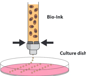
Cytoscribing was born in 1988, opening the door for bioprinting development.27 Since then, many kinds of
bioprinters have been developed, each with specific properties, and they can be combined together to design a multilayer construct. The extrusion-based bioprinter schematically drops the bio-ink through a syringe under automatized pressure (Fig 2). It is useful for designing the volume of the construct, and specifically the 3D matrix environment by distributing precisely layer-by-layer the organic components (including cells). For example, this technique can be used conveniently for printing a cellularized hydrogel that mimics soft tissues. A second technique is the laser-assisted bioprinting (Fig 3).
Int J Maxillofac Prosthetics 2019;1:20-26. DOI: 10.26629/ijmp.2019.05 23
use of sacrificial inks has shown to be effective for designing arteries and muscles. By combining all these techniques, one could in theory obtain the multilayered epithelium, the basal membrane, the soft connective tissue with vessels, the muscles, the soft periosteum and the bone scaffold.
Naveau et al.
Fig 1. Capillary supply in nutrient and oxygen to the surrounding
tissue relies on a limited diffusion. Dark pink: diffusion range.
Fig 2. Extrusion-based bioprinting. Nutrients (culture medium)
appear in pink, hydrogel in orange, and cells in brown.
The laser, precisely driven by the computer, hits a metal plate and generates locally a vapor bubble. The underneath bio-ink, composed of a medium with high cell density, is projected down on the substrate. The volume of the droplets is so small that cells can be dropped by group of 4-5. This process is very useful for example for the prevascularization of the construct, as aligning endothelial cells will conduct them to interact and form a capillary-like network.28 Some other techniques
can also be used for printing bio-inks, such as fused deposition modeling or jet-binding (Fig 4). These are often used for printing bone-like scaffold, and in this particular case, can be considered as (mineral) 3D printers rather than (organic) bioprinters. Moreover, the
Fig 3. Laser-assisted bioprinting. Nutrients (culture medium)
appear in pink, hydrogel in orange, and cells in brown.
Fig 4. Inkjet bioprinting. Nutrients (culture medium) appear in
pink, hydrogel in orange, and cells in brown.
3D bioprinted maxillofacial applications
The bioprinting technologies promise a vast array of potential maxillofacial applications, but their entrance on the healthcare market is undefined.24,29
The maturation level of bioprinting technologies is still too low to garner the investment required for proper development and movement to significant clinical trials. Some clinical applications have been described, but rather fall within the scope of 3D printing than of bio-printing, as the inks contained no organic compound.
For example, customized implants for bony defects are being used for cranioplasty.28 The protocol requires
using tomographic data of the patient to design the cutting guide and the implant shape. Then, the synthetic bony structure is printed in vitro to fill the patient defect. This procedure has been performed with printed hydroxyapatite, polyetherketoneketone (PEKK) or polycaprolactone (PCL) inks.30,31
Considering palatal defect reconstruction, oral bone-mucosa composites have also been studied.32
However, these models also relied on a 3D printed bone scaffold with tissue engineered oral mucosal model. At this point, the primary objective of this bone-mucosa composite is to provide an in vitro model for the investigation of oral cancer mechanisms, diagnosis and therapy.
Still in the maxillofacial field, ear and nose reconstruction benefited from the same 3D-printing approach, replacing cartilage with a printed acrylonitrile/ butadiene/styrene (ABS) scaffold secondarily coated with fibronectin for biocompatibility, or PCL coated with hydrogel and chondrocytes.33-35 In China, some of
these 3D printed scaffolds for ear were recently transplanted in 5 microtia patients, with follow-up periods ranging from 6 months to 2.5 years.20 The
team reported achieving satisfactory aesthetical outcome with mature cartilage formation in those children during the trial, and expected interesting long term stability. The protocol used expanded microtia chondrocytes, polyglycolic acid (PGA), polylactic acid (PLA) and PCL biodegradable scaffold to engineer patient-specific ear-shaped cartilage in vitro. However, the 3D-printed objects were only the 2D PCL mesh and a pair of 3D ear molds. On the bioprinting side, the most mature studies reported ears that were fabricated with PCL, gelatin, fibrinogen, hyaluronic acid and auricular chondrocytes.36 Another team even printed
an ear with a chondrocyte-seeded alginate hydrogel and combined it with an conductive electronic antenna perceiving sounds that normal human ear cannot.37
The nanoelectronic elements consisted of infused silver nanoparticles, enabling readout of signals from cochlea-shaped electrodes. This proof of concept ear exhibited enhanced auditory sensing for radio frequency reception, and a possibility of listening stereo audio music. Although encouraging results were shown in vitro by the bioprinted constructs, human grafting was not considered yet. However, at some point, one can imagine this engineering tissue to be printed in situ, directly in the patient to fill the defect and regenerate the missing tissues. Then the next step will be to improve the patient with organic elements aggregated on artificial structures, transforming him/her in a robotic cybernetic organism.
TOWARDS A MAXILLOFACIAL HYBRIDIZATION
ETHICS
In conclusion, the future evolution of maxillofacial prosthetics will require a constant guidance from ethics. Unfortunately, researchers and engineers called upon to invent tomorrow’s biotechnologies are not necessarily aware of their invention impact on the human evolutionary destiny. Contemporary artists raise relevant questions through their works and performances, by highlighting the fact that converging technologies will not only supplement motor and sensory deficits. For the neurophysiologist and engineer Alain Berthoz, the concept of vicariance (i.e. the ability to replace one function with another or to delegate an action to a virtual avatar) “no longer seems to be limited to a mere replacement because it refers undeniably to the notion of overrun: technologies that transform us into cyborgs, such as those that replace a sensory deficit, open up new possibilities. It is possible to increase the efficiency of a process for the human brain. Here we can see a strange evolutionary direction: in addition to random mutations of the genome that lead to modifications of the organism, the living one uses technologies outside the brain to increase its effectiveness”.38 In addition to
playing “the sorcerer’s apprentice” by manipulating genes, does not man fit in a process of creating evolutionary essence leading our species to "mutate" to a new technological genus, the Homo orthopedicus?39 The “care vs amplification” dichotomy
should not be reduced to a simplistic equation of “good vs evil” because amplification can be an opportunity for an individual to fully live and even to access other levels of understanding through the transcendent action of the bio-prosthesis. This principle directly refers to the concept of meliorism advocated by philosopher William James: progress is a very real and concrete concept leading to a progressive improvement of the world. Indeed, humans can interfere with nature through biological or technological processes to improve over the human condition and its perfectibility.40
ACKNOWLEDGEMENTS
The authors declare that there is no conflict of interest regarding the publication of this article.
Naveau et al.
REFERENCES
1. Naveau A, Bou C, Sharma A. Evolution of topics in maxillofacial prosthetics publications. Int J Prosthodont 2018;31:565-568.
2. Elbashti ME, Sumita YI, Kelimu S, Aswehlee AM, Awuti S, Hattori M, et al. Application of digital technologies in maxillofacial prosthetics literature: A 10-year observation of five selected prosthodontics journals. Int J Prosthodont 2018;32:45-50.
3. Ariani N1, Visser A, van Oort RP, Kusdhany L, Rahardjo TB, Krom BP, et al. Current state of craniofacial prosthetic rehabilitation. Int J Prosthodont 2013;26:57-67.
4. Haraway D. A cyborg manifesto: Science, technology, and socialist-feminism in the late twentieth century. In: Simians, Cyborgs and Women: The Reinvention of Nature. New York: Routledge; 1991:149-181.
5. Heudin J. Robots et Avatars. Paris: Odile Jacob; 2009. 6. Glauser W. Doctors among early adopters of Google
Glass. CMAJ 2013;185:1385.
7. Muensterer OJ, Lacher M, Zoeller C, Bronstein M, Kübler J. Google Glass in pediatric surgery: An exploratory study. Int J Surg 2014;12:281-289.
8. Bionic Man | National Institute of Biomedical Imaging and
Bioengineering.
https://www.nibib.nih.gov/science-education/bionic-man. Accessed September 16, 2019. 9. Lv Z, Feng L, Li H, Feng S. Hand-free motion interaction
on Google glass. Siggraph Asia 2014 Mobile Graphics and Interactive Applications. December 3, 2014:21.
10. Park J, Kim J, Kim SY, Cheong WH, Jang J, Park YG, et al. Soft, smart contact lenses with integrations of wireless circuits, glucose sensors, and displays. Sci Adv. 2018;4:eaap9841.
11. Wyatt Jr JL, Kelly S, Ziv Ofer, Wu YC, Drohan B, Doyle P, et al. The retinal implant project. In: Research Laboratory of Electronics (RLE) Report at the Massachussetts Institute of Technology (MIT) 2011;19:1-11.
12. Rizzo JF. Update on Retinal Prosthetic Research: The Boston Retinal Implant Project: J Neuroophthalmol 2011;31:160-168.
13. Millas JJ. Reportaje | El ciborg del tercer ojo. El País. https://elpais.com/diario/2012/01/15/eps/1326612415_850 215.html. Published January 15, 2012. Accessed September 18, 2019.
14. Garcia FC. Nace una fundación dedicada a convertir humanos en ciborgs. La Vanguardia. February 2011. https://www.lavanguardia.com/vida/20110301/5412153796 8/nace-una-fundacion-dedicada-a-convertir-humanos-en-ciborgs.html. Accessed September 18, 2019.
15. Gutiérrez J, Horrillo MC. Advances in artificial olfaction: sensors and applications. Talanta 2014;124:95-105. 16. Rekimoto J. Future and Alternative Nows. In: Designing
Interactions. MIT Press. Bill Moggridge; 2006:626-639. 17. Miller RJ. It’s time we listened to our teeth: the SoundBite
hearing system. Am J Orthod Dentofacial Orthop. 2010;138:666-669.
18. Loizeau J, Auger J. Audio tooth implant.
http://www.sciencemuseum.org.uk/about_us/press_and_m edia/press_releases/2002/06/134.aspx?keywords=Audio+t ooth+implant. Published February 1, 2002. Accessed September 18, 2019.
19. Qaisi M, Kolodney H, Swedenburg G, Chandran R, Caloss R. Fibula jaw in a day: State of the art in maxillofacial reconstruction. J Oral Maxillofac Surg 2016;74:1284.e1-1284.e15.
20. Levine JP, Bae JS, Soares M, Brecht LE, Saadeh PB, Ceradini DJ, et al. Jaw in a day: Total maxillofacial reconstruction using digital technology. Plast Reconstr Surg. 2013;131:1386-1391.
21. Wijbenga JG, Schepers RH, Werker PMN, Witjes MJH, Dijkstra PU. A systematic review of functional outcome and quality of life following reconstruction of maxillofacial defects using vascularized free fibula flaps and dental rehabilitation reveals poor data quality. J Plast Reconstr Aesthet Surg 2016;69:1024-1036.
22. Roser SM, Ramachandra S, Blair H, Grist W, Carlson GW, Christensen AM, et al. The accuracy of virtual surgical planning in free fibula mandibular reconstruction: Comparison of planned and final results. J Oral Maxillofac Surg 2010;68:2824-2832.
23. Sharaf B, Levine JP, Hirsch DL, Bastidas JA, Schiff BA, Garfein ES. Importance of computer-aided design and manufacturing technology in the multidisciplinary approach to head and neck reconstruction. J Craniofac Surg 2010;21:1277-1280.
24. Naveau A, Smirani R, Catros S, de Oliveira H, Fricain J-C, Devillard R. A bibliometric study to assess bioprinting evolution. Appl Sci 2017;7:1331.
25. Groll J, Boland T, Blunk T, Burdick JA, Cho DW, Dalton PD, et al. Biofabrication: reappraising the definition of an evolving field. Biofabrication 2016;8:013001.
26. Guillemot F, Mironov V, Nakamura M. Bioprinting is coming of age: Report from the International Conference on Bioprinting and Biofabrication in Bordeaux (3B’09). Biofabrication 2010;2:010201.
27. Klebe RJ. Cytoscribing: a method for micropositioning cells and the construction of two- and three-dimensional synthetic tissues. Exp Cell Res 1988;179:362-373. 28. Bonda DJ, Manjila S, Selman WR, Dean D. The recent
revolution in the design and manufacture of cranial implants: Modern advancements and future directions. Neurosurgery. 2015;77(5):814-824; discussion 824. 29. Wu C, Wang B, Zhang C, Wysk RA, Chen Y-W.
Bioprinting: An assessment based on manufacturing readiness levels. Crit Rev Biotechnol 2017;37(3):333-3544.
30. Staffa G, Barbanera A, Faiola A, Fricia M, Limoni P, Mottaran R, et al. Custom made bioceramic implants in complex and large cranial reconstruction: a two-year follow-up. J Craniomaxillofac Surg 2012;40:e65-70. 31. Teo L, Teoh SH, Liu Y, Lim L, Tan B, Schantz JT, et al.
A Novel bioresorbable implant for repair of orbital floor fractures. Orbit 2015;34:192-200.
32. Almela T, Al-Sahaf S, Brook IM, Khoshroo K, Rasoulianboroujeni M, Fahimipour F, et al. 3D printed tissue engineered model for bone invasion of oral cancer. Tissue Cell. 2018;52:71-77.
33. Cai H, Azangwe G, Shepherd DET. Skin cell culture on an ear-shaped scaffold created by fused deposition modelling. Biomed Mater Eng 2005;15:375-380. 34. Zopf DA, Mitsak AG, Flanagan CL, Wheeler M, Green
GE, Hollister SJ. Computer aided-designed,
3-dimensionally printed porous tissue bioscaffolds for craniofacial soft tissue reconstruction. Otolaryngol Head Neck Surg 2015;152:57-62.
35. Zhou G, Jiang H, Yin Z, Liu Y, Zhang Q, Zhang C, et al. In vitro regeneration of patient-specific ear-shaped cartilage and its first clinical application for auricular reconstruction. EBioMedicine 2018;28:287-302.
36. Kang H-W, Yoo JJ, Atala A. Bioprinted scaffolds for cartilage tissue engineering. Methods Mol Biol 2015;1340:161-169.
37. Mannoor MS, Jiang Z, James T, Kong YL, Malatesta KA, Soboyejo WO, et al. 3D Printed Bionic Ears. Nano Lett 2013;13:2634-2639. 38.Berthoz A. Vicariance (La): Le cerveau créateur
de mondes. Odile Jacob; 2013.
39. Roelens N. HOMO ORTHOPEDICUS: Le corps et ses prothèses à l’époque (post)moderniste. Editions L’Harmattan; 2002.
40. Stroud SR. William James on meliorism, moral ideals, and business ethics. Transactions of the Charles S Peirce Society. 2009;45(3):378-401.