Cellular Microbiology (2006) 8(3), 471–484 doi:10.1111/j.1462-5822.2005.00639.x First published online 28 October 2005
© 2005 The Authors
Blackwell Science, LtdOxford, UKCMICellular Microbiology 1462-5814© 2005 The Authors; Journal compilation © 2005 Blackwell Publishing Ltd83471484Original ArticleM.-A. Bringer et al.AIEC strain LF82 replicates in mature phagolysos-omes
Received 31 May, 2005; revised 30 August, 2005; accepted 31 August, 2005. *For correspondence. E-mail a-lise.glasser@ u-clermont1.fr; Tel. (+33) 4 73 17 83 76; Fax (+33) 4 73 17 83 71.
The Crohn’s disease-associated adherent-invasive
Escherichia coli
strain LF82 replicates in mature
phagolysosomes within J774 macrophages
Marie-Agnès Bringer,1 Anne-Lise Glasser,1*
Ching-Hsuan Tung,2 Stéphane Méresse3 and
Arlette Darfeuille-Michaud1
1Pathogénie Bactérienne Intestinale, Laboratoire de
Bactériologie, USC INRA 2018, Université d’Auvergne, CBRV, Clermont-Ferrand, France.
2Center for Molecular Imaging Research, Harvard Medical
School, Boston, MA, USA.
3Centre d’Immunologie de Marseille-Luminy,
CNRS-INSERM-University Méditerranée, Parc Scientifique de Luminy, Marseille, France.
Summary
Adherent-invasive Escherichia coli (AIEC) bacteria
isolated from Crohn’s disease patients are able to extensively replicate within macrophages in large vacuoles. The mechanism by which AIEC bacteria sur-vive within phagocytic cells is unknown. This report describes the maturation of AIEC LF82-containing phagosomes within J774 macrophages. LF82-containing phagosomes traffic through the endocytic pathway as shown by the sequential acquisition and loss of EEA1 and Rab7 and by accumulation of Lamp-1, Lamp-2 and cathepsin D. We demonstrated that AIEC LF82-containing phagosomes mature into active phagolysosomes where bacteria are exposed to low pH and to the degradative activity of cathepsin D. Finally, we showed that an acidic environment is nec-essary for replication of AIEC LF82 bacteria within J774 macrophages. Thus, evidence is provided that AIEC LF82 bacteria do not escape from the endocytic pathway but undergo normal interaction with host endomembrane organelles and replicate within acidic and cathepsin D-positive vacuolar phagolysosomes. Introduction
Crohn’s disease (CD) is an inflammatory bowel disease of unknown aetiology in humans (Duchmann and Zeitz, 1999). CD has features that might be the result of a microbial process in the gut (Sartor et al., 1996; Elson,
2000; Podolsky, 2002; Shanahan, 2002). Some character-istic pathological elements of CD, including aphthous ulcers of the mucosa, mural abscesses, and macrophage and epithelioid cell granulomas also occur in well-recognized infectious disease such as shighellosis, salmonellosis and Yersinia enterocolitis, in which invasive-ness is an essential virulence factor of the bacteria involved (Zumla and James, 1996). However, these patho-genic bacteria have not been found associated with CD.
Escherichia coli DNA was detected in 80% of
microdis-sected granulomas of CD patients, which suggests a pos-sible role for E. coli in CD lesions (Ryan et al., 2004). Besides, we recently reported that the ileal mucosa of 36.7% of CD patients is abnormally colonized by patho-genic E. coli strains termed AIEC for adherent-invasive E.coli, which are able to adhere to and to invade intestinal epithelial cells (Darfeuille-Michaud et al., 1998; 2004; Boudeau et al., 1999). They are also able to replicate extensively in large vacuoles within macrophages without triggering host cell death (Glasser et al., 2001; Darfeuille-Michaud et al., 2004).
Macrophages serve as the first line of defence by elim-inating indesirable microorganisms. These professional phagocytic cells engulf bacteria within phagosomes that rapidly evolve into bactericidal organelles termed phagoly-sosomes. As they mature into digestive organelles, phagosomes progressively acidify and interact with the endosomal network and/or the biosynthetic pathway (Des-jardins et al., 1994; Desjardins, 1995; Claus et al., 1998; Ullrich et al., 1999; Deretic et al., 2004; Becker et al., 2005). Two predominant models exist to explain the bio-genesis of phagolysosomes: the ‘vesicle shuttle’ model, which supports that transport intermediates deriving from endocytic organelles are targeted to phagosomes, and the ‘kiss and run’ model, which proposes that phago-somes undergo transient and partial fusion with endocytic organelles (Desjardins et al., 1994; Desjardins, 1995; Gu and Gruenberg, 1999; Gruenberg, 2001; Harrison et al., 2003). This dynamic process is modulated by the sequen-tial appearance and disappearance of proteins on the membrane. First, plasma membrane proteins, including the transferrin receptor (TfR), which initially compose the phagosome, disappear and are replaced sequentially by proteins present in early endosomes (e.g. early endo-somes antigen 1 or EEA1, and Rab5 GTPase). The
fol-472 M.-A. Bringer et al.
lowing event leads to the late endosome stage of maturation, characterized by the acquisition of specific markers such as the Rab7 GTPase and peripheral trans-membrane glycoproteins Lamps. Finally, as the phago-somes continue to acidify and accumulate different Rabs, more Lamps, cathepsin D and other acid hydrolases, they develop lysosomal traits. In addition to this well-character-ized endocytic pathway, cells possess another defensive mechanism against invading pathogens, the autophagic pathway (Mizushima et al., 2002; Reggiori and Klionsky, 2002; Yoshimori, 2004).
Inside host cells, certain intracellular pathogens control the fate of their membrane-bound compartments, and escape host degradation by interfering with the endocytic pathway or by infiltrating the autophagic route (Meresse et al., 1999; Dorn et al., 2002). Intravacuolar pathogens have evolved several different strategies of finding a suc-cessful intracellular replication niche. One strategy devel-oped by Mycobacteria and Salmonella enterica serovar Typhimurium within phagocytic cells is the establishment of vacuoles specifically retaining or excluding proteins that govern phagosome maturation. Indeed, aberrant distribu-tion of the EEA1/Rab5 and Rab7 GTPases has been correlated with virulence for these pathogens (Clemens and Horwitz, 1995; Via et al., 1997; Hashim et al., 2000; Knodler and Steele-Mortimer, 2003). A second strategy used by a subgroup of vacuolar pathogens such as Bru-cella abortus (Pizarro-Cerda et al., 1998a,b), Coxiella bur-netii (Beron et al., 2002) and Porphyromonas gingivalis (Dorn et al., 2001) is to infiltrate the autophagic pathway of host cells. The transit through autophagic pathway allows B. abortus and Legionella pneumophila to gain access to the endoplasmic reticulum (ER) (Pizarro-Cerda et al., 1998a,b; Amer and Swanson, 2005).
The strategy used by AIEC bacteria to resist macroph-age degradation is unknown. The aim of the present study was to define the biogenesis of the large phagosomes containing numerous live AIEC LF82 bacteria within J774 macrophages. Our results demonstrate that AIEC LF82-containing phagosomes transit along the classical endocytic pathway, and do not infiltrate the autophagic route. AIEC LF82 bacteria are able to survive and to replicate within a compartment harbouring hostile features of mature phagolysosomes including acid pH and active cathepsin D. Moreover, the acidic vacuolar environment is necessary for replication of AIEC LF82 bacteria within J774 macrophages.
Results
AIEC LF82-containing phagosomes rapidly acquire early endosomal markers
Experiments using an antibody directed against the early endocytic antigen 1, EEA1, were performed to investigate
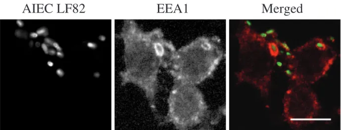
the interactions of AIEC LF82-containing phagosomes with early endocytic compartments within J774 macroph-ages. EEA1 was found on 52.8% ±2.9% of LF82-containing phagosomes after 10 min of infection, and at this time point the protein appeared to be evenly distrib-uted around bacteria-containing phagosomes (Table 1 and Fig. 1). After 20 min of infection, 30.0% ± 3.0% of the LF82-containing phagosomes were still EEA1-positive but this immunostaining decreased rapidly as at 60 min post-infection EEA1 was detected only on 17.1% ± 1.9% of LF82-containing phagosomes and less than 1.4% ± 0.5% of them were EEA1-positive at 24 h post-infection. These experiments show that EEA1 was delivered to and rapidly removed from LF82-containing phagosomes. The local-ization of EEA1 on AIEC LF82-containing phagosomes may represent a prerequisite for the subsequent acquisi-tion of late endocytic characteristics.
AIEC LF82-containing phagosomes acquire features of late endosomes
In order to further determine the nature of the AIEC LF82-replicating compartment within J774 macrophages, immu-nofluorescence experiments were performed using anti-bodies directed against late endosomal specific markers, the Rab7 GTPase and the membrane glycoprotein Lamp-1. After 20 min of infection, 54.4% ± 2.9% of LF82-containing phagosomes were Rab7-positive (Table 1 and Fig. 2). The interaction between LF82-containing phago-somes and Rab7 was transient because at 60 min post-infection only 8.5% ± 0.9% of LF82-containing phago-somes were Rab7-positive. Lamp-1 was detected only on
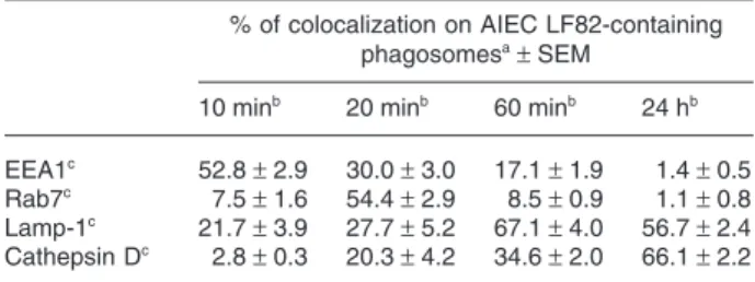
Table 1.Kinetics of the acquisition of endosomal/lysosomal markers by AIEC LF82-containing phagosomes.
% of colocalization on AIEC LF82-containing phagosomesa± SEM 10 minb 20 minb 60 minb 24 hb EEA1c 52.8 ± 2.9 30.0 ± 3.0 17.1 ± 1.9 1.4 ± 0.5 Rab7c 7.5 ± 1.6 54.4 ± 2.9 8.5 ± 0.9 1.1 ± 0.8 Lamp-1c 21.7 ± 3.9 27.7 ± 5.2 67.1 ± 4.0 56.7 ± 2.4 Cathepsin Dc 2.8 ± 0.3 20.3 ± 4.2 34.6 ± 2.0 66.1 ± 2.2 a. For each point, at least 100 AIEC LF82-containing vacuoles were counted and scored for the presence or absence of markers. The results shown are the mean percentage of marker colocalization on AIEC LF82-containing phagosomes ± standard error of the mean (SEM). At least three independent experiments were performed. b. J774 macrophages grown on coverslips were infected with GFP-expressing AIEC strain LF82 as described in Experimental procedures.
c. The endosomal/lysosomal markers were localized using goat anti-bodies against EEA1, chicken antianti-bodies against Rab7, rat antianti-bodies against Lamp-1 and rabbit antibodies against cathepsin D, followed by corresponding secondary antibodies and visualized by confocal microscopy.
AIEC strain LF82 replicates in mature phagolysosomes 473
© 2005 The Authors
Fig. 1. The early endosome-associated antigen 1, EEA1, localizes on AIEC LF82-containing phagosomes immediately after infection. J774 macrophages were infected with GFP-expressing AIEC strain LF82 for 10 min and monolayers were then fixed immediately. Labelling of EEA1 was performed using goat antibodies to EEA1 and Alexa 594-conjugated donkey anti-goat IgG secondary antibodies.
AIEC LF82
EEA1
Merged
Fig. 2. The Rab7 GTPase transiently localizes on AIEC LF82-containing phagosomes after 20 min of infection and is then removed at later times. J774 macrophages were infected with GFP-expressing AIEC strain LF82 for 20 min (20 min post-infection, p.i.) and then fixed immediately, or fixed after a further 40 min incubation period in fresh cell culture medium containing gentamicin (60 min p.i.). Labelling of Rab7 was performed using rabbit antibodies against Rab7 and secondary antibodies Alexa 594-conjugated donkey anti-rabbit IgG.
20 min p.i.
60 min p.i.
474 M.-A. Bringer et al.
27.7% ±5.2% of LF82-containing phagosomes after 20 min of infection (Table 1 and Fig. 3). At 60 min post-infection, 67.1% ± 4.0% of the bacteria-containing phago-somes were Lamp-1-positive. Taken together, these results demonstrate that during early time post-infection, LF82-containing phagosomes progressed from EEA1-positive compartments to Lamp-1-EEA1-positive compartments, with a rapid loss of EEA1 and a transient acquisition of Rab7.
AIEC LF82-containing phagosomes harbour markers of phagolysosomes
At the end of the normal maturation, phagosomes pos-sess lysosomal traits. They accumulate Lamps, continue to acidify, and acquire hydrolytic enzymes, such as cathe-psin D, which are required for the efficient degradation of the phagosomal contents. Analysis of infected macroph-ages at 8 h and 24 h post-infection demonstrated that Fig. 3. The lysosomal membrane protein Lamp-1 localizes on AIEC LF82-containing phagosomes at 60 min post-infection and is maintained until 24 h post-infection. J774 macrophages were infected with GFP-expressing AIEC strain LF82 for 20 min and then incubated for 40 min, 8 h or 24 h (60 min, 8 h and 24 h post-infection, p.i.) in fresh cell culture medium containing gentamicin. Labelling of Lamp-1 was performed using rat antibodies to the murine Lamp-1 and Alexa 594-conjugated goat anti-murine IgG secondary antibodies.
AIEC LF82
Lamp-1
Merged
60 min p.i.
8 h p.i.
AIEC strain LF82 replicates in mature phagolysosomes 475
© 2005 The Authors
AIEC LF82-containing phagosomes were positive for both Lamp-1 and Lamp-2 (Table 1, Figs 3 and 4B).
AIEC LF82-containing phagosomes were further anal-ysed by monitoring the presence of cathepsin D using an
antibody directed against this luminal hydrolase (Fig. 4A). After 20 min of infection, 20.3% ± 4.2% of AIEC LF82-containing phagosomes were positive for cathepsin D. The percentage of LF82-containing phagosomes that
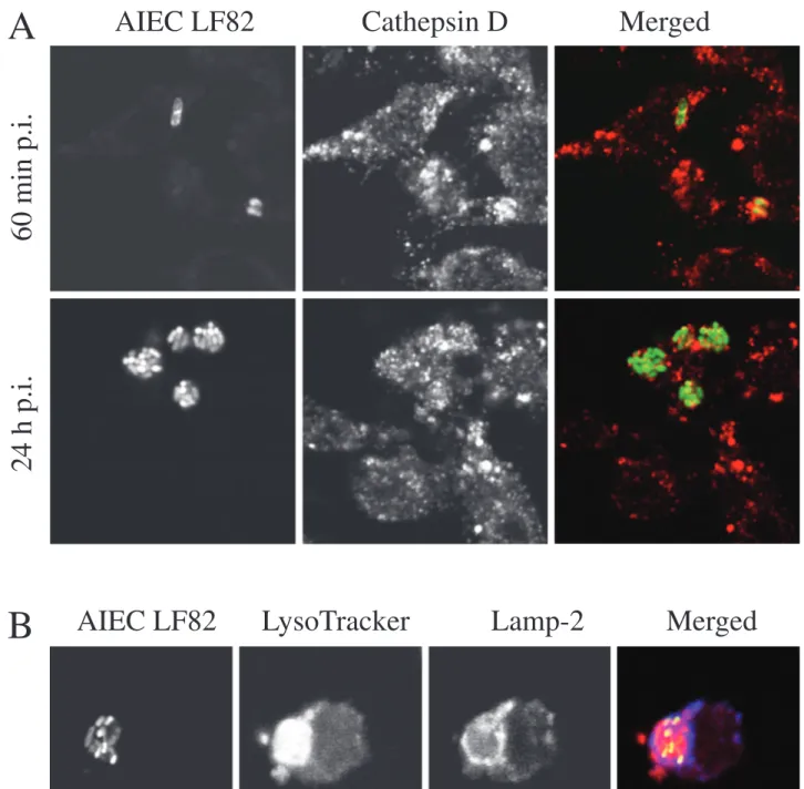
Fig. 4. AIEC LF82 bacteria replicate within acid phagosomes harbouring characteristic markers of phagolysosomes.
A. J774 macrophages were infected with GFP-expressing AIEC strain LF82 for 20 min and then incubated for 40 min (60 min post-infection, p.i.) or for 24 h (24 h p.i.) in fresh cell culture medium containing gentamicin. Labelling of cathepsin D was performed using rabbit anti-cathepsin D antibodies and Alexa 594-conjugated donkey anti-rabbit secondary antibodies.
B. J774 macrophages were infected with GFP-expressing AIEC strain LF82 for 20 min and then incubated for 24 h (24 h p.i.) in fresh cell culture medium containing gentamicin. Labelling of Lamp-2 was performed using rat antibodies to the murine Lamp-2 and Alexa 594-conjugated goat anti-murine secondary antibodies. Colocalization of LysoTracker Red DND-99 with GFP expressing AIEC LF82 bacteria was performed as described in Experimental procedures.
AIEC LF82
LysoTracker
Lamp-2
Merged
A
B
60 min p.i.
24 h p.i.
476 M.-A. Bringer et al.
are cathepsin D-positive increased with time and 66.1% ± 2.2% of them were positively labelled at 24 h post-infection (Table 1 and Fig. 4A). Thus, the progressive acquisition in time of Lamps on the membrane of the AIEC LF82-containing phagosomes was accompanied by an accumulation of the intraluminal cathepsin D, suggesting that these phagosomes mature into phagolysosomes.
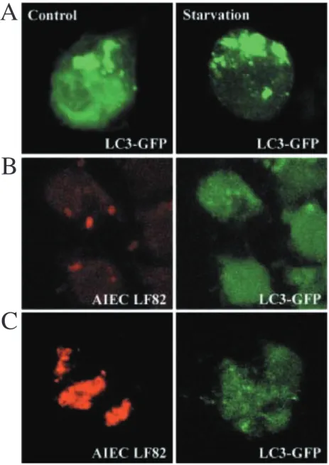
Maturation of AIEC LF82-containing phagosomes does not involve interaction with the autophagic pathway To investigate the possible involvement of the autophagic pathway in the maturation of AIEC LF82-containing pha-gosomes, the localization of LC3, an autophagosome-specific membrane marker, was analysed. For this
pur-pose, J774 macrophages were transiently transfected with the plasmid pEGFP-LC3. As a positive control, macroph-ages were subjected to starvation, a conventional inducer of autophagy (Fig. 5A). Numerous GFP-LC3-labelled vac-uoles were observed in starved cells. As shown in Fig. 5B and C, GFP-LC3 was not recruited on AIEC LF82-containing phagosomes neither at 60 min post-infection nor at 24 h post-infection. No colocalization between GFP-LC3 and LF82 bacteria-containing phagosomes was observed at very early (10 min and 20 min) or later (4 h and 8 h) post-infection times (data not shown). These results suggest that AIEC LF82-containing phagosomes do not interact with the autophagic pathway. However, a transient association of LC3 with autophagosomes cannot be ruled out.
A
B
C
Fig. 5. AIEC LF82-containing phagosomes do not colocalize with LC3, the specific marker of the autophagic pathway. J774 macrophages were transiently transfected with the plasmid pEGFP-LC3.
A. Transfected J774 cells were incubated in complete medium (control, left panel) or in star-vation medium for 4 h (starstar-vation, right panel). B. Transfected J774 cells were infected with AIEC strain LF82 for 20 min and then incubated for 40 min in fresh cell culture medium contain-ing gentamicin.
C. Transfected J774 cells were infected with AIEC strain for 20 min and then incubated for 24 h in fresh cell culture medium containing gentamicin. Bacteria were revealed using rabbit antiserum directed against lipopolysaccharide O83 and Alexa 594-conjugated donkey anti-rabbit secondary antibodies.
AIEC strain LF82 replicates in mature phagolysosomes 477
© 2005 The Authors
AIEC LF82 bacteria replicate within mature
phagolysosomes with low pH and active cathepsin D As AIEC LF82-containing phagosomes were cathepsin D-positive, we performed experiments to define if this pro-tease was in its immature or active form. An acid pH is necessary for a functional and optimal activity of cathep-sin D, thus the pH of bacteria-containing phagosomes was sensed using LysoTracker Red DND-99, a fluorescent acidotropic component used to probe acid organelles. At 24 h post-infection, the large phagosomes within which AIEC LF82 bacteria had extensively replicated were pos-itively marked with the LysoTracker Red (Fig. 4B), indicat-ing that the LF82-containindicat-ing phagosomes were acidic compartments.
To determine the activity of cathepsin D, which localized within AIEC LF82 bacteria-containing phagosomes, experiments using NIRF probe were performed (Fig. 6). NIRF probe is a dual fluorochrome (Cy5.5 and FITC) cathepsin D-activable probe. If cathepsin D is immature, the close spatial proximity of the multiple fluorochromes results in quenching of Cy5.5 fluorochromes and only a FITC signal is detected, which indicates a well internal-ization of the probe into the cell. Besides, in the presence of active cathepsin D, peptidic substrates present on the NIRF probe are cleaved resulting in a release of free Cy5.5 and thus by detection of a Cy5.5 signal (Tung et al., 1999). As a positive control, Texas Red dextran conju-gates were used (Fig. 6A). The distribution of the Cy5.5
fluorescence of the NIRF probe revealed the presence of an active proteolytic form of cathepsin D within LF82-containing phagosomes at 24 h post-infection, suggesting that AIEC bacteria have the ability to replicate in an envi-ronment with active protease (Fig. 6B).
Replication ability of AIEC LF82 bacteria is impaired when the acid pH of the vacuolar compartment is neutralized We studied the effect of vacuolar pH-neutralizing reagents on the replication ability of AIEC LF82 bacteria within J774 macrophages. Two reagents raising the pH of acid com-partments were used, ammonium chloride (NH4Cl) and
chloroquine, which are lysosomotropic components that are membrane permeative in their non-protonated forms but impermeative when protonated in acid phagosomes. None of the reagents tested was directly lethal to either the bacteria or the host cells at the concentrations used. Growth curves of AIEC LF82 in the presence of chloro-quine and NH4Cl are presented in Fig. 7A. As chloroquine
and NH4Cl are lysosomotropic drugs, they presumably
reach higher concentrations in the phagocytic vacuoles than those added originally. To exclude the possibility that higher concentrations of these drugs could be bacteri-cidal, LF82 bacteria were also grown in RPMI medium supplemented with drugs at 100-fold higher concentra-tions (chloroquine 200 µM and NH4Cl 200 mM) than those
used during experiments with macrophages. In the
pres-A
B
Fig. 6. In situ measurement of cathepsin D activity: AIEC strain LF82 bacteria colocalize with active protease.
A. J774 macrophages were incubated with Texas Red dextran conjugates (blue) for 60 min.
B. J774 macrophages were infected with AIEC strain LF82 for 20 min and then incubated for 24 h in culture medium containing gentamicin. Bacteria were revealed using rabbit antiserum directed against lipopolysaccharide O83 and Alexa 594-conjugated donkey anti-rabbit IgG secondary antibodies (blue). NIRF probe was added to cells for 1 h before the end of the experiment. The colocalization of Texas Red dextran conjugates or bacteria with NIRF probe (FITC fluorescence, green) was examined with a confocal microscope (left panel). The Cy5.5 fluorescence (red) corresponds to active cathe-psin D (right panel).
478 M.-A. Bringer et al.
ence of chloroquine at 2 µM or NH4Cl at 2 mM, growth
curves were identical to those of bacteria grown in the absence of these compounds. At concentrations 100-fold of those used in experiments with macrophages, only slight decreases in bacterial growth were observed.
Treatments with NH4Cl and chloroquine decreased the
replication of intracellular AIEC LF82 bacteria in a dose-dependent manner (Fig. 7B). When macrophages were treated with chloroquine at 2 µM or NH4Cl at 2 mM,
inhi-bitions of LF82 intracellular bacteria replication were observed. Percentages of intracellular bacteria at 24 h post-infection relative to those at 1 h post-infection, taken
as 100%, were 69.2% ± 16.8% in the presence of chloro-quine and 109.9% ± 18.8% in the presence of NH4Cl
compared with 361.9% ± 33.0% in absence of treatment. The decrease in bacterial replication of intramacrophagic LF82 bacteria after 24 h of cell treatment with chloroquine or NH4Cl was confirmed by confocal microscopic
exami-nation. At 24 h post-infection chloroquine or NH4
Cl-treated and -infected macrophages showed phagosomes containing only a few bacteria compared with untreated macrophages, which exhibited large phagosomes con-taining numerous bacteria (Fig. 7C). These data demon-strated that an acidic environment is required for Fig. 7. Effect of vacuolar pH-neutralizing agents and iron supplementation on AIEC LF82 intracellular replication within J774 macrophages. A. Growth of AIEC strain LF82 in RPMI medium supplemented with chloroquine at 2 or 200 µM or NH4Cl at 2 or 200 mM.
B. Treatment of AIEC LF82-infected J774 macrophages with chloroquine (1 and 2 µM) or with NH4Cl (1 and 2 mM) with or without ferric citrate at 10 µM. Results are expressed as the number of viable intracellular bacteria at 24 h infection relative to that obtained after 1 h post-infection, taken as 100%. Each value is the mean ± SEM of three independent experiments.
C. Confocal microscopic images of J774 macrophages infected with GFP-expressing AIEC strain LF82 (green) untreated with drug (left panel), treated with 2 µM of chloroquine (middle panel) or treated with 2 mM of NH4Cl (right panel). Actin cytoskeleton of J774 cells was stained using TRITC-labelled phalloidin (red).
Untreated
1.0
0.8
0.6
OD
6200.4
0.2
0
0
4
Time of growth (hours)
% of intracellular
bacteria at
24 h/l h post-infection
8
12
16
20
24
1
2
Chloroquine (
mM)
NH
4Cl (mM)
2
1
2
2
0
100
200
300
400
No ferric citrate Ferric citrate 10 mM Chloroquine 2 mM Chloroquine 200 mM NH4Cl 200 mM NH4Cl 2 mMC
A
B
AIEC strain LF82 replicates in mature phagolysosomes 479
© 2005 The Authors
intracellular replication of AIEC strain LF82 within J774 macrophages.
Exogenous iron supplementation does not restore replication of AIEC LF82 within chloroquine or NH4Cl
treated-macrophages
The intracellular transport and mobilization of iron are dependent upon an acidic environment in endocytic ves-icles. As vacuolar pH-neutralizing reagents such as chlo-roquine and NH4Cl have been reported to interfere with
iron availability for intracellular bacteria (Baynes et al., 1987), we investigated if the decrease in intracellular LF82 replication could be restored following supplementation with exogenous iron. The addition of ferric citrate at 10 µM did not restore the replication level of the AIEC LF82 bacteria within macrophages (Fig. 7B). A very slight increase in bacterial replication was observed with chlo-roquine-treated macrophages but higher concentrations of ferric citrate did not induce any increase in bacterial replication (data not shown). These results indicated that the decrease in the intracellular replication of LF82 bac-teria observed after intraphagosomal pH neutralization was not attributed to impaired iron acquisition.
Discussion
The ileal mucosa of patients with CD is abnormally colo-nized by pathogenic AIEC strains, which have the ability to extensively replicate within J774 macrophages in spa-cious phagosomes, without inducing host cell death (Glasser et al., 2001; Darfeuille-Michaud et al., 2004). The aim of the study was to investigate whether AIEC refer-ence strain LF82 avoid the bactericidal degradative activ-ities of macrophages, by creating a specific replicative niche or by adapting to the hostile phagolysosomal envi-ronment. As biogenesis of AIEC-containing phagosomes may play a crucial role in creating a replicative niche, interactions of these phagosomes with endocytic and autophagic pathways were followed from early to late events within J774 cells.
After infection, AIEC LF82-containing phagosomes rap-idly acquire EEA1, as previously observed with S. enterica serovar Typhimurium (Steele-Mortimer et al., 1999; Knodler and Steele-Mortimer, 2003). As EEA1 is a major effector of the Rab5 GTPase, and as these proteins are associated with vesicle tethering and SNARE docking, which are involved in the process of membrane fusion, transient acquisition of EEA1 can confer to AIEC-phago-somes the ability to reach the late endosomal stage of maturation (Desjardins, 1995; Simonsen et al., 1998; Duc-los et al., 2003). This event involves in particular the acqui-sition of the Rab7 GTPase (Desjardins et al., 1994; Desjardins, 1995). Pathogenic bacteria such as
Mycobac-terium tuberculosis and MycobacMycobac-terium avium inhibit the phagosome–lysosome fusion and replicate within an immature phagosome by blocking vesicle maturation between the stages controlled by Rab5 and Rab7 (Clem-ens and Horwitz, 1996; Via et al., 1997). In the case of AIEC strain LF82, the association of Rab7 with LF82-containing phagosomes occurred but little to no colocal-ization of Rab7 was detected on AIEC LF82-containing phagosomes at 60 min post-infection. This indicates that in contrast to what observed with Mycobacteria, the Rab7 GTPase was not retained on LF82-containing phagosomes, suggesting that they could evolved into phagolysosomes.
Salmonella-containing vacuoles also acquire Rab7 and further Lamp-1, but by bypassing the mannose-6-phosphate receptor (M6PR) compartment, they do not acquire lysosomal hydrolytic enzymes including cathepsin D (Buchmeier and Heffron, 1991; Rathman et al., 1997; Hashim et al., 2000; Garvis et al., 2001; Knodler and Steele-Mortimer, 2003). A gradual increased amount of Lamp-1 on LF82-containing phagosomes was observed at 60 min post-infection, and this Lamp-1 level was main-tained until 24 h post-infection. At this time point, LF82-containing phagosomes were also Lamp-2-positive. In contrast to Salmonella-containing vacuoles, the progres-sive acquisition of Lamps on LF82-containing phago-somes was accompanied by an increased accumulation of the intraluminal cathepsin D. However, the mechanism by which cathepsin D is delivered to the LF82-containing phagosomes remains to be investigated. Indeed, this hydrolase can be delivered either by the endocytic path-way or via the biosynthetic pathpath-way by involving the trans-Golgi network (Claus et al., 1998; Deretic et al., 2004).
As AIEC LF82-containing phagosomes display lysosomal features, LF82 bacteria could reside in an autophagolysosome-like compartment. Indeed, autoph-agy participates to the establishment of replicative com-partments for pathogens such as B. abortus, C. burnetii, L. pneumophila and P. gingivalis (Pizarro-Cerda et al., 1998a,b; Dorn et al., 2001; Beron et al., 2002; Amer and Swanson, 2005). No translocation of the highly specific autophagosomal marker LC3 from the cytosol to the membrane of AIEC LF82-containing phagosomes was observed within J774 macrophages expressing GFP-LC3 at times post-infection studied. This suggests that AIEC LF82-containing phagosomes do not divert to a compart-ment with autophagic characteristics. However, LC3 asso-ciation with autophagosomes can be transient and the involvement of autophagy in the process of phagosomal maturation, though unlikely, cannot be definitively excluded.
As most of the AIEC LF82-containing phagosomes were cathepsin D-positive at 24 h post-infection, and as immunocytological techniques do not address the
biolog-480 M.-A. Bringer et al.
ical activity of cathepsin D, experiments were performed to investigate whether this protease was active. Cathepsin D matures by an internal cleavage induced by the low luminal pH of late endocytic compartments (Rijnboutt et al., 1992). At 24 h post-infection, spacious cathepsin D-positive vacuoles containing LF82 bacteria were D-positive for LysoTracker, a fluorescent acidotropic component used to probe acid organelles, which indicates that these compartments had low pH. Moreover, the dual fluoro-chrome NIRF probe was used to image cathepsin D intraphagolysosomal activity. Confocal microscopy analy-sis revealed the presence of an active proteolytic form of cathepsin D within LF82-containing phagosomes. Thus, AIEC LF82-containing phagosomes mature into active phagolysosomes where bacteria are exposed to acid pH and also to degradative proteases. The way in which AIEC bacteria resist the attacks of protease activities is under current investigation. Bacterial cytoplasmic membrane is one of the targets for most antimicrobial peptides (Epand and Vogel, 1999; Hancock and Rozek, 2002). However, recent studies suggest that lipopolysaccharide (LPS) con-stitues the first protective layer that controls the binding of peptides and their insertion into the cytoplasmic mem-brane (Papo and Shai, 2003; 2005). Thus, it can be spec-ulated that the LPS by itself or the expression of specific proteins on the cell surface could protect AIEC bacteria from antimicrobial peptides. Nor can it be excluded that AIEC bacteria may exploit these proteases to generate amino acids that contribute to their high replication within the phagolysosomes.
Acid vacuolar pH could play a key role in the AIEC LF82 replication within macrophages. Treatment of J774 mac-rophages with vacuolar pH-neutralizing agents such as chloroquine and NH4Cl inhibited the replication of
intrac-ellular LF82 bacteria. However, as these vacuolar pH-neutralizing agents are known to interfere with iron trans-port and mobilization, we checked that the defect in intra-cellular AIEC LF82 bacteria replication was not attributed to impaired iron availability. Iron supplementation has been reported to restore intracellular replication of Fran-cisella tularensis and L. pneumophila in choloroquine and/ or NH4Cl-treated cells (Byrd and Horwitz, 1991; Fortier
et al., 1995), but we observed no increase in AIEC LF82 intracellular replication. Thus, although it is clear that neu-tralizing the endosomal pH interferes with AIEC LF82 bacteria replication, the mechanism of this inhibition needs to be investigated. As already observed with C. burnetii, LF82 bacteria may take advantage of the proton gradient to drive biochemical reactions or nutrient/metab-olite transports that are essential for growth (Hackstadt and Williams, 1981; 1983; Chen et al., 1990). Interest-ingly, we have already reported that low pH represents a key signal for AIEC LF82 bacteria to induce the expres-sion and/or regulation of genes required for
intraphagoso-mal replication (Bringer et al., 2005). Indeed, the stress protein HtrA plays an essential role in the adaptation of intracellular AIEC LF82 bacteria to low pH conditions. Moreover, the LF82-∆htrA isogenic mutant has a reduced rate of growth in an acid medium that partly reproduces the microenvironment of the phagosome, and htrA gene expression was highly upregulated in bacteria grown under these in vitro conditions and also in intramacroph-agic bacteria. The importance of phagosomal acidification for intramacrophagic survival and/or replication has already been reported. An acidic environment constitutes the physiological signal mediating the virB operon expres-sion in B. abortus and the gene expresexpres-sion of the SPI-2 type III secretion system and of the two-component sys-tem PhoP/PhoQ in Salmonella Typhimurium (Alpuche Aranda et al., 1992; Beuzon et al., 1999; Steele-Mortimer et al., 2000; Boschiroli et al., 2002).
In summary, we have provided evidence that in contrast to many pathogens that escape from the normal endocytic pathway or infiltrate autophagy, AIEC LF82 bacteria are taken up by macrophages within phagosomes, which mature without diverting from the classical endocytic path-way, and which share features with phagolysosomes. To survive and replicate in the harsh environment encoun-tered inside these compartments, including acid pH and proteolytic activity of cathepsin D, AIEC LF82 bacteria may have evolved from non-pathogenic bacteria by elab-orating adaptation mechanisms for which acid pH consti-tutes a key signal to activate the expression of virulence genes.
Experimental procedures
Bacterial strain, plasmids and culture condition
AIEC strain LF82 was isolated from a chronic ileal lesion of a patient with CD and belongs to E. coli serotype O83:H1. The plasmid pFPV25.1, which harbours the green fluorescent protein (GFP), was used to visualize bacteria for confocal microscopy analysis (Valdivia and Falkow, 1997). The plasmid pEGFP-LC3 was kindly provided by Tamotsu Toshimori, National Institute of Genetics, Mishima-Shizuoka, Japan (Kabeya et al., 2000). Luria– Bertani (LB) broth or LB agar plates were used for standard cultivation (Institut Pasteur Production).
Culture and infection of J774 macrophages
J774 is a mouse macrophage cell line that was derived from a tumour in a female BALB/c mouse and has been shown to possess characteristics of macrophages. The murine macroph-age-like cell line J774 (American Type Culture Collection No. TIB67) was maintained in an atmosphere containing 5% CO2 at 37°C in RPMI 1640 medium (Biowhittaker Cambrex Company, Verviers, Belgium) supplemented with 10% (v/v) fetal calf serum (FCS; Biowhittaker) and 1% L-Glutamine (Invitrogen, Cergy-Pontoise, France). Cells were seeded in 24-well tissue culture plates at a density of 1 × 10⁵ cells cm−2 and were grown for 18 h
AIEC strain LF82 replicates in mature phagolysosomes 481
© 2005 The Authors
in an atmosphere containing 5% CO2 at 37°C. Before infection, cell monolayers were washed twice with PBS and the medium was replaced with 1 ml of RPMI 1640 supplemented with 10% heat inactivated FCS. J774 monolayers were infected at a multi-plicity of infection (moi) of 100 bacteria per macrophage. After 10 min of centrifugation at 1000 g, corresponding to 10 min of infection and after supplementary 10 min incubation at 37°C with 5% CO2, corresponding to 20 min of infection, infected macroph-ages were washed twice with PBS, and fresh cell culture medium containing 20 µg ml−1 of gentamicin (Gm) was added for 40 min, corresponding to the 60 min post-infection time point, or was added for 8 h or for 24 h, corresponding to the 8 h and 24 h post-infection time point.
Macrophage survival assay
Bacterial uptake, survival and replication were measured by Gm protection assay. Cells were infected as described above. After 10 min of centrifugation at 1000 g and a 10 min incubation period at 37°C with 5% CO2, infected macrophages were washed twice with PBS, and fresh cell culture medium containing 20 µg ml−1 of Gm was added for a 1 h or a 24 h period. To determine the number of intracellular bacteria, cell monolayers were washed once with PBS, and 0.5 ml of 1% Triton X-100 (Sigma Chemical, St Louis, MO) in deionized water was added to each well for 5 min to lyse eukaryotic cells. This concentration of Triton X-100 had no effect on bacterial viability for at least 30 min. Samples were mixed, diluted and plated onto LB agar plates to determine the number of colony-forming units (cfu) recovered from the lysed monolayers. The number of intracellular bacteria was determined after 1 h and 24 h of Gm treatment. Bacterial replication was expressed as the mean percentage of bacteria recovered at 24 h post-infection relative to the number of bacteria recovered after 1 h of Gm treatment, defined as 100%.
Antibodies
Goat antibodies against EEA1 (sc-6415) were obtained from Santa Cruz biotechnology, CA, USA. Rat antibodies against Lamp-1 (No. 1D4B) and rat antibodies against Lamp-2 (No. ABL-93) were obtained from the Developmental Studies Hybridoma Bank, Baltimore, MD, USA. Chicken antibodies against Rab7 were kindly provided by Angela Wandinger-Ness (University of New Mexico, Albuquerke, USA) and rabbit antibodies against Rab7 have been described elsewhere (Meresse et al., 1997). Rabbit antibodies against cathepsin D were purchased from DAKO Corporation (Carpinteria, CA, USA). Rabbit antiserum against E. coli LPS O83 was generously provided by Lothar Beutin (Department of Biological Safety, Robert Koch Institut, Berlin, Germany). Secondary antibodies used were: Alexa 594-conjugated goat anti-murine IgG, Alexa 594-594-conjugated donkey anti-goat IgG, Alexa 594-conjugated donkey anti-rabbit IgG (Jackson ImmunoResearch Laboratories, West Grove, PA, USA) and Fluo Probes 546-conjugated goat anti-chicken IgG (Molecular Probes, Interchim, Montluçon, France).
LysoTracker probe
The fluorescent basic amine probe LysoTracker Red DND-99 was obtained from Molecular Probes. J774 macrophages were
infected with GFP expressing-AIEC strain LF82 as described above. LysoTracker was diluted at a final concentration of 50 nM in complete culture medium containing 20 µg ml−1 of Gm. One hour before the end of the experiment, the medium was replaced by a medium containing LysoTracker. Cells were washed five times with PBS and then fixed with 3% paraformaldehyde (PFA), pH 7.4, in PBS for 10 min at room temperature. Colocalization of LysoTracker with bacteria was determined using confocal microscopy.
Cathepsin D activity measurement
J774 macrophages were infected as described above. The cathe-psin D-sensitive near-infrared fluorescence (NIRF) probe was prepared as previously described (Tung et al., 1999; 2000). It was conjugated with FITC to monitor probe internalization and with Cy5.5 marker that became fluorescent in the near-infrared spec-trum after cathepsin D activation. NIRF at 0.1 µM was added to infected cells 1 h before the end of the experiment. J774 cells were then washed to remove the free NIRF probe and were fixed with 3% PFA, pH 7.4, in PBS for 10 min at room temperature. As a positive control, Texas Red dextran conjugates, 40 000 MW (Molecular Probes) were used. The colocalization of bacteria and dextran conjugates with the NIRF probe was examined by con-focal microscopy.
Transient transfection with pEGFP-LC3
Adherent cells at 90% confluence were transfected during 5 h with the plasmid pEGFP-LC3 (3 µg DNA) using lipofectamine reagent (Invitrogen) according to the manufacturer’s instructions. After transfection, cells were washed twice with PBS, and com-plete RPMI medium was added until infection. Transfected-cells were infected with AIEC strain LF82 as described above and processed for confocal microscopy.
Induction of autophagy
Autophagy was induced by starvation (Mortimore and Schworer, 1977; Munafo and Colombo, 2001). LC3-GFP transfected cells were washed three times with PBS at 37°C and incubated in 1 ml of Opti-MEM I reduced Medium (Invitrogen) for 4 h.
Addition of vacuolar-pH-neutralizing reagents and ferric citrate to infected cells
Reagents used to neutralize the vacuolar pH were: ammonium chloride (NH4Cl; Sigma) used at 1 or 2 mM and chloroquine (Sigma) used at 1 or 2 µM. Ferric citrate (Sigma) was used at 10, 50 and 100 µM. Macrophages were infected as described below. The reagents were added to cells at 1 h post-infection. Infected and untreated cells were analysed in parallel. All experiments were performed in duplicate. Results are expressed as the number of viable intracellular bacteria at 24 h post-infection relative to that obtained after 1 h post-infection, taken as 100%. Macrophage viability was checked by trypan blue dye exclusion assays and by measurement of lactate dehydrogenase (LDH) release. To test that these reagents were not bactericidal, growth curves were performed in RPMI
482 M.-A. Bringer et al.
medium supplemented with chloroquine at 2 or 200 µM or NH4Cl at 2 or 200 mM.
Immunofluorescence and confocal microscopy
Fixation was performed with 3% PFA, pH 7.4, in PBS for 10 min at room temperature. Fixed cells were washed three times in PBS and permeabilized by incubation in PBS containing 0.1% saponin (Sigma). Primary and secondary antibodies were diluted in PBS containing 0.1% saponin and 5% horse serum (Sigma). Cover-slides were incubated with primary antibodies for 30 min at room temperature, washed in PBS containing 0.1% saponin and then incubated for 30 min in obscurity with secondary antibodies. Cov-erslides were mounted onto glass slides using Mowiol (Aldrich, Steinheim, Germany). When necessary, the actin cytoskeleton was stained for 15 min with TRITC-labelled-phalloidin at 1 µg ml−1 (Sigma). Cells were observed with Leica TCS 4DA confocal microscope (CNRS Marseille Luminy, France) and with Olympus Fluoview (IFR Santé Université d’Auvergne, Clermont-Ferrand, France). To determine the percentage of positive AIEC LF82-containing phagosomes for a specific marker, at least 100 bac-teria-containing phagosomes were counted and scored for the presence or absence of the marker protein. Each confocal microscopy image is representative of three independent experiments.
Statistical analysis
Student’s t-test was used for comparison of the two groups of data. All experiments were repeated at least three times. A
P-value less than or equal to 0.05 was considered statistically
significant.
Acknowledgements
This study was supported by a grant from the Ministère de la Recherche et de la Technologie and by grants from the Associ-ation F. Aupetit (AFA) and Institut de Recherche des Maladies de l’Appareil Digestif (IRMAD; Laboratoire Astra France). The con-focal microscope Olympus fluoview was property of Institut Fédéral de Recherche (IFR) Santé Auvergne. We thank L. Beutin (Department of Biological Safety, Robert Koch Institut, Berlin, Germany) for rabbit antiserum against E. coli lipopolysaccharide O83, T. Yoshimori (National Institute of Genetics Mishima, Shizuoka-Ken, Japan) for the plasmid pEGFP-LC3 and A. Wandinger-Ness (University of New Mexico, Albuquerque, USA) for Rab7 antibody.
References
Alpuche Aranda, C.M., Swanson, J.A., Loomis, W.P., and Miller, S.I. (1992) Salmonella typhimurium activates viru-lence gene transcription within acidified macrophage pha-gosomes. Proc Natl Acad Sci USA 89: 10079–10083. Amer, A.O., and Swanson, M.S. (2005) Autophagy is an
immediate macrophage response to Legionella
pneumo-phila. Cell Microbiol 7: 765–778.
Baynes, R., Bukofzer, G., Bothwell, T., Bezwoda, W., and Macfarlane, B. (1987) Transferrin receptors and transferrin
iron uptake by cultured human blood monocytes. Eur J Cell
Biol 43: 372–376.
Becker, T., Volchuk, A., and Rothman, J.E. (2005) Differential use of endoplasmic reticulum membrane for phagocytosis in J774 macrophages. Proc Natl Acad Sci USA 102: 4022– 4026.
Beron, W., Gutierrez, M.G., Rabinovitch, M., and Colombo, M.I. (2002) Coxiella burnetii localizes in a Rab7-labeled compartment with autophagic characteristics. Infect Immun 70: 5816–5821.
Beuzon, C.R., Banks, G., Deiwick, J., Hensel, M., and Holden, D.W. (1999) pH-dependent secretion of SseB, a product of the SPI-2 type III secretion system of Salmonella
typhimurium. Mol Microbiol 33: 806–816.
Boschiroli, M.L., Ouahrani-Bettache, S., Foulongne, V., Michaux-Charachon, S., Bourg, G., Allardet-Servent, A.,
et al. (2002) The Brucella suis virB operon is induced intra-cellularly in macrophages. Proc Natl Acad Sci USA 99: 1544–1549.
Boudeau, J., Glasser, A.L., Masseret, E., Joly, B., and Dar-feuille-Michaud, A. (1999) Invasive ability of an Escherichia
coli strain isolated from the ileal mucosa of a patient with
Crohn’s disease. Infect Immun 67: 4499–4509.
Bringer, M.A., Barnich, N., Glasser, A.L., Bardot, O., and Darfeuille-Michaud, A. (2005) HtrA stress protein is involved in intramacrophagic replication of adherent and invasive Escherichia coli strain LF82 isolated from a patient with Crohn’s disease. Infect Immun 73: 712–721.
Buchmeier, N.A., and Heffron, F. (1991) Inhibition of mac-rophage phagosome-lysosome fusion by Salmonella
typh-imurium. Infect Immun 59: 2232–2238.
Byrd, T.F., and Horwitz, M.A. (1991) Chloroquine inhibits the intracellular multiplication of Legionella pneumophila by limiting the availability of iron. A potential new mechanism for the therapeutic effect of chloroquine against intracellular pathogens. J Clin Invest 88: 351–357.
Chen, S.Y., Vodkin, M., Thompson, H.A., and Williams, J.C. (1990) Isolated Coxiella burnetii synthesizes DNA during acid activation in the absence of host cells. J Gen Microbiol 136: 89–96.
Claus, V., Jahraus, A., Tjelle, T., Berg, T., Kirschke, H., Faulstich, H., and Griffiths, G. (1998) Lysosomal enzyme trafficking between phagosomes, endosomes, and lysos-omes in J774 macrophages. Enrichment of cathepsin H in early endosomes. J Biol Chem 273: 9842–9851.
Clemens, D.L., and Horwitz, M.A. (1995) Characterization of the Mycobacterium tuberculosis phagosome and evidence that phagosomal maturation is inhibited. J Exp Med 181: 257–270.
Clemens, D.L., and Horwitz, M.A. (1996) The Mycobacterium
tuberculosis phagosome interacts with early endosomes
and is accessible to exogenously administered transferrin.
J Exp Med 184: 1349–1355.
Darfeuille-Michaud, A., Neut, C., Barnich, N., Lederman, E., Di Martino, P., Desreumaux, P., et al. (1998) Presence of adherent Escherichia coli strains in ileal mucosa of patients with Crohn’s disease. Gastroenterology 115: 1405–1413. Darfeuille-Michaud, A., Boudeau, J., Bulois, P., Neut, C.,
Glasser, A.L., Barnich, N., et al. (2004) High prevalence of adherent-invasive Escherichia coli associated with ileal mucosa in Crohn’s disease. Gastroenterology 127: 412– 421.
Deretic, V., Vergne, I., Chua, J., Master, S., Singh, S.B., Fazio, J.A., and Kyei, G. (2004) Endosomal membrane
AIEC strain LF82 replicates in mature phagolysosomes 483
© 2005 The Authors
traffic: convergence point targeted by Mycobacterium
tuberculosis and HIV. Cell Microbiol 6: 999–1009.
Desjardins, M. (1995) Biogenesis of phagolysosomes: the ‘kiss and run’ hypothesis. Trends Cell Biol 5: 183–186. Desjardins, M., Huber, L.A., Parton, R.G., and Griffiths, G.
(1994) Biogenesis of phagolysosomes proceeds through a sequential series of interactions with the endocytic appa-ratus. J Cell Biol 124: 677–688.
Dorn, B.R., Dunn, W.A., Jr, and Progulske-Fox, A. (2001)
Porphyromonas gingivalis traffics to autophagosomes in
human coronary artery endothelial cells. Infect Immun 69: 5698–5708.
Dorn, B.R., Dunn, W.A., Jr, and Progulske-Fox, A. (2002) Bacterial interactions with the autophagic pathway. Cell
Microbiol 4: 1–10.
Duchmann, R., and Zeitz, M. (1999) Crohn’s disease. In
Mucosal Immunology. Ogra, P.L., Mestecky, J., Lamm,
M.E., Strober, W., Bienenstock, J., and McGhee, J.R. (eds). San Diego, CA: Academic Press, pp. 1055–1080. Duclos, S., Corsini, R., and Desjardins, M. (2003)
Remodel-ing of endosomes durRemodel-ing lysosome biogenesis involves ‘kiss and run’ fusion events regulated by rab5. J Cell Sci 116: 907–918.
Elson, C.O. (2000) Commensal bacteria as targets in Crohn’s disease. Gastroenterology 119: 254–257.
Epand, R.M., and Vogel, H.J. (1999) Diversity of antimicrobial peptides and their mechanisms of action. Biochim Biophys
Acta 1462: 11–28.
Fortier, A.H., Leiby, D.A., Narayanan, R.B., Asafoadjei, E., Crawford, R.M., Nacy, C.A., and Meltzer, M.S. (1995) Growth of Francisella tularensis LVS in macrophages: the acidic intracellular compartment provides essential iron required for growth. Infect Immun 63: 1478–1483. Garvis, S.G., Beuzon, C.R., and Holden, D.W. (2001) A role
for the PhoP/Q regulon in inhibition of fusion between lyso-somes and Salmonella-containing vacuoles in macroph-ages. Cell Microbiol 3: 731–744.
Glasser, A.L., Boudeau, J., Barnich, N., Perruchot, M.H., Colombel, J.F., and Darfeuille-Michaud, A. (2001) Adher-ent invasive Escherichia coli strains from patiAdher-ents with Crohn’s disease survive and replicate within macrophages without inducing host cell death. Infect Immun 69: 5529– 5537.
Gruenberg, J. (2001) The endocytic pathway: a mosaic of domains. Nat Rev Mol Cell Biol 2: 721–730.
Gu, F., and Gruenberg, J. (1999) Biogenesis of transport intermediates in the endocytic pathway. FEBS Lett 452: 61–66.
Hackstadt, T., and Williams, J.C. (1981) Biochemical strata-gem for obligate parasitism of eukaryotic cells by Coxiella
burnetii. Proc Natl Acad Sci USA 78: 3240–3244.
Hackstadt, T., and Williams, J.C. (1983) pH dependence of the Coxiella burnetii glutamate transport system. J
Bacte-riol 154: 598–603.
Hancock, R.E., and Rozek, A. (2002) Role of membranes in the activities of antimicrobial cationic peptides. FEMS
Microbiol Lett 206: 143–149.
Harrison, R.E., Bucci, C., Vieira, O.V., Schroer, T.A., and Grinstein, S. (2003) Phagosomes fuse with late endo-somes and/or lysoendo-somes by extension of membrane pro-trusions along microtubules: role of Rab7 and RILP. Mol
Cell Biol 23: 6494–6506.
Hashim, S., Mukherjee, K., Raje, M., Basu, S.K., and Mukho-padhyay, A. (2000) Live Salmonella modulate expression
of Rab proteins to persist in a specialized compartment and escape transport to lysosomes. J Biol Chem 275: 16281– 16288.
Kabeya, Y., Mizushima, N., Ueno, T., Yamamoto, A., Kir-isako, T., Noda, T., et al. (2000) LC3, a mammalian homo-logue of yeast Apg8p, is localized in autophagosome membranes after processing. EMBO J 19: 5720–5728. Knodler, L.A., and Steele-Mortimer, O. (2003) Taking
posses-sion: biogenesis of the Salmonella-containing vacuole.
Traffic 4: 587–599.
Meresse, S., Andre, P., Mishal, Z., Barad, M., Brun, N., Desjardins, M., and Gorvel, J.P. (1997) Flow cytometric sorting and biochemical characterization of the late endo-somal rab7-containing compartment. Electrophoresis 18: 2682–2688.
Meresse, S., Steele-Mortimer, O., Moreno, E., Desjardins, M., Finlay, B., and Gorvel, J.P. (1999) Controlling the mat-uration of pathogen-containing vacuoles: a matter of life and death. Nat Cell Biol 1: E183–E188.
Mizushima, N., Ohsumi, Y., and Yoshimori, T. (2002) Autoph-agosome formation in mammalian cells. Cell Struct Funct 27: 421–429.
Mortimore, G.E., and Schworer, C.M. (1977) Induction of autophagy by amino-acid deprivation in perfused rat liver.
Nature 270: 174–176.
Munafo, D.B., and Colombo, M.I. (2001) A novel assay to study autophagy: regulation of autophagosome vacuole size by amino acid deprivation. J Cell Sci 114: 3619– 3629.
Papo, N., and Shai, Y. (2003) Can we predict biological activity of antimicrobial peptides from their interactions with model phospholipid membranes? Peptides 24: 1693– 1703.
Papo, N., and Shai, Y. (2005) A molecular mechanism for lipopolysaccharide protection of Gram-negative bacteria from antimicrobial peptides. J Biol Chem 280: 10378– 10387.
Pizarro-Cerda, J., Moreno, E., Sanguedolce, V., Mege, J.L., and Gorvel, J.P. (1998a) Virulent Brucella abortus prevents lysosome fusion and is distributed within autophagosome-like compartments. Infect Immun 66: 2387–2392. Pizarro-Cerda, J., Meresse, S., Parton, R.G., van der Goot,
G., Sola-Landa, A., Lopez-Goni, I., et al. (1998b) Brucella
abortus transits through the autophagic pathway and
rep-licates in the endoplasmic reticulum of nonprofessional phagocytes. Infect Immun 66: 5711–5724.
Podolsky, D.K. (2002) Inflammatory bowel disease. N Engl J
Med 347: 417–429.
Rathman, M., Barker, L.P., and Falkow, S. (1997) The unique trafficking pattern of Salmonella typhimurium-containing phagosomes in murine macrophages is independent of the mechanism of bacterial entry. Infect Immun 65: 1475– 1485.
Reggiori, F., and Klionsky, D.J. (2002) Autophagy in the eukaryotic cell. Eukaryot Cell 1: 11–21.
Rijnboutt, S., Stoorvogel, W., Geuze, H.J., and Strous, G.J. (1992) Identification of subcellular compartments involved in biosynthetic processing of cathepsin D. J Biol Chem 267: 15665–15672.
Ryan, P., Kelly, R.G., Lee, G., Collins, J.K., O’Sullivan, G.C., O’Connell, J., and Shanahan, F. (2004) Bacterial DNA within granulomas of patients with Crohn’s disease-detection by laser capture microdissection and PCR.
484 M.-A. Bringer et al.
Sartor, R.B., Rath, H.C., Lichtman, S.N., and van Tol, E.A. (1996) Animal models of intestinal and joint inflammation.
Baillieres Clin Rheumatol 10: 55–76.
Shanahan, F. (2002) Crohn’s disease. Lancet 359: 62–69. Simonsen, A., Lippe, R., Christoforidis, S., Gaullier, J.M.,
Brech, A., Callaghan, J., et al. (1998) EEA1 links PI(3)K function to Rab5 regulation of endosome fusion. Nature 394: 494–498.
Steele-Mortimer, O., Meresse, S., Gorvel, J.P., Toh, B.H., and Finlay, B.B. (1999) Biogenesis of Salmonella
typhimurium-containing vacuoles in epithelial cells involves
interactions with the early endocytic pathway. Cell
Micro-biol 1: 33–49.
Steele-Mortimer, O., St-Louis, M., Olivier, M., and Finlay, B.B. (2000) Vacuole acidification is not required for survival of Salmonella enterica serovar Typhimurium within cultured macrophages and epithelial cells. Infect Immun 68: 5401– 5404.
Tung, C.H., Bredow, S., Mahmood, U., and Weissleder, R. (1999) Preparation of a cathepsin D sensitive near-infrared fluorescence probe for imaging. Bioconjug Chem 10: 892– 896.
Tung, C.H., Mahmood, U., Bredow, S., and Weissleder, R. (2000) In vivo imaging of proteolytic enzyme activity using a novel molecular reporter. Cancer Res 60: 4953– 4958.
Ullrich, H.J., Beatty, W.L., and Russell, D.G. (1999) Direct delivery of procathepsin D to phagosomes: implications for phagosome biogenesis and parasitism by Mycobacterium.
Eur J Cell Biol 78: 739–748.
Valdivia, R.H., and Falkow, S. (1997) Fluorescence-based isolation of bacterial genes expressed within host cells.
Science 277: 2007–2011.
Via, L.E., Deretic, D., Ulmer, R.J., Hibler, N.S., Huber, L.A., and Deretic, V. (1997) Arrest of mycobacterial phagosome maturation is caused by a block in vesicle fusion between stages controlled by rab5 and rab7. J Biol Chem 272: 13326–13331.
Yoshimori, T. (2004) Autophagy: a regulated bulk degradation process inside cells. Biochem Biophys Res Commun 313: 453–458.
Zumla, A., and James, D.G. (1996) Granulomatous infec-tions: etiology and classification. Clin Infect Dis 23: 146– 158.