HAL Id: inserm-01135586
https://www.hal.inserm.fr/inserm-01135586
Submitted on 25 Mar 2015HAL is a multi-disciplinary open access archive for the deposit and dissemination of sci-entific research documents, whether they are pub-lished or not. The documents may come from teaching and research institutions in France or abroad, or from public or private research centers.
L’archive ouverte pluridisciplinaire HAL, est destinée au dépôt et à la diffusion de documents scientifiques de niveau recherche, publiés ou non, émanant des établissements d’enseignement et de recherche français ou étrangers, des laboratoires publics ou privés.
Type, density, and presentation of grafted adhesion
peptides on polysaccharide-based hydrogels control
preosteoblast behavior and differentiation.
Jing Jing, Audrey Fournier, Anna Szarpak-Jankowska, Marc Block, Rachel
Auzély-Velty
To cite this version:
Jing Jing, Audrey Fournier, Anna Szarpak-Jankowska, Marc Block, Rachel Auzély-Velty. Type, den-sity, and presentation of grafted adhesion peptides on polysaccharide-based hydrogels control pre-osteoblast behavior and differentiation.. Biomacromolecules, American Chemical Society, 2015, 16 (3), pp.715-22. �inserm-01135586�
1
Type, density, and presentation of grafted adhesion peptides on
1polysaccharide based hydrogels control pre-osteoblast behavior and
2differentiation
34
Jing Jinga, Audrey Fourniera, AnnaSzarpak-Jankowskaa, Marc R. Blockb and Rachel
Auzély-5
Veltya*
6
aUniv. Grenoble Alpes; CERMAV, F-38000 Grenoble, France; CNRS, CERMAV, F-38000
7
Grenoble, France
8
bUniv. Grenoble Alpes; Institut Albert Bonniot, CR INSERM U823; team 4; BP170, F-38042
9 Grenoblecedex 09, France 10 11 *Corresponding author 12
Tel: +33 4 76 03 76 71. E-mail: rachel.auzely@cermav.cnrs.fr
13 14
ABSTRACT:
15
In this work, cell-responsive polysaccharide hydrogels were prepared by a simple procedure
16
based on the sequential bioconjugation and crosslinking of the polysaccharide backbone with
17
bioactive peptides and poly(ethylene glycol)-bis(thiol) (PEG-(SH)2), respectively. Using
thiol-18
ene reactions, we successfully functionalized hyaluronic acid (HA) and carboxymethylcellulose
19
(CMC) with short and long peptides (5-mer and 15-mer derivatives, respectively) derived from
20
adhesive proteins of bone extracellular matrix. The resulting HA-peptide and CMC-peptide
2
conjugates with varying degrees of substitution were then carefully characterized by 1H NMR
1
spectroscopy to precisely control the peptide density into the hydrogels crosslinked with
PEG-2
(SH)2. Pre-osteoblast seeded on the hydrogels with controlled identical stiffness spread in a
3
manner that was strongly dependent on ligand density. Surprisingly, increasing the density of the
4
adhesive peptide anchors did not result in a plateau of initial cell spreading but rather in a
bell-5
shaped cell response which varies with the nature of both polysaccharide backbone and
6
functional peptide. Placing the cells under optimal conditions for cell/hydrogel interaction, we
7
showed that in HA hydrogels, the polysaccharide moiety is not solely a passive scaffold that
8
presents the active peptides but is an active player in cell microenvironment to control and sustain
9
cell activity.
10
11
Keywords: hyaluronic acid, hydrogel, thiol-ene, cell-adhesion, osteoblasts
12 13
INTRODUCTION
14
Bio-instructive, -specific, and -responsive hydrogels mimicking the extracellular matrix (ECM)
15
environment have emerged out of the traditional landscape of inert biomaterials.1-3They are often
16
made of natural polysaccharides that act as scaffold for cellular guidance and wound healing
17
while they may also be subjected to biodegradation and can be replaced by bona fide extracellular
18
matrix over time. In addition, their biophysical properties such as compliance can be easily tuned
19
to match the physiological cellular environment and have been of broad use such as in vitro
20
culture and maintenance of stem cells, or direct stem cell specification.4-5 Indeed, changes in
21
tissue and organ stiffness are frequently symptoms of diseases such as cancer.6
3
Hyaluronic acid is a high molar mass(6-7000 kg/mol) linear glycosaminoglycan found in soft
1
tissues such as connective, epithelial and neuronal tissues, and synovial fluid as well. It consists
2
of a repeating disaccharide unit made of N-acetyl-D-glucosamine and D-glucuronic acid residues.
3
7-8
This polysaccharide can be obtained with a high degree of purity (pharmaceutical grade) on a
4
large scale by bacterial fermentation which makes it very attractive for designing soft materials in
5
the biomedical field. Various functional groups such as sulfate, (meth)acrylate, alkyl chains but
6
also host molecules, and peptides can be introduced along the polysaccharide backbone in order
7
to control gelation or compliance and trigger cell specific interactions with cell surface
8
receptors.9-12 Therefore this polysaccharide has been widely used to produce a variety of tailor
9
made hydrogels.11, 13-14 Other hydrogels relying on polysaccharides that are not present in human
10
tissues (carboxymethylcellulose, chitosan, alginates…), may be chosen for their longer resistance
11
to in vivo environment while keeping their biocompatibility.
12
The cell surface receptors integrins are viewed as major players of cell adhesion to the
13
extracellular matrix and transmit signals to cells upon occupancy with extracellular ligands as
14
well as acting as mechano-transduction sensors.15 Their roles have been emphasized in many
15
biological processes such as development, immune response, migration, and hemostasis.16-19 In
16
addition, on mammalian cells, four major classes of HA cell receptors have been described:
17
CD44 standard or variants,20 their co-receptor RHAMM,21 layilin,22 and ICAM-123. So far, little
18
is known about the role of HA receptors in cell signaling as well as their putativecrosstalk with
19
other well-established partners of cell/ECM interactions such as integrins, although recent reports
20
have described some interplay between HA and integrin substrate in controlling cell behavior.24
21
On the other hand, many cell types can attach and spread onto HA hydrogels in a
stiffness-22
dependent manner. This indicates that beside integrins, HA receptors may act as
mechano-23
sensors as well.25 However this adhesion is cell type dependent and is weaker than integrin
4
mediated adhesion. This is in contrast to non-mammalian polysaccharide-based gels which are
1
generally resistant to cell-adhesion. Such hydrogel matrices are often used to provide a blank
2
canvas in which peptides containing consensus sequences recognized by integrins such as the
3
Arg-Gly-Asp (RGD) sequence are immobilized, thereby providing a defined chemical
4
environment to regulate cell functions. Naturally derived alginate is an example of such hydrogel
5
in which ligand density was found to be a critical regulator of adhesion, spreading, proliferation,
6
and differentiation of pre-osteoblasts.26 Yet, for properly probing cell responses to these soft
7
matrices, polysaccharide hydrogels with independently tunable stiffness and biochemical ligand
8
density must be carefully designed.5We have previously shown that ene-functional
9
polysaccharides esterified with pentenoic anhydride can be rapidly cross-linked with
di-10
functional thiols via thiol-ene photochemistry under physiological conditions.27 Cell-responsive
11
polysaccharide hydrogels were thus prepared by a simple procedure based on the sequential
12
bioconjugation and crosslinking of the polysaccharide backbone with a bioactive peptide and
13
poly(ethylene glycol)-bis(thiol), respectively. By varying the initial reactant stoichiometry as well
14
as the nature of the biomolecules, this strategy offers enormous possibilities in the design of
15
reproducible, highly tunable gels of which the cell-adhesive and mechanical properties are
16
independently controlled.
17
In this work, we used this procedure to investigate cell adhesion, spreading, and proliferation in
18
short term culture as well as differentiation in long term culture of pre-osteoblasts on hydrogels
19
depending on theirpolysaccharide component (hyaluronic acid vs carboxymethylcellulose) as
20
well as on peptide type and density.The peptides used are derived from adhesive proteins present
21
in the ECM of bone, namely fibronectin (Fn) and bone sialoprotein (bsp). They consist of
22
GRGDS (a fragment fromFn)28 and Ac-CGGNGEPRGDTYRAY-NH2 (a 15-mer derived from
23
bone sialoprotein, and here denoted as bsp-RGD)29. These two RGD peptides were selected
5
because they have shown promise for use in orthopedic tissue engineering applications by
1
improving the adhesion and function of a number of osteoblastic cell lines.30-31While several
HA-2
based hydrogels with tunable stiffness and RGD-ligand density have been reported in the
3
literature,32-36 the potential regulatory role of presentation of adhesion peptides in terms of
4
polysaccharide backbone, type and density on 2D cell culture has never been examined in detail.
5
Herein, we demonstrated ability to form HA-based gels that either permit or inhibit cell spreading
6
according to the ligand density at fixed stiffness. Moreover, by comparing cell responses to
HA-7
based gels with those to CMC-gels, at optimal peptide density allowing to support efficient
8
spreading of pre-osteoblasts, HA-peptide hydrogels versus CMC peptide hydrogels allowed a
9
better differentiation of pre-osteoblasts into osteoblasts (using increased mineralization
10
capabilities as a read-out). Therefore in addition to peptide-induced integrin signaling, the HA
11
backbone triggers an additional signal that was required for differentiation.
12 13
MATERIALS AND METHODS
14
Materials.Bacterial hyaluronic acid under the sodium salt form (Mw = 120 kg/mol) was
15
purchased from HTL (Javené, France). The sample of carboxymethylcellulose (CMC, marketed
16
under the name Blanose®, grade 7LF PH, DS = 0.7, Mw = 70 kg/mol) was kindly provided by
17
Hercules (France). The molecular weight distribution and the weight-average molecular weight
18
of the HA and CMC samples were determined by size exclusion chromatography using a Waters
19
GPC Alliance chromatograph (USA) equipped with a differential refractometer and a light
20
scattering detector (MALLS) from Wyatt (USA); the solutions were injected at a concentration of
21
1 mg/mL in 0.1 M NaNO3. The polydispersity index of the samples is Mw/Mn ~ 1.5-2.Pentenoic
22
anhydride, N,N-dimethylformamide (DMF), phosphate buffer saline (PBS, pH ~ 7.4),
2-(N-23
morpholino)ethanesulfonic acid (MES), (3-aminopropyl)trimethoxysilane (APTS),
6
dimethylaminopropyl)carbodiimide hydrochloride (EDC), N-hydroxysulfosuccinimide
(sulfo-1
NHS)were purchased from Sigma-Aldrich-Fluka (L'isled'Abeau, France).
2-hydroxy-1-[4-(2-2
hydroxyethoxy)phenyl]-2-methyl-1-propanone (Irgacure 2959) was kindly provided by Ciba
3
Specialty Chemicals (Basel, Switzerland). All chemicals were used without any further
4
purification.
5
The peptides GRGDS, GRGDE, Ac-CGGNGEPRGDTYRAY-NH2 and
Ac-6
CGGNGEPRGETYRAY-NH2 with >85% purity (from manufacturer HPLC analysis), were
7
obtained from GeneCust Europe (Luxemburg). Poly(ethylene glycol)-bis(thiol) (Mn = 3400
8
g/mol) was purchased from PEGWorks (USA). All chemicals were used without any further
9
purification. The water used in all experiments was purified by aElgaPurelab purification system,
10
with a resistivity of 18.2 MΩ·cm.
11
Synthesis of pentenoate-modified HA (HA-p). HA (0.30 g, 0.75 mmol) was dissolved in
12
ultrapure water (15 mL) at 4 °C and the resulting mixture was kept at 4 °C under continuous
13
stirring overnight for complete dissolution. DMF (10 mL) was then added dropwise in order to
14
have a water/DMF ratio of (3:2, v/v).Pentenoic anhydride (0.411 g, 2.25 mmol) was added while
15
maintaining the pH between 8 and 9 (by adding 0.5 M NaOH) for 4 hours. The reaction was kept
16
at 4 °C under continuous stirring for one night. After this time, NaCl was added to the reaction
17
mixture to have a NaCl concentration of 0.5 M. The polymer was precipitated by addition of
18
ethanol (water/ EtOH (v/v) ratio of 2:3). After removal of the supernatant, the precipitate was
19
successively washed with mixtures of water/EtOH (3:7, 1:4, v/v) and finally dissolved in
20
ultrapure water for a final purification by diafiltration (ultramembraneAmicon YM10) with
21
ultrapure water. The purified product was recovered by freeze-drying and characterized by 1H
22
NMR spectroscopy. The degree of substitution (DS, the average number of moles of substituents
23
per repeating unit of the polysaccharide)was determined to be 0.50 0.05.
7
Synthesis of pentenoate-modified CMC. CMC (0.30 g, 1.34 mmol) was dissolved in ultrapure
1
water (15 mL) at 4 °C and the resulting mixture was kept at 4 °C under continuous stirring
2
overnight for complete dissolution. DMF (10 mL) was then added dropwise in order to have a
3
water/DMF ratio of (3:2, v/v). Pentenoic anhydride (0.980 g, 5.36 mmol) was added while
4
maintaining the pH between 8 and 9 (by adding 0.5 M NaOH) for 4 hours. The reaction was kept
5
at 4 °C under continuous stirring for one night. After this time, NaCl was added to the reaction
6
mixture to have a NaCl concentration of 0.5 M. The polymer was precipitated by addition of
7
ethanol (water/ EtOH (v/v) ratio of 2:3). After removal of the supernatant, the precipitate was
8
successively washed with mixtures of water/EtOH (3:7, 1:4, v/v) and finally dissolved in
9
ultrapure water for a final purification by diafiltration (ultramembraneAmicon YM10) with
10
ultrapure water. The purified product was recovered by freeze-drying and characterized by 1H
11
NMR spectroscopy. The degree of substitution was determined to be 0.20 ± 0.02.
12
Synthesis of HA-peptide and CMC-peptide conjugates.
13
Pentenoate-modified HA or CMC (0.100 g) was dissolved in ultrapure water (10 mL). 50 µL of
14
an aqueous solution of Irgacure 2959 (10 mg/mL) was then added to the polysaccharide solution
15
to obtain a final photoiniator concentration of 5 % (w/v), followed by thepeptide containing a
16
thiol function. The amount of the peptide was adapted to the desired degree of substitution (see
17
Table 1). The mixture was exposed to light with a UV intensity of 20 mW/cm2 for 5 min under
18
stirring. The product was purified by diafiltration (ultramembraneAmicon YM10) with ultrapure
19
water and was recovered by freeze-drying. The degree of substitution of the
polysaccharide-20
peptide conjugates was determined by 1H NMR.
21
NMR spectroscopy
8
The 1H and 13C NMR spectra of the polysaccharide derivatives dissolved in deuterium oxide
1
were performed at 25 or 80 °C (depending of the viscosity of the solution) using a Bruker
2
AVANCE III HD spectrometer operating at 400 MHz (1H) and at 100 MHz (13C). All 1H NMR
3
spectra were recorded by applying a 45° tip angle for the excitation pulse, and a 10 s recycle
4
delay for accurate integration of the proton signals. The 13C NMR spectra were recorded by
5
applying a 45° tip angle for the excitation pulse, and a 3 s recycle delay. All 2D experiments were
6
acquired using 2K data points and 256 time increments. Chemical shifts are given relative to
7
external tetramethylsilane (TMS = 0 ppm) and calibration was performed using the signal of the
8
residual protons or carbons of the solvent as a secondary reference. Deuterium oxide was
9
obtained from SDS (Vitry, France). Details concerning experimental conditions are given in the
10
figure captions.
11
Photorheometry.
12
An AR2000Ex rheometer (TA Instruments Inc.) fitted with a UV-curing cell ( = 365 nm) and an
13
aluminum plate (diameter 19 mm) was used for the in situ measurement of the viscoelastic
14
properties of the HA- and CMC-based hydrogels. Following deposition of 250 µL of a mixture of
15
HA-peptide conjugate (or CMC-peptide conjugate), PEG-(SH)2 and photoinitiator, the gap
16
between the flat quartz plate and the aluminum plate was initially 0.7 mm (measuring ambient
17
temperature). It was controlled during the experiments by maintaining the normal force at 0 0.1
18
N. On each hydrogel, oscillatory time sweep and frequency sweep experiments were performed.
19
They were carried out at 25 °C, with a film of silicone to avoid solvent evaporation. All the
20
dynamic rheological data were checked as a function of strain amplitude to ensure that the
21
measurements were performed in the linear viscoelastic region. In the oscillatory time sweep
22
experiments, the storage modulus (G’) and loss modulus (G’’) were measured during a period of
9
25-30 min at a fixed frequency of 1 Hz and a fixed deformation of 3.5 %. Typically, after
1
deposition of the solution of the mixture of polymers in PBS between the plates and equilibration
2
for 1 min, the solution was illuminated ( = 365 nm) for 25-30 min at a fixed light power (20
3
mW/cm2) leading to gelation. All measurements were done in triplicate. The photoinduced
4
crosslinking reaction was performed by adjusting the PEG-(SH)2 amount with respect to HA and
5
CMC to obtain hydrogels having a similar elastic modulus (G’) of 15000 1500 Pa.The
6
thiol:CMC molar ratio used for gel formation was approximately two-times lower than the
7
thiol:HA molar ratio as the repeating unit of CMC is a monosaccharide whereas that of HA is a
8
disaccharide (see next section).
9
Hydrogel immobilization
10
For the cell culture experiments, hydrogels were covalently linked to the coverslips during
photo-11
crosslinking reactions by grafting pentenoate-modified HA or CMC on the coverslip prior to use.
12
The coverslip was modified with APTS as previously described.37 The modified slip with –NH2
13
groups was immersed in 2 mL of a solution of pentenoate-modified HA or CMC (3 mg/mL in 0.2
14
M MES, pH 4.75). A solution of sulfo-NHS (30 g/L, 100 µL) and a solution of EDC (50g/L, 100
15
µL) were subsequently added. The coverslip was gently stirred at room temperature for 4 h. The
16
slip was then rinsed with water and dried under a flow of nitrogen before use. The HA- and
17
CMC-based hydrogels were prepared by deposition of a solution of HA or CMC modified with
18
pentenoate groups and peptides (75µL, [HA] = 30 g/L and [CMC] = 30 g/L) containing Irgacure
19
2959 (5 % w/v)and PEG-(SH)2(2.1 mg (0.61× 10⁻³mmol)for HA and 0.9 mg (0.26 × 10⁻³mmol
20
for CMC),[SH]/[pentenoate] = 0.5 for HA and for CMC)to the glass slip. The mixture was then
21
coated with a glass slide using 0.4 mm-thick glass plate spacers and then exposed to
UV-22
irradiation (20mW/cm2, 20 min).Under such conditions, hydrogel disks with and without
10
adhesive peptides (G' = 15000 1500 Pa)were prepared.After preparation, hydrogels were
1
transferred to cell culture media (Dulbecco's modified minimal essential medium, DMEM,
2
Invitrogen) supplemented with 50 U/mL penicillin, and 50 mg/mL streptomycin and left for 12 h
3
for equilibrium before cell culture studies. Media was refreshed after 12 h to remove any
4
remaining monomer or initiator.
5
Generation of preosteoblast cell line.
6
To generate preosteoblast cell lines,primary osteoblasts (passage 2) were immortalized by
7
infection with adenovirus expressing the large SV40 T antigen,38Immortalized cells were cloned
8
and each clone tested for its ability to differentiate as viewed alkaline phosphataseexpression and
9
aptitude to mineralize ECM,39 as previously described40. They were cultivated under the standard
10
conditions in DMEM supplemented in 10% fetal calf serum in a 5% CO2 atmosphere.
11
Cell labeling and quantification of cell spreading
12
For quantifying the projected areas of spread cells, the cells were fixed with a solution of 4%
13
(w/v) of paraformaldehyde in 0.2 M phosphate buffer (pH= 7.2) for 10 min at room temperature.
14
After 3 washes in TRIS saline buffer (TBS), the cells were incubated in DMEM supplemented
15
with 0.5% of Vybrant™ DIL cell labeling solution for 15 min at 37°C according to the
16
manufacturer's instructions (Life Technologies, St Aubin, France). The cells were washed 3 times
17
in PBS and mounted under a coverslip with 10 µL of mounting solution for epifluorescence
18
observations. Observation were carried out with anOlympus BX 51 microscope (Olympus
19
Europe Hamburg, Germany) equipped with a Plan NeoFluar 20X (N.A. 0.5) objective. Areas
20
were determined with Metamorph software (Molecular Devices, Sunyvale, CA USA) at a
21
magnification of 200X. Statistical analyses were carried out with R software (The R Project for
22
Statistical Computing, http://www.r-project.org/).
11
EdU cell proliferation assay
1
DNA synthesis detection in proliferating cells was based on the incorporation of
5-ethynyl-2-2
deoxyuridine (EdU) and its subsequent detection by a fluorescent azide through a Cu(I)-catalyzed
3
[3 + 2] cycloaddition reaction.41Cells were grown on hydrogel substrates for 24 hin DMEM
4
supplemented with 10% fetal calf serum, penicillin, and streptomycin. EdU was added to the
5
culture media at 10 μM (Stock 10mM), for 1-2 hour. After labeling, cells were washed with PBS.
6
Cells were fixed by using a standard paraformaldehyde 4% for 10 min. Subsequently, cells were
7
rinsed once with TBS and stained by incubating for 30 min with 0.5-1mL/well with 6 mL of
8
afreshly prepared staining mix made of 110 mMTris pH 8.5, 1 mM CuSO4,60mM ascorbic acid,
9
10µM Alexa azido (from stock solution in DMSO).After staining, the cells were washed three
10
times with TBS with 0.2% Triton X-100. The fraction of fluorescent (dividing) cells was
11
estimated on at least 10 independent frames.
12
Alizarin Red-S Protocol for Mineralization
13
Pre-osteoblasts 80-90% confluent were placed into osteogenic media made of regular DMEM
14
medium with 10 % FetalCloneII (Hyclone), supplemented with 10mM-glycerophosphate, and
15
100µM of ascorbic acid. Culture was continued for 21 days. Mineralization was already observed
16
at 10 days. The calcified mineral deposits were stained with Alizarin Red-S (AR-S) using the
17
protocol developed by Gregory et al.42 Briefly, separate 12-well plates after 8, 11, 15 and 24 days
18
in culture were washed gently with PBS and fixed in 10 % (v/v) formaldehyde for 15 min at
19
room temperature. After washing twice with excess deionized water, the samples were immersed
20
in 0.5 mL of 40 mM AR-S (pH 4.1) for 20 min with gentle shaking. Then, the samples were
21
washed four times with excess deionized water for 5 min each with shaking. The wet samples
22
were subsequently imaged using a stereo microscope Olympus SZX10v with an angled
12
illumination source (Schott K1500). For quantification, threshold of the staining on digital
1
images allowed the measurement of mineralized areas using Metamorph software.
2 3
RESULTS
4
Synthesis of HA-peptide and CMC-peptide conjugates. To fabricate HA and CMC hydrogels
5
(Figure 1), we first functionalized HA and CMC with pentenoate groups by reaction of the
6
hydroxyl groups of the polysaccharides with 4-pentenoic anhydride as previously described.27In
7
these reactions, the feed molar ratios of anhydride to the polysaccharide repeat unit were adjusted
8
in a way to obtain HA-pentenoate (HA-p) and CMC-pentenoate (CMC-p) derivatives having a
9
degree of substitution (DS) of 0.50 and 0.20, respectively (see experimental section). The DS was
10
approximately two-times lower in the case of CMC because the repeating unit of this
11
polysaccharide is only composed of one sugar.
12
Then, the resulting HA-p and CMC-p derivatives were reacted withthe 5-mer peptide GRGDS
13
and the 15-mer peptide bsp-RGD via photoinducedthiol-ene reactions between the thiol of the
14
terminal mercaptopropionic acid in GRGDS and the terminal cysteine in bsp-RGD and, the
15
alkene function of pentenoate. In order to tune the cell-adhesive ligand density while maintaining
16
a constant elastic modulus, the peptides were added in equivalents of no greater than 50% of the
17
total number of alkene groups of HA and CMC.In this way, the double bonds after peptide
18
conjugation are in sufficient numbers to maintain the same crosslinking density while increasing
19
the peptide density. Integrin specific cell response was controlled by grafting non-adhesion
20
peptides containing the RGE sequence instead of RGD: GRGES (Gly-Arg-Gly-Glu-Ser) and
bsp-21
RGE (Ac-Cys-Gly-Gly-Asn-Gly-Glu-Pro-Arg-Gly-Glu-Thr-Tyr-Arg-Ala-Tyr-NH2). Following
22
this coupling step, the HA-peptide and CMC-peptide conjugates were purified by diafiltration
23
and analyzed by 1H NMR to check their structural integrity and precisely determine their DS (see
13
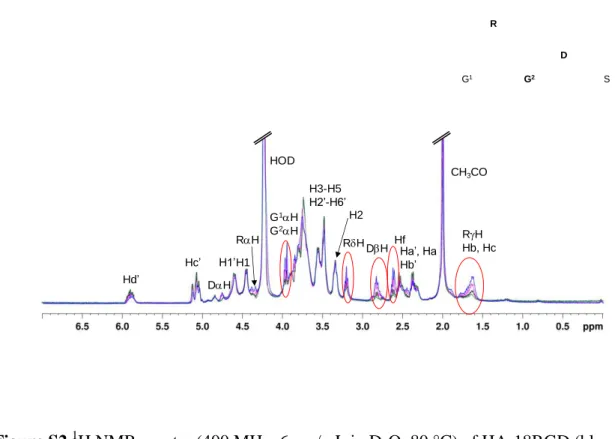
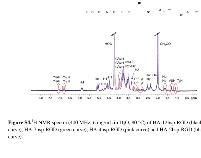
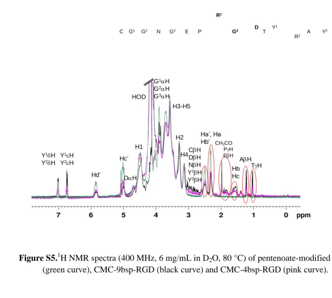
Table 1 and 1H NMR spectrain Supporting Information (Figures S1-S10)). The DS was
1
calculated by digital integration of well-defined signals of the peptide and of the polysaccharides
2
(Figures S3, S6, S8, S10). Of note, the DS values could be confirmed by analyzing the
3
disappearance of the signals of the protons associated with the double bonds (Figures S3, S6, S8,
4
S10).Comparison between the feed molar ratios of peptide to pentenoate and the DS of the
5
modified polysaccharides (Table 1) indicated that the peptides were grafted on HA and on CMC
6
in quite satisfactory yields (66-91 %). The final crosslinking reaction step was performed by
7
adjusting the PEG-(SH)2 amount at a fixed amount of HA or CMC. The HA- and CMC-based
8
hydrogels were prepared using, respectively, a thiol:HA disaccharide repeating unit molar ratio of
9
0.25 and a thiol:CMC monosaccharide repeating unit molar ratio of 0.10 in order to obtain
10
hydrogels having a similar elastic modulus (G’) of 15000 ±1500 Pa. This value was chosen based
11
on our previous results showing the ability of pre-osteoblasts to attach and partially spread on a
12
HA-based hydrogel having an elastic modulus of 17000 Pa.
13
Pre-osteoblast spreading.Preliminary biological studies were performed to investigate the effect
14
of the nature of the peptide ligand and its density as well as of the polysaccharide backbone on
15
cell spreading at constant stiffness. To this end, pre-osteoblasts were seeded on the hydrogels
16
prepared from the HA and CMC derivatives listed in Table 1 as well as from HA-p and CMC-p,
17
and cultured overnight in DMEM supplemented with 10% fetal calf serum. Cell spreading was
18
analyzed by statistical measurements of the cell projected area using Vybrant™ DIL staining and
19
Metamorph image analysis software (see experimental section).
20
Figure 2A shows that the HA hydrogellacking peptides was able to sustain some but limited cell
21
spreading (in good agreement with previous reports25) while under identical conditions,
pre-22
osteoblasts seeded on the gel derived from CMC-p remained rounded up. In contrast, significant
23
spreading on CMC and HA gels was obtained by incorporation of the GRGDS and bsp-RGD
14
peptides. A marked difference in cell spreading efficiency could be observed for the two peptides
1
with hydrogels based on CMC hydrogels (Figures2A and 2B); Thebsp-RGD-endowed gels
2
sustained cell spreading in a more effective way. Surprisingly, for all hydrogels bell shaped
3
adhesion curves were obtained depending on peptide densities, and optimalpeptide concentration
4
varied according to the polysaccharide backbone used andthepeptide sequence. For instance,
5
optimal GRGD peptide density was higher than the bsp-RGD one to induce the same degree of
6
spreading (Figure 2B). Since the control GRGES or bsp-RGE peptides promote little or
7
nosignificant spreading when incorporated on HA and CMC, respectively,similarly to non grafted
8
gels (Figure 2B), one can conclude that the observed pre-osteoblast responses are
9
mostlydependent on the grafted peptide and likely due to integrin surface receptors.
10
The decrease in cell binding at high ligand concentrations may result from a negative effect of
11
neighboring ligands on binding as previously reported43.Assuming that the cells can only access
12
the top 10 nm of the hydrogel, the peptide surface concentrations were found to be between 0.7
13
and 12.2 pmol/cm2 (tableau 1). Rezania et al. determined the minimum concentration of
bsp-14
RGD required for spreading of rat-derived calvarial osteoblasts on quartz surfaces functionalized
15
with bsp-RGD to be ~ 0.6 pmol/cm2.29Although the actual minimum concentration of bsp-RGD
16
ligands required at the HA and CMC-based gel surfaces for spreading of pre-osteoblasts was not
17
identified, one can however notethat the optimal concentration required for maximum spreading
18
on the bsp-RGD endowed HA- and CMC hydrogels is 4.5 fold the limit literature value.
19
All together, these results highlight that by exploiting the design flexibility of the characteristics
20
offered by these biomimetic hydrogels, it may be possible to selectively engineer their biological
21
properties. The gels based on HA-11RGD and on HA-4bsp-RGDappeared to bethe most efficient
22
substrates for promoting cell spreadingafter one night (marked in yellow in Figure 2B). The
23
CMC-based substrates were found to be effective in promoting cell spreading only if they
15
contained bsp-RGD ligands. Since the optimal cell/substrate interaction depends not only on both
1
the nature of the peptide used for functionalization and its surface concentration, but also on the
2
polysaccharide backbone, these experimental conditions must clearly be determined when
3
comparing the biological properties of hydrogels.
4
Cell response proliferation and differentiation during long term culture on hydrogels. To
5
better understand how the types of biomimetic modification and polysaccharide matrix might be
6
used for best biocompatibility, proliferation assays where carried out after a 24 h pre-culture and
7
measured by EdU staining of dividing cells (see experimental section). The results presented in
8
Figure 3, clearly indicated that at the optimal DS allowing to support efficient cell spreading,
9
both RGD and bsp-RGD triggered a high proliferation rate, and that RGD-grafted HA gels
10
allowed a better cell proliferation rate that the CMC counterpart.Analysis of matrix
11
mineralization in long term culture (a read-out of osteogenic differentiation) was carried out with
12
hydrogels that support optimized cell spreading, namely gel substrates based on HA-11RGD,
13
HA-4bsp-RGD and CMC-4bsp-RGD for 21 days. Observing surface-bound pre-osteoblasts
14
during this period, it was found that cells not bound to surfaces died after a few days, the others
15
were proliferating and formed a monolayer before stopping dividingand initializing
16
differentiation.
17
At different time points ECM mineralization was examined on representative cell cultures by
18
staining calcium deposition using Alizarin Red S (Figure 4). Significantly more mineral
19
deposition (red to dark brown staining depending on the amount of calcium) on the gel surfaces
20
was detected for HA-based gels after 10 and 21 days (Figure 4). This was confirmed by a
21
statistically analysis of 10 distinct fields for quantification of the areas of positively stained
22
deposits (Figure 5). Significantly larger areas of mineral deposits on HA-based gels compared to
23
the CMC-based hydrogel were observed after 10 and 21 days although mineralization nodules
16
were quite visible on CMC gels at 21 days. Furthermore, comparison between the HA-4bsp-RGD
1
and HA-11RGD substrates indicates a higher degree of mineralization for the gel displaying
bsp-2
RGD ligands. Overall these results indicate that HA-based hydrogels promote osteogenic
3
development when combined with peptide ligands that facilitate integrin-mediated binding matrix
4
mineralization. Considering the significant difference in the optimal DS of the two HA-peptide
5
conjugates, it can clearly be concluded that the bsp-RGD-endowed hydrogel sustained cellular
6
proliferation and mineralization in a more effective way.
7 8
DISCUSSION
9
In this study, we synthesized and characterized a new class of HA- and CMC-based hydrogels
10
with independently tunable stiffness and adhesive peptide functionality and used these soft
11
hydrogels to study the dependence of spreading and long-term function of pre-osteoblast cells on
12
changes in matrix chemical composition at fixed gel stiffness. Taking advantage of the flexibility
13
of thiol-ene reactions, we showed the ability to incorporate short peptides (5-mer derivatives) as
14
well as long peptides (15-mer derivatives) in nearly 100 % yields and precisely quantify their
15
incorporation into hydrogels by a careful characterization of peptide-modified polysaccharides by
16
1
H NMR spectroscopy. Pre-osteoblast seeded on the hydrogels spread in a manner that was
17
strongly dependent on ligand density. Surprisingly, increasing the density of the adhesive peptide
18
anchors did not result in a plateau of initial cell spreading but rather in a bell shaped cell response
19
which varies with the nature of both polysaccharide backbone and functional peptide. When
bsp-20
RGD was incorporated into HA and CMC gels instead of GRGDS, the optimal average peptide
21
density for cell spreading was shifted to lower values, which can be attributed either to a better
22
accessibility of the bsp-RGD possibly due the increased chain length, or to a better integrin
23
signaling or/and clustering. It is therefore very important to determine key matrix design
17
relationships such as the effects of presentation of adhesion peptides in terms of type and density,
1
nature of polysaccharide backboneused for gel formation and crosslink density leading to optimal
2
cell response, before drawing conclusions on the biocompatibility of a specific hydrogel.
3
We also demonstrated that for pre-osteoblasts of which adhesion to fibronectin is mediated by
4
significant interactions with both the α5β1 and αv integrins,44hyaluronic acid synergizes with
5
integrin occupancy not only to promote cell spreading under short term culture, but also
6
osteoblast differentiation and mineralization under long term culture. This specific cell response
7
to endowed HA gels suggests that there is an interplay between HA receptors and
RGD-8
specific integrins(i.e.51 and v3).24Although several investigators havepreviously reported on
9
the development of HA-based hydrogelswith tunable stiffness and ligand density,32-36the likely
10
involvement of HA receptors namely CD44 and RHAMM along with RGD-binding integrins has
11
only recently been reported.24, 45Our work clearly shows that in HA hydrogels, the polysaccharide
12
moiety is not solely a passive scaffold that presents the active peptides but is an active player in
13
cell microenvironment to control and sustain cell activity.
14
In this regard, our strategy described here for synthesizing well-defined hydrogels that combine
15
the intrinsic properties of mammalian polysaccharides and cell adhesive ligands isolated from
16
extracellular matrix proteins, provides a versatile platform to get a better insight into the
17
molecular mechanisms that underlie cell-specific responses to naturally-derived hydrogels with
18
respect to key chemical and mechanical attributes.
19 20
CONCLUSIONS
21
The strategy for the synthesis of bioactive polysaccharide hydrogels described in this work
22
represents a versatile platformto evaluate the effect of peptides, their density and
18
thepolysaccharide matrix on cellular adhesion and function with minimal contribution from
non-1
specific interactions. Using these substrates, the residuesflanking the RGD sequence in peptides
2
was shown to significantly influence the spreading and long-term function of pre-osteoblast cells.
3
Future studies will focus on engineering gels with controlled porosity to promote cell infiltration
4
into gels as a bioactive scaffold for osteogenesis. In addition, naturally-derived hydrogels with
5
controlled biological interactions conferred by tissue-specific ligands may be applied to the
6
engineering of a variety of other tissue types.
7 8
ACKNOWLEDGMENTS
9
This work was financially supported by the Joseph Fourier University (Grenoble) and the
10
Agencenationale de la recherche(ANR–TecSan 2009 program). R.A.V. is a Junior Member of the
11
InstitutUniversitaire de France, whose support is gratefully acknowledged. The authors thank
12
Isabelle Jeacomine for technical help in the NMR analysis of the peptides and the
peptide-13
modified polysaccharides.
14
15
Supporting Information Available:
16
1
H NMR spectra of GRGDS, GRGDE, HA-RGD with different DS, two-dimensional 1H,13
C-17
HSQC spectrum of bsp-RGD, HA-bsp-RGD with different DS, CMC-RGD with different DS,
18
CMC-bsp-RGD with different DS. This material is available free of charge via the Internet at
19 http://pubs.acs.org. 20 21 REFERENCES 22
19
1. Hunt, J. A.; Chen, R.; van Veen, T.; Bryan, N. J. Mater. Chem. B 2014, 2, 5319-5338.
1
2. Seliktar, D. Science 2012, 336, 1124-1128.
2
3. Slaughter, B. V.; Khurshid, S. S.; Fisher, O. Z.; Khademhosseini, A.; Peppas, N. A. Adv.
3
Mater. 2009, 21, 3307-3329.
4
4. Engler, A. J.; Sen, S.; Sweeney, H. L.; Discher, D. E. Cell 2006, 126, 677-689.
5
5. Trappmann, B.; Chen, C. S. Curr. Opin. Biotechnol. 2013, 24, 948-953.
6
6. Paszek, M. J.; Zahir, N.; Johnson, K. R.; Lakins, J. N.; Rozenberg, G. I.; Gefen, A.;
7
Reinhart-King, C. A.; Margulies, S. S.; Dembo, M.; Boettiger, D.; Hammer, D. A.; Weaver,
8
V. M. Cancer Cell 2005, 8, 241-254.
9
7. Dicker, K. T.; Gurski, L. A.; Pradhan-Bhatt, S.; Witt, R. L.; Farach-Carson, M. C.; Jia, X.
10
Acta Biomater. 2014, 10, 1558-1570.
11
8. Fraser, J. R.; Laurent, T. C.; Laurent, U. B. J. Intern.Med.1997, 242, 27-33.
12
9. Charlot, A.; Auzely-Velty, R. Macromolecules 2007, 40, 9555-9563.
13
10. Kadi, S.; Cui, D.; Bayma, E.; Boudou, T.; Nicolas, C.; Glinel, K.; Picart, C.; Auzély-Velty,
14
R. Biomacromolecules 2009, 10, 2875-2884.
15
11. Lam, J.; Truong, N. F.; Segura, T. Acta Biomater. 2014, 10, 1571-1580.
16
12. Leach, J. B.; Bivens, K. A.; Patrick, C. W., Jr.; Schmidt, C. E. Biotechnol. Bioeng. 2003, 82,
17
578-589.
18
13. Burdick, J. A.; Prestwich, G. D. Adv. Mater.2011, 23, H41-H56.
19
14. Xu, X.; Jha, A. K.; Harrington, D. A.; Farach-Carson, M. C.; Jia, X. Soft Matter 2012, 8,
20
3280-3294.
21
15. Ross, T. D.; Coon, B. G.; Yun, S.; Baeyens, N.; Tanaka, K.; Ouyang, M.; Schwartz, M. A.
22
Curr. Opin. Cell Biol. 2013, 25, 613-618.
23
16. Rivera, J.; Lozano, M. L.; Navarro-Nunez, L.; Vicente, V. Haematologica 2009, 94,
700-24
711.
25
17. Kinashi, T. Methods Mol Biol 2012, 757, 261-78.
26
18. Vicente-Manzanares, M.; Choi, C. K.; Horwitz, A. R. J. Cell Sci. 2009, 122, 199-206.
27
19. De Arcangelis, A.; Georges-Labouesse, E. Trends Genet. 2000, 16, 389-395.
28
20. Lesley, J.; Hyman, R.; Kincade, P. W. Adv. Immunol. 1993, 54, 271-335.
29
21. Turley, E. A.; Austen, L.; Vandeligt, K.; Clary, C. J. Cell Biol.1991, 112, 1041-7.
30
22. Borowsky, M. L.; Hynes, R. O. J. Cell Biol. 1998, 143, 429-442.
31
23. McCourt, P. A. G.; Ek, B.; Forsberg, N.; Gustafson, S. J. Biol. Chem. 1994, 269, 30081-4.
32
24. Chopra, A.; Murray, M. E.; Byfield, F. J.; Mendez, M. G.; Halleluyan, R.; Restle, D. J.;
Raz-33
Ben Aroush, D.; Galie, P. A.; Pogoda, K.; Bucki, R.; Marcinkiewicz, C.; Prestwich, G. D.;
34
Zarembinski, T. I.; Chen, C. S.; Pure, E.; Kresh, J. Y.; Janmey, P. A. Biomaterials 2014, 35,
35
71-82.
36
25. Hachet, E.; Van Den Berghe, H.; Bayma, E.; Block, M. R.; Auzély-Velty, R.
37
Biomacromolecules 2012, 13, 1818-1827.
38
26. Alsberg, E.; Anderson, K. W.; Albeiruti, A.; Franceschi, R. T.; Mooney, D. J. J. Dent. Res.
39
2001, 80, 2025-2029.
40
27. Mergy, J.; Fournier, A.; Hachet, E.; Auzély-Velty, R. J. Polym. Sci. Polym. Chem. 2012, 50,
41
4019-4028.
42
28. Hersel, U.; Dahmen, C.; Kessler, H. Biomaterials 2003, 24, 4385-4415.
43
29. Rezania, A.; Healy, K. E. J. Biomed. Mater. Res. 2000, 52, 595-600.
44
30. MacNeil, R. L.; Berry, J.; D'Errico, J.; Strayhorn, C.; Piotrowski, B.; Somerman, M. J.
45
Connect Tissue Res 1995, 33, 1-7.
20
31. Drevelle, O.; Bergeron, E.; Senta, H.; Lauzon, M.-A.; Roux, S.; Grenier, G.; Faucheux, N.
1
Biomaterials 2010, 31, 6468-76.
2
32. Seidlits, S. K.; Khaing, Z. Z.; Petersen, R. R.; Nickels, J. D.; Vanscoy, J. E.; Shear, J. B.;
3
Schmidt, C. E. Biomaterials 2010, 31, 3930-3940.
4
33. Marklein, R. A.; Burdick, J. A. Soft Matter 2010, 6, 136-143.
5
34. Lei, Y.; Gojgini, S.; Lam, J.; Segura, T. Biomaterials 2010, 32, 39-47.
6
35. Ghosh, K.; Pan, Z.; Guan, E.; Ge, S.; Liu, Y.; Nakamura, T.; Ren, X.-D.; Rafailovich, M.;
7
Clark, R. A. F. Biomaterials 2006, 28, 671-679.
8
36. Ananthanarayanan, B.; Kim, Y.; Kumar, S. Biomaterials 2011, 32, 7913-23.
9
37. Destaing, O.; Planus, E.; Bouvard, D.; Oddou, C.; Badowski, C.; Bossy, V.; Raducanu, A.;
10
Fourcade, B.; Albiges-Rizo, C.; Block, M. R. Mol. Biol. Cell 2010, 21, 4108-4119.
11
38. Faessler, R.; Pfaff, M.; Murphy, J.; Noegel, A. A.; Johansson, S.; Timpl, R.; Albrecht, R. J.
12
Cell Biol. 1995, 128, 979-88.
13
39. Mansukhani, A.; Bellosta, P.; Sahni, M.; Basilico, C. J. Cell Biol. 2000, 149, 1297-1308.
14
40. Bouvard, D.; Aszodi, A.; Kostka, G.; Block, M. R.; Albiges-Rizo, C.; Fassler, R.
15
Development2007, 134, 2615-2625.
16
41. Salic, A.; Mitchison, T. J. Proc. Natl. Acad. Sci. U. S. A. 2008, 105, 2415-2420.
17
42. Gregory, C. A.; Gunn, W. G.; Peister, A.; Prockop, D. J. Anal. Biochem. 2004, 329, 77-84.
18
43. Kantlehner, M.; Schaffner, P.; Finsinger, D.; Meyer, J.; Jonczyk, A.; Diefenbach, B.; Nies,
19
B.; Holzemann, G.; Goodman, S. L.; Kessler, H. ChemBioChem 2000, 1, 107-114.
20
44. Lee, M. H.; Adams, C. S.; Boettiger, D.; DeGrado, W. F.; Shapiro, I. M.; Composto, R. J.;
21
Ducheyne, P. J. Biomed. Mater. Res., Part A 2007, 81A, 150-160.
22
45. Gares, S. L.; Pilarski, L. M. Dev. Immunol. 2000, 7, 209-225.
23 24 25
21
Figure 1. Schematic representation of the synthesis of polysaccharide-peptide conjugates and
1
their cross-linking to form hydrogels using thiol-ene photochemistry.
2 3
Figure 2.Pre-osteoblast attachment and spreading on HA- and CMC-based hydrogels as a
4
function of presentation of adhesion peptides in terms of type and density.A)phase contrast
5
observations carried out on living cells with a Zeiss 100M Axiovert microscope equipped with a
6
plan 10X objective (N.A 0.25). Bar is 60 µm. B)cell spreading quantification. After cell fixation
7
with paraformaldehyde, cells were labeled with Vybrant™ DIL lipophilic tracer. Observations of
8
the fluorescent cells were carried out with an Olympus BX51 epifluorescent microscope
9
equipped with a Plan NeoFluar 20X objective (N.A. 0.50). The images threshold value was
10
adjusted to fit the cells limits and the projected cell areas were determined with Metamorph
11
software. Statistical analyses and box plots were performed with the R software (n=60). The
12
bottom and top of the box are the 25th and 75th percentile (the lower and upper quartiles,
13
respectively), and the band near the middle of the box is the 50th percentile (median). The ends of
14
the whiskers represent the lowest datum still within 1.5 Inter Quantile Range of the lower
15
quartile, and the highest datum still within 1.5 Inter Quantile Range of the upper quartile. The DS
16
of the HA-peptide and CMC-peptide conjugates is indicated above the boxes in red color. The
17
DS values giving rise to the highest degree of spreading are marked in yellow.
18 19
Figure 3. Cell proliferation on peptide-modified HA and CMC hydrogels with DS promoting cell
20
spreading: HA-11RGD, HA-4bsp-RGD and CMC-4bsp-RGD (see Figure 2)after 24 h of
pre-21
culture.Error bar represents mean standard deviation. The analysis of 10 independent fields
22
under each experimental conditions represented the analysis of more than 100 cells. Data on two
1
experiments were pooled to allow student tests.
2
Figure 4. Digital photographs of representative Alizarin Red-S stained cell monolayers on
3
peptide-modified hydrogels after 0, 10, and 21 days in culture in the osteogenic medium. The red
4
to dark brown areas represent the calcified deposits. Color intensity reflects the amount of
5
hydroxyapatite deposition.After 21 days the HA-based hydrogels showed higher degrees of
6
mineralization than the CMC-based hydrogel. Bar is 200 µm.
7 8
Figure 5. Area quantification of the calcified ECM after 0, 10, and 21 days in culture on 10
9
independent fields.The HA-based hydrogels displayed significantly greater mineralization than
10
the CMC-hydrogel on days 10 and 21. Error bar represents mean standard deviation.
11 12 13 14
23
Table 1. List of peptide sequences and of polysaccharide (PS)-peptide conjugates prepared by
1 thiol-ene chemistry 2 PS Peptide/ Peptide sequence [peptide]/ [PS repeat unit] DS (% peptide)a Name Coupling yieldb Peptide surface densityc HA-p GRGDS HS-CH2-CH2-CO-NH- GRGDS 0.04 0.07 0.12 0.20 3 6 11 18 HA-3RGD HA-6RGD HA-11RGD HA-18RGD 75 85 91 90 2.0 4.1 7.5 12.2 bsp-RGD Ac-CGGNGEPRGDTYRAY 0.015 0.03 0.06 0.09 0.15 1 2 4 7 12 HA-1bsp-RGD HA-2bsp-RGD HA-4bsp-RGD HA-7bsp-RGD HA-12bsp-RGD 66 66 66 77 80 0.7 1.2 2.7 5.5 8.2 GRGES HS-CH2-CH2-CO-NH- GRGES 0.08 6 HA-6RGE 75 4.1 bsp-RGE Ac-CGGNGEPRGETYRAY 0.06 4 HA-4bsp-RGE 66 2.7 CMC-p GRGDS 0.04 0.07 3 6 CMC-3RGD CMC-6RGD 75 85 3.9 7.7 bsp-RGD 0.015 0.03 0.05 0.08 0.12 1 2 4 6 9 CMC-1bsp-RGD CMC-2bsp-RGD CMC-4bsp-RGD CMC-6bsp-RGD CMC-9bsp-RGD 66 66 80 75 75 1.2 2.5 5.0 7.4 11.2 bsp-RGE 0.05 3 CMC-3bsp-RGE 60 3.7 a
determined from 1H NMR spectroscopy;bcalculated by dividing (0.01 DS) by the feed ratio 3
[peptide]/[PS repeat unit];ccalculated by assuming that the cells can only access the top 10 nm of the 4
hydrogel. 5
6 7
24 1 2 3 4 5 Figure 1 6 7 8 SH S Photoinitiator Water, RT SH SH S S S S Photoinitiator Water, RT SH HS SH HS ≡ SH HA-p or CMC-p Pre-osteoblasts Ac-CGGNGEPRGDTYRAY (denoted as “bsp-RGD“) HS-CH2-CH2-CO-NH- GRGDS (denoted as "RGD") or
Ac-CGGNGEPRGETYRAY (denoted as “bsp-RGE“) or
HS-CH2-CH2-CO-NH- GRGES (denoted as "RGE")
or SH HS ≡ HA-p CMC-p SH SH
25 1 2 3 4 Figure 2 5 6 7 8 A) HA-bsp-RGD p CMC-RGD CMC-bsp-RGD HA-RGD
HA-p HA-RGE CMC-bsp-RGE
HA-BSP(RGE) 6 11 3 18 1 2 4 7 1 2 4 12 6 0 0 6 4 6 9 3 3 11 4 4 B)
26 1 2 3 4 5 Figure 3 6 7 8 0 10 20 30 40 50 60 70 80 90 100 HA-RGD CMC-RGD HA-BSP Ed U P O SI T IV E N U C LE I( % ) p=0.0040 p=0.0001 n.s.
27 1 2 3 4 Figure 4 5 6 7
Day 0
Day 10
Day 21
HA-11GRGDS
HA-4bsp-RGD
28 1 2 3 4 Figure 5 5 6 7 0 10 20 30 40 50 60 70 80
Day 0 Day 10 Day 21
% M in e ra li z e d A re a HA-11RGD HA-4bsp-RGD CMC-4bsp-RGD
29 For Table of Contents Use Only
1 2 3 4 5 6
Figure S1.1H NMR spectra (400 MHz, 3 mg/mL in D2O, 25 °C) of GRGDS (a) and GRGDE (b).
7 8 9 10 11 12 13 14 15 R G1 G2 E S R G1 G2 D S Ha Hb EH SH EH RH G1H G2H (b) Ha Hb (a) SH RH EH RHRH RHRH DH RH G1H G2H SH SH RH DH
30 1
31 1
2
Figure S2.1H NMR spectra (400 MHz, 6 mg/mL in D2O, 80 °C) of HA-18RGD (blue curve),
3
HA-11RGD (pink curve), HA-6RGD (black curve) and HA-3RGD (green curve).
4 5 6 7 8 Hf RH Hb, Hc Hd’ Hc’ G1H G2H DH RH DH CH3CO HOD H3-H5 H2’-H6’ H1’H1 H2 R G1 G2 D S RH Ha’, Ha Hb’
32 1 R1 Y1 C G1 G2 N G3 E P G3 D T R2 A Y 2 N H D H R 1H R2H PH CH PH EH TH TH G1H G2H G3H Y1 H Y2 H CH DH NH Y1H Y2H EH EH AH TH CH3CO PH EH R1H R2H R1H R2H Y1H Y2H Y1H Y2H
33
Figure S3.Complete and partial contour plots of a 1H,13C-HSQC experiment performed on
bsp-1
RGD (4 mg/mL in D2O, 25 °C). The 1H and 13C DEPT135 NMRprojection spectra of the peptide
2
are shown along the horizontal and vertical axes, respectively.
3
4 5
Figure S4.1H NMR spectra (400 MHz, 6 mg/mL in D2O, 80 °C) of HA-12bsp-RGD (black
6
curve), HA-7bsp-RGD (green curve), HA-4bsp-RGD (pink curve) and HA-2bsp-RGD (blue
7 curve). 8 9 10 R1 Y1 C G1 G2 N G3 E P G3 D T R2 A Y 2 Hb Hc Hd’ Hc’ G1H G2H G3H CH3CO HOD H3-H5 H2’-H6’ H1’ H2 R1,H R2,H H1 DH Y1H Y2H Y1H Y2H Ha’, Ha Hb’ AH TH
34 1
2
Figure S5.1H NMR spectra (400 MHz, 6 mg/mL in D2O, 80 °C) of pentenoate-modified CMC
3
(black curve), CMC-6RGD (green curve) and CMC-3RGD (pink curve).
4 5 6 7 R G1 G2 D Hf Hd’ Hc’ G1H G2H DH HOD H3-H5 H1 Ha’, Ha Hb’ H2H4 RH RH Hb, Hc DH RH
35 1
2 3
Figure S5.1H NMR spectra (400 MHz, 6 mg/mL in D2O, 80 °C) of pentenoate-modified CMC
4
(green curve), CMC-9bsp-RGD (black curve) and CMC-4bsp-RGD (pink curve).
5 6 7 8 ppm R1 Y1 C G1 G2 N G3 E P G3 D T R2 A Y 2 Hb Hc Hd’ Hc’ G1H G2H G3H HOD H1 DH Y1H Y2H Y1H Y2H AH TH Ha’, Ha Hb’ CH3CO PH EH CH DH NH Y1H Y2H H2 H4 H3-H5
36 1 2 3 4 5 6 7
Figure S6.1H NMR spectra (400 MHz, 6 mg/mL in D2O, 80 °C) of HA-12bsp-RGD (black
8
curve), HA-7bsp-RGD (green curve), HA-4bsp-RGD (pink curve) and HA-2bsp-RGD (blue
9 curve). 10 11 12 Cell seeded hydrogels SH Photoinitiator Water, RT Alkene-modified
hyaluronic acid (HA) or carboxymethylcellulose (CMC)
Polysaccharide hydrogels with controlled peptide
![Table 1. List of peptide sequences and of polysaccharide (PS)-peptide conjugates prepared by 1 thiol-ene chemistry 2 PS Peptide/ Peptide sequence [peptide]/ [PS repeat unit] DS (% peptide) a Name Coupling yieldb Peptide surface densityc HA-p](https://thumb-eu.123doks.com/thumbv2/123doknet/14701447.564935/24.918.113.887.167.754/sequences-polysaccharide-conjugates-prepared-chemistry-peptide-peptide-coupling.webp)