HAL Id: hal-01655696
https://hal.univ-lorraine.fr/hal-01655696
Submitted on 12 Dec 2017
HAL is a multi-disciplinary open access
archive for the deposit and dissemination of
sci-entific research documents, whether they are
pub-lished or not. The documents may come from
teaching and research institutions in France or
abroad, or from public or private research centers.
L’archive ouverte pluridisciplinaire HAL, est
destinée au dépôt et à la diffusion de documents
scientifiques de niveau recherche, publiés ou non,
émanant des établissements d’enseignement et de
recherche français ou étrangers, des laboratoires
publics ou privés.
Legume Symbiotic Nitrogen Fixation by
β-Proteobacteria Is Widespread in Nature
Wen-Ming Chen, Lionel Moulin, Cyril Bontemps, Peter Vandamme, Gilles
Béna, Catherine Boivin-Masson
To cite this version:
Wen-Ming Chen, Lionel Moulin, Cyril Bontemps, Peter Vandamme, Gilles Béna, et al.. Legume
Symbiotic Nitrogen Fixation by β-Proteobacteria Is Widespread in Nature. Journal of Bacteriology,
American Society for Microbiology, 2003, 185 (24), pp.7266-7272. �10.1128/JB.185.24.7266-7272.2003�.
�hal-01655696�
0021-9193/03/$08.00⫹0 DOI: 10.1128/JB.185.24.7266–7272.2003
Copyright © 2003, American Society for Microbiology. All Rights Reserved.
Legume Symbiotic Nitrogen Fixation by
-Proteobacteria Is
Widespread in Nature
Wen-Ming Chen,
1Lionel Moulin,
2† Cyril Bontemps,
2,3Peter Vandamme,
4Gilles Be´na,
2and Catherine Boivin-Masson
2,3*
Laboratory of Microbiology, Department of Seafood Science, National Kaohsiung Institute of Marine Technology, Kaohsiung City 811, Taiwan1; LSTM, IRD-INRA-CIRAD-ENSAM, TA 10/J, Baillarguet, 34 398
Montpellier Cedex 5,2and Laboratoire des Interactions Plantes Micro-Organismes, INRA-CNRS, 31 326 Castanet-Tolosan Cedex,3France; and Laboratorium voor
Microbiologie, Universiteit Gent, B-9000 Ghent, Belgium4
Received 20 June 2003/Accepted 24 September 2003
Following the initial discovery of two legume-nodulating Burkholderia strains (L. Moulin, A. Munive, B. Dreyfus, and C. Boivin-Masson, Nature 411:948–950, 2001), we identified as nitrogen-fixing legume symbionts at least 50 different strains of Burkholderia caribensis and Ralstonia taiwanensis, all belonging to the-subclass of proteobacteria, thus extending the phylogenetic diversity of the rhizobia. R. taiwanensis was found to represent 93% of the Mimosa isolates in Taiwan, indicating that-proteobacteria can be the specific symbionts of a legume. The nod genes of rhizobial-proteobacteria (-rhizobia) are very similar to those of rhizobia from the␣-subclass (␣-rhizobia), strongly supporting the hypothesis of the unique origin of common nod genes. The -rhizobial nod genes are located on a 0.5-Mb plasmid, together with the nifH gene, in R. taiwanensis and
Burkholderia phymatum. Phylogenetic analysis of available nodA gene sequences clustered -rhizobial se-quences in two nodA lineages intertwined with␣-rhizobial sequences. On the other hand, the -rhizobia were grouped with free-living nitrogen-fixing-proteobacteria on the basis of the nifH phylogenetic tree. These findings suggest that-rhizobia evolved from diazotrophs through multiple lateral nod gene transfers. Members of the Leguminosae, comprising about 18,000
spe-cies, play an important ecological role, with representatives in nearly every type of plant on Earth. Most species are able to form nitrogen-fixing symbioses with specific bacteria known as rhizobia. The recent identification of two -proteobacterial strains of the genus Burkholderia able to nodulate legumes (10) changed the long-held dogma that only bacteria of the␣ sub-division are able to nodulate legumes (18, 23). These two strains were subsequently described as Burkholderia tuberum and Burkholderia phymatum (24). In addition, eight strains isolated from root nodules of Mimosa spp. were recently de-scribed as Ralstonia taiwanensis, also classified as -proteobac-teria (1), although their nodulation capacity was not con-firmed. The terms ␣- and -rhizobia were proposed to distinguish the rhizobial␣- and -proteobacteria, respectively (10). This unexpected discovery raised the question as to whether nodulation by -proteobacteria is an extremely rare phenomenon or whether it had simply been overlooked until now. Moreover, the fact that the first two nodulating
Burkhold-eria strains were isolated from Aspalathus and Machaerium
spp., which are known to be associated with Bradyrhizobium (2, 12), may suggest that these-proteobacteria are not the spe-cific partners of the respective host legumes.
In this article, we confirm the widespread phylogenetic di-versity of nitrogen-fixing legume symbionts by identifying as
-rhizobia an additional 2 Burkholderia strains from the spe-cies Burkholderia caribensis and a collection of at least 44 R.
taiwanensis strains. These data increase to four the number of
different-rhizobial species identified so far, originating from three different continents. Moreover, we show that R.
taiwan-ensis is the preferred partner of Mimosa pudica and Mimosa diplotricha in Taiwan. -Rhizobia possess nod and nif genes which are very similar to those of ␣-rhizobia and which are located on a symbiotic plasmid. Phylogenetic analysis of avail-able nodA and nifH genes from␣- and -proteobacteria sug-gests that -rhizobia have evolved from diazotrophs through multiple lateral gene transfers.
MATERIALS AND METHODS
Bacterial strains and growth conditions.The Mimosa strains used in these studies are listed in Table 1. B. phymatum STM815, isolated from Machaerium
lunatum in French Guiana, was previously described (10, 24). Mimosa strains
were isolated from root nodules collected at 14 sites in Taiwan (Fig. 1) by using a previously described isolation procedure (1). Strains were maintained and grown on yeast extract-mannitol medium (18) at 28°C.
DNA manipulation.For pulsed-field gel electrophoresis (PFGE) genotyping, agarose plugs containing intact bacterial genomic DNA were digested with XbaI (Boehringer Mannheim) and subjected to electrophoresis on 1.2% SeaKem GTG agarose (FMC) gels in 0.5⫻ Tris-borate-EDTA buffer for 24 h at 14°C with a pulse ramp of 5 to 35 s at 200 V (LKB 2015 system; Pharmacia). For PFGE genome organization analysis, intact genomic DNA in agarose plugs was elec-trophoresed on an 0.8% agarose gel in Tris-agarose-EDTA for 41 h with a pulse time of 500 s at 100 V (CHEF-Mapper XA system; Bio-Rad). PFGE agarose gels were blotted on nylon membranes (Hybond), hybridized with32P-labeled nodA,
nodC, and nifH PCR products for 17 h at 65°C, and washed at 55°C in 2⫻ SSC (1⫻ SSC is 0.15 M NaCl plus 0.015 M sodium citrate)–0.5% sodium dodecyl sulfate.
Nearly full-length 16S ribosomal DNA (rDNA) was amplified and sequenced as previously described (1). 16S rDNA PCR-restriction fragment length poly-morphism (RFLP) analysis was performed as described previously (1), except * Corresponding author. Mailing address: Laboratoire des
Interac-tions Plantes-Microorganismes, INRA-CNRS, BP 27, 31 326 Castanet-Tolosan Cedex, France. Phone: (33) 5 61 28 54 49. Fax: (33) 5 61 28 50 61. E-mail: boivin@toulouse.inra.fr.
† Present address: Department of Biology 3, University of York, P.O. Box 373, York YO10 5YW, United Kingdom.
7266
at University of York on May 11, 2009
jb.asm.org
TABLE 1. Rhizobial strains and relevant characteristics Strain(s) (no. of isolates) Host plant Geographicalorigina
PCR-RFLP pattern PFGE
pattern Reference orsource 16S rDNA nodA
Ralstonia taiwanensis
LMG 19425 Mimosa diplotricha Pingtung I V E 1
TJ1 to TJ4 (4) Mimosa pudica Pingtung I I A This study
TJ12 (1) Mimosa pudica Taoyuan I I A This study
TJ13 to TJ18 (6) Mimosa pudica Miaoli I V A This study
TJ19 to TJ28 (10) Mimosa pudica Taichung I I A This study
TJ29 (1) Mimosa pudica Nantou I II B This study
TJ30 to TJ35 (6) Mimosa pudica Nantou I I A This study
TJ36 to TJ39 (4) Mimosa pudica Tainan I V A This study
TJ41 to TJ48 (8) Mimosa pudica Kaohsiung I I A This study
TJ49 to TJ57 (9) Mimosa pudica Hengchuen I V A This study
TJ60 and TJ61 (2) Mimosa pudica Eluanbi I V I This study
TJ62 to TJ64 (3) Mimosa pudica Hualien I I A This study
TJ65 to TJ77 (13) Mimosa pudica Penghu I I A This study
TJ78 to TJ86 (9) Mimosa diplotricha Pingtung I I A This study
TJ87 (1) Mimosa diplotricha Pingtung I V E This study
TJ89 to TJ97 (9) Mimosa diplotricha Taipei I V A This study
TJ99 to TJ107 (9) Mimosa diplotricha Taoyuan I V A This study
TJ108 to TJ110 (3) Mimosa diplotricha Taichung I III O This study TJ112 to TJ119 (8) Mimosa diplotricha Taichung I IV P This study
TJ120 to TJ122 (3) Mimosa diplotricha Taichung I V N This study
TJ123 and TJ124 (2) Mimosa diplotricha Wuchi I V A This study
TJ125 (1) Mimosa diplotricha Wuchi I V D This study
TJ128 to TJ130 (3) Mimosa diplotricha Nantou I IV D This study
TJ131 and TJ132 (2) Mimosa diplotricha Nantou I V E This study
TJ137 and TJ138 (2) Mimosa diplotricha Nantou I V A This study
TJ141 (1) Mimosa diplotricha Yunlin I V D This study
TJ142 to TJ144 (3) Mimosa diplotricha Tainan I V B This study
TJ145 to TJ153 (9) Mimosa diplotricha Kaohsiung I I A This study
TJ154 (1) Mimosa diplotricha Kaohsiung I I J This study
TJ160 (1) Mimosa diplotricha Hengchuen I V L This study
TJ161 and TJ162 (2) Mimosa diplotricha Hengchuen I V A This study TJ163 and TJ164 (2) Mimosa diplotricha Hengchuen I IV M This study
TJ190 to TJ199 (10) Mimosa diplotricha Penghu I I A This study
LMG 19426 Mimosa pudica Pingtung II IV K 1
TJ40 (1) Mimosa pudica Kaohsiung II II K This study
TJ58 and TJ59 (2) Mimosa pudica Eluanbi II II K This study
TJ111 (1) Mimosa diplotricha Taichung II IV G This study
TJ126 and TJ127 (2) Mimosa diplotricha Nantou II IV C This study TJ133 and TJ134 (2) Mimosa diplotricha Nantou II IV F This study TJ135 and TJ136 (2) Mimosa diplotricha Nantou II IV B This study TJ139 and TJ140 (2) Mimosa diplotricha Yunlin II IV G This study TJ155 to TJ159 (5) Mimosa diplotricha Hengchuen II IV K This study
LMG 19424 Mimosa pudica Pingtung III V H 1
TJ5 to TJ10 (6) Mimosa pudica Pingtung III V H This study
TJ98 (1) Mimosa diplotricha Taipei III V H This study
LMG 19430 Mimosa diplotricha Pingtung IV V R 1
TJ11 (1) Mimosa pudica Pingtung IV V Q This study
TJ88 (1) Mimosa diplotricha Pingtung IV V R This study
Rhizobium sp.
TJ167bto TJ169 (3) Mimosa diplotricha Taoyuan V ND T This study
TJ173bto TJ176 (4) Mimosa diplotricha Nantou V ND X This study
TJ171b(1) Mimosa diplotricha Hualien VI ND V This study
TJ172b(1) Mimosa diplotricha Hualien VII ND W This study
TJ189 (1) Mimosa diplotricha Pingtung VII ND W This study
Burkholderia caribensis
TJ182b(1) Mimosa diplotricha Pingtung VIII ND Y This study
TJ183 (1) Mimosa pudica Pingtung VIII ND Y This study
Sinorhizobium sp. strain TJ170b(1) Mimosa pudica Penghu IX ND U This study
aSites were located in Taiwan (Fig. 1).
b16S rDNA of the indicated strain was sequenced as part of this study. ND, not determined.
VOL. 185, 2003 SYMBIOSIS OF -RHIZOBIA AND LEGUMES 7267
at University of York on May 11, 2009
jb.asm.org
that AluI, CfoI, HinfI, and MspI were used. nodA amplification and sequencing were performed with pairs of primers with the sequences 5⬘-TGGARVBTNYSY TGGGAAA-3⬘ and 5⬘-TCAYARYTCNGRNCCRTTYC-3⬘ (strains LMG 19424, LMG 19425, TJ171, and TJ182), 5⬘-TGGARVBTNYSYTGGGAAA-3⬘ and 5⬘-GGRTKNGGNCCRTCRTCRAANGT-3⬘ (strains TJ167, TJ172, and TJ173), and 5⬘-TGCRGTGGAARNTRBVYTGGGAAA-3⬘ and 5⬘-TCACARC TCKGGCCCGTTCCG-3⬘ (strain STM815). nodA PCR-RFLP analysis was per-formed with a 531-bp PCR product obtained with a primer pair with the se-quences 5⬘-ATCTTGAACTCTCCGACC-3⬘ and 5⬘-GTTCGATTGTTTCGCC G-3⬘ and digested with AluI, CfoI, HinfI, MspI, and NdeII.
A 520-bp fragment containing part of the nodH and nodA genes of strain LMG 19424 was amplified and sequenced with primers with the sequences 5⬘-GCCA TCCACATCATCGATG-3⬘ and 5⬘-CGGCTTCGCATTGAAAGGC-3⬘. A 2.1-kb fragment containing the nodB gene and part of the nodC gene of strain LMG 19424 was amplified with primers with the sequences 5⬘-CAGATCNAG DCCBTTGAARCGCA-3⬘ and 5⬘-CTNCGNGCCCARCGNAGTTG-3⬘. A 1.2-kb overlapping fragment containing part of the nodC and nodI genes was amplified with primers with the sequences 5⬘-GTATGTTCCTAACGCTATCG CGGC-3⬘ and 5⬘-TCTTCCATVAWRTGVGTNGTCA-3⬘. These fragments were further sequenced with pairs of degenerate primers based on available
nodB, nodC, and nodI alignments.
A 640-bp fragment containing part of the nifH gene was amplified and se-quenced with primers with the sequences 5⬘-CGCIWTYTACGGIAARGGIG G-3⬘ and 5⬘-GGIKCRTAYTSGATIACIGTCAT-3⬘.
PCR products of 440 to 636 bp, used as probes for PFGE hybridization, were amplified from LMG 19424 and STM815 with primer pairs with the sequences 5⬘-AARGGNGGNATYGGHAARTC-3⬘ and 5⬘-GCRTAVAKNGCCATCATY TC-3⬘ (for nifH), 5⬘-GGTTCCACGTAAGCTTCCCTCWCCGAYCAYWTSG ARTTGGC-3⬘ and 5⬘-GCGATTACCCTGTACACCCACAGSTYKGGYCCC CGTTCCG-3⬘ (for nodA), and 5⬘-GGTTCCACGTAAGCTTCCCGACATGG AGTACTGGCTCGC-3⬘) and 5⬘-GCGATTACCCTGTACACCCGACAGCCA ATCGCTATTTCCG-3⬘ (for nodC).
Phylogenetic analysis.Multiple alignments were performed with CLUSTAL X (19) and manually corrected by using GeneDoc (11). Phylogenetic analysis was carried out with a maximum-likelihood (ML) approach by using PAUP version 4.0b10 (17). Two types of substitution (the substitution matrix being estimated by ML), three classes of site rate variation based on the codon structure of the DNA sequence, and base frequencies were estimated from the data by the ML ap-proach. The same model was applied for both nifH and nodA phylogenies. Node robustness was esimated by bootstrap analysis by combining the Seqboot and DnaML programs from the PHYLIP package (5).
Plant tests.Seeds were surface sterilized with concentrated sulfuric acid for 10 min and then with 3% sodium hyperchlorite for 10 min. Plant cultivation and
nodulation tests were carried out as described previously (8). Nitrogen fixation was estimated by visual observation of the vigor and foliage color of 60-day-old plants. Fresh nodules were observed under an Olympus SHZ 10 stereomicro-scope. Sections 80m thick were prepared by using a Leica VT1000S Vibratome. Microscopic preparations were cleared with sodium hypochlorite and stained with methylene blue as described by Truchet et al. (21).
Nucleotide sequence accession numbers.EMBL accession numbers for the 16S rRNA genes are as follows: AJ505296 (TJ167), AJ505297 (TJ170), AJ505298 (TJ171), AJ505299 (TJ172), AJ505300 (TJ173), and AJ505301(TJ182). Acces-sion numbers for the nifH genes are as follows: AJ505312 (TJ173), AJ505313 (TJ172), AJ505314 (TJ171), AJ505315 (TJ170), AJ505316 (TJ167), AJ505317 (TJ182), AJ505319 (STM815), AJ505320 (LMG 19424), and AJ505321 (LMG 19425). Accession numbers for the nodA genes are as follows: AJ505304 (LMG 19425), AJ505305 (TJ167), AJ505306 (TJ171), AJ505307 (TJ172), AJ505308 (TJ173), AJ505309 (TJ182), AJ55310 (STM1441), AJ505311 (LMG 19424), and AJ505318 (STM815). The accession number for nodBCI of LMG 19424 is AJ505303.
RESULTS
Most rhizobia isolated from M. pudica and M. diplotricha in Taiwan belong to the genus Ralstonia.To further examine the taxonomic diversity of Mimosa nodule isolates, 190 new
iso-FIG. 1. Sampling sites for M. pudica and M. diplotricha in Tai-wan.
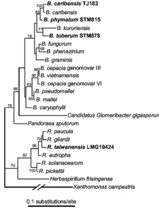
FIG. 2. 16S rDNA tree showing phylogenetic positions of legume-nodulating Ralstonia and Burkholderia species within the -proteobac-teria. The ML tree (base frequencies estimated, mutation rates drawn from an␥ ⫹ INV distribution, four classes of mutations) was recon-structed by using PAUP. Xanthomonas campestris was used as an outgroup. Legume symbionts are shown in bold type. Nodulating
Burk-holderia strains are named according to Vandamme et al. (24).
Gen-Bank/EMBL accession numbers for the 16S rDNA sequences were AF175314 (B. cepacia genomovar VI), AF148556 (B. cepacia genomo-var III), U96928 (B. vietnamensis), U91839 (B. pseudomallei), AF110188 (B. mallei), AB021423 (B. carophylli), AJ302312 (B.
phyma-tum), AJ505301 (B. caribensis), Y17009 (B. caribensis), AF215705 (B. fungorum), AB021394 (B. phenazinium), U96939 (B. graminis),
AB024310 (B. kuruiensis), AJ302311 (B. tuberum), AF139176 (P.
spu-torum), AF139176 (G. gigasporum), AJ238359 (H. frisingense),
AL646072 (R. solanacearum), AB004790 (R. pickettii), AF085226 (R.
paucula), AF300324 (R. taiwanensis), AF076645 (R. gilardii), M32021
(R. eutropha), and AF188831 (X. campestris).
at University of York on May 11, 2009
jb.asm.org
lates were recovered from root nodules of M. pudica and M.
diplotricha plants growing in 14 different areas in Taiwan
(Ta-ble 1).
16S rDNA PCR-RFLP analysis grouped 177 isolates to-gether with R. taiwanensis reference strains (Table 1). Addi-tional PFGE and nodA PCR-RFLP (see below) analyses showed that these R. taiwanensis isolates represented at least 44 different strains (Table 1). The remaining 13 isolates fell into six PFGE pattern groups. 16S rDNA sequencing of rep-resentative strains of these groups showed that they belonged to the genus Burkholderia (strains TJ182 and TJ183) (Fig. 2), the genus Rhizobium (TJ167 to TJ169, TJ171 to TJ176, and TJ189) (data not shown), or the genus Sinorhizobium (TJ170) (data not shown) (Table 1). Further taxonomical analysis iden-tified TJ182 and TJ183 as B. caribensis (24) (Fig. 2).
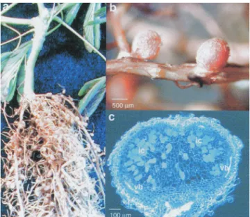
Representatives of the Mimosa isolate collection were dou-ble-checked for their ability to nodulate M. pudica under ax-enic laboratory conditions. The four R. taiwanensis strains tested, LMG 19424, LMG 19425, LMG 19426, and LMG 19430, formed nitrogen-fixing nodules (Fig. 3a and b) from which the original inoculated bacteria could be reisolated. The nodules displayed a genuine nodule structure, with central infected tissue containing cells with intracellular bacteria and peripheral tissue with vascular bundles (Fig. 3c). B. caribensis TJ182 and TJ183, as well as Rhizobium sp. strains TJ167 and TJ173, were able to effectively nodulate M. pudica (data not shown). On the other hand, Sinorhizobium sp. strain TJ170 was unable to nodulate either M. pudica or M. diplotricha (data not shown).
Ralstonia and Burkholderia nod and nif genes are very
simi-lar to those of␣-rhizobia. In ␣-rhizobia, the ability to nodulate requires the nodABC genes, responsible for the synthesis of the Nod factor core structure (13, 14). These three genes are all present in the first-rhizobium identified, B. tuberum STM678.
To investigate whether the other-rhizobial species identified so far also possess essential nod genes, we searched for the presence of nodA in a collection of-rhizobial strains. So far, sequences homologous to nodA have not been identified in nonrhizobial bacteria, and this gene therefore constitutes— together with the nodBC genes—a molecular signature for rhizobia. We found that an internal nodA sequence (from bp 44 to 574) could be amplified from 181 Ralstonia isolates (177 new isolates and 4 R. taiwanensis reference strains) (data not shown). The nodA genes of R. taiwanensis LMG 19424 and LMG 19425 were sequenced, as were those of B. caribensis TJ182 and B. phymatum STM815. Partial nodA sequences of four␣-rhizobial isolates were also determined. Sequence sim-ilarities between ␣- and -rhizobial NodA proteins ranged from 60% (B. caribensis and Azorhizobium caulinodans) to 72% (B. phymatum and Rhizobium sp. strain TJ172) (Table 2), confirming that the nodA genes of-proteobacteria are very similar to those of␣-rhizobia. Interestingly, ␣- and -rhizobial symbionts of M. diplotricha possess unrelated NodA sequences (Table 2).
We also searched for the presence of other nodulation genes, besides nodA, in R. taiwanensis LMG 19424 by PCR amplification (see Materials and Methods). Analysis of the amplified DNA sequences revealed the presence of the com-mon nodBC genes preceded by a NodD-dependent regulatory sequence (nod box), as well as part of the nodH gene, involved in Nod factor sulfation, and the nodI gene, presumably in-volved in Nod factor transport. Sequencing indicated that
nodA is separated from nodBC. Such genetic nonlinkage of nodABC was found previously for B. tuberum (10) as well as for
several rhizobia (25). Sequence similarity with rhizobial Nod proteins available in databases ranged from 32% (A.
caulino-dans) to 58% (Mesorhizobium sp. strain N33) for NodB and
from 49% (Rhizobium gallicum) to 74% (Rhizobium etli) for NodC.
Part of the nifH gene, encoding dinitrogenase reductase, a key enzyme in nitrogen fixation, was also amplified and se-quenced in representative strains of R. taiwanensis, B.
cariben-sis, B. phymatum, and Rhizobium spp. (data not shown).
nod and nif genes are located on a plasmid in R. taiwanensis
and B. phymatum. Genes required for nodulation and symbi-otic nitrogen fixation are often clustered and located on large plasmids (9) or mobile symbiotic islands (15). To determine
FIG. 3. Nodules of M. pudica 4 weeks after inoculation with R.
taiwanensis LMG 19424. (a) Nodulated roots. (b) Root segment with
pink nodules. (c) Longitudinal section showing the structure of a nodule. Plant tissue was cleared with sodium hypochlorite and stained with methylene blue as described by Truchet et al. (21). ic, infected cells; vb, vascular bundles.
TABLE 2. Sequence identities among NodA amino acid sequencesa
Sequence % Identity to the following sequence:
Bca Bp Ac Sme WM9 TJ167 TJ172 Rt 99.4 79.8 60.1 63.0 66.5 69.4 68.2 Bca 80.2 60.0 63.2 66.8 70.0 67.9 Bp 64.3 68.7 69.2 69.8 72.0 Ac 55.6 56.1 57.4 58.4 Sme 61.7 64.5 61.6 WM9 69.5 70.0 TJ167 81.6
aRt, R. taiwanensis LMG 19425; Bca, B. caribensis TJ182; Bp, B. phymatum
STM815; Ac, A. caulinodans ORS571; Sme, Sinorhizobium meliloti 2011; WM9,
Bradyrhizobium sp. strain WM9; TJ167, Rhizobium sp. strain TJ167; TJ172, Rhizobium sp. strain TJ172. NodA sequence lengths ranged from 170 amino
acids (partial sequences) to 197 amino acids (complete sequences). Percent identities were calculated from 170-amino-acid sequences.
VOL. 185, 2003 SYMBIOSIS OF -RHIZOBIA AND LEGUMES 7269
at University of York on May 11, 2009
jb.asm.org
the locations of symbiotic genes in the genomes of the new -rhizobia, we first examined the genome organization of
Ral-stonia and Burkholderia representatives by using PFGE (Fig.
4). Two high-molecular-weight replicons with apparent sizes of 3.5 and 2.4 Mb and a smaller replicon of about 0.5 Mb were identified for R. taiwanensis LMG 19424, while B. phymatum STM815 possesses replicons of approximately 3.5, 2.8, 2.1, and 0.5 Mb. No readable PFGE profile could be obtained with B.
tuberum. To determine which replicons carry the symbiotic
genes, Southern blots of PFGE agarose gels were hybridized with nifH and either nodA or nodC probes. Both nod and nif probes hybridized with the smallest 0.5-Mb replicons of R.
taiwanensis and B. phymatum (Fig. 4). These symbiotic
repli-cons did not hybridize with parental strain 16S rRNA, suggest-ing that they are genuine plasmids (data not shown).
Hence, the clustering of nodulation and nitrogen fixation genes is a common feature of␣- and -rhizobia.
Phylogenetic analysis of nodA and nifH genes of ␣- and -rhizobia. Phylogenetic analysis of 42 nodA sequences—in-cluding most available ␣-rhizobial and four -rhizobial se-quences—resulted in the ML tree shown in Fig. 5. The four -proteobacteria fell into two strongly supported clades. B.
phymatum, B. caribensis, and R. taiwanensis strains clustered in
the same clade. The nodA sequence closest to this clade comes from the highly divergent and atypical A. caulinodans, although this finding may have resulted from a long branch attraction artifact (4). B. tuberum and Methylobacterium nodulans fell into a separate and strongly supported cluster. Interestingly, the -rhizobia R. taiwanensis and B. caribensis and the ␣-rhizobia
Rhizobium sp. strains TJ167 and TJ172 isolated from M. dip-lotricha clustered separately in the nodA tree, suggesting that
their nodulation genes have different origins.
The clustering of the-rhizobial sequences in different nodA lineages intertwined with␣-rhizobial sequences suggested that
multiple nod gene transfers have occurred between the two subclasses of proteobacteria. Indeed, a single transfer of nod-ulation genes between␣- and -proteobacteria would have led to a single branch of-proteobacteria within the rest of the tree, which is composed of␣-proteobacteria. Constraining the four-rhizobial symbionts to the same clade led to a tree that was only marginally less likely than the ML tree (P value, 0.083, as determined by the Shimodeira-Hasegawa test implemented in PAUP) and thus did not clearly support or infer the hypoth-esis of multiple nod gene transfers. On the other hand, the different lengths of the NodA proteins from B. tuberum and R.
taiwanensis that clustered in two clades support the hypothesis
of different origins for the corresponding genes. The NodA sequence from B. tuberum possesses at the N terminus an additional 13-amino-acid segment that is characteristic of bra-dyrhizobial NodA sequences (9a), while R. taiwanensis and the genera Azorhizobium, Sinorhizobium, Mesorhizobium, and
Rhi-zobium all lack this NodA N-terminal extension. Moreover, the
similarity between B. caribensis TJ182 and R. taiwanensis LMG 19424 nodA sequences (97.4% identity) indicates that nod gene transfer may have occurred between -proteobacteria, as al-ready suggested for␣-proteobacteria (16).
Interestingly, the phylogeny of the nitrogen fixation gene
nifH provides a representation of the rhizobia different from
that of the phylogeny of nodA (Fig. 6). Indeed, some of the groupings within the nifH tree corresponded to the phylogeny of the organisms as deduced from comparative 16S rDNA analysis, although ␣- and -proteobacteria did not form dis-tinct and monophyletic clades. Moreover, the nifH tree grouped together free-living and symbiotic nitrogen-fixing
Burkholderia and Ralstonia strains. An example of a
represen-tative organism is B. tuberum, which grouped with M. nodulans in the nodA phylogeny but grouped with other -proteobacte-ria in the nifH phylogeny. Constraining either␣-and -rhizobia or M. nodulans and B. tuberum to the same clade led to a tree that was statistically less probable than the ML tree (both with
P values of ⬍10⫺4). These results suggest that nod and nif
genes of-rhizobia have different origins. DISCUSSION
In this study, we have confirmed and extended the phyloge-netic diversity of rhizobia initially presented in articles by Mou-lin et al. (10) and Chen et al. (1). We have identified as rhizobia two additional Burkholderia strains as well as at least 48 dif-ferent R. taiwanensis strains isolated from M. pudica and M.
diplotricha. Representative Burkholderia and Ralstonia strains
fix nitrogen in symbiosis with their respective host plants, dem-onstrating that the root nodule -proteobacteria are indeed true rhizobia. Detailed studies have shown that R. taiwanensis-induced nodule ontogeny and development are similar to those described for other, mimosa-like legumes (1a). Moreover, R.
taiwanensis is the favored partner of M. pudica and M. dip-lotricha in Taiwan, indicating that nodulation by -proteobac-teria is not a rare phenomenon exhibited by certain opportu-nistic strains. In this respect, it should be noted that R.
taiwanensis strains also have been isolated from M. pudica in
India (20).
The widespread character of nodulation by-proteobacteria
FIG. 4. Locations of nodA and nifH genes on replicons of R.
tai-wanensis LMG 19424 (A) and B. phymatum STM815 (B). Lane 1,
PFGE of undigested genomic DNA stained with ethidium bromide; lanes 2 and 3, autoradiographs of blotted PFGE gels hybridized with
nodA and nifH probes, respectively. Sizes of replicons are indicated on
the left.
at University of York on May 11, 2009
jb.asm.org
is also attested to by the phylogenetic diversity of the -rhizo-bia identified so far, including one Ralstonia species (1) and three Burkholderia species (B. caribensis, B. tuberum, and B.
phymatum) (24), as well as the fact that they have been isolated
from Asia, Africa, and South America. Since many legumes and environments remain to be explored, it is highly likely that further characterization of rhizobia will reveal an even greater diversity. For many decades, standard isolation procedures have been used for rhizobia, and identification as legume sym-bionts through nodulation tests has required the availability of host plant seeds. These traditional approaches, coupled with the difficulty of obtaining seeds for many tropical legumes, have probably contributed to masking of the natural diversity of rhizobia.
Nitrogen fixation, which is widespread in eubacteria and archaea, is thought to be an ancestral function now lost by many bacteria (3). Conversely, nodulation is thought to have appeared recently in evolution, at the same time as the ap-pearance of legumes on Earth, about 70 to 130 millions years ago. At that period of history, the␣- and -proteobacteria and the different rhizobial lineages already had diverged (22). The genes required for legume nodulation are thought to have
been acquired subsequently by lateral transfer from undefined sources, thus converting soil saprophytes into symbionts (7). This hypothesis has been confirmed by recent data (6, 15). The presence in␣- and -rhizobia of very similar and phylogeneti-cally related nodABC genes strongly supports the hypothesis of a unique origin for the common nod genes. However, it is not clear whether a single transfer event was responsible for the spread of nodulation genes from one subclass to the other or whether recurrent transfers occurred between the two sub-classes. Our phylogenetic and NodA length analyses together suggest the occurrence of at least two lateral transfers between these two unrelated subclasses of proteobacteria, although sta-tistical analysis did not allow this hypothesis to be ascertained. Further identification of other -rhizobia may be useful for confirming such a hypothesis. Moreover, a comparative anal-ysis of the nodA, nifH, and 16S rDNA trees suggests that -rhizobia emerged through the transfer of nod genes to dia-zotrophic -proteobacteria. Since the nod and nif genes are located on the same plasmid in the-rhizobia investigated, it is possible that exogenous nod and nif genes were cotransferred prior to the replacement of the exogenous nifH gene by the indigenous gene. This level of complexity is in line with the
FIG. 5. Unrooted nodA phylogenetic tree of rhizobia. -Proteobacterial strains are in shown in bold type and underlined. The tree was reconstructed by using an ML approach based on a 597-bp alignment (excluding the additional segment at the N-terminal part). Sequence lengths included ranged from 558 bp (B. phymatum STM815; partial sequence) to 597 bp (most strains). Values along branches indicate bootstrap percentages higher than 50%, based on 100 replicates. nodA sequences for published bacteria are available from GenBank. EMBL accession numbers and nodA sequences for unpublished bacteria were AJ505318 (B. phymatum), AJ505311 (R. taiwanensis), AJ505309 (B. caribensis), AJ300229 (S. terangae bv. acaciae), AJ300249 (M. plurifarium), AJ505307 (TJ172), AJ300234 (BR816), AJ505305 (TJ167), AJ300242 (A. undicola), AJ302321 (B. tuberum), AJ303088 (STM259), AJ430707 (WU425), AJ430730 (CBP70), AJ430715 (ORS938), AJ430712 (USDA3139), AJ430728 (CCT6220), AJ300260 (STM270), AJ300247 (M. ciceri), AJ300228 (S. terangae bv. sesbaniae), and J300235 (R. huautlense).
VOL. 185, 2003 SYMBIOSIS OF -RHIZOBIA AND LEGUMES 7271
at University of York on May 11, 2009
jb.asm.org
highly complex evolutionary history of the legume-rhizobium symbiosis.
ACKNOWLEDGMENTS
We thank Y. Prin for help in microscopy studies and M. Dukhan for help with photographs. We also thank D. Barker and J. Batut for comments and suggestions.
W.-M. Chen was supported by a grant from the National Science Council, Taipei, Taiwan, Republic of China (NSC 91-2320-B-022-001).
REFERENCES
1. Chen, W. M., S. Laevens, T. M. Lee, T. Coenye, P. de Vos, M. Mergeay, and P. Vandamme.2001. Ralstonia taiwanensis sp. nov., isolated from root nod-ules of Mimosa species and sputum of a cystic fibrosis patient. Int. J. Syst. Evol. Microbiol. 51:1729–1735.
1a.W.-M. Chen, E. K. James, A. R. Prescott, M. Kierans, and J. I. Sprent. Nodulation of Mimosa spp. by the-proteobacterium Ralstonia taiwanensis. Mol. Plant-Microbe Interact., in press.
2. Deschodt, C. C., and B. W. Strijdom. 1976. Effective nodulation of
Aspal-athus linearis by rhizobia from other AspalAspal-athus species. Phytophylactica
8:103–104.
3. Fani, R., R. Gallo, and P. Lio. 2000. Molecular evolution of nitrogen fixation:
the evolutionary history of the nifD, nifK, nifE, and nifN genes. J. Mol. Evol. 51:1–11.
4. Felsenstein, J. 1978. Cases in which parsimony or compatibility methods will be positively misleading. Syst. Zool. 27:401–410.
5. Felsenstein, J. 1993. PHYLIP (phylogeny inference package), version 3.5c. University of Washington, Seattle.
6. Galibert, F., T. M. Finan, S. R. Long, A. Puhler, P. Abola, F. Ampe, F. Barloy-Hubler, M. J. Barnett, A. Becker, P. Boistard, G. Bothe, M. Boutry, L. Bowser, J. Buhrmester, E. Cadieu, D. Capela, P. Chain, A. Cowie, R. W. Davis, S. Dreano, N. A. Federspiel, R. F. Fisher, S. Gloux, T. Godrie, A. Goffeau, B. Golding, J. Gouzy, M. Gurjal, I. Hernandez-Lucas, A. Hong, L. Huizar, R. W. Hyman, T. Jones, D. Kahn, M. L. Kahn, S. Kalman, D. H. Keating, E. Kiss, C. Komp, V. Lalaure, D. Masuy, C. Palm, M. C. Peck, T. M. Pohl, D. Portetelle, B. Purnelle, U. Ramsperger, R. Surzycki, P. Thebault, M. Vandenbol, F. J. Vorholter, S. Weidner, D. H. Wells, K. Wong, K. C. Yeh, and J. Batut.2001. The composite genome of the legume symbiont
Sinorhizo-bium meliloti. Science 293:668–672.
7. Hirsch, A. M., M. R. Lum, and J. A. Downie. 2001. What makes the rhizobia-legume symbiosis so special? Plant Physiol. 127:1484–1492.
8. Lortet, G., N. Me´ar, J. Lorquin, B. Dreyfus, P. de Lajudie, C. Rosenberg, and C. Boivin.1996. Nod factor thin-layer chromatography profiling as a tool to characterize symbiotic specificity of rhizobial strains: application to
Sinorhi-zobium saheli, S. teranga and RhiSinorhi-zobium sp. strains isolated from Acacia and Sesbania. Mol. Plant-Microbe Interact. 9:736–747.
9. Martinez-Romero, E., and J. Caballero-Mellado. 1996. Rhizobium phylog-enies and bacterial genetic diversity. Crit. Rev. Plant Sci. 15:113–140. 9a.L. Moulin, G. Be´na, C. Boivin-Masson, and T. Stpkowski. Phylogenetic
analyses of symbiotic nodulation genes support vertical and lateral gene co-transfer within the Bradyrhizobium genus. Mol. Phylogenet. Evol., in press.
10. Moulin, L., A. Munive, B. Dreyfus, and C. Boivin-Masson. 2001. Nodulation of legumes by members of the-subclass of Proteobacteria. Nature 411:948– 950.
11. Nicholas, K. B., H. B. Nicholas, Jr., and D. W. Deerfield. 1997. GeneDoc: analysis and visualization of genetic variation. EMBNEW News 4:14. 12. Parker, M. A., and A. Lunk. 2000. Relationships of bradyrhizobia from
Platypodium and Machaerium (Papilionoideae: tribe Dalbergieae) on Barro
Colorado Island, Panama. Int. J. Syst. Evol. Microbiol. 50:1179–1186. 13. Perret, X., C. Staehelin, and W. J. Broughton. 2000. Molecular basis of
symbiotic promiscuity. Microbiol. Mol. Biol. Rev. 64:180–201.
14. Schultze, M., and A. Kondorosi. 1998. Regulation of symbiotic root nodule development. Annu. Rev. Genet. 32:33–57.
15. Sullivan, J. T., and C. W. Ronson. 1998. Evolution of rhizobia by acquisition of a 500-kb symbiosis island that integrates into a phe-tRNA gene. Proc. Natl. Acad. Sci. USA 95:5145–5149.
16. Suominen, L., C. Roos, G. Lortet, L. Paulin, and K. Lindstrom. 2001. Identification and structure of the Rhizobium galegae common nodulation genes: evidence for horizontal gene transfer. Mol. Biol. Evol. 18:907–916. 17. Swofford, D. 1998. PAUP*: Phylogenetic Analysis Using Parsimony (and
other methods), version 4. Sinauer Associates, Sunderland, Mass. 18. Sy, A., E. Giraud, P. Jourand, N. Garcia, A. Willems, P. de Lajudie, Y. Prin,
M. Neyra, M. Gillis, C. Boivin-Masson, and B. Dreyfus.2001. Methylotro-phic Methylobacterium bacteria nodulate and fix nitrogen in symbiosis with legumes. J. Bacteriol. 183:214–220.
19. Thompson, J. D., T. J. Gibson, F. Plewniak, F. Jeanmougin, and D. G. Higgins.1997. The CLUSTAL X windows interface: flexible strategies for multiple sequence alignment aided by quality analysis tools. Nucleic Acids Res. 25:4876–4882.
20. Tripathi, A. K. 2002. Rhizobia of the-subclass of Proteobacteria: a tale of losing the race. Curr. Sci. 82:8–9.
21. Truchet, G., S. Camut, F. de Billy, R. Odorico, and J. Vasse. 1989. The
Rhizobium-legume symbiosis. Protoplasma 149:82–88.
22. Turner, S. L., and J. P. W. Young. 2000. The glutamine synthetases of rhizobia: phylogenetics and evolutionary implications. Mol. Biol. Evol. 17: 309–319.
23. van Berkum, P., and B. D. Eardly. 1998. Molecular evolutionary systematics of the Rhizobiaceae, p. 1–24. In H. P. Spaink, A. Kondorosi, and P. J. J. Hooykaas (ed.), The Rhizobiaceae. Molecular biology of plant-associated bacteria. Kluwer Academic Publishers, Dordrecht, The Netherlands. 24. Vandamme, P., J. Goris, W. M. Chen, P. de Vos, and A. Willems. 2002.
Burkholderia tuberum sp. nov. and Burkholderia phymatum sp. nov., nodulate
the roots of tropical legumes. Syst. Appl. Microbiol. 25:507–512. 25. Zhang, X. X., S. L. Turner, X. W. Guo, H. J. Yang, F. Debelle´, G. P. Yang,
J. De´narie´, J. P. W. Young, and F. D. Li.2000. The common nodulation genes of Astragalus sinicus rhizobia are conserved despite chromosomal diversity. Appl. Environ. Microbiol. 66:2988–2995.
FIG. 6. nifH phylogenetic tree. The tree was reconstructed by using an ML approach based on an 800-bp alignment matrix (partial and full sequence lengths ranged from 336 to 797 bp). Values along branches indicate bootstrap percentages higher than 50%. The tree was rooted by using sequences from V. diazotrophicus, K. pneumoniae, and A.
vinelandii. Rhizobia are shown in bold type, and the␣-, -, or ␥-pro-teobacterial classification is indicated in parentheses. Clusters 1 and 3 contain␣-rhizobia only, while cluster 2 includes both symbiotic and nonsymbiotic diazotrophic-proteobacteria. nifH sequences for pub-lished bacteria are available from GenBank EMBL. EMBL accession numbers and nifH sequences for unpublished bacteria were AJ302315 (B. tuberum), AJ505320 (R. taiwanensis), AJ512206 (B. vietnamensis), AJ512207 (B. caryophylli), AJ505317 (B. caribensis), AJ505319 (B.
phy-matum), and AJ512205 (M. nodulans). nifH sequences from B. fungo-rum, R. palustris, and R. leguminosarum were from partially completed
genome Web sites.
at University of York on May 11, 2009
jb.asm.org