HAL Id: tel-03029246
https://tel.archives-ouvertes.fr/tel-03029246
Submitted on 28 Nov 2020
HAL is a multi-disciplinary open access archive for the deposit and dissemination of sci-entific research documents, whether they are pub-lished or not. The documents may come from teaching and research institutions in France or abroad, or from public or private research centers.
L’archive ouverte pluridisciplinaire HAL, est destinée au dépôt et à la diffusion de documents scientifiques de niveau recherche, publiés ou non, émanant des établissements d’enseignement et de recherche français ou étrangers, des laboratoires publics ou privés.
Functional, structural and evolutionary aspects of the
auxin transcriptional response
Raquel Martin-Arevalillo
To cite this version:
Raquel Martin-Arevalillo. Functional, structural and evolutionary aspects of the auxin
transcrip-tional response. Biomolecules [q-bio.BM]. Université Grenoble Alpes, 2017. English. �NNT :
THÈSE
Pour obtenir le grade de
DOCTEUR DE LA COMMUNAUTE UNIVERSITE
GRENOBLE ALPES
Spécialité : Biologie Structurale Arrêté ministériel : 25 mai 2016
Présentée par
Raquel MARTÍN-AREVALILLO
Thèse dirigée par Renaud DUMAS préparée au sein du
Laboratoire de Physiologie Cellulaire et Végétale dans l'École Doctorale de Chimie et Sciences du Vivant
Aspects fonctionnels, structuraux et
évolutifs de la réponse
transcriptionnelle à l’auxine
Thèse soutenue publiquement le 27 Novembre 2017 devant le jury composé de :Dr. Catherine BELLINI
Directrice de recherche, INRA, Rapportrice Pr. Miguel BLÁZQUEZ
Professeur, Université de Valencia, Rapporteur Pr. Eva PEBAY-PEYROULA
Professeure, Université Grenoble Alpes, Présidente Dr. Teva VERNOUX
Directeur de recherche, CNRS, Examinateur Dr. François PARCY
Directeur de recherche, CNRS, Examinateur Dr. Renaud DUMAS
3
None but those who have experienced them can
conceive of the enticements of science. In other studies,
you go as far as others have gone before you, and there
is nothing more to know; but in a scientific pursuit
there is continual food for discovery and wonder.
5
Abstract
Auxin is a plant hormone implicated in almost all plant developmental stages, since the embryo formation till flowering, determining the position of the organs in the plant and thus, its whole structure. As for any other hormone, auxin perception is followed by a signal transduction that finishes in a series of changes in a plant cell, including transcriptional changes. This thesis is divided in 3 chapters, each with a focus on the structural, molecular and evolutionary aspects of different proteins involved in the regulation of auxin genes response.
First, we focused our studies on TOPLESS (TPL), a co-repressor implicated, not only in auxin responsive genes repression, but also in many other plant processes due to its interactions with numerous transcriptional repressors in plants. Our determination of the TPL N-terminal structure allowed us to understand that TPL can interact with different partners through the same binding site. Moreover, it revealed that TPL is a tetrameric protein, with the tetramerization interface formed by a newly identified domain, the CRA domain, that is also part of the binding site. The high residues conservation in both tetramerization interface and TPL binding site since m.y.a indicates the importance of TPL role since the origin of plants. This work also shows that the structural similarities between TPL and other co-repressor with similar domains but different function nicely exemplify how evolution plays with common features for creating new functions.
Second, we studied ARF proteins, the transcription factors of the auxin transcriptional response, with a focus on their DNA binding preferences. For this, we used a combination of bioinformatic analyses of DAP-seq ARFs genomic binding, with in vitro DNA binding tests and structure modelling. Our results point out that different ARFs can have different preferential binding sites within the genome, with these preferences being determined by the orientation and spacing of the binding motifs. Moreover, our studies suggest that depending on the binding site, ARFs could bind with different conformations using dimerization interfaces not yet discovered. These results can explain how different ARFs co-expressed inside a plant cell can collaborate to the specificity and robustness of auxin transcriptional response by differential bindings to the genome.
Finally, we travelled back in time to position the origin of auxin signalling pathway in the evolution of plants. Here we looked for protein homologues of the auxin signalling pathway in charophyte green algae, the most ancient plants ancestor (450 M years). This search retrieved an ARF and a TPL homologue in the first multicellular charophyte algae (Chlorokybus atmophyticus). The biochemical characterization of C. atmophyticus ARF indicated that it presented already the same properties of the ARFs from land plants and that it was able to interact with TPL protein, as it is the case for some ARFs. The absence of auxin receptor homologues in these primitive algae indicates however that auxin-dependency appeared with the acquisition of TIR1/AFB-Aux/IAA coreceptor system, after charophytes divergence into land plants.
6
Résumé
L’auxine est une hormone végétale impliquée dans presque toutes les étapes du développement des plantes, de la formation de l’embryon jusqu’à la floraison, déterminant la position des organes et donc la structure de la plante. Comme pour les autres hormones, la perception de l’auxine est suivie par une transduction du signal qui produit une série de changements dans les cellules végétales dont des régulations transcriptionnelles. Cette thèse est divisée en 3 chapitres, chacun d’eux étant focalisé sur des aspects structuraux, moléculaires et évolutifs de différentes protéines impliquées dans la régulation des gènes de réponse à l’auxine.
Nous avons tout d’abord centré nos études sur TOPLESS (TPL), un corépresseur qui agit au niveau de la répression des gènes de réponse à l’auxine, mais aussi dans d’autres processus végétaux compte tenu de son interaction avec de nombreux répresseurs transcriptionnels. Nous avons déterminé la structure de la partie N-terminale de TPL et compris comment TPL interagit avec différents partenaires au niveau d’un même site de liaison. Nous avons alors démontré que TPL forme un tétramère à l’aide d’une surface de tétramérisation constituée par un nouveau domaine, le domaine CRA, qui fait aussi partie du site de liaison. Les résidus impliqués dans la tétramérisation et l’interaction avec des partenaires sont très conservés depuis des centaines de millions d’années montrant ainsi l’importance du rôle de TPL depuis l’origine des plantes. Enfin, les similarités de structure entre TPL et d’autres corépresseurs qui possèdent des domaines similaires mais possédant une fonction différente montrent un bel exemple de la manière dont l’évolution joue avec des domaines protéiques pour créer de nouvelles fonctions.
Nous avons ensuite étudié les préférences de liaison à l’ADN des facteurs de transcription de la réponse à l’auxine (ARF). Pour cela nous avons utilisé une combinaison d’analyses bio-informatiques de données de DAP-seq sur la liaison des ARFs sur le génome, des tests d’interaction ADN-protéine in vitro et de la modélisation de structures. Nos résultats indiquent que les différents ARFs ont des sites préférentiels de liaison sur le génome et que ces préférences sont déterminées par l’orientation et l’espacement entre motifs de liaison. Enfin, ces études suggèrent qu’en fonction du site de liaison, les ARFs pourraient se lier avec différentes conformations à l’aide de surfaces de dimérisation qui ne sont pas encore décrites. Ces résultats permettent d’expliquer comment différents ARFs coexprimés dans la même cellule peuvent fonctionner ensemble pour contribuer à une réponse transcriptionnelle à l’auxine spécifique et robuste.
Finalement, nous avons remonté le temps pour positionner l’origine de la voie de signalisation de l’auxine chez les plantes. Pour cela, nous avons recherché des homologues des protéines de la voie de signalisation de l’auxine dans des algues vertes charophytes, les ancêtres les plus lointains (450 Millions d’années) des plantes. Nous avons alors trouvé un homologue des ARFs et TPL chez les premières algues multicellulaires (Chlorokybus atmophyticus). La caractérisation biochimique de
7
plantes terrestres et était aussi capable d’interagir avec TPL comme certains ARFs. L’absence d’homologues du récepteur de l’auxine chez ces algues primitives indique cependant que la dépendance à l’auxine aurait été acquise plus tard avec l’apparition du système corécepteur TIR1/AFB-Aux/IAA après la divergence des charophytes vers les plantes terrestres.
9
Acknowledgements
It might be my name which appears in the cover of this manuscript and it will be me the person that will have to defend these results in front of everybody (trying not to freak out), but all this it is not just my work. It would not have been possible without the help of many people and therefore, I need to say many, many thanks to you all.
First, thank you to the components of my thesis jury, Miguel Blázquez, Catherine Bellini, Eva Pebay-Peyroula and Teva Vernoux for having taken the time to evaluate this work and discuss it with me.
Thank you, Norbert Rolland, Laurent Blanchoin, Eric Maréchal and François Parcy for having welcome me in your laboratory.
François, to you I have to say thanks for many other things. Thank you for having shared with me your experience, knowledge, enthusiasm, ideas and jokes. You have always been available for me for whatever need I had, no matter if it was for discussing results, making “sexier” presentations or trying to help me with my incompetence with computers. I learnt a lot from you both at the professional and the personal levels. Merci beaucoup.
Renaud… I can’t believe how lucky I am to have had you as my supervisor. I don’t think I can thank you enough for everything you’ve done for me during these 3 years. You have taught me protein biochemistry and structure. You taught me to be critic with myself and my results at the same time that you gave me freedom for doing things the way I thought they should be done. You were always available for discussing with me. And I guess all this was part of your work, an amazing work. But it is not just that; you have done for me many other things that were not part of your work. When things weren’t going good, you’ve always had a joke to say to make me smile again. When you’ve seen me stressed, tired, sad or ill you have been worried about me and you have always offered me your help. You have even tried to find a husband for me! And this is the only thing in which you failed (:P) because with everything else you helped me to feel like home. Merci, merci et merci pour tout, Renaud.
Thomasito and Manuelito, thank you for all the help with the experiments. I remember arriving to the lab, not being able to find anything (not even my own office) and you were always willing to help and to teach me new stuffs. You taught me more than science, you also taught me French! Thanks to you two and Gregounette and PH, now I can go as rude as I want when speaking French. I had a lot of fun with you guys 😉.
Euge! Luckily you were there, in the bench next to mine helping me not to forget how to speak my own language. It’s been really “con padre” and “con madre” to have our Hispanic side of the lab.
10
Muchas gracias por haberme dedicado tiempo, paciencia y una sonrisa cuando estaba a punto de subirme por las paredes.
Arnaud and Adrien, A&A, you’ve turned my last months here into a nightmare of oligos. Just kidding. I’ve loved to bother you every 5 minutes with all my questions about bioinformatics and I had a lot of fun discussing with you.
Marie and Gaby, thank you for always transmitting so much happiness. You’re always in such a good mood and so ready to help everybody that it’s been a huge pleasure to share lunches, time and jokes with you. Marie, please, don’t give up in our task of hiring a handsome cleaning gentleman. Send pictures!
Sophie, Tiffany and Danielle, thank you for your good mood and efficiency. You helped me a lot not being such a disaster with the administrative issues.
Leonie, unluckily it’s been just a few months that we could spend together but still, it’s been enough to realize your passion for research. Good luck with your crystals!
Cristel, Gilles, Agnes, Vero, Aditya and Chloe, I didn’t have the chance to work with you directly but it’s been very nice to meet you all. Thank you for all your help and advices.
Max Nanao and David Cobessi, thank you for the crystallographic work and for inviting me over to the lines. It’s been really exciting to see our crystals turning around in the robot.
Teva, Antoine and David, les Lyonnais, thanks for all the amazing work you have developed in Lyon, for the discussions, the ideas and for inviting us over for beers. It has been very nice collaborating with you, both at the professional and personal levels.
Thank you to all my friends here. We’ve travelled, we’ve danced, we’ve eaten and we’ve drunk. We’ve had so much laughter and fun in a country that was not ours but that we made ours, somehow. Thank you for helping me to not miss my family and culture that much by turning yourselves into my family and culture.
Thank you to my family, the real one this time, the one in Spain. Gracias por el apoyo incondicional que siempre me brindáis, decida lo que decida y vaya donde vaya. Si estoy aquí es indudablemente por vosotros. Os he echado mucho de menos.
Along the manuscript you’ll find conclusions for the results, but here you have my conclusion, the summary of these three years of my life: I have been happy and this is thanks to each and all of you.
Thank you.
Merci.
11
Abbreviations
2,4-D 2,4-Dichlorophenoxyacetic Acid
4-Cl-IAA 4-Chloroindole-3-Acetic Acid
ABA Abscisic Acid
ABC ATP-Binding Cassette
ABI3 Abscisic Acid Insensitive 3
ABP1 Auxin Binding Protein 1
AG AGAMOUS
AP2 APETALA 2
ARF Auxin Responsive Factor
Aux/IAA Auxin/ Indole-3-Acetic Acid Protein
AUX1 Auxin Transporter Protein 1
AuxRE
Auxin Response Element
BR Brassinosteroid
BRM
BRAHMA
cDNA Complementary Deoxyribonucleic Acid
CK Cytokinin
COI1 Coronatine Insensitive 1
CRA CT11 RanBPM
CTLH C-terminal to LisH
DAP-seq DNA-affinity Purification Sequencing
DBD DNA-Binding Domain
DD Dimerization Domain
DNA Deoxyribonucleic Acid
DNA
MTase DNA Methyltransferases
DR Direct Repeat
EAR Ethylene-responsive element binding factor-associated Amphiphilic
Repression
EMSA Electrophoretic Mobility Shift Assay
ER Endoplasmic reticulum
12
ERF Ethylene-Responsive Factors
FD Flanking Domain
FL Full-Length
GA Gibberellins
gDNA genomic Deoxyribonucleic Acid
GH3 Gretchen Hagen 3
Gro
Groucho
GST Glutathione-S-transferase
HAT
Histone Acetyltransferases
HDA
Histone Deacetylases
HTRF Homogeneous Time Resolved Fluorescence
IAA Indole-3-Acetic Acid
IAA-Ala IAA-Alanine IAA-Asp IAA-Aspartate IAA-Glu IAA-Glutamate IAAld Indole-3-Acetaldehyde IAA-Leu IAA-Leucine IAA-Trp IAA-Tryptophan IAM Indolacetamide IAN Indole-3-Acetonitrile IAOx Indole-3-Acetaldoxine
IGP Indole-3-Glycerol Phosphate
IPTG Isopropyl-β-D-1-thyogalactopiranoside
IPyA Indole-3-Pyruvic Acid
IR Inverted Repeat
JA Jasmonic Acid
JAZ Jasmonate-ZIM Domain
LAX Auxin Transporter-like Protein
LEC2 Leafy Cotyledon 2
LisH
Lissencephaly
LRR Leucine Rich Repeat
13
MBP Maltose Binding Protein
mRNA
Messenger Ribonucleic Acid
NAA 1-Naphtaleneacetic Acid
NAP Nuclear Auxin Pathway
NCoR
Nuclear Receptor Co-repressor
NO Nitric Oxide
PAA Phenylacetic Acid
PB1 Phox/Bemp1
PWM Position Weight Matrixes
RAV Related to ABI3/VP1
REM Reproductive Meristem
SA Salicylic Acid
SDM Site Directed Mutagenesis
SEC Size Exclusion Chromatography
SEC-MALLS SEC-Multi-Angle Laser Light Scattery
SEU SEUSS
SKP2a S-Phase Kinase-Associated Protein 2a
SL Strigolactone
SMRT
Silencing Mediator of Retinoic cid and Thyroid hormone receptor
SYD
SPLAYED
TAA1 Tryptophan Aminotransferase 1
TBL1 Transducin Beta-like 1
TF Transcription Factor
TIR1/AFB Transport Inhibitor Resistant 1/Auxin Signalling F-box
TPL/TPR
TOPLESS/TOPLESS-related
VAL VP1/ABI3-like
WUS WUSCHEL
Y2H
Yeast-2-hybrid
15
Contents
General introduction to auxin, the hormone growth ... 19
Hormones in plants! Really? Why? ... 19
A little bit of history… ... 20
Hormones inside a plant cell ... 21
Auxins: the hormone that influences almost everything ... 22
Auxin synthesis ... 23 Auxin inactivation ... 24 Auxin transport ... 25 Auxin signalling ... 27 Objectives ... 35 Chapter I ... 37 Introduction ... 39
Gene expression control in eukaryotic organisms ... 39
Co-repressors ... 40
Co-repression in plants ... 43
TOPLESS co-repressor mechanism ... 45
TPL in the specific context of auxin signalling ... 47
Article 1 ... 51
Supplementary Information ... 57
SI Figures ... 58
SI Tables ... 67
SI Materials and Methods ... 72
SI References ... 78
Complementary results and discussion ... 80
Conclusions ... 83
Chapter II ... 85
Introduction ... 87
DNA-TFs interplay in the control of gene expression ... 87
ARFs, transcription factors of the auxin response ... 88
ARFs DNA binding sites and mechanism ... 92
16
Discussion ... 112
Conclusions ... 117
Chapter III ... 119
Introduction ... 121
Charophyte organisms: from water to land ... 121
B3 family ... 122
Auxin signalling: from bryophytes to flowers ... 125
Auxin signalling clues in charophytes? ... 126
Results ... 128
Discussion ... 141
General discussion and conclusions... 147
Materials & Methods ... 151
Bibliography ... 161
17
General introduction to auxin,
the hormone growth
19
General introduction to auxin, the hormone growth
Hormones in plants! Really? Why?
Whereas most animals can move around escaping from danger or from undesired environmental conditions, plants, as sessile organisms, are subjected for their whole life to an ever-changing (and often adverse) environment from which they will receive very different stimuli. Diverse light, temperature, atmosphere and soil conditions, pathogens or abiotic stresses are some of them. Therefore, plants need to coordinate all these external cues with their physiological and developmental state. Contrary to animals, plants have continuous sources of stem cells, called meristems. Meristems allow the de novo formation of organs all along a plant’s life conferring them a high developmental plasticity that helps them coping with external and internal signals (Jaillais and Chory, 2010).
Plant hormones, also called phytohormones, are a structurally and chemically diverse group of plant secondary metabolites with a main role in the integration of all these external and internal signs. Instead of being produced in specific organs, as it happens in animals, phytohormones can be produced and sensed by all or almost all plant cells where they will have a range of physiological effects at specific stages of a plant’s life (Dharmasiri et al., 2013; Kumar et al., 2016; Rigal et al., 2014).
In order to more specifically determine if a molecule corresponds to a plant hormone or not several criteria have been established (Leyser, 1998):
• It must be active at concentrations lower than 10-6M • It must be synthesized by the plant
• It must be transported some distance (at least one cell diameter) • It must have some important physiological effect on the plant
• It must act by non-covalent binding to a specific receptor, remain non-covalently bound and non-covalently modified while acting.
According to these criteria, nowadays 10 hormones have been or are being described: auxins (IAA, Indole-3-acetic acid), cytokinins (CKs), gibberellins (GAs), abscisic acid (ABA), ethylene, brassinosteroids (BRs), jasmonic acid (JA), salicylic acid (SA), strigolactones (SLs) and nitric oxide (NO) (Fig 1).
20 General introduction to auxin, the hormone growth
A little bit of history…
At the end of the 18th century a pineapple farmer in the Azores discovered that the fumes he was using to try to kill insects in the greenhouses were promoting an early flowering of pineapple trees. Since then, smoke started being used as a synchronizer of flowering in agriculture without people knowing that the phytohormone ethylene was behind these effects.
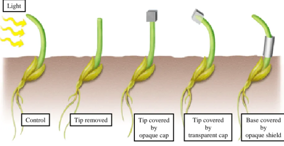
The first evidences of some kind of mobile signal within plants came from the Darwin family. In the end of the 18th century, Charles and Francis Darwin discovered that light induced a differential elongation in grass coleoptiles and proposed that this was mediated by a signal transported towards the roots creating an unequal redistribution that regulated plant curvature towards the light (Sauer et al., 2013) (Fig 2).
It took around 50 years more (Went, 1926) to discover that the mobile signal causing these effects was auxin, and even a few more for auxin to be isolated from higher plants (1946), being this the first plant hormone to be discovered.
Simultaneously, in 1926 in Japan, the scientist Eiichi Kurosawa was studying the “bakanae” (in Japanese “foolish seedling”) disease in rice that caused an excessive growth of the plants which could no longer stand their own weight -quite foolish, indeed. He discovered that these effects were due to the infection by the fungus Gibberella fujikuroi. Years later, the molecule responsible for
Fig 1. Chemical structure of plant hormones.
Indole-3-acetic acid
Cytokinins Gibberelins Abscisic acid Ethylene
Brassinolide
Jasmonic acid Salycilic acid Strigolactones Auxins
Zeatin GA1
Brassinosteroids
Strigol
21
this was isolated from fungus extracts and was named gibberellins. Gibberelins were first isolated from plants and thus defined as plant hormones in 1956 (Baca and Elmerich, 2007).
But if there is a winner in the weirdest story, those are cytokinins. Carlos Miller, a postdoc in Folke Karl Skoog’s lab (the same Skoog to whom we must thank for Murashige&Skoog medium) was working in the improvement of plant tissues growing medium by adding different extracts. Surprisingly, the additive that seemed to work the best as a growth promoter was autoclaved herring sperm, so he ordered a whole keg of it! Cytokinins were then, the third of the “classical phytohormones” to be discovered, as adenine-derivatives that resulted from DNA degradation (Baca and Elmerich, 2007).
Even if we have been using plant hormones effects on agriculture for decades, it is now that we are starting to understand phytohormones and the molecular mechanisms below their actions mainly thanks to the recent advances in Arabidopsis thaliana model plant genetics, that have allowed an extensive characterization of many of the components involved in the production, sensing and response of plants to phytohormones.
Hormones inside a plant cell
The response of a plant cell to a phytohormone will depend on two aspects. First, on the local concentration of a hormone and its availability, which in turn can be modulated through different processes: its synthesis, conjugation, degradation, compartmentalization within the cell and transport within the whole plant.
Fig 2. Darwin’s phototropic response experiments. Grass seedlings grow towards light. When the tip is removed or covered by an opaque cap the shoots grow straight up. When the opaque cap is displaced by a transparent one the curvature still takes places. If the opaque cap is placed on the base of the seedling instead of on the tip there is still a phototropic response. These results suggested the presence of a mobile signal induced by light on the tip that is transported down to the growing region of the shoot.
Control Tip removed Tip covered
by opaque cap Tip covered by transparent cap Base covered by opaque shield Light
22 General introduction to auxin, the hormone growth
On the other hand, it will be dependent on the sensitivity of a cell to the available hormone concentration: a hormone signal will trigger a series of changes and responses in a cell that can be transcriptional changes (slow response) or non-transcriptional changes (quick response). The hormonal signal will be detected by receptors either in the plasma membrane or inside the cell that will, normally, provoke changes in transcription factors (stabilization, degradation, activity modification or post-translational modifications) which will induce activation or repression of genes responding to this hormone. Depending on the signalling components present in the cell at that particular moment the response to this hormone concentration can vary (Dharmasiri et al., 2013) (Fig 3).
In most of the cases, part of the signalling process includes a feedback regulation by the hormone itself, which contributes to the hormone homeostasis.
Auxins: the hormone that influences almost everything
As previously mentioned, auxins were the first hormones to be discovered and since, they have been the most studied ones due to its importance in plant development. Auxin effects are apparent along all the plant and its developmental stages, from embryogenesis, during which it controls apical-basal polarization, to senescence and from tip of the root to tip of the shoot (Dharmasiri et al., 2013; Ruiz Rosquete et al., 2012). It has been involved in hypocotyl elongation, tropic responses and responses to pathogens or abiotic stresses (Sauer et al., 2013; Wang and Estelle, 2014). Furthermore, it induces organogenesis at the meristems, which determines plant architecture. At the
Fig 3. Hormonal signalling cascade. Hormone perception by receptors in the membrane or inside the cell (1) triggers changes in transcription factors (2) which in turn will regulate expression of hormone-responsive genes (3). (1) HORMONE PERCEPTION
(2) SIGNAL TRANSDUCTION
(3) CHANGES IN GENE EXPRESSION TFs Stabilization Degradation Activity modification Post-translational modification TFs TFs
23
cellular level auxins have been related to cell division, cell expansion and cell differentiation (Paque and Weijers, 2016).
Chemically speaking, auxins are weak organic acids that present an aromatic ring coupled to a side chain containing a terminal carboxyl group (Fig 4) (Ljung, 2013; Sauer et al., 2013).
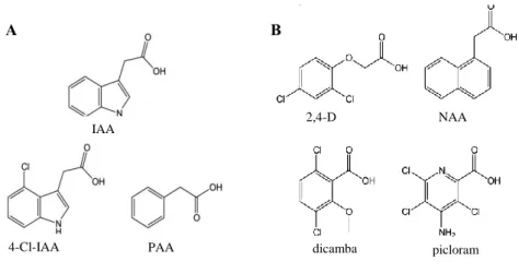
A more physiological definition describes active auxins as molecules that when exogenously supplied to a plant, induce in it an auxin response. In this sense, several naturally occurring auxin forms have been identified: indole-3-acetic acid (IAA), 4-chloroindole-3-acetic acid (4-Cl-IAA) and phenylacetic acid (PAA) (Fig 4.A). On the other hand, they also exist synthetic active auxins compounds: 2,4-dichlorophenoxyacetic acid (2,4-D), 1-naphtaleneacetic acid (NAA), 3-6-dichloro-2-methoxy-benzoic acid (dicamba) and 4-amino-3,5,6-trichloropicolinic acid (picloram) (Fig4.B) (Korasick et al., 2013). Synthetic auxins are frequently used in agriculture as herbicides or rooting agents and in plant tissue cultures, due to its higher stability and easier absorption by cells (Ljung, 2013).
Same as it happens with any other hormone, the physiological auxin response is first subjected to the auxin content inside a cell -in turn dependent on auxin synthesis, conjugation, compartmentalization and transport- and then to the combination of components involved in auxin signalling present at that precise moment inside the cell nucleus (Wang and Estelle, 2014).
Auxin synthesis
Although many cell types can produce auxin, it is preferentially synthesized in the aerial parts of the plants, especially in young leaves and meristems from which it can be transported to other parts of the plant (Paque and Weijers, 2016; Ruiz Rosquete et al., 2012).
Fig 4. Chemical structures of naturally occurring (A) and artificial (B) auxin compounds. Adapted from (Korasick et al., 2013). A B IAA 4-Cl-IAA PAA 2,4-D NAA dicamba picloram
24 General introduction to auxin, the hormone growth
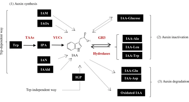
L-Tryptophan (L-Trp)-dependent biosynthesis is thought to be the main way of IAA production in plants. L-Trp is produced from chorismate, the final compound of the shikimate pathway, that takes place in the plastid. Later, in the cytosol L-Trp can be transformed into IAA through different precursors: indole-3-pyruvic acid (IPyA), indolacetamide (IAM), indole-3-acetaldoxine (IAOx), indole-3-acetonitrile (IAN) and indole-3-acetaldehyde (IAAld) (Korasick et al., 2013; Ljung, 2013). Although some of the enzymes that produce these different compounds have been identified, IAA synthesis from IPyA is the only fully described auxin synthetic pathway in plants. It involves a two-step process: first the transformation of L-Trp in IPyA by a tryptophan aminotransferase (TAA1) followed by its conversion into IAA by flavin mono-oxygenases from the YUCCA family (Finet and Jaillais, 2012) (Fig 5).
However, it has been observed that Arabidopsis mutants impaired in Trp synthesis present comparable levels of IAA to a wild-type plant, being this indicative of the existence of a Trp-independent IAA production. Although little is known about this other synthetic pathway it is thought to derive from another product of the shikimate route, indole-3-glycerol phosphate (IGP) (Korasick et al., 2013) (Fig 5).
Alterations in the different biosynthetic pathways have been observed in response to diverse environmental cues, indicating this that the redundant auxin pathways could collaborate to combinatorial responses for the integration of external signals (Ruiz Rosquete et al., 2012). Auxin inactivation
IAA levels can also be attenuated by its inactivation, which can happen by conjugation to other molecules (normally reversible) or through IAA catabolism (irreversible).
IAA conjugates are considered as temporary storage forms from which, by hydrolysis, active IAA can be recovered. Its importance has been proven in response to seed imbibition, auxin-mediated symbiosis, nodule organogenesis and auxin toxic levels. Although the specific role of these compounds is not clear yet they are thought to antagonize auxin effects competing with its receptors or transporters (Korasick et al., 2013; Ruiz Rosquete et al., 2012).
25
IAA can be ester-linked to sugars or amide-linked to amino acids (low molecular weight conjugates) or to peptides or proteins (high molecular weight conjugates). Although the composition of IAA conjugates varies between different species, IAA amides are the most common forms of conjugation, more specifically IAA-Alanine (IAA-Ala), IAA-Leucine (IAA-Leu), IAA-Tryptophan (IAA-Trp), IAA-Aspartate (IAA-Asp) and IAA-Glutamate (IAA-Glu). These reactions are carried out by the Gretchen Hagen 3 (GH3) protein family and, except for IAA-Asp and IAA-Glu compounds, they can be reversed back by amidohydrolases. GH3 genes are auxin-early response genes, indicating this another point for the feedback control of available auxin inside a cell (Finet and Jaillais, 2012; Korasick et al., 2013; Ruiz Rosquete et al., 2012) (Fig 5).
On the other hand, IAA and its conjugated forms IAA-Asp and IAA-Glu can be oxidized which leads to the irreversible degradation of the hormone. The molecular mechanisms behind these processes are not well described till now (Ruiz Rosquete et al., 2012) (Fig 5).
Auxin transport
Same as it happens with nutrients and photosynthetic assimilates, plant hormones can be transported long distances in a fast-non-directional way along the phloem. However, auxin is the only
IAM IAOx IAN IAAld (1) Auxin synthesis (2) Auxin inactivation IAA-Glucose IAA-Ala IAA-Leu IAA-Trp IAA-Glu IAA-Asp Oxidated IAA (3) Auxin degradation IGP IPA Trp TAAs YUCs Trp-independent way T rp -d ep en d en t w ay IAA GH3 Hydrolases
Fig 5. Auxin synthetic and inactivation pathways. Auxin can be synthetized in a Trp-dependent (from IAM, IAOx, IPA, IAN or IAAld precursors) or a Trp-independent way (through IGP) (1). Auxin free levels can be regulated by the hormone conjugation to sugars or amino acids (2) or by its degradation (3). Continuous arrows indicate known steps and the enzymes that mediate them (in red). Discontinuous arrows show not yet characterized reactions. Double arrows indicate reversible steps (Adapted from (Ljung, 2013)).
26 General introduction to auxin, the hormone growth
phytohormone known to also be transported short distances from cell to cell. This type of transport is known as polar transport as it happens in a directional way. It has been proven to be fundamental for several plant developmental processes such as apical-basal early embryo determination, hypocotyl elongation, lateral roots formation or flower development, processes all of them based in peaks of minimums and maximums of auxin levels (Adamowski and Friml, 2015). Indeed, alteration of polar auxin transport causes dramatic effects on plant development leading to the formation of naked shoots (Fig 6.A) (Vernoux et al., 2000).
Cell-to-cell auxin transport happens according to the chemiosmotic model: in the apoplast of the plant cell, where the pH is around 5, IAA will predominate in its protonated form (IAAH) that can diffuse across the cell wall, or can enter the cell by active transport (influx carriers). Once inside the cell, given the neutral pH of the cytosol, IAA will be found mostly as IAA- that can only exit the cell by efflux carriers (Fig 6.B).
There are four main classes of active auxin transporters in A. thaliana: auxin transporter protein 1 (AUX1) and auxin transporter-like protein (LAX) families of proteins constitute auxin influx carriers. On the other hand, auxin efflux carriers are members of the ATP-binding cassette (ABC) transporters family and the pin-formed proteins (PIN). Whereas ABC are uniformly localized in the plasma membrane, PIN proteins are polarly present in one side of the plant cell. This polar localization of PIN efflux carriers is what drives cell-to-cell auxin transport in a polar way (Grones and Friml, 2015) (Fig 6.B).
Out of the 8 PIN proteins found in A. thaliana, PIN5, 6 and 8 are localized in the endoplasmic reticulum (ER). This positioning is thought to contribute to auxin homeostasis by auxin compartmentalization. As for the rest of the PIN proteins, they can agglomerate in the basal or apical part of the cell in a dynamic manner: PIN proteins go through continuous endocytosis and recycling processes that will control the abundance of the protein in the plasma membrane and thus the auxin efflux. PIN maintenance in the plasma membrane is highly regulated. Post-translational modifications of PIN, such as phosphorylation or ubiquitination, environmental signals, microtubules orientation and other phytohormones (strigolactones, citokynins, gibberelins and auxins themselves) will influence PIN presence in the plasma membrane (Adamowski and Friml, 2015; Habets and Offringa, 2014).
27
PINs are also regulated at the transcriptional level. Different expression patterns exist for each PIN gene within different tissues of the plant. Auxin is one of the signals that contribute to the transcriptional regulation of these proteins through Auxin Responsive Factors (ARFs) transcription factors (Habets and Offringa, 2014).
Auxin signalling
The auxin response inside the cell starts with the detection of the hormone by auxin receptors. Three possible auxin perception mechanisms have been described till now: the TIR1/AFB (Transport Inhibitor Resistant 1/Auxin Signalling F-box)-Aux/IAA co-receptor mechanism, the SKP2a (S-Phase Kinase-Associated Protein 2a) and ABP1 (Auxin Binding Protein 1).
• SKP2a
SKP2a involvement in auxin response has been recently proposed when auxin binding to this protein was found. SKP2a is a component of the SCF (Skp, Cullin, F-box) complex that participates in the G1/S checkpoint in cell cycle mediating the degradation of E2FC and DPB factors. This relieves repression of cell cycle control genes. It presents a F-box and a Leucine Rich Repeats (LRR) domain, which mediates binding to auxin (Jurado et al., 2010; Peer, 2013; Powers and Strader, 2016). By structure modelling with human SKP2 and further comparison of the modelled SKP2a with TIR1 structure (Fig 10.C), the SKP2a auxin binding pocket was identified. SKP2a mutants in this site were not able to induce E2FC and DPB degradation showing that auxin binding was necessary for SKP2a ubiquitin ligase role. SKP2a mutants were auxin-resistant and overexpression of the protein gave auxin-related phenotypes (Jurado et al., 2010; Powers and Strader, 2016), proving all this SKP2a implication in auxin response. SKP2a auxin perception could partially explain auxin roles in cell division.
Fig 6. Auxin polar transport. A. Wt A. thaliana plant (left) vs pin1 mutant (right) affected in auxin transport. Adapted from (Vernoux et al., 2000). B. Auxin transport scheme: in the apoplast IAA predominates as IAAH which can enter the cell by diffusion (1) or by active transport driven by influx carriers (2). Inside the cytosol, IAA is found in its deprotonated form, IAA- , and it will only exit the cell with the help of efflux carriers: ABC (3); uniformly distributed along the plasma membrane or PIN (4) polarized on one side of the cell.
PIN AUX1/LAX ABC
B IAA -IAAH IAAH (1) (2) (3) (4) A
28 General introduction to auxin, the hormone growth
• ABP1
ABP1 auxin receptor role is a controversial matter in the auxin field nowadays. Even if it was the first auxin receptor to be discovered, its study became complicated due to the lack of auxin-related phenotypes associated to these receptors. In 2001, the first ABP1 mutant, abp1-1, was generated in
A. thaliana and this allowed the generation of other mutant lines in downstream elements associated
with this signalling cascade. However, recent genetic studies suggest that ABP1 mutations give no phenotype and that the already described ones are the consequence of off-targets mutations (Feng and Kim, 2015).
ABP1 protein is localized in the plasma membrane or in the ER. Although ABP1 has been proven to bind to auxin (Woo et al., 2002), the conditions in the ER are not favourable for the binding of the hormone, so the role of the ABP1 localized in the organelle is still to be studied.
On the other hand, ABP1 located in the plasma membrane has been associated mostly with the auxin non-transcriptional response, among others with the ROP-GTPase pathway that leads to clathrin-mediated endocytosis of PIN proteins (Peer, 2013).
• TIR1/AFB-Aux/IAA
Finally, the TIR1/AFB-Aux/IAA co-receptor mechanism has been extensively studied together with the transcription factors and the transcriptional response downstream auxin perception. All these proteins constitute the Nuclear Auxin Pathway (NAP). NAP reconstitution in a yeast system proved that only three components conform the core auxin response module: the TIR1/AFB and Aux/IAA co-receptors and the Auxin Response Factors, ARFs; and that these are enough for obtaining an auxin basic transcriptional response (Pierre-Jerome et al., 2014). However, the auxin transcriptional response in plants is everything but basic: different auxin inputs and gradients controlled by cell type, developmental stages and environmental cues give rise to a complex set of outputs in auxin response (Paque and Weijers, 2016; Pierre-Jerome et al., 2013). In the next lines, we will see how auxin is able to play its magic always through the same repertoire of proteins: TIR1/AFB, Aux/IAAs and ARFs (Fig 7). Due to the interactions established between them, it is difficult to talk about these proteins individually and thus we will treat them as proteinic modules.
29
o Module ARF-DNA
ARF proteins are transcription factors that bind to DNA sequences named Auxin Response Elements (AuxREs) located in the promoters of auxin responsive genes. Only in the model plant A. thaliana, 23 ARF proteins have been found. Out of the 23, just 5 of them (ARF5-8 and ARF19) are considered as ARF activators. The rest have been classified as repressors although there is not yet evidence of a repressor role for all of them (Peer, 2013).
ARFs-DNA interaction is mediated by a B3 domain located in their N-terminus. The B3 domain is flanked by regions that mediate dimerization of the protein (Dimerization Domains, DD), thanks to which ARFs bind to double AuxREs as dimers. In AtARF1 and AtARF5 DBD structures, a third domain, Flanking Domain (FD) was identified but its role remains unknown (Fig 7-8) (Boer et al., 2014)1.
o Module ARF-Aux/IAA
On the other hand, in their C-terminal ARF proteins present a Phox/Bem1p (PB1) domain which is shared with Aux/IAAs (corresponding to domains III/IV, DIII/IV) (Fig 7). PB1s are protein-protein interaction domains that have been shown to mediate homo or hetero-oligomerization of ARFs or Aux/IAAs and ARFs-Aux/IAAs, respectively (Farcot et al., 2011; Guilfoyle and Hagen, 2012; Li et al., 2011; Piya et al., 2014; Trigg et al., 2017).
1ARFs-DNA interaction and the details of ARFs structure and domains organization will be properly explained in
Chapter II of this manuscript.
PB1 DD B3 DD FD DBD PB1 DI DII 18 LRRs F-box ARFs Aux/IAAs TIR1/AFB
Fig 7. Domains organization of the protein families involved in the Nuclear Auxin Pathway. DBD (DNA-binding domain); DD (Dimerization Domain); FD (Flanking Domain); MR (Middle Region); DIII/IV (Domains III/IV) also called PB1 (Phox/Bemp1); DI (Domain I, containing EAR-motifs); DII (Domain II, containing degron motif); LRR (Leucine Rich Repeat).
DIII/IV DIII/IV MR
30 General introduction to auxin, the hormone growth
These interactions take place in a front-to-back manner due to the presence of two interaction interfaces in these domains: a negative and a positive interface, that allow chaining a high number of molecules by means of electrostatic interactions (Chandrasekaran et al., 2015; Kim et al., 2015; Korasick et al., 2014; Nanao et al., 2014; Parcy et al., 2016; Wang et al., 2015) (Fig 9). A high-throughput Yeast-2-hybrid (Y2H) experiment proved that most Aux/IAA proteins can homo-oligomerize or hetero-oligomerize mainly with ARF activators. ARF activators were also able to homo-oligomerize but ARFs repressors showed barely any interactions among themselves or with Aux/IAA proteins (Farcot et al., 2011).
The specificity and affinity of Aux/IAA-ARF and ARF-ARF interactions is related to the residues present in the acidic and basic interfaces of their PB1 domains (Parcy et al., 2016), meaning that certain combinations of PB1 domains are more favourable than others (Han et al., 2015). Also, post-translational modifications have been shown to affect these interactions (Dinesh et al., 2016; Hill, 2015; Vert et al., 2008; Wang and Estelle, 2014).
It is necessary to point out that ARF PB1 domains can also interact with other transcription factors such as MYB77, PIF4, bZIP or BZR1/2. These interactions might help the cross-talk between auxin and other signalling pathways (Dinesh et al., 2016).
Fig 8. ARFs DBD structure. Structure of AtARF1 DBD (light pink, 4LDX) in complex with DNA (black) overlapped with ARF5 DBD structure (purple, 4LDU) showing the high similarity between them. FD, Flanking Domain; DD, Dimerization Domain. DNA-interactions are mediated by two loops within the B3 domain. Adapted from (Boer et al., 2014).
DD FD
31 o Module Aux/IAA-TIR1
Apart from the DIII/IV in Aux/IAAs, that mediates interactions with ARFs, in their N-terminal Aux/IAAs present two other domains I and II (DI and DII). DII, also called degron motif, mediates the interaction with TIR1/AFB auxin receptor.
TIR1/AFB are a family of proteins that belong to the E3 ubiquitin ligase systems. They present a F-box domain and a Leucine Rich Repeats domain (LRRs) composed by 18 LRRs (Fig 7). TIR1 was the first plant hormone receptor which structure was solved, helping this to the understanding of auxin perception: auxin binding takes places in an auxin binding pocket located within the structure formed by the LRRs. The hormone does not induce any conformational change in the TIR1/AFB receptors but it acts as a “molecular glue” between TIR1/AFB proteins and DII of Aux/IAAs (Tan et al., 2007) that leads to Aux/IAAs ubiquitination and further proteosomal-mediated degradation (Salehin et al., 2015) (Fig 10).
Recent studies have shown that TIR1 can also form oligomers and that the receptor oligomerization could help binding and poly-ubiquitination of Aux/IAAs (Dezfulian et al., 2016).
In A. thaliana 6 TIR1/AFB and 29 Aux/IAA proteins have been described. All of them have different patterns of gene expression in different plant tissues (Farcot et al., 2011; Wang and Estelle, 2014). In addition to this, TIR1/AFB-Aux/IAA combinations present
A
B C D
Fig 9. PB1 structures. 4 PB1 domains structure have been determined till now. (A) AtARF5-PB1 oligomer (4CHK,(Nanao et al., 2014)) showing the alternance of positive and negative interfaces that mediate the front-to-back interactions. Each PB1 monomer shown in a different violet intentsity. (B) AtARF7-PB1 (4NJ6, (Korasick et al., 2014)). (C) PsIAA4 (Peasum sativum) (2M1M, (Dinesh, D.C. et al, 2015)) . (D) AtIAA17 (2MUK, (Han et al., 2015)). Positive and negative interface indicated for each.
32 General introduction to auxin, the hormone growth
different affinities between themselves and for auxin hormone, which is translated into different degradation rates of Aux/IAA proteins, with half-lives that can range from 6 to 80 min in the presence of the hormone (Dinesh et al., 2016).
Although residues GWPPV within Aux/IAA-DII are the ones necessary for this interaction and further Aux/IAA degradation other parts inside the DII and in between DI and DII have been shown to influence the degradation rate of the protein, contributing this to the diversity of affinities (Calderón Villalobos, L; Lee, S; De Oliveira, C; Ivetac, A;Brandt, W; Armitage, L; Sheard, LB; Tan, X; Parry, G; Mao, H; Zheng, N; Napier, R; Kepinski, 2012).
Finally, post-translational modifications have been described for TIR1/AFB proteins that can modify their stability and interaction with Aux/IAA proteins. Altogether, transcriptional regulation, post-translational modifications and the diversity of TIR/AFB-Aux/IAA complexes contributes to the control and fine-tuning of auxin response (Wang and Estelle, 2014).
The combinatorial action of all these modules and the diversity of combinations within each, allows the repression or activation of auxin transcriptional response in a fine-tuned and spatial-temporal way. We must add a fourth component to the equation, though: the co-regulators. Although not specific to auxin signalling pathway, and thus not included into the so-called NAP, the co-repressors
Fig 10. TIR1 structure. A-B. AtTIR1 structure in complex with IAA7-DII (red) and auxin (green) (2P1N). (B) Rotated 90º with respect to (A). LRR (grey) and F-box domains (orange) form a mushroom-shaped structure, with the LRR mediating the interaction with both the hormone and the degron in the same binding pocket. The structure was also obtained in complex with ASK1 (ubiquitin ligase system) and Ins6P, identified as a co-factor of unknown function (structure not shown here) (Tan et al., 2007). (C) Structure model of SKP2a (green) aligned to LRR domain of TIR1 that allowed SKP2 binding site for auxin (purple) (Adapted from (Jurado et al., 2010)).
A B
LRR
F-box
33
TOPLESS (TPL) and the chromatin remodelling ATPases SPLAYED (SYD) and BRAHMA (BRM) collaborate to the repression or activation of the signal2.
Putting all the pieces together they create the following, precise, and stylish puzzle (Fig 11): under low auxin concentrations, ARF activators are blocked by Aux/IAA repressor proteins that bind to them through the DIII/IV present in both proteins. At the same time, the Aux/IAA domain I (DI) can recruit TOPLESS co-repressor (TPL) that will in turn recruit chromatin modifier enzymes, forming a repressor complex that will avoid the transcription of auxin responsive genes through chromatin compaction. However, when auxin levels increase in the plant cell Aux/IAA proteins will bind auxin together with TIR1/AFB which leads to Aux/IAA degradation and thus all the co-repressor protein complex will be taken away from the DNA. This leaves ARF activators free to do their transcriptional activator role, for which they will recruit chromatin remodelling proteins (such SYD and BRM) that will help opening chromatin so that transcription of auxin responsive genes can take place (Lavy and Estelle, 2016; Wu et al., 2015b).
This mechanism is relatively well described for ARF activators. However, ARF repressors are much more of a mystery. Most of them are unable to interact with Aux/IAA repressors (Farcot et al., 2011) and therefore they are thought to work in an auxin-independent manner. Three possible mechanisms are proposed for ARF repressors working mode: “kidnapping” of ARF activators by binding to them, competition with ARF activators for AuxREs and/or TOPLESS recruitment (Chandler, 2016).
2TPL involvement in auxin signalling with be further discussed in Chapter I of this manuscript
DII
DI
DII
DI
Auxin responsive gene OFF
DII
Auxin responsive gene ON
- auxin + auxin ARF activator PB1 DBD DII DI PB1
Aux/IAA TPL SYD/BRM TIR1
F-box LRR
IAA 26S proteosomal degradation
34 General introduction to auxin, the hormone growth
Fig 11. Nuclear Auxin Pathway. In the absence of auxin (left), Aux/IAA repressors bind to ARF activators through their PB1 domains and recruit TPL through its DI preventing auxin responsive genes transcription. In the presence of auxin (right) a TIR1/AFB-Aux/IAA auxin complex is formed and triggers the degradation of Aux/IAAs and to the release of ARF activators, now free to recruit SYD and BRM chromatin remodellers, leading to auxin responsive genes expression. The domains or proteins pictured as structures represent real structures already determined for some of these proteins (Adapted from (Nanao et al., 2014)).
35
Objectives
This manuscript has a focus on the structural and biochemical mechanisms that lie beyond the proteins involved in the NAP. Along the following chapters, we will try first, to unravel the structural secrets of TPL co-repressor function and its interaction with Aux/IAA repressor proteins. We will immerse ourselves into the details of ARFs-DNA binding mysteries. And last but not least, we will try to answer to a big intriguing question: where did all this come from?
Chapter I: Function, structure and evolution of TOPLESS co-repressor
TOPLESS is a co-repressor protein in plants able to interact with a huge diversity of plant repressors. These interactions are mediated through conserved sequences of amino acids present in all these repressors, the Ethylene-responsive element binding factor-associated Amphiphilic Repression (EAR) motifs.
The first chapter of this manuscript aims at answering:
• how TOPLESS interacts with such a diversity of transcriptional repressors
• how TOPLESS-Aux/IAA interactions happen and how they affect the repression of auxin responsive genes
For this, we followed a “from structure to function” strategy mainly based on in vitro studies comprising a wide variety of protein-protein interaction techniques.
Chapter II: ARFs-DNA preferences, players of the specificity of auxin transcriptional response
ARF transcription factors have been classically classified into activators or repressors. Either one or the other they all present DNA binding domains (DBD) that mediate ARFs interaction with Auxin Response Elements, AuxREs. Surprisingly, the residues mediating DNA-interactions are highly conserved among activators and repressors and they have both been shown to bind to the same nucleotide sequences. AuxREs are normally formed by two repeats of ARF DNA binding. Depending on their orientation and spacing different types of AuxREs have been described in literature.
In Chapter II we tried to go in depth into the specificity of the different classes of ARF proteins for the different types of AuxRE repeats. To do this we used an integrated strategy of bioinformatic analysis and in vitro studies of protein-DNA interactions.
36 Objectives
Chapter III: Back to the water looking for ARF ancestors
Land plants evolution from algae happened hand in hand with the adaptation to terrestrial stresses and the development of a certain body complexity. Given the importance of auxin hormone in plant growth and architecture, the acquisition of the auxin signalling pathway must have been a breaking point in the transition from water to terrestrial life. However, not much is known about how and when this happened. ARF proteins have been traced back till bryophyte organisms but till now no sign of them has been found in charophyte algae, the proposed ancestors of land plants.
Swimming among the algae, we found by sequences homology and protein structure modelling two possible candidates of ARF ancestors in the charophyte algae Klebsormidium nitens and
Chlorokybus atmophyticus. Both proteins presented a predicted DBD and a PB1 domain with high
similarities to those found in plant ARF proteins. Furthermore, as it is the case for some ARF repressors in between both predicted domains we found potential motifs that might recruit TPL.
Along the third and final chapter of this manuscript I will describe the structural and biochemical characterization of these different domains in terms of their DNA binding specificity, oligomerization potential and TPL interactions.
37
Chapter I
Function, structure and evolution of
TOPLESS co-repressor
39
Introduction
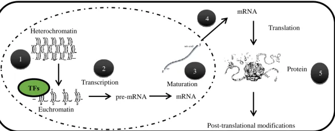
Once upon a time (1958) Francis Crick proposed what till now we know as the “Central Dogma of Biology” according to which, the genetic information is stored inside cells as deoxyribonucleic acid, DNA, that will be transcribed into messenger ribonucleic acid, mRNA, which in turn will be translated into proteins, the elements that will in the end, develop a specific function inside a cell.
Decades after Crick’s proposal we can affirm that the transition from DNA to proteins is not that elemental -dear Watson- with each step of the process being susceptible to different levels of regulation (Fig 12).
Gene expression control in eukaryotic organisms
Gene expression is fundamental in every living organism and thus, it is one of the key regulated points in all this process. Eukaryotic organisms have developed more evolved and complex regulatory systems of gene expression in comparison with prokaryotes.
Bacteria’s DNA is “naked” and “free” inside the cell (Payankaulam et al., 2010). On the contrary, eukaryotes conceal their genetic information inside the nucleus. Thanks to this, transcription
TFs Heterochromatin Euchromatin Transcription Maturation pre-mRNA mRNA mRNA Translation Protein Post-translational modifications 1 2 3 4 5
Fig 12. Regulation levels from chromatin to proteins. (1) Chromatin compaction state can allow or avoid the access of proteins (transcription factors or transcriptional machinery). (2) Euchromatin is a transcriptionally active state in which transcription will be controlled by regulatory sequences in the promoters of genes and the proteins bound to them. (3) Eukaryotic organisms have developed a machinery of processing and splicing of the transcripts that can influence their stability or generate different combinations of exons. (4) Exit of the mature mRNA from the nucleus can control its availability in the cytosol for its translation into proteins. (5) Once the mRNA is translated into proteins the function or availability of these proteins can be also modulated by its tertiary or quaternary structure or by post-translational modifications (Adapted from (Alonso and Wilkins, 2005)).
40
Introduction Chapter I Chapter I
(nuclear) and translation (cytosolic) are physically separated. Therefore, the processing and transport of mRNA from the nucleus to the cytosol adds one level of control with respect to bacteria (Fig 12).
On the other hand, eukaryotic DNA is wrapped around nucleosomes -octamers of histone proteins - that form a structure named chromatin (Liu et al., 2014; Payankaulam et al., 2010) (Fig 12). Chromatin can be in two different forms: euchromatin, that is lightly packed and transcriptionally active and heterochromatin, with a high level of compaction in which transcription cannot happen. Dynamic marks in the DNA or in histone tails influence chromatin packing level. DNA methylation and histones deacetylation are considered as repressive marks whereas histones acetylation is thought to cause relaxation of the chromatin structure. Several families of enzymes have been involved in the deposition or removal of the different marks, including DNA Methyltransferases (DNA MTase), Histone Deacetylases (HDA) or Histone Acetyltransferases (HAT) (Liu et al., 2014; Xiao et al., 2017). Other types of enzymatic histone modifications, such as ubiquitination, methylation or phosphorylation have been described (Liu et al., 2014; Perissi et al., 2010; Xiao et al., 2017). Chromatin state -and all the proteins involved in its dynamics- is consequently another point of control since it constitutes a physical barrier for the access of transcriptional regulatory proteins.
Among these regulatory proteins, transcription factors, either activator or repressors, have the ability to bind to specific DNA sequences in the regulatory regions within genes and from there they will contribute to the activation or repression of these genes.
Transcription factors are present in both prokaryote and eukaryote organisms. In the formers, the regulation of transcription by transcription factors is “direct”: they interact with the basal transcriptional machinery. All over again, eukaryotes went a bit further and they evolved “intermediate proteins”, named coregulators (Payankaulam et al., 2010).
Co-repressors
Co-regulators, either co-activators or co-repressors, are proteins that bridge transcription factors to chromatin modifying enzymes, allowing the formation of a coregulator complex with enzymatic activity that will alter chromatin conformation in specific regions in the DNA (Mottis et al., 2013). They do not have DNA binding capacity themselves. Instead, they are recruited to specific regions in the genome by transcription factors that do have DNA specificity. One single co-regulator can be recruited to the chromatin environment by many different transcription factors helping to the regulation of a wide set of genes (Watson et al., 2012).
41
These two facts (co-regulators binding to a diversity of transcription factors and to chromatin remodeller enzymes) bring us to consider co-regulators as “hub” or “scaffolding” proteins that integrate in one single flexible complex diverse protein functions (Watson et al., 2012). Transcriptional repressors present repressor domains that mediate the recruitment of co-repressor proteins to specific regulatory sequences within genes (Kagale et al., 2010a). There, co-repressors will inhibit the expression of these genes by inducing chromatin compaction. Several mechanisms have been described that aid explaining how chromatin closing is triggered. Recruitment of HDA or other chromatin modifying enzymes, direct or indirect -through the Mediator complex- interactions with the transcriptional machinery or blocking of the activation sites of transcription factors are some of them (Agarwal and Mathew, 2015).
On the other hand, co-repressors disposition on chromatin is also thought to be determinant for chromatin compaction. Co-repressors have been classified into long-range and short-range (Payankaulam et al., 2010). By “spreading” along chromatin, long-range co-repressors are suggested to help the deposition of repressor marks along long regions of the DNA. On the contrary, short-range co-repressors are locally positioned inducing the formation of DNA loops that antagonize chromatin opening (Sekiya and Zaret, 2007).
Co-repressor examples have been described in a diversity of eukaryotic organisms. Among the most studied ones are the Gro (Groucho) family in Drosophila and the SMRT (Silencing Mediator of Retinoic Acid and Thyroid hormone receptor)/NCoR (Nuclear Receptor Co-repressor) family in humans. Orthologues of these proteins have been found in lower organisms indicating this their importance along evolution (Mottis et al., 2013; Watson et al., 2012).
• SMRT/NCoR family
SMRT/NCoR co-repressors are associated with nuclear receptors to which they will bind in the absence of ligand. Upon ligand interaction, nuclear receptors will dissociate from co-repressors and will bind coactivators instead. SMRT/NCoR complexes have been implicated in diseases such as cancer or diabetes (Mottis et al., 2013). 10-12 proteins were found to be part of the human SMRT/NCoR complex (Yoon et al., 2003), among them HDA3, GPS2, TBL1 and obviously, SMRT/NCoR. TBL1 “hubs” this complex by establishing interactions with both GPS2 and SMRT/NCoR. TBL1 presents a Lissencephaly (LisH) domain in its N-terminal region and WD40 domains in its C-terminal (Fig 13.A). Structural determination of TBL11-90 N-ter revealed that TBL1 is a tetrameric protein with the tetramer established as a dimer of dimers. The LisH domain and a third expanded helix mediate both dimerization and tetramerization and they generate in between a hydrophobic groove that mediates interaction with GPS2 and SMRT/NCoR proteins (Fig 13.B)
42
Introduction Chapter I Chapter I
(Oberoi et al., 2011). The LisH domain has been proven to be fundamental for TBL1 binding to histones and for repression (Choi et al., 2008; Yoon et al., 2005, 2003).
SET3 complex is thought to be the orthologue of the SMRT/NCoR complex in yeast. Among the proteins integrating SET3, Sif2 is the equivalent to TBL1, presenting a LisH N-terminal domain and WD40 C-terminal repeats (Mottis et al., 2013; Yoon et al., 2005) (Fig13. A). Same as for TBL1, Sif2 LisH domain mediates its tetramerization (Cerna and Wilson, 2005). The function of the WD40 in these proteins remains unknown.
LUFS TBL1/Sif2 Gro/Tup/TLE/Grg LUG TPL B C D A
43 • Groucho family
Groucho, present in Drosophila, was the first co-repressor protein of this family to be identified. Gro mutation resulted in clumps of extra bristles over the eyes of the poor flies that after this, had an apparently striking resemblance with the famous American humourist Groucho Marx. I bet he did not find this funny (Fig 14.A).
Nowadays, orthologues of Gro have been described in other organisms: Tup1 in yeast, Grg in mouse or TLE in humans. All these proteins present a glutamine (Q)-rich region in their N-ter and a WD40 repeats domain in their C-ter separated by a non-conserved glycine (G) and proline (P)-rich region (Liu and Karmarkar, 2008) (Fig 13.A). Structural studies have proven that Gro family members are tetrameric proteins which tetramerization is mediated by their N-ter. The tetramers are formed as dimers of dimers (Chodaparambil et al., 2014; Matsumura et al., 2012) (Fig 13.C-D) and have been shown to be needed for binding to histones and for repression (Chodaparambil et al., 2014; Flores-Saaib and Courey, 1999; Sekiya and Zaret, 2007). As for the WD40 in the C-terminal regions of Gro proteins, they have been implicated in interactions with transcription factors and in chromatin condensation (Jennings et al., 2006; Sekiya and Zaret, 2007).
Co-repression in plants
Plants are not less than flies or humans and so, they have also evolved stylish transcriptional repressor mechanisms that involve the action of co-repressor proteins. LEUNIG/LEUNING-HOMOLOGUE (LUG/LUH) and TOPLESS/TOPLESS-related (TPL/TPRs) are the two most studied families of plant co-repressors (Lee and Golz, 2012; Liu and Karmarkar, 2008).
LUG was the first co-repressor to be identified in plants. lug mutants presented floral defects associated with an ectopic expression of the floral regulator AGAMOUS (AG) (Conner and Liu, 2000) (Fig 14.B). Further biochemical studies revealed that LUG protein presented a LisH and a Q-rich region in its N-ter and WD40 domains in its C-terminal region (Lee and Golz, 2012; Liu and Karmarkar, 2008). The LisH domain is enclosed into the so-called LUFS domain, responsible for mediating interactions with transcription factors through the intermediate protein SEUSS (SEU) (Sridhar et al., 2004, 2006) (Fig 13.A). LUG repressor capacity has been shown to be dependent on
Fig 13. Structural features of co-repressors. A. Domains distribution of co-repressor proteins from diverse organisms: TBL1 (human)/Sif2 (yeast) of the SMRT/NCoR co-repressor complex, Gro (Drosophila)/Tup1 (Saccharomyces)/TLE (Homo sapiens)/Grg (Mus musculus), LUG (A. thaliana), TPL (A. thaliana) (adapted from (Liu and Karmarkar, 2008)). B. Structure of the human TBL1 tetramer (2XTC) with LisH domain in red in one monomer (Oberoi et al., 2011). C. Structure of the human TLE N-terminal tetramer (4OM3) (Chodaparambil et al., 2014). D. Structure of the yeast Tup1p (3VP8) N-terminal tetramer (Matsumura et al., 2012). Tetramerization interfaces framed inside a black rectangle.


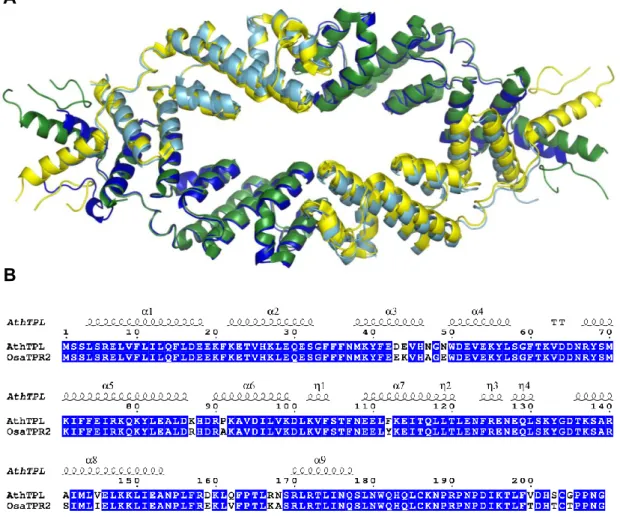
![Figure S1: TPL has a CRA domain. Superimposition of two proteins possessing the LisH- LisH-CTLH-CRA domains (the human RAN-Binding protein 9 [Uniprot Q96S59, green], and a protein from the unicellular algae C](https://thumb-eu.123doks.com/thumbv2/123doknet/12850144.367847/59.892.256.619.152.615/figure-superimposition-proteins-possessing-domains-binding-uniprot-unicellular.webp)