RESEARCH OUTPUTS / RÉSULTATS DE RECHERCHE
Author(s) - Auteur(s) :
Publication date - Date de publication :
Permanent link - Permalien :
Rights / License - Licence de droit d’auteur :
Bibliothèque Universitaire Moretus Plantin
Institutional Repository - Research Portal
Dépôt Institutionnel - Portail de la Recherche
researchportal.unamur.be
University of Namur
Structural analysis of Brucella abortus RicA substitutions that do not impair
interaction with human Rab2 GTPase
Nkengfac, Bernard; Pouyez, Jenny; Bauwens, Emilie; Vandenhaute, Jean; Letesson,
Jean-Jacques; Wouters, Johan; De Bolle, Xavier
Published in: BMC biochemistry DOI: 10.1186/1471-2091-13-16 Publication date: 2012 Document Version
Publisher's PDF, also known as Version of record Link to publication
Citation for pulished version (HARVARD):
Nkengfac, B, Pouyez, J, Bauwens, E, Vandenhaute, J, Letesson, J-J, Wouters, J & De Bolle, X 2012, 'Structural analysis of Brucella abortus RicA substitutions that do not impair interaction with human Rab2 GTPase', BMC
biochemistry, vol. 13, no. 1, 16, pp. 16. https://doi.org/10.1186/1471-2091-13-16
General rights
Copyright and moral rights for the publications made accessible in the public portal are retained by the authors and/or other copyright owners and it is a condition of accessing publications that users recognise and abide by the legal requirements associated with these rights. • Users may download and print one copy of any publication from the public portal for the purpose of private study or research. • You may not further distribute the material or use it for any profit-making activity or commercial gain
• You may freely distribute the URL identifying the publication in the public portal ?
Take down policy
If you believe that this document breaches copyright please contact us providing details, and we will remove access to the work immediately and investigate your claim.
R E S E A R C H A R T I C L E
Open Access
Structural analysis of Brucella abortus RicA
substitutions that do not impair interaction with
human Rab2 GTPase
Bernard Nkengfac
1,3, Jenny Pouyez
2, Emilie Bauwens
1, Jean Vandenhaute
1, Jean-Jacques Letesson
1,
Johan Wouters
2and Xavier De Bolle
1*Abstract
Background: Protein-protein interactions are at the basis of many cellular processes, and they are also involved in the interaction between pathogens and their host(s). Many intracellular pathogenic bacteria translocate proteins called effectors into the cytoplasm of the infected host cell, and these effectors can interact with one or several host protein(s). An effector named RicA was recently reported in Brucella abortus to specifically interact with human Rab2 and to affect intracellular trafficking of this pathogen.
Results: In order to identify regions of the RicA protein involved in the interaction with Rab2, RicA was subjected to extensive random mutagenesis using error prone polymerase chain reaction. The resulting allele library was selected by the yeast two-hybrid assay for Rab2-interacting clones that were isolated and sequenced, following the “absence of interference” approach. A tridimensional model of RicA structure was used to position the substitutions that did not affect RicA-Rab2 interaction, giving a“negative image” of the putative interaction region. Since RicA is a bacterial conserved protein, RicA homologs were also tested against Rab2 in a yeast two-hybrid assay, and the C. crescentus homolog of RicA was found to interact with human Rab2. Analysis of the RicA structural model
suggested that regions involved in the folding of the“beta helix” or an exposed loop with the IGFP sequence could also be involved in the interaction with Rab2. Extensive mutagenesis of the IGFP loop suggested that loss of interaction with Rab2 was correlated with insolubility of the mutated RicA, showing that“absence of interference” approach also generates surfaces that could be necessary for folding.
Conclusion: Extensive analysis of substitutions in RicA unveiled two structural elements on the surface of RicA, the most exposedβ-sheet and the IGFP loop, which could be involved in the interaction with Rab2 and protein folding. Our analysis of mutants in the IGFP loop suggests that, at least for some mono-domain proteins such as RicA, protein interaction analysis using allele libraries could be complicated by the dual effect of many substitutions affecting both folding and protein-protein interaction.
Keywords: Protein-protein interaction, Yeast two-hybrid, Mutagenesis, Brucella
* Correspondence:xavier.debolle@fundp.ac.be 1
Molecular Biology Research Unit (URBM), Narilis, University of Namur, 61 Rue Bruxelles, Namur B-5000, Belgium
Full list of author information is available at the end of the article
© 2012 Nkengfac et al.; licensee BioMed Central Ltd. This is an Open Access article distributed under the terms of the Creative Commons Attribution License (http://creativecommons.org/licenses/by/2.0), which permits unrestricted use, distribution, and reproduction in any medium, provided the original work is properly cited.
Background
Brucella abortus is a facultative intracellular pathogen responsible for a worldwide zoonosis [1]. Like other intracellular bacteria such as Legionella spp [2-4] and Salmonella spp[5], B. abortus probably depends on pre-cisely orchestrated interactions with host cell proteins for its infectious process. Remarkably, these intracellular pathogens secrete proteins regulating host small GTPases [4-7]. Small GTPases of the Ras super family are signaling proteins that cycle between a GDP-bound inactive state and a GTP-bound active state. These two states are regulated by guanine-nucleotide exchange fac-tors, which facilitate the conversion of GDP to GTP; GTPase activating proteins, which facilitate the hydroly-sis of the GTP and Guanine-nucleotide-dissociation inhibitors, which negatively regulate the exchange activ-ity of the GTPase and dislocate them from membranes. Rab GTPases are small GTPases playing a critical role in the control of membrane trafficking. Specifically, Rab2 has been shown to control membrane trafficking be-tween the Golgi apparatus and the endoplasmic reticulum [8], Rab2 was also putatively associated with the phagosome [9] but without any known function in phagosomal maturation in mammalian cells [10]. RicA is an effector recently identified in B. abortus, which inter-acts with human Rab2 [6]. This interaction was detected using yeast 2-hybrid (Y2H) and confirmed by GST-pull-down. RicA has a preference for GDP-bound GST-Rab2 compared to GTPγS-bound GST-Rab2 [6]. Active Rab2 is known to be required for B. abortus intracellular pro-liferation [11]. A B. abortus ΔricA strain recruits less Rab2 on the Brucella containing vacuole, suggesting that RicA is playing a role during the intracellular trafficking of the bacterium [6].
RicA is predicted to belong to the superfamily of LβH proteins, comprising acetyltransferases, acyltransferases,
carbonic anhydrases, ferripyochelin binding proteins, as well as many proteins of unknown functions. Their structure is characterized by the assembly of three β sheets in a left-handed“β helix” structure. In this paper, we attempted to localize the Rab2 interaction surface on the RicA predicted structure. We performed the “ab-sence of interference” approach [12] previously used to map the interface of the catalytic domain of the DNA methylase Dnmt3a and its regulatory factor Dnmt3L. Mapping of the substitutions that do not disrupt the RicA-Rab2 interaction, on the predicted model of RicA structure, revealed two possible interfaces, a beta sheet and a loop called IGFP. The data reported here sug-gested that, of these two structural elements, at least the IGFP loop is also involved in RicA folding.
Results
Prediction of RicA three-dimensional structure
A His6tagged version of RicA (His6-RicA) was
overpro-duced, purified to homogeinity and tested in several crystallization protocols that failed (data not shown). We therefore modelized the RicA structure by homology and verified for model correctness using EsyPred3D [13] and verify3D [14] programs respectively. The three-dimensional (3D) structure of Bacillus cereus BC4754 sequence (1XHD code in protein databank, 41.9% iden-tity) was used as the template for the homology model-ing. The function of this B. cereus protein is unknown. Modeling using other templates (2EG0, 1V3W and 1THJ codes in protein databank) generated very similar models (data not shown). Conserved domain analysis of amino acid sequences of RicA and 1XHD revealed tandemly-repeating hexapeptide repeats (Hex-motif; [LIV]-[GAED]-X-X-[STAV]-X), indicating that the over-all conformation of RicA contains a left-handedβ-helical component (LβH) characteristic of acetyltransferases
Figure 1 Predicted three-dimensional structure of RicA. The left-handedβ-helix is composed of three β sheets connected by short loops. One of these loops is the IGFP loop (Ile69 to Pro73). The structure is shown as a ribbon aligned to the backbone of the model (made using the MacPyMol program), comprising residues Ile3 to Arg170. A“top view” is shown in the middle part of the figure. The three β sheets form a triangle in this view. On the right part of the figure, the accessible surface of the predicted trimer is depicted, with a monomer ribbon shown by transparency.
Nkengfac et al. BMC Biochemistry 2012, 13:16 Page 2 of 8
superfamily [15]. The RicA monomers were assembled as trimers. Indeed several homologs are trimeric, and the three histidine residues involved in zinc binding be-tween the monomers, in the structure of carbonic anhy-drase from Methanosarcina thermophila (1THJ code in protein databank), are conserved in RicA (His67, His84 and His89), suggesting that the trimeric structure is con-served in RicA. The predicted structure of RicA is pre-sented in Figure 1.
RicA mutagenesis and selection of alleles allowing interaction with Rab2
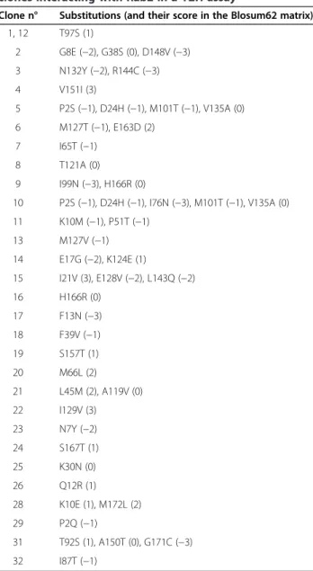
RicA specifically interacted with Rab2 in the Y2H system as indicated by the induction of the HIS3 (cell growth on plates lacking histidine in the presence of 3-amino-triazole, 3AT) and the lacZ (blue color when assayed with 5-bromo-4-chloro-3-indolyl-β-D-galactopyranoside) reporter genes (data not shown). In this interaction assay, RicA was fused with the transactivation coding se-quence (AD) of Gal4p. Since fusion of RicA with the DNA binding domain (DB) of Gal4p was autoactivating the HIS3 and lacZ reporters, the simple selection of ‘edgetic’ (interaction defective) alleles [16] was not pos-sible. We therefore decided to use the previously pro-posed “absence of interference” approach [12], in which a possible interface is mapped on a three-dimensional (3D) structure thanks to the absence of interaction-disruptive substitutions in this region of the protein. Mutated RicA (525 bp) was synthesized by error-prone polymerase chain reaction (PCR) [17] from the pDEST-AD-RicA expression clone. A mutant library of about 10,000 clones was prepared in E. coli, by BP recombina-tional cloning of the PCR products in the pDONR201 vector. Five independent clones randomly selected were sequenced to check the mutation load. We found 36 mutations for the 2625 sequenced bases, i.e. a mutation frequency of 1.4%. The mutant library was transferred to the pDEST-AD vector and assayed with Rab2 in the Y2H to assess the influence of the mutations introduced into RicA. Among 1200 yeast clones, only 32 were posi-tive for lacZ and HIS3 reporters, indicating that only ap-proximately 3% were able to interact with wild-type Rab2. A screening made with a slightly higher mutation rate for RicA coding sequence did not yield any positive interaction (data not shown), suggesting that 1.4% is close to the maximum mutation rate still allowing the recovery of interacting proteins, for this experimental setting. The ricA coding sequence was amplified from the 32 interacting clones by PCR and sequenced. We observed that a selective pressure occurred as the muta-tional load of 1.4% in the unselected library decreased to 0.5% in the selected clones. Among the 32 interacting clones, only two had the wild-type sequence, and all sub-stitutions observed in other clones are reported in
Table 1. A total of 29 substitutions were collected. As expected, some mutations are found in several clones, consistent with the hypothesis of their generation at dif-ferent stages of the mutagenic PCR. For example, clones 5 and 10 are very similar, with four common substitu-tions and one additional substitution in clone 10.
Rab2 interaction assay with RicA homologs
Since B. abortus RicA is conserved in many other bac-teria, we tested the interaction of RicA homologs with human Rab2, using Y2H. Interestingly, by fusing Caulo-bacter crescentusRicA homolog to the AD of Gal4p, we detected interaction with human Rab2 in a Y2H assay using the HIS3 and URA3 reporters. The C. crescentus
Table 1 Distribution of substitutions in the mutated RicA clones interacting with Rab2 in a Y2H assay
Clone n° Substitutions (and their score in the Blosum62 matrix) 1, 12 T97S (1) 2 G8E (−2), G38S (0), D148V (−3) 3 N132Y (−2), R144C (−3) 4 V151I (3) 5 P2S (−1), D24H (−1), M101T (−1), V135A (0) 6 M127T (−1), E163D (2) 7 I65T (−1) 8 T121A (0) 9 I99N (−3), H166R (0) 10 P2S (−1), D24H (−1), I76N (−3), M101T (−1), V135A (0) 11 K10M (−1), P51T (−1) 13 M127V (−1) 14 E17G (−2), K124E (1) 15 I21V (3), E128V (−2), L143Q (−2) 16 H166R (0) 17 F13N (−3) 18 F39V (−1) 19 S157T (1) 20 M66L (2) 21 L45M (2), A119V (0) 22 I129V (3) 23 N7Y (−2) 24 S167T (1) 25 K30N (0) 26 Q12R (1) 28 K10E (1), M172L (2) 29 P2Q (−1) 31 T92S (1), A150T (0), G171C (−3) 32 I87T (−1)
Clones 27 and 30 did not contain substitutions. Clones 1 and 12 contained the same substitutions.'
RicA homolog is sharing 52% identities with B. abortus RicA, indicating that it has a similar fold but with many substitutions, strongly suggesting that only conserved residues contribute to the interaction between RicA and Rab2.
Structural analysis of the substitutions that do not impair RicA-Rab2 interaction
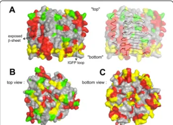
The substitutions that do not impair mutated RicA bind-ing to Rab2 in the Y2H assay were positioned on the RicA 3D model. However, it is predictable that substitu-tions involving very similar residues, within the interface region, would not impair RicA-Rab2 interaction. The mapping of such substitutions could thus prevent the localization of the interface on the surface of RicA. We therefore arbitrarily removed substitutions with a score >1 in the Blosum62 score matrix, since a substitu-tion reversing charge (K-E) has a score of 1 in this matrix. The remaining“low scoring” (LS) substitutions were posi-tioned on the surface of the RicA 3D model (Figure 2). The same procedure was applied to substitutions occur-ring between B. abortus RicA and the C. crescentus RicA homolog, that were also positioned on the model (Figure 2, Additional file 1: Movie S1 and Additional file 2: Movie S2). The LS substitutions are less frequent in
the regions predicted to be at the interface between mono-mers within the trimeric structure (Figure 3, Additional file 3: Movie S3). When all such LS substitutions are indicated on the trimeric RicA model, almost all the surface of the model is covered by substitutions, except for two regions (Figure 2A, Additional file 1: Movie S1 and Additional file 2: Movie S2). The first is the most exposed β-sheet in the RicA trimeric structure (Figure 2A). This re-gion is probably conserved because it is involved in the folding of the β-helical component of the structure. Substitutions in this region are thus suspected to interfere with folding of the protein, which is consistent with the absence of mutations in this region in clones that still allow interaction with Rab2, since unfolded proteins are very likely unable to interact with Rab2 in the Y2H assay. The second region, smaller than the first, is the IGFP loop (Figure 1 and Figure 2A). It is a loop of theβ-helical com-ponent of the structure. Since it was conceivable that mu-tagenesis of this loop could generate loss of interaction without affecting folding, we generated a collection of mutants in this loop.
Mutagenesis of the IGFP loop
The IGFP loop is part of a sequence of 8 amino acids in length flanked on each side by a β strand of the LβH (Figure 1). The sequence of four amino acids Ile-Gly-Phe-Pro (IGFP) is exposed to the surface of RicA in the
Figure 2 Position of the LS substitutions that do not prevent RicA binding to Rab2. The predicted accessible surface of the trimeric structure is depicted in each view. The position of the LS substitutions (score > 1 in the Blosum62 matrix) is indicated with the following color code : the substitutions found in mutated alleles only are shown in green, substitutions found in C. crescentus homolog only are shown in red, residues substituted in the mutated alleles and in the C. crescentus homolog are shown in yellow. In each picture, the ribbon of one monomer (as depicted in Figure 1) is shown by transparency, but in (A) on the right, the transparency is increased to allow the correspondence with Figure 1. (A) Position of the exposedβ-sheet and the IGFP loop, in a “side view”. The arbitrary“top” and “bottom” of the structure are indicated. (B) and (C) Top and bottom views of the substitutions on the predicted trimeric structure of RicA, respectively.
Figure 3 Position of the LS substitutions on the monomeric model of RicA. The structure is presented in the same orientation as in the middle pannel of Figure 1. Three possible views are possible. The view 1 is corresponding to the exposed face of the RicA monomer in the RicA tetramer. In views 2 and 3, the dashed line surrounds the region predicted to be involved in trimer formation. These regions are poor in LS substitutions, which is consistent with a trimeric structure preserved in the yeast two-hybrid assay and in the C. crescentus RicA homolog. As in Figure 2, the substitutions found in mutated alleles only are shown in green, substitutions found in C. crescentus homolog only are shown in red, residues substituted in the mutated alleles and in the C. crescentus homolog are shown in yellow.
Nkengfac et al. BMC Biochemistry 2012, 13:16 Page 4 of 8
3D model of the trimer (Figures 1 and 2). Loop regions of the LβH-containing proteins are known to contribute most of the residues that interact with binding partners [18,19] and surface hydrophobicity has been used to identify regions of a protein surface most likely to inter-act with a binding ligand [20]. We therefore proposed that this exposed IGFP loop could be involved in the recognition of Rab2.
Since the Gly and Pro residues could adopt particular ϕ and ψ torsion angles and thus their substitution may affect folding of neighboring regions, mutagenesis was limited to the Ile-70 and Phe-72 residues that were replaced with random amino acids (amino acid switches from IGFP to XGXP, where X may be one of the 20 pos-sible amino acids). The basic procedure is described in the materials and methods section. A library of XGXP mutants (around 8,000 clones) was constructed, fused to AD domain of Gal4p and assayed for interaction with DB-Rab2 using Y2H. We observed that XGXP mutagen-esis resulted in loss of interaction in about 80% of the clones (inability to drive the expression of the HIS3 and lacZreporters in the Y2H assay). We sequenced alleles generating or not interaction between RicA and Rab2, and the sequence of the XGXP loop is given in Table 2. It is detectable that the amino acid (aa) composition at the first position (70) is more variable in the RicA mutants that still interact with Rab2, compared to the second position (72). Analysis of the mutated sequences shows that slight variations at both positions, e.g. in
clones 10 and 11 (Table 2), where Ile to Leu substitution occurs at position 70, and Phe to Trp and His at pos-ition 72 respectively, disrupt interaction with Rab2 in the Y2H assay.
In order to test a possible alteration of RicA-Rab2 interaction using GST pulldown, we attempted to over-produce XGXP clones n°2, 3, 4, 5, 6 and 8 as His6-RicA
fusions. Among the 6 clones tested, none were found in the soluble extract and all were detected in the insoluble pellet, while the wild type control was found exclusively in the soluble fraction. This observation strongly sug-gests that mutations in the IGFP loop contribute to the proper folding of His6-RicA, at least in E. coli.
Discussion
The objective of this study was to experimentally iden-tify and characterize protein-protein interaction site of RicA for Rab2, to provide a better understanding of the structural basis of a human small GTPase recognition by a bacterial effector protein. In the 3D model of RicA, the protein may be divided in two parts: a N-terminal LβH component and a C-terminal α helix. Within the LβH, there are three β sheets, two embedded in the trimeric structure and one exposed to the exterior. The two in-ternal β sheets are predicted to form the interfaces be-tween monomers and are rarely substituted (Figure 3) in mutants generated and still able to interact with Rab2, or in the C. crescentus homolog. However, the loops involved in the formation of the central pore of the RicA model (visible in Figure 2B and 2C) are often mutated. The residues of the C-terminal α helix in contact with the LβH component are rarely mutated, while many exposed residues of this α helix are substituted in the mutated RicA or in the C. crescentus homolog. These data are consistent with the proposed 3D model of RicA. Our data suggest that mutations in the IGFP loop that impair interaction with Rab2 also generate a folding problem. This is rather surprising because the IGFP loop is not very well conserved (except for the G and P resi-dues, see Additional file 4: Figure S1), and moreover it is exposed to the surface of the homologous proteins of known structure (Additional file 4: Figure S2). The role of the IGFP loop is unknown but it seems to be needed for the generation of a correct tertiary or quaternary structure, since the 6 XGXP mutants unable to interact with Rab2 are found to be insoluble when expressed in E. coli, while the wild type RicA is soluble. This data indicates that regions necessary for folding could overlap the regions necessary for interaction between RicA and Rab2, unless the LS substitutions do not affect inter-action between RicA and Rab2. Indeed, we cannot ex-clude that the RicA-Rab2 interaction is sufficiently stable to be resistant to point mutations, which would preclude
Table 2 Sequenced IGFP replacements that disrupted (clones 1 to 12) or not (clones 13 to 15) RicA-Rab2 interaction
Clone n° X70GX72P sequence Y2H assay with Rab2
1 YGGP His-, lacZ
-2 WGVP His-, lacZ
-3 TGVP His-, lacZ
-4 RGGP His-, lacZ
-5 AGHP His-, lacZ
-6 VGNP His-, lacZ
-7 LGEP His-, lacZ
-8 SG*P His-, lacZ
-9 AGSP His-, lacZ
-10 LGWP His-, lacZ
-11 LGHP His-, lacZ
-12 AGYP His-, lacZ
-13 SGFP His+, lacZ+
14 VGFP His+, lacZ+
15 KGLP His+, lacZ+
The * indicates a stop codon. His
-and His+
refer to the phenotype generated by the HIS3 reporter, and LacZ
-and LacZ+
to the phenotype generated by the lacZ reporter.
most of the strategies targeting loss-of-interaction mutants.
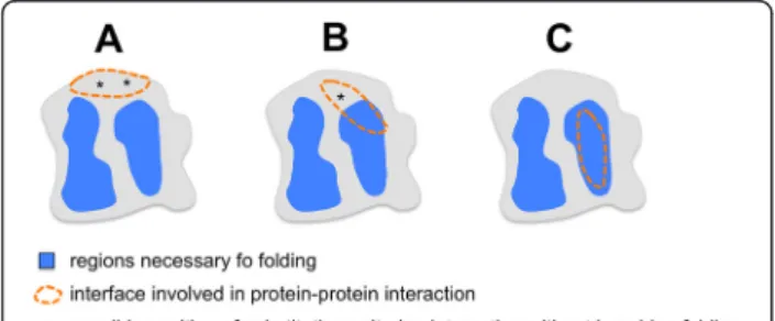
To our knowledge, the identification of“edgetics” alleles (also called “interaction defective” alleles) is the easiest way to identify interaction surfaces on the structure of the proteins involved in a given interaction [16]. However, this method is only applicable if the protein to be mapped is not an autoactivator in the Y2H assay. Our example of the IGFP loop suggests that in some instances, regions neces-sary for folding could overlap regions involved in the protein-protein interaction (Figure 4). Such a situation could lower the probability to get“edgetics” alleles. Also, the “absence of interference” approach will generate a similar situation since the negative image produced by the absence of substitutions in a given region of the structure could simply reflect the absence of substitutions that do not affect folding of the protein.
Conclusion
In conclusion, we predict that for a fraction of mono-domain proteins, including B. abortus RicA, some struc-tural element(s) like the IGFP loop could be bifunctional, involved in both protein folding and protein-protein inter-action, as depicted in Figure 4.
Methods
Plasmidic constructs
B. abortus2308 ricA coding sequence was cloned in the Gateway entry vector pDONR201 and sequenced before sub-cloning in pAD vector as a fusion protein with Gal4 activation domain (AD) and served as the prey plasmid (pAD-RicA). Human Rab2 coding sequence was cloned in pDB vector as a fusion protein with Gal4 DNA bind-ing domain (BD) and used as the bait (pDB-Rab2). The ricAcoding sequence was also cloned in pET15b vector (Novagen pET expression system, pET15b-RicA) as a fu-sion protein with N-terminal hexahistidine tag (His6
-RicA). The ricA coding sequence was PCR amplified
with primers that introduced N-terminal NdeI site and a C-terminal BamHI site (NdeI RicA-F: 5’CAT ATG CCG ATC TAT AAC GG; BamHI RicA-R: 5’GGA TCC TCA GGC AGG CTC CAT). The pET15b-RicA construct was checked by restriction diagnosis and sequencing (primer, T7: 5’TAA TAC GAC TCA CTA TAG GG).
Generation of random mutagenesis, site directed mutagenesis and sequencing
The ricA mutagenesis fragment (525 bp) was synthesized by error-prone PCR [17] on the expression clone pAD-RicA with primers that hydridize to attB1 and attB2 sites that flank ricA in the pAD-RicA (attB1-F 50ACA AGT TTG TAC AAA AAA GCA G-30; attB2-R 5’ AC CAC TTT GTA CAA GAA AGC T-30). Following PCR, DNA was purified and cloned into pDONR201 and the attL1 site primer (50-CTGAAGCTTGGATCTCGGGC-30) was used for sequencing. The generated random mutant library (entry clones) was sub-cloned into pAD expression vector.
Site directed mutations were incorporated into the IGFP motif using the MutagenexTM Library method. The residues Ile-70 and Phe72 were replaced with ran-dom amino acids (amino acid switches from IGFP to XGXP, where X may be one of the 20 possible amino acids). The pAD-RicA plasmid was used as template for a PCR with four synthetic oligonucleotide primers, two containing the desired mutations (FM 50 ATG CAC ACC GAT NNK GGC NNK CCG CTG ACC ATC 30; RM 50 GAT GGT CAG CGG MNN GCC MNN ATC GGT GTG CAT 30) (where N is any of A, C, G, or T; K is G or T; M is A or C) which are complementary to op-posite strands of the insert, and two hybridizing to attB1 and attB2. PCR amplifications were performed for up-stream and downup-stream regions of the mutations. A third assembly PCR was performed with attB1 and attB2 primers, using upstream and downstream PCR frag-ments as initial substrates. The final PCR products were cloned into pDONR201 to generate a large pool of entry-clones. We sequenced 5 randomly picked clones using the attL1 site primer to confirm the expected site directed mutagenesis of Ile-70 and Phe-72 codons. The entry-clone plasmidic DNA library was prepared and sub-cloned into pAD destination vector. DNA sequen-cing experiment was performed with the “Standard Se-quencing Run" on an ABI PRISMW 3100 Genetic Analyser (Applied Biosystems). Y2H-AD (50-CGC GTT TGG AAT CAC TAC AGG G 30 and Y2H-Term (50 -GGA GAC TTG ACC AAA CCT CTG GCG 30) primers were used to sequence RicA mutants still interacting with Rab2 in the Y2H.
Y2H assays
RicA or RicA allele libraries and Rab2 were transformed into MaV203 yeast strain. MaV203 contains single copies
Figure 4 General model illustrating the overlap between regions necessary for folding and regions required for protein-protein interaction. If the overlap between folding and interaction regions is high, the probability to isolate substitutions impairing protein-protein interaction without preventing folding may be high (A), low (B) or very low (C).
Nkengfac et al. BMC Biochemistry 2012, 13:16 Page 6 of 8
of each three reporter genes (HIS3, URA3 and lacZ) that are stably integrated at different loci in the yeast gen-ome. The interaction between RicA or its allele and Rab2 reconstituted an active transcription factor, hence the expression of reporter genes. HIS3 gene expression was detected by plating transformants on selective medium lacking leucine, trytophan and histidine in the presence of 3AT (20 mM). The lacZ reporter was tested by β-galactosidase filter assay. All controls were carried out with appropriate co-transformed vectors.
Mapping and display of mutations on the surface of the protein
The mutations were mapped on the proposed RicA structure using MacPymol (http://www.pymol.org/). The proposed structure was obtained using EsyPred3D pro-gram [13] and verified for correctness using verify3D [14] server (http://nihserver.mbi.ucla.edu/Verify_3D/).
Expression and purification of His6-RicA fusion
Purified His6-RicA was obtained from E. coli BL21 (DE3)
over-expression clone. A filtered lysate was loaded into a chromatography column (Econo-PacW cat. n° 732–1010, Biorad) pre-loaded with Ni-NTA His-Bind superflow (cat n° 70691–3, NovagenW) resin followed by wash steps and
elution (45 mM Tris–HCl pH 7.9, 500 mM NaCl, 200 mM imidazole pH 7.9 and 10% glycerol).
Additional file
Additional file 1: Movie S1. Is showing the 3D structure of the RicA trimer model. The color code is the same as Figures 2 and 3. The movement (120°) around the Y axis allows the distinction of zones free of substitutions (in grey). A ribbon is shown for one monomer, by transparency.
Additional file 2: Movie S2. Is showing the 3D structure of the RicA trimer model. The color code is the same as Figures 2 and 3. The movement (120°) around the X axis allows the distinction of zones free of substitutions (in grey). A ribbon is shown for one monomer, by transparency.
Additional file 3: Movie S3. Is showing the 3D structure of the RicA monomer model. The color code is the same as Figures 2 and 3. The movement (360°) around the Y axis allows the distinction of zones free of substitutions (in grey). The Cα trace is shown by transparency.
Additional file 4: Figure S1. Is a multiple sequence alignment of RicA with 17 homologs. Figure S2 shows the exposed loop corresponding to IGFP in RicA homologs of known structure.
Abbreviations
AD: Gal4p transactivation domain; DB: Gal4p DNA binding domain; 3AT: 3-aminotriazole; LβH: Left-handed β-helical; 3D: Three-dimensional; Y2H: Yeast two-hybrid.
Competing interests
The authors declare that they have no competing interests. Authors’ contributions
BN participated in the experimental design, performed all experiments unless otherwise indicated below and participated in writing the manuscript. EB tested the interaction between RicA homologs and human Rab2. JP
made the gel permeation with purified RicA and tested the stability of RicA mutated in the IGFP loop. JV, JJL, JW and XDB supervised the work. All authors read and approved the final manuscript.
Acknowledgments
This work was supported by FRFC (Fonds de la Recherche Fondamentale Collective, conventions n°2.4521.04 and 2.4541.08) from FRS-FNRS (Fonds de la Recherche Scientifique– Fonds National de la Recherche Scientifique) as well as ARC programs (Actions de Recherches Concertées, conventions 04/ 09-325 and 08/13-015, French Speaking Community of Belgium). Bernard Nkengfac was recipient of a PhD fellowship from the University of Namur. Author details
1Molecular Biology Research Unit (URBM), Narilis, University of Namur, 61 Rue
Bruxelles, Namur B-5000, Belgium.2Laboratory of Structural Biological Chemistry (CBS), member of Narilis, University of Namur, 61 Rue Bruxelles, Namur B-5000, Belgium.3Current address: McGill University, McGill University Health Centre, Royal Victoria Hospital, 687 Pine Ave. W., Room L3.05, Montreal H3A 1A1, Canada.
Received: 24 July 2012 Accepted: 30 July 2012 Published: 14 August 2012
References
1. Boschiroli ML, Foulongne V, O’Callaghan D: Brucellosis: a worldwide zoonosis. Curr Opin Microbiol 2001, 4(1):58–64.
2. Kagan JC, Stein MP, Pypaert M, Roy CR: Legionella subvert the functions of Rab1 and Sec22b to create a replicative organelle. J Exp Med 2004, 199(9):1201–1211.
3. Kagan JC, Murata T, Roy CR: Analysis of Rab1 recruitment to vacuoles containing Legionella pneumophila. Methods Enzymol 2005, 403:71–81. 4. Ingmundson A, Delprato A, Lambright DG, Roy CR: Legionella pneumophila
proteins that regulate Rab1 membrane cycling. Nature 2007, 450(7168):365–369.
5. Stebbins CE, Galan JE: Modulation of host signaling by a bacterial mimic: structure of the Salmonella effector SptP bound to Rac1. Mol Cell 2000, 6(6):1449–1460.
6. de Barsy M, Jamet A, Filopon D, Nicolas C, Laloux G, Rual JF, Muller A, Twizere JC, Nkengfac B, Vandenhaute J, et al: Identification of a Brucella spp. secreted effector specifically interacting with human small GTPase Rab2. Cell Microbiol 2011, 13(7):1044–1058.
7. Stender S, Friebel A, Linder S, Rohde M, Mirold S, Hardt WD: Identification of SopE2 from Salmonella typhimurium, a conserved guanine nucleotide exchange factor for Cdc42 of the host cell. Mol Microbiol 2000, 36(6):1206–1221.
8. Tisdale EJ, Balch WE: Rab2 is essential for the maturation of pre-Golgi intermediates. J Biol Chem 1996, 271(46):29372–29379.
9. Garin J, Diez R, Kieffer S, Dermine JF, Duclos S, Gagnon E, Sadoul R, Rondeau C, Desjardins M: The phagosome proteome: insight into phagosome functions. J Cell Biol 2001, 152(1):165–180.
10. Stenmark H: Rab GTPases as coordinators of vesicle traffic. Nat Rev Mol Cell Biol2009, 10(8):513–525.
11. Fugier E, Salcedo SP, de Chastellier C, Pophillat M, Muller A, Arce-Gorvel V, Fourquet P, Gorvel JP: The glyceraldehyde-3-phosphate dehydrogenase and the small GTPase Rab 2 are crucial for Brucella replication. PLoS Pathog 2009, 5(6):e1000487.
12. Dhayalan A, Jurkowski TP, Laser H, Reinhardt R, Jia D, Cheng X, Jeltsch A: Mapping of protein-protein interaction sites by the‘absence of interference’ approach. J Mol Biol 2008, 376(4):1091–1099.
13. Lambert C, Leonard N, De Bolle X, Depiereux E: ESyPred3D: Prediction of proteins 3D structures. Bioinformatics 2002, 18(9):1250–1256.
14. Luthy R, Bowie JU, Eisenberg D: Assessment of protein models with three-dimensional profiles. Nature 1992, 356(6364):83–85.
15. Raetz CR, Roderick SL: A left-handed parallel beta helix in the structure of UDP-N-acetylglucosamine acyltransferase. Science 1995, 270(5238):997–1000. 16. Dreze M, Charloteaux B, Milstein S, Vidalain PO, Yildirim MA, Zhong Q,
Svrzikapa N, Romero V, Laloux G, Brasseur R, et al:‘Edgetic’ perturbation of a C. elegans BCL2 ortholog. Nature methods 2009, 6(11):843–849. 17. Spee JH, de Vos WM, Kuipers OP: Efficient random mutagenesis method
with adjustable mutation frequency by use of PCR and dITP. Nucleic Acids Res 1993, 21(3):777–778.
18. Sugantino M, Roderick SL: Crystal structure of Vat(D): an acetyltransferase that inactivates streptogramin group A antibiotics. Biochemistry 2002, 41(7):2209–2216.
19. Wang XG, Olsen LR, Roderick SL: Structure of the lac operon galactoside acetyltransferase. Structure 2002, 10(4):581–588.
20. Young L, Jernigan RL, Covell DG: A role for surface hydrophobicity in protein-protein recognition. Protein Sci 1994, 3(5):717–729.
doi:10.1186/1471-2091-13-16
Cite this article as: Nkengfac et al.: Structural analysis of Brucella abortus RicA substitutions that do not impair interaction with human Rab2 GTPase. BMC Biochemistry 2012 13:16.
Submit your next manuscript to BioMed Central and take full advantage of:
• Convenient online submission
• Thorough peer review
• No space constraints or color figure charges
• Immediate publication on acceptance
• Inclusion in PubMed, CAS, Scopus and Google Scholar
• Research which is freely available for redistribution
Submit your manuscript at www.biomedcentral.com/submit
Nkengfac et al. BMC Biochemistry 2012, 13:16 Page 8 of 8


