HAL Id: tel-01371862
https://tel.archives-ouvertes.fr/tel-01371862
Submitted on 26 Sep 2016
HAL is a multi-disciplinary open access
archive for the deposit and dissemination of sci-entific research documents, whether they are pub-lished or not. The documents may come from teaching and research institutions in France or abroad, or from public or private research centers.
L’archive ouverte pluridisciplinaire HAL, est destinée au dépôt et à la diffusion de documents scientifiques de niveau recherche, publiés ou non, émanant des établissements d’enseignement et de recherche français ou étrangers, des laboratoires publics ou privés.
Signalling and morphogenesis during Drosophila dorsal
closure
Antoine Ducuing
To cite this version:
Antoine Ducuing. Signalling and morphogenesis during Drosophila dorsal closure. Morphogenesis. Université de Lyon, 2016. English. �NNT : 2016LYSEN002�. �tel-01371862�
THÈSE DE DOCTORAT
DE L’UNIVERSITE DE LYON
Préparée à l’Ecole Normale Supérieure de LyonEcole Doctorale n°340 - Biologie Moléculaire Intégrative et Cellulaire (BMIC) Discipline : Sciences de la Vie
par
Antoine Ducuing
Signalling and morphogenesis during
Drosophila
dorsal closure
Thèse présentée et soutenue publiquement à l’Ecole Normale Supérieure de Lyon, le 11 mars 2016
Directeur de thèse : Dr. Stéphane Vincent
Devant le jury composé de:
Dr. Yohanns Bellaïche , Directeur de Recherche, Institut Curie, Paris Rapporteur
Dr. Krzysztof Jagla, Directeur de Recherche, GReD, Clermont-Ferrand Rapporteur
Dr. Stéphane Vincent, Maître de Conférences, LBMC, ENS de Lyon Directeur de Thèse
Pr. Arezki Boudaoud, Professeur, RDP, ENS de Lyon Examinateur
Dr. Muriel Grammont, Chargée de Recherche, LBMC, ENS de Lyon Examinatrice
Dr. Raphaël Rousset, Chargé de Recherche, IbV, Nice Examinateur
On a deux vies. La deuxième commence le jour où on réalise qu'on en a juste une. — Confucius
Acknowledgement
First of all, I would like to thank my two rapporteurs, Yohanns Bellaiche and Krzysztof Jagla for spending their time on reading my thesis. I also thank all my jury for letting me defend my thesis, for listening to my scientific presentation, and for future exciting scientific discussions.
I would also like to thank Stéphane for his mentoring, technical advice, exiting scientific discussions and patience over these four years spent together. I hope that the number and the quality of publications in which the two of us are involved will confirm that our association was stimulating and fructuous.
I also thank Muriel Grammont and Raphaël Rousset for participating to my “Comités de suivi de thèse” during which they provided me useful technical, conceptual and human feedbacks.
Many thanks to Dali Ma, who helped me to greatly improve my scientific writing.
I also thank all the persons that helped me during my thesis: Djamel Belgarbi (creating the embryo collection vials for instance), Christophe Chammot, Claire Lionnet and Elodie Chatre (members of the PLATIM facility, they helped me many times on diverse microscope and ImageJ plugins), all the fly persons for advice and discussion.
I also thank my wife Jennifer for critical and extensive reviewing of my thesis, for assistance when going to the lab on Sunday was mandatory, for scientific discussions about the overall relevance of my work from a non-specialist point of view and for her general support.
Unless otherwise indicated, cartoons are home-made, confocal pictures have been generated by me.
ABSTRACT
Drosophila dorsal closure is a key embryonic process during which the dorsal-most epidermal cells called leading edge cells differentiate and act in a coordinated manner to close a transient dorsal hole covered by the amnioserosa in a process reminiscent of wound healing. During dorsal closure, leading edge cells have a highly specialized cytoskeleton: leading edge cells are polarized, display strong adherent junctions, accumulate a dense microtubule network and produce a trans-cellular acto-myosin cable and filopodia. Leading edge cells receive both JNK and DPP (TGF-ß homolog) inputs where JNK induces DPP. These two signalling pathways are crucial for dorsal closure since embryos mutants for either JNK or DPP pathway components fail to undergo correct dorsal closure and exhibit a “dorsal open” phenotype. However, how JNK and DPP contribute to dorsal closure and how these signals are integrated in a robust manner remained unclear. I showed that JNK and DPP are wired in a network motif called ‘feed-forward loop’ (FFL) that controls leading edge cell specification and differentiation. The DPP branch of the FFL filters unwanted JNK activity that occurs during thermal stress. DPP here buffers against environmental challenges and canalizes cell identity, which is a novel function from its well-established ability to spread spatial information.
Next, I focused on the actin cable, a supra-cellular structure produced by the leading edge cells during dorsal closure or wound healing from fly to humans. Using Zasp52, one of the JNK/DPP feed-forward loop targets I identified, I noticed that the actin cable is a discontinuous structure and is dispensable for both dorsal closure and wound healing. This questions the main model in which the actin cable acts as a contractile purse string. My data suggest that the actin cable does not provide a major contractile force. Rather, the actin cable balances forces and stabilizes cell geometry so that closure resolves in a perfectly structured and scar-free tissue. The
absence of the cable leads to cell shape irregularities as well as patterning and planar cell polarity defects that are reminiscent of scarring. We propose that the cable prevents scaring by acting as a mechanical freeze field that protects fine cellular structures from the major closure forces that operate at tissue level.
I also showed that during dorsal closure, DPP does not prevent JNK-induced cell death but rather that the physiological cell death of the amnioserosa participates to the onset of the dorsal open phenotype in DPP signalling mutant embryos.
Last, I found that over time, abnormal tensions / stress can trigger ectopic JNK activity. This stress-induced JNK activity is crucial for embryonic wound healing.
Altogether, my work brings new insights on the signalling and morphogenesis during dorsal closure.
RESUME
La fermeture dorsale est un événement majeur de l’embryogénèse de la drosophile durant lequel les cellules les plus dorsales de l’épiderme se différencient et agissent de concert pour refermer une ouverture dorsale temporairement recouverte par l’amnioséreuse. Ce processus présente de nombreuses similarités avec la cicatrisation cellulaire. Pendant la fermeture dorsale, les cellules de la marge active ont un cytosquelette extrêmement dynamique : les cellules sont polarisées, elles accumulent de fortes jonctions adhérentes et un réseau de microtubule dense. Les cellules de la marge active produisent également un câble d’actine ainsi que des protrusions appelées filopodes. Pendant la fermeture dorsale, les cellules de la marge active sont régulées par les voix JNK et DPP (homologue à la voie TGB-ß), où JNK induit DPP. Ces deux voies sont nécessaires à la fermeture dorsale. En effet, dans les mutants de la voie JNK ou DPP, la fermeture dorsale ne se fait pas. Les embryons présentent un phénotype d’ouverture dorsale. Cependant, on ne connaît pas comment les signaux de la voie JNK et DPP sont intégrés par les cellules de la marge active pour permettre une fermeture dorsale robuste. J’ai montré que les voies JNK et DPP forment une boucle cohérente appelée « feed-forward loop » (boucle d’anticipation) qui contrôle la différentiation des cellules de la marge active. La branche DPP de cette boucle filtre les signaux non désirés de la voix JNK quand les embryons sont soumis à un stress thermique. DPP joue un rôle ici de tampon contre les variations environnementales, ce qui est une nouvelle fonction par rapport à son rôle bien décrit de morphogène.
Je me suis ensuite concentré sur le câble d'actine, une structure supra-cellulaire produite par les cellules de la marge active lors de la fermeture dorsal. Les cellules autour d’une plaie dans des embryons de Drosophile, de poulet ou même de souris produisent également ce câble d’actine. En me servant de Zasp52, l'une des
cibles de la boucle de régulation JNK / DPP, j’ai montré que le câble d’actine est une structure discontinue qui n’est pas nécessaire pour la fermeture dorsale ou pour la cicatrisation cellulaire. Ceci remet en cause le modèle principal selon lequel le câble d’actine agit comme un cordon de bourse qui se ferme. J’ai montré que le câble ne confère par une force contractile pendant la fermeture. Le câble d’actine homogénéise les forces et stabilise la géométrie cellulaire pour que la fermeture se fasse de manière parfaite et sans cicatrice. Sans le câble, les cellules ont une forme irrégulière, associé à des défauts de patterning et des défauts de polarité planaire qui ressemblent aux défauts que l’on trouve lors de la formation d’une cicatrice. Nous proposons donc que le câble empêche la formation de cicatrice en « congelant » les propriétés mécaniques des cellules afin de les protéger des forces qui agissent au niveau tissulaire lors de la fermeture dorsale.
J’ai également montré que lors de la fermeture dorsale, DPP ne protège pas contre la mort cellulaire induite par JNK. J’ai également montré que c’est plus vraisemblablement la mort cellulaire dans l’amnioséreuse qui participe à l'apparition du phénotype d’ouverture dorsale dans les mutants de la voie DPP.
Enfin, j’ai montré que les tensions anormales / le stress peuvent déclencher l’activation de la voie JNK. Cette activité de JNK induite par le stress est cruciale pour la cicatrisation cellulaire chez l’embryon.
En conclusion, mon travail apporte un regard neuf sur la signalisation et la morphogenèse lors de la fermeture dorsale de l’embryon de Drosophile.
INDEX ABSTRACT... 7 RESUME... 9 INDEX ... 11 FIGURES INDEX... 15 ABBREVIATIONS ... 17 INTRODUCTION ... 21
I. From Thomas Hunt Morgan to Today: Drosophila melanogaster as a powerful model organism to study signalling and morphogenesis ... 21
I.1 Thomas Morgan and his pioneer ‘Fly Room’ ... 21
I.2 Easy to grow, easy to keep: Drosophila as a versatile tool ... 22
I.3 A small but instructive genome... 23
I.4 Drosophila genetics ... 25
I.5 Live imaging and in vivo techniques... 26
II. Drosophila embryonic development ... 29
II.1 Early embryogenesis (Stage 1 – Stage 5) ... 29
II.2 Gastrulation (Stage 6 – Stage 7) ... 32
II.3 Germ band extension (Stage 8 – Stage 10) ... 35
II.5 Germ Band retraction (Stage 12) ... 41
II.6 Dorsal closure (Stage 13 – Stage 15) ... 43
II.7 Late embryogenesis (Stage 16 – Stage 17)... 44
III. Morphogenesis during dorsal closure ... 45
III.1 An overview of dorsal closure ... 47
III.2 The amnioserosa ... 50
III.3 The actin cable... 53
III.4 The filopodia... 59
III.6 Dorsal closure has a model for wound healing... 63
IV. Signalling during dorsal closure... 65
IV.1 The JNK pathway: a stress-response and developmental pathway. ... 65
IV.2 The DPP pathway: a patterning and morphogenetic pathway... 70
V. Jupiter, Jaguar and Zasp52: 3 cytoskeletal-associated proteins that define the leading edge identity during dorsal closure ... 78
V.1 Jupiter, a microtubule-associated protein ... 79
V.2 JAGUAR, the Myosin VI homolog ... 82
V.3 Z band alternatively spliced PDZ-motif containing protein 52 ... 84
RESULTS... 88
1. A DPP-mediated feed-forward loop canalizes morphogenesis during Drosophila dorsal closure... 88
1.1. The Article... 88
2. Zasp52 paper ... 106
3. Stress-induced JNK story ... 142
4. Cell death paper... 155
ADDITIONAL PAPERS... 197
1. Absolute requirement of cholesterol binding for Hedgehog gradient formation in Drosophila... 198
2. Cholesterol-free and cholesterol-bound Hedgehog: Two sparring-partners working hand in hand in the Drosophila wing disc?... 200
DISCUSSION ... 213
1. JNK and DPP form a coherent feed-forward loop during dorsal closure. ... 216
2. What is the leading edge? ... 220
3. Zasp52 is an upstream regulator of the actin cable... 222
4. The actin cable: do not call me purse string... 224
5. Is JNK acting as a stress-mediator pathway in the embryo? ... 228
MATERIALS AND METHODS ... 234 1. Embryo collection ... 234 2. Fly stocks ... 236 3. Immunofluorescence ... 240 3.1 Regular immunofluorescence... 240 3.2. Phalloidin stainings ... 242 3.3 Antibodies list ... 245
4. Live imaging and in vivo techniques ... 248
4.1. Aligning embryos for the Spinning disc... 248
4.2 Setting up the Spining disc ... 250
4.3. Laser ablation experiments. ... 254
5. Quantifications... 256
5.1. Closure dynamics ... 256
5.2. Recoil experiments ... 256
5.3. Leading edge straightness. ... 257
5.4 Quantification and statistical analyses. ... 257
6. Image processing ... 258
6.1 Live imaging... 258
6.2 Immunofluorescence ... 259
FIGURES INDEX
Figure 1. The Drosophila melanogaster life cycle... 22
Figure 2. WT and TM6 balancer chromosomes... 24
Figure 3. The UAS-Gal4 system... 25
Figure 4. Early Drosophila embryogenesis... 30
Figure 5 Antero-posterior axis specification... 31
Figure 6. Dorso-ventral axis specification. ... 32
Figure 7. Ventral furrow formation during Drosophila gastrulation... 33
Figure 8. Cell intercalation during germ-band extension. ... 36
Figure 9. Drosophila embryos during germ-band extension... 37
Figure 10. Drosophila segmentation. ... 39
Figure 11. Drosophila segment organization. ... 40
Figure 12. Stage 11 and 12 Drosophila embryos. ... 41
Figure 13. Trachea metamer. ... 42
Figure 14. Drosophila embryonic stages... 45
Figure 15. Drosophila dorsal closure... 47
Figure 16. Microtubules accumulation and cell elongation at the leading edge during dorsal closure... 48
Figure 17. Amnioserosa cell oscillations during dorsal closure. ... 50
Figure 18. Amnioserosa during the slow and fast phases... 51
Figure 19. Amnioserosa cell delamination... 52
Figure 20. Actin cable during dorsal closure... 53
Figure 21. Ena, Ed and Baz expression pattern during dorsal closure... 54
Figure 22. Filopodia during dorsal closure. ... 60
Figure 23. Proposed model of zipping. ... 60
Figure 24. Actin cable and filopodia formation during human and Drosophila wound healing... 63
Figure 25. JNK activity during Drosophila larval wound healing... 64
Figure 26. Simplified view of the JNK pathway during dorsal closure. ... 67
Figure 27. The JNK signalling pathway during dorsal closure... 70
Figure 28. DPP gradient in the wing imaginal disc... 71
Figure 29. A simplified view of the DPP pathway... 74
Figure 30. The DPP pathway during dorsal closure... 76
ABBREVIATIONS
Abl Abelson
Aop Anterior open
AS Amnioserosa
Baz Bazooka
Bnl Branchless
BMP Bone Morphogenetic Protein
Brk Brinker
Bsk Bsk
Btl Breathless
Chic Chicadee
Ci Cubitus interruptus
Dad Daughters against DPP
DB Dorsal branch
DC Dorsal closure
Dfd Deformed
Dia Diaphanous
Dl Dorsal
Dok Downstream kinase
DPP Decapentaplegic
Dsh Dishevelled
DTa Dorsal Trunk anterior
Ed Echinoid
En Engrailed
Ena Enabled
FGF Fibroblast growth factor
GFP Green Fluorescent Protein
Hep Hemipterous
Hh Hedgehog
Hnt Hindsight
JNK Jun N-terminal Kinase
Jra Jun-related antigen
Kay Kayak
Kr Kruppel
LE Leading Edge
LT Lateral Trunk
Mad Mother against DPP
MAPK Mitogen-activated protein kinase
Msn Misshapen
Omb Optomotor-blind
Pnr Pannier
Puc Puckered
RFP Red Fluorescent Protein
ROS Reactive oxygen species
Rpr Reaper Salm Spalt SB Stublle Scaf Scarface Shn Schnurri Slpr Slipper
Sog Short gastrulation
Sqh Spaghetti Squash
Tkv Thickveins
Tld Tolloid
TM6 Third Multipular 6
UAS Upstream Activating Sequences
Ush U-shaped
VB Ventral Branch
Wg Wingless
YFP Yellow Fluorescent Protein
INTRODUCTION
I. From Thomas Hunt Morgan to Today: Drosophila melanogaster as a powerful model organism to study signalling and morphogenesis
I.1 Thomas Morgan and his pioneer ‘Fly Room’
Drosophila melanogaster has been a widely used model organism for more than a century. In the early 1920, Thomas Morgan's lab, nicknamed the ‘Fly Room’, pioneered the use of Drosophila as a model organism to understand genetics. At the origin, Morgan’s lab was said to be has big as a broom closet. Thanks to Drosophila, Thomas Morgan made key contributions working on heredity, and sex-linked traits. This historic photo from the Betsey Bridges Family Collection is showing Calvin Bridges, one of Thomas Morgan’s disciple in the fly room where they discovered white (w), the first X-linked mutation in Drosophila (Morgan et al., 1915).
I.2 Easy to grow, easy to keep: Drosophila as a versatile tool
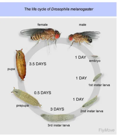
Since Thomas Morgan, many scientists have been using Drosophila as a model organism. Indeed, Drosophila is cheap, easy to breed and has a short life-time generation that lasts only 10 days at 25°C. The embryogenesis starts right after egg laying and lasts for 24h to give rise to a ready-to-live larva. The newborn larvae go through 3 different stages during which they grow. Specifically, the imaginal discs will undergo cell proliferation and differentiation throughout the larval life to form the adult appendages. After a phase of growth, larvae enter into a quiescent pupal stage, during which metamorphosis takes place followed by the emergence of the new adult fly (Figure 1).
Flies are tiny (< 3 mm), and are therefore easy to keep. A female can produce between 750 and 1,500 eggs during its life when harvest with a good food medium. Therefore it is quite easy to generate hundreds of flies quickly.
1 DAY! 1 DAY! 1 DAY! 3 DAYS! 0.5 DAYS! 3.5 DAYS!
Figure 1. The Drosophila melanogaster life cycle.
At 25°C, the life cycle last ten days.
Source: http://flymove.uni-muenster.de
I.3 A small but instructive genome
The Drosophila genome is composed of four pairs of chromosomes: X/Y, 2, 3 and 4. The fourth chromosome is very small and is not often studied, although some important genes are located on the fourth chromosome (e.g. eyeless, cubitus interruptus). Each chromosom (exept the X that has a single arm) is divided into a left and a right arm, and each arm is subdivided into segments. The Drosophila genome has little redundancy: by affecting a single gene, a complete function can be affected. Drosophila is therefore an excellent model for genetic screens. In 1980, Christiane Nüsslein-Volhard and Eric Wieschaus induced mutations in the entire genome and discovered key developmental genes including patched or hedgehog for instance (Nusslein-Volhard and Wieschaus, 1980). For these major discoveries, Christiane Nüsslein-Volhard and Eric Wieschaus are the 1995 recipients of the Physiology and Medicine Nobel Prize.
Although the Drosophila genome is simpler than more complex model organisms such as the mouse genome, 50% of Drosophila genes have a human homolog. There are multiple examples where Drosophila and mammalian genes display functional homology. A striking example is that in the absence of the BMP-4/BMP-2 Drosophila homologue called Decapentaplegic (Dpp), BMP4 ligand sequences can function in lieu of DPP in the Drosophila embryo (Padgett et al., 1993). Thus, human and Drosophila genes can display functional homologies.
A problem with mutations is that they are often homozygous lethal, and can only be maintained at heterozygous state. The issue is that, by breeding heterozygous flies, a third of the emerging progeny will not carry any copy of the mutation. Thus, over time, two populations – wild-type and heterozygous flies – can coexist. Considering that the mutation brings a natural disadvantage, even at heterozygous state, it is likely that over time, only the wild-type flies will remain in
the stocks. To overcome this difficulty, the fly community has set up the so-called ‘balancer chromosomes’. Balancers are chromosomes that carry numerous chromosomal inversions, which prevent any meiotic recombination. Balancers carry a dominant marker and a recessive mutation. Balancers are therefore homozygous lethal (or sterile) and carry a visible marker.
Here are the advantages of the system:
- Because balancers are homozygous lethal, the only genotype maintained over the generations will be Mutation over Balancer (The combinations Balancer/Balancer and Mutation/Mutation are lethal, only the combination Mutation/Balancer is viable).
- Because balancers carry a dominant marker, the mutation can be counter selected when making crosses (the offspring either gets the mutation and therefore no makers, or the balancer AND the marker).
- Since flies, and especially females undergo meiotic recombination, the inversion of chromosomic sequences in the balancer prevents any recombination. This way, one can be sure that the mutation will never be on the same chromosome than the marker or the balancer.
TM6, Sb is a Balancer chromosome located on the third chromosome. TM stands
for Third Multipular. TM6 carries a recessive mutation, and the marker Sb.
Sb (Stubble) is a homozygous lethal marker. It is not a balancer. Sb flies have short
bristles, like the regrowth of a shaven beard (Figure 2).
61 62 63 !64 65 66 !67 68 69 !70 71 72!73 74 75!76 77 78!79 80! 81 82 83 !84 85 86 !87 88 89 !90 91 92!93 94 95!96 97 98!99! 81 82 83 !84 85 86 ! 87 88 89 ! 78 79 80 ! 75 79 77! 94 95 96!97 98 99! 100! 100! 62 63 ! 90 91 92! 73 74 93! 64 65 66!67 68 69 !70 71 72 ! WT III! TM6! 61!
Figure 2. WT and TM6 balancer chromosomes.
The configuration of the TM6 balancer chromosome prevents recombination, apart from the 61 and 100 extremities.
I.4 Drosophila genetics
In addition of the mutant collections available, several genetic tools are available in Drosophila. Among them, Andrea Brand and Norbert Perrimon set up the UAS-Gal4 system, a powerful genetic tool that allows specific gene over-expression with temporal and spatial resolution (Brand et al., 1994).
The yeast transcriptional activator Gal4 is expressed under the control of an endogenous Drosophila enhancer. Cells within this domain can therefore activate transgenes controlled by Upstream Activating Sequences (UAS) (Figure 3). The system has many advantages:
- Expression of a given cDNA with temporal and spatial specificity. - Almost infinite combination between Gal4 and UAS lines.
- Avoid the toxicity (the system is only active in the progeny, since UAS and Gal4 sequences are from yeast are therefore not interpreted by the fly genome). GAL4% GAL4! UAS! cDNA! enhancer! Tissue-specific expression of GAL4!
cDNA under the control of the UAS sequences!
Specific expression of the cDNA! in the GAL4 expressing cells!
X!
PARENT 1! PARENT 2!
PROGRENY!
Prd-Gal4, UAS-Ena!
Figure 3. The UAS-Gal4 system.
Top: Cartoon depicting the UAS-Gal4 system. Inspired from (St Johnston, 2002).
Bottom: Prd-Gal4, UAS-Ena embryo
marked with anti enabled (grey).
Prd-Gal4 drives expression in epidermal stripes. Ena is therefore over-expressed in epidermal stripes.
I.5 Live imaging and in vivo techniques
The genetic power of Drosophila also resides in the use of fluorescent-tagged proteins. The isolation of the green fluorescent protein (GFP) from the jelly fish aequorea victoria by Osamu Shimomura enabled Martin Chalfie to tag C. Elegans proteins with GFP and follow their behaviour in vivo (Chalfie et al., 1994). GFP and other derivatives (RFP, etc.) have been widely used in Drosophila. Expression of fluorescent reporters constitutes a convenient way to decipher or to mark the expression pattern of various Gal4 lines. GFP-exon trap screen also allowed the characterization of previously unknown genes for instance (Morin et al., 2001). In addition, the development of fluorescent balancers to easily sort out the mutant and the non mutant populations constitute a convenient – if not crucial – advance for in vivo studies (Le et al., 2006).
Importantly, the expression of GFP-tagged cytoskeletal markers has been extremely useful to better understand a variety of morphogenetic processes. For instance, it allowed a better understanding of cell junction rearrangement (Bardet et al., 2013), cell-mixing process like during tumour invasion (Levayer et al., 2015), local forces induced by apoptosis (Monier et al., 2015) or mechanical control of growth in the wing disc (Legoff et al., 2013). It also enabled the characterisation of actin-based protrusion called cytonemes, that appear more and more as a major mechanism of paracrine signalling (Roy et al., 2011; Roy et al., 2014). In the embryo, these reporters have been notably used to better understand the behaviour of various tissues during dorsal closure (Jacinto et al., 2000; Kiehart et al., 2000; Jacinto et al., 2001; Jacinto et al., 2002; Kaltschmidt et al., 2002; Franke et al., 2005; Jankovics and Brunner, 2006; Laplante and Nilson, 2006; Fernandez et al., 2007; Millard and Martin, 2008; Rodriguez-Diaz et al., 2008; Toyama et al., 2008; Solon et al., 2009; Wells et al., 2014; Ducuing et al., 2015). In addition, single junction cuts with a UV
laser has become a standard way to assess local tensions cells are subjected to, or to perturb ongoing morphogenesis.
New techniques are also emerging such as the development of light-sheet microscopy system (Saias et al., 2015), or the development of super-resolution microscopy.
II. Drosophila embryonic development
The Drosophila embryonic development is a complex process that lasts 22 hours at 25°C. All the embryonic stages are depicted at the end of this section in the Figure
14. In this section, I will describe the most important developmental processes,
except dorsal closure that will be described in greater details in the next section.
II.1 Early embryogenesis (Stage 1 – Stage 5)
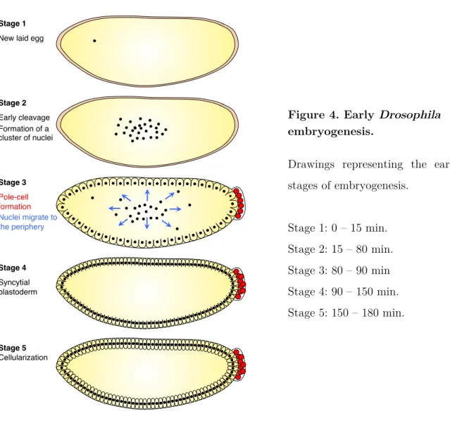
During the five first embryonic stages, the egg will undergo 13 round of synchronous nuclear divisions without cellular division. Nuclei then migrate to the periphery to eventually undergo a simultaneous cellularization to form a 8,000-cell blastula.
Initially, the egg is composed of a homogenous cytoplasm and contains yolk granules. The first stage of embryogenesis usually starts after the egg laying and last until the completion of the two first cleavages (Stage 1).
Then, the 5 next nuclear divisions (without cellular division) occur predominantly in the anterior part of the egg, leading to the formation of a cluster of nuclei (Stage 2). The nuclei progressively move towards the posterior pole of the embryo while the embryo constricts, leading to the formation of an unfilled space both at the anterior and the posterior part of the egg.
From the 8th nuclear division, the nuclei migrate progressively at the periphery to relocate under the vitelline membrane (Stage 3). The first 3 nuclei to reach the posterior pole divide and then cellularize to become the pole cells. These pole cells fill the posterior space created earlier and will constitute the germ line.
At Stage 4, the nuclei are migrating at the periphery, leading to the formation of a syncytial blastoderm: they share the same cytoplasm, but are excluded from the
Stage 1! New laid egg!
Stage 2! Early cleavage! Formation of a! cluster of nuclei! Stage 3! Pole-cell ! formation! Nuclei migrate to the periphery! Stage 4! Syncytial blastoderm! Stage 5! Cellularization!
central part of the egg due to the presence of the yolk. The duration of cleavage divisions 10-13 increases progressively, from approximately 8 min to 20 min.
Cellularization occurs during stage 5. Cellularization starts with the invagination of membrane furrows from the periphery towards the centre of the egg. Blastoderm cells are not completely isolated since they still connect with the yolk cytoplasm through cytoplasmic bridges. These bridges are lost later, during gastrulation. After cellularization, the blastoderm cells have a homogenous shape and size (Figure 4).
During cellularization, the antero-posterior and the dorso-ventral axis are established. The antero-posterior axis is established by the formation of opposite gradients of four maternal-effect genes. Bicoid and Hunchback regulate the
Figure 4. Early Drosophila embryogenesis.
Drawings representing the early stages of embryogenesis. Stage 1: 0 – 15 min. Stage 2: 15 – 80 min. Stage 3: 80 – 90 min Stage 4: 90 – 150 min. Stage 5: 150 – 180 min.
production of anterior structures (Driever and Nusslein-Volhard, 1988; Struhl et al., 1992), while Nanos and Caudal regulate the formation of the posterior part of the embryo (Macdonald and Struhl, 1986; Nusslein-Volhard et al., 1987). The classical view is that bicoid mRNA is actively transported via microtubules towards the anterior part of the egg while nanos mRNA remains in the posterior part of the egg. Bicoid and Nanos then establish an opposite protein gradient. Nanos inhibits Hunchback transcription. Hunchback therefore adopts a gradient opposite to Nanos gradient. Similarly, Bicoid represses Caudal transcription. Caudal thus adopts a gradient opposite to Bicoid gradient (Figure 5).
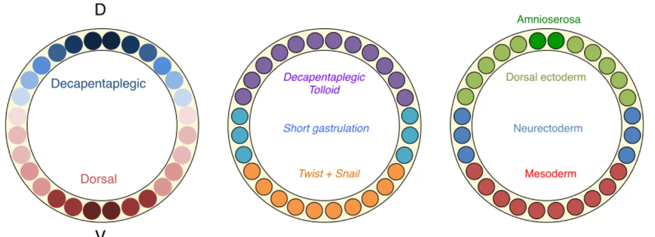
The patterning of the dorsoventral axis is regulated by the mutually exclusive action of the two morphogenes Dorsal (Dl) and Decapentaplegic (Dpp). Dl is the determinant of the ventral axis and establishes a dorsoventral nuclear gradient with peak levels in the ventral nuclei (Roth et al., 1989; Steward, 1989). The ventral-most cells that display the highest nuclear concentration of Dl express twist and snail, two transcription factors that will specify the mesoderm. Specifically, snail represses the expression of short gastrulation (sog), a determinant of the neurodermal fate. In more ventral cells where Snail in not expressed, lower nuclear levels of Dl can activate sog. Sog prevents in turn cells from becoming dorsal ectodermal cells by sequestering
A" P" P" Bicoid! Hunchback! Caudal! Nanos! A" Pro te in co n ce n tra ti o n!
Figure 5 Antero-posterior axis specification.
Top: Bocoid and Nanos protein gradients. Bottom: Bicoid inhibits Caudal while Nanos inhibits Hunchback, leading to the formation of opposite gradients.
Decapentaplegic! Dorsal! Decapentaplegic! Tolloid! Short gastrulation! Twist + Snail! Dorsal ectoderm! Neurectoderm! Mesoderm! Amnioserosa! D! V!
Dpp. In the dorsal-most regions, Dl is absent from the nucleus: Dpp and Tolloid (Tld), a metalloprotease that cleaves Sog are expressed and secreted. In the future dorso ectodermal cells, Tld prevents Sog-dependant Dpp sequestration, thus allowing Dpp to specify the dorsal ectoderm. The amnioserosa is specified by zerknullt (zen), a transcription-factor that is initially broadly expressed like Dpp, but that becomes restricted to the dorsal-most region in a Dpp-dependent manner (Doyle et al., 1986; Rushlow et al., 1987). Interestingly, Zelda is a uniformly expressed factor that would potentiate Dl gradient interpretation (Foo et al., 2014). Specifically, Zelda opens the chromatin of the genes that are induced by Dorsal. However, the number of Zelda binding sites per gene varies, thus modulating the ability of these genes to respond to various concentration of nuclear Dl (Figure 6).
Figure 6. Dorso-ventral axis specification.
Cartoons represent cross-sections of a Stage 5 Drosophila embryo.
Left: opposite Dorsal and Decapentaplegic gradients. Nuclear (for Dorsal) and extra cellular (for Dpp) gradients are represented in a similar manner for the sake of simplicity.
Middle: High levels of nuclear Dorsal induce Twist and Snail (orange). Snail represses Short gastrulation. Medium levels of nuclear Dorsal in the absence of Snail induce Short gastrulation (blue). In the absence of Dorsal in the nucleus DPP and Tolloid are produced. Short gastrulation sequesters Dpp while Tolloid inhibit this sequestration, leading to the establishment of a Dpp gradient.
Right: Twist and Snail expressing cells form the mesoderm (red). Short gastrulation expressing cells form the neuro ectoderm. Dpp receving cells form the dorsal ectoderm. When Dpp activity pattern refines, zerknullt expression pattern refines to the dorsal-most cells, where the amnioserosa
II.2 Gastrulation (Stage 6 – Stage 7)
Gastrulation is a developmental phase during which a single-layered embryo becomes a three-layered embryo with formation of the ectoderm (future epidermis and nervous system), the mesoderm (future muscles) and the endoderm (future intestine). Gastrulation starts at Stage 6 by the formation of the three distinct furrows. The cephalic furrow, located in the first third of the embryo starts to fold. Second, the pole cells at the posterior part of the embryo progressively shifts dorsally and are engulfed in a pocket.
The most striking process during gastrulation is the formation of a ventral furrow (Figure 7). During this process, about 1000 future mesodermal cells progressively invaginate from the surface of the embryo in a coordinated manner to eventually form the mesodermal tube (Leptin, 1999). As the ventral furrow forms, the invaginating cells constrict apically and undergo cell elongation. At a morphogenetic level, the non-muscle myosin II (spaghetti squash, sqh) is localized apically. Sqh associates with actin to promote the apical constriction of the cells and allow their flattening via the association of the acto-myosin cytoskeleton to the apical adherens junctions (Dawes-Hoang et al., 2005). Once the furrow is formed, the future mesodermal cells go back to their original length, to end up in a wedge–like shape.
Figure 7. Ventral furrow formation during Drosophila gastrulation.
This figure is composed of surface sections (left) and cross-sections (right) of the ventral epithelium of the Drosophila embryo during ventral furrow formation and furrow invagination. This figure is taken
from (Spahn and Reuter, 2013). Nrt: Neurtactin (surface glycoprotein). Spider-GFP is a casein kinase
I encoded by the gene gilgamesh that associates with the plasma membrane and secretory vesicles destined for the plasma membrane.
The formation of the ventral furrow is controlled by the two transcription factors twist and snail. snail is a transcriptional repressor required for the initiation of the ventral furrow formation. It acts by repressing the neuroectodermal fate in the invaginating mesoderm. twist is a transcriptional activator that will control the proper expression of mesodermal genes. Interestingly, snail expression needs to be synchronous for correct gastrulation thanks to RNA Polymerase II pausing mechanism (Lagha et al., 2013). The mechanism of “paused Polymerase II” is a mechanism by which the RNA polymerase starts the initiation of the transcription, but does not proceed further to elongation due to the lack of additional factors. This way, the RNA Polymerase II is linked to the nascent RNA in a “ready-to-go” state (Adelman and Lis, 2012). The RNA Polymerase II pausing is essential for fast and synchronous snail expression in the presumptive mesoderm. Importantly, the paused RNA polymerase II mechanism determines the ‘‘time to synchrony’’, which is the time necessary for coordinating gene expression across a tissue (Lagha et al., 2013).
During the second part of gastrulation (Stage 7), the pole cells that are engulfed in a pocket of about 150 cells adopt a horizontal position compared with the dorsal egg surface. The cells that are immediately anterior to this pocket start to form a deep groove that becomes continuous with the ventral furrow. This is the proctodeum invagination. In addition, the stomodeum, composed of the anterior midgut primordium, invaginates. The mitosis are now non-synchronous and occur in so-called “mitotic domains” (Foe, 1989). The embryo is ready for the extension of its germ-band.
Shrinking junction! New junction!
T1! T2! T3!
A! P!
D!
V!
II.3 Germ band extension (Stage 8 – Stage 10)
At the end of stage 7, gastrulation is completed. The ventral furrow is closed, and the mesodermal tubes composed of a regular and structured epithelium. During stage 8, the mesodermal tube starts to disaggregate and the mesodermal cells undergo mitosis. In parallel, germ-band elongation (or germ-band extension) occurs.
Germ-band elongation is a morphogenetic process during which the epidermis doubles in length along the anterior-posterior axis while reducing its width along the dorsal-ventral axis thanks to medio-lateral to antero-posterior cell intercalation (Irvine and Wieschaus, 1994). During germ-band elongation, the posterior half of the trunk reaches the dorsal side of the embryo, while the anterior half constitutes the ventral side of the embryo (Irvine and Wieschaus, 1994). Germ-band elongation is divided into two phases: a first fast phase (25 minutes) during which most of the elongation occurs, and a slow phase (70 minutes) after which elongation is completed (da Silva and Vincent, 2007). The process of cell intercalation is the main driver of germ-band extension, as no cell division occurs during this period (Figure 8).
At the cellular level, the junction that undergoes the dorso-ventral orientated shrinkage has less E-Cadherin than their neighbours, but accumulates myosin-II and the Rho-kinase in response to tensions (Bertet et al., 2004; Fernandez-Gonzalez et al., 2009). Specifically, the polarized flow of actomyosin bursts towards dorso-ventral orientated junctions would be the key driver factor of cell-membrane shrinkage (Rauzi et al., 2010). In addition, the myosin phosphorylation constitutes an instructive cue to generate the proper tensions during cell rearrangement (Kasza et al., 2014).
In parallel, formation of the amnioserosa, an extra-embryonic tissue involved in dorsal closure occurs. As germ-band extension proceeds, the proctodeal invagination that was formed during gastrulation and that was containing amnioserosa primordium becomes deeper. The cells for the amnioserosa primordium become flat, elongated and are progressively engulfed between the tip of tail and the head (Campos-Ortega and Hartenstein, 1985; Frank and Rushlow, 1996).
During stage 9, germ-band extension continues. In parallel, the first neuroblasts start to delaminate from the ectoderm in three distinct waves (Campos-Ortega and Hartenstein, 1985). The mesoderm also rearrange to form a monolayer while undergoing mitosis (Campos-Ortega and Hartenstein, 1985).
The extension of the germ-band ends at Stage 10. At this stage, the stomodeum invaginates, which will give rise to the foregut. Neuroblasts start to undergo asymmetric cell divisions. The first signs of parasegmentation are also visible (Figure 9).
Figure 8. Cell intercalation during germ-band extension.
Left: Figure taken from Irvine and Wieschaus, 1994 showing the cell intercalation phenomenon during germ-band extension.
Right: Drawings representing the cell-intercalation process, in a T1-T2-T3 mechanism, with the shortening of the D/V orientated junction (T1), the formation of a rosette (T2), and the formation of a new A/P orientated junction (T3).
A!
C! D!
B!
E! F!
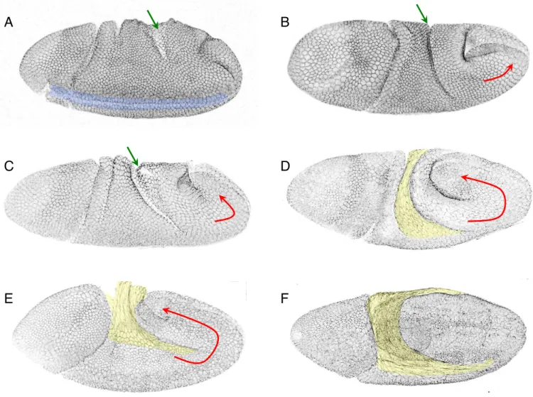
Figure 9. Drosophila embryos during germ-band extension.
All the embryos are marked with E-Cadherin. Green arrows indicate the proctodeal invagination that gets deeper over time. Red arrows indicate the extension of the germ band. The amnioserosa is in yellow. A = Beginning of germ-band elongation (Stage 8), with the end of the ventral furrow invagination visible (blue)
B, C, D = Fast phase of elongation (Stage 8), E = Slow phase of elongation (Stage 9), F = End of elongation (Stage 9/10).
II.4 Segmentation and trachea invagination (Stage 11)
At stage 11, the metameric organisation of the embryo becomes apparent. The embryo is composed of 3 thoracic and 8 abdominal segments. Segmentation occurs by the progressive refining of the expression patterns of key determinant of the antero/posterior axis of each segment. Segmentation is therefore initiated earlier in development.
Initially, the egg contains maternal genes (inherited by the mother) such as bicoid or nanos. These maternal genes adopt a graded distribution to establish the antero-posterior axis.
The combination and the concentration of these morphogens regulate the expression pattern of ‘gap genes’ that divide the embryo into large regions. Mutations in these genes create ‘gaps’ in the segmentation. For instance, krüppel mutant embryos display only the 3 most-posterior abdominal segments (Nusslein-Volhard and Wieschaus, 1980).
These gap genes then control the expression of so-called ‘pair-rule’ genes that are expressed in large stripes and that establish pairs of segments. The pair-rule genes mutants lack either odd or even segments. For instance, embryos mutant for fushi tarazu exhibit only odd thoracic segments and even abdominal ones (Kankel et al., 2004).
The pair-rule genes finally control the ‘segment polarity genes’ that are expressed in narrow stripes in each segment and that control the antero-posterior organisation of the segments (Figure 10). A simplified view is to consider that during embryogenesis, these different classes of genes have a temporal hierarchy. However, the reality is more complex: for instance, seven stripes of the pair-rule gene odd skipped can be detected before the extension of the germ-band (Stage 7), but 14 stripes are detected at least until dorsal closure (Stage 14), when the segment
polarity genes are present (Vincent et al., 2008; Ducuing et al., 2013). Therefore, the temporal hierarchy of these classes of genes should be taken with caution.
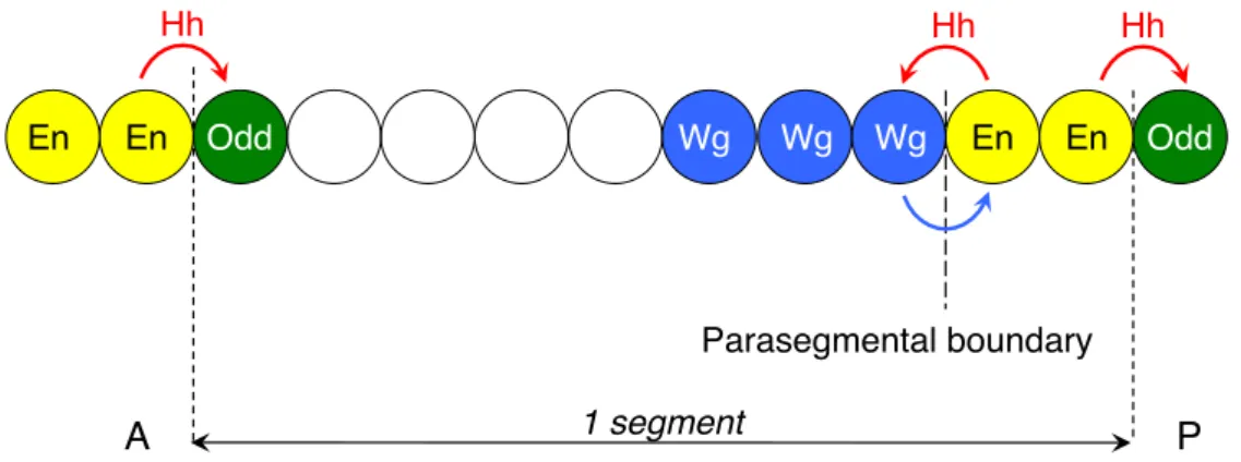
The fine organisation of each segment is achieved by complex crosstalks between segment-polarity genes. Each segment is divided into the anterior and posterior compartment by analogy to the disc organisation ƒsignalwhere García-Bellido and colleagues proved the existence of a non physical boundary that divides the wing disc into an anterior and a posterior compartment (Garcia-Bellido et al., 1973). As it is the case in the wing disc, the posterior compartment of each segment in the Drosophila embryo expresses the transcription factor engrailed (en) (Fjose et al., 1985; Kornberg et al., 1985) and secretes Hedgehog (Hh), a double-lipid modified ligand (Kornberg et al., 1985; Tabata and Kornberg, 1994). Hh diffuses and induces its targets in the Cubitus interruptus (Ci)-expressing domains that border the En-expressing cells. Since Ci is the transcription factor of the Hh pathway and is never expressed in the En-domain, the En-expressing cells are competent to produce but not to interpret Hh (Aza-Blanc et al., 1997). In response to Hh signal, the
Ci-A" P"
D"
V" Maternal-effect genes!
(bicoid, nanos, hunchback)!
Segment polarity genes!
(hedgehog, wingless, engrailed)!
Paire-rule genes!
(odd skipped, even skipped, fushi tarazu)! Gap genes! (kruppel, knirps, giant)!
Figure 10. Drosophila segmentation.
Drawings representing maternal-effect, gap, pair-rule and segment-polarity genes.
Examples listed for each class of genes is not exhaustive.
expressing cells that are anterior to the En cells maintain Wingless (Wg) expression, another secreted ligand (Baker, 1987; Alexandre et al., 1999). Wingless diffuses and in return maintains Engrailed in the posterior cells. The Wg and En cells form therefore a feedback loop and constitute the parasegmental organizer, by analogy to the Spemann organizer (Martinez-Arias and Lawrence, 1985). Posterior to the engrailed-cells, Hh diffuses and maintains the expression of the pair-rule gene odd skipped in the next segment that will constitute the groove cells (Vincent et al., 2008). Therefore, En and Odd cells define the segmental boundary (Figure 11).
During Stage 10, cells that constitute the tracheal placodes divide and invaginate at Stage 11 to form the tracheal pits (80 cells per pit). The anterior-most pits will give rise to the anterior spiracles while the posterior-most pits will form the posterior spiracles. The remaining pits will give rise to the tracheal tree without any cell division.
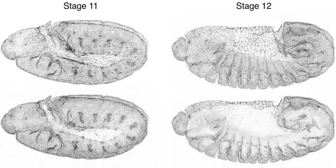
In parallel, cell death located between the epidermis and the nervous system occurs, leading to the formation of large clusters of neurons. It continues until Stage 12 (Figure 12). En! En! Hh! Hh! Wg! Wg! Wg! Odd! Hh!
En! En! Odd!
1 segment!
Parasegmental boundary!
A! P!
Figure 11. Drosophila segment organization.
En = Engrailed ; Wg = Wingless ; Odd = Odd skipped ; Hh = Hedgehog. Wg and En cells constitute the parasegmental organizer: En cells produce Hh that maintains Wg expression, while Wg maintains En cells. Posterior to the En cells, Hh maintains Odd expression that marks the groove cells.
II.5 Germ Band retraction (Stage 12)
At stage 12, the tail of the embryo retracts. The amnioserosa, wrinkled like an accordion at the end of Stage 11 starts to deploy to cover a transient dorsal gap. Grooves also start to form. These groove cells have a specific shape and cytoskeleton: groove cells display a ladder-like organisation, and accumulate adherent junction molecules such as Crumbs, aPKC or Ena (Vincent et al., 2008).
While neuronal cell death still occurs at Stage 12, the ventral cord separates from the epidermis and the first axons in the ventral nerve cord are visible.
During Stage 12, the invaginated trachea pits start to elongate and form the trachea. The tracheal metameres are composed of 5 branches: the dorsal branch (DB), the dorsal trunk anterior (DTa), the visceral branch (VB), the lateral trunk (LT) and the ganglionic branch (GB) (Samakovlis et al., 1996) (Figure 13).
Stage 11! Stage 12!
Figure 12. Stage 11 and 12 Drosophila embryos.
Confocal pictures of a stage 11 and a stage 12 embryo marked with E-Cadherin. The second row represents lower Z-section to highlight the trachea.
During trachea formation, the migration of the dorsal branches depends on the action of the FGF homolog Branchless (Bnl) and DPP (Vincent et al., 1997). The tracheas cell express the FGF receptor Breathless (Btl) and are therefore capable to interpret the Bnl produced by the organs that are “attracting” the migration of the branches.
DPP plays a dual role for the DB, LT and GB specification and migration. First, DPP repress spalt while activating knirps expression, whereas in other branches, the situation is the opposite: knirps is inhibited while spalt is transcribed (Vincent et al., 1997). Second, DPP controls Bnl expression for the proper migration of the DB, LT and GB. Consistently, in embryos where DPP signalling is impaired such as in the thickveins (tkv) mutant embryos, dorsal branches as well as lateral and ganglionic branches are absent (Vincent et al., 1997).
DB!
DTa!
VB!
LT!
GB!
Figure 13. Trachea metamer.
Drawing representing a trachea metamer. DB = Dorsal Branch
DTa = Dorsal Trunk anterior VB = Ventral Branch
LT = Lateral Branch GB = Ganglionic Branch
II.6 Dorsal closure (Stage 13 – Stage 15)
After the completion of the retraction of the germ band, dorsal closure takes place from Stage 13 to Stage 15. During dorsal closure, the transient dorsal gap covered by the amnioserosa is progressively closed by the fusion of the first row of dorsal epidermal cells called the leading edge at each extremity. I will detail dorsal closure in the next section. In parallel to dorsal closure, the head of the embryo invaginates.
During these stages, other layers continue to develop. The central and the peripheral nervous system (including sensilla and motor neurons) start to differentiate. The ventral nerve cord starts to condensate at Stage 14. The mesoderm also progressively differentiates: by stage 13, the fusion of myoblasts is completed. As the segments stretch during closure, the muscle fibres become distinguishable. The visceral mesoderm, attached to the somatic mesoderm at stage 12 is progressively attached to the midgut primordial at stage 13. It later spreads to encircle the developing gut. The trachea continue to migrate at Stage 13, the anterior-directed dorsal branches of all segments fuse to form the dorsal longitudinal tracheal trunk.
II.7 Late embryogenesis (Stage 16 – Stage 17)
The two last embryonic stages will give rise to a ready-to-live larva. The epidermis starts to secrete the cuticle, including the denticle belts that are enriched with actin. The diverse organs terminate their differentiation. The condensation of the ventral nerve cord, initiated at stage 14 continues until stage 17. During these last steps, the trachea become filled with air. The sensilla are differentiated and sensory axons are connected with the central nervous system. The motor axons are now connected to the muscles, leading to spontaneous movement of the embryo at mid-stage 17, until its eventual hatching (Figure 14).
Stage Time Developemental Events 5 3h00 Cellulariza:on 6‐7 3h – 3h20 Gastrula:on 8‐9 3h20 – 4h20 Germ band extension 10 4h20 – 5h20 End of germ band extension Stomodeum invagina:on 11 5h20 – 7h20 Trachea invagina:on Parasegmenta:on 12 7h20 – 9h40 Germ band retrac:on 13 9h40 – 10h20 14 10h20 – 11h Dorsal closure and head involu:on 15 11h – 13h 16‐17 13h – 22h Condensa:on of the nervous system Embryo ready to hatch Figure 14. Drosophila embryonic stages.
Homemade drawings adapted from the Atlas of Drosophila development written by Volker Hartenstein. This figure depicts the key Drosophila embryonic stages at 25°C.
III. Morphogenesis during dorsal closure
III.1 An overview of dorsal closure
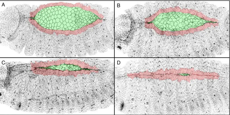
Dorsal closure is a key embryonic process during which the dorsal gap covered by the amnioserosa progressively disappears. As dorsal closure proceeds, the dorsal-most epidermal cells that constitute the leading edge, elongate dorso-ventrally, meet and zip at each extremity called canthus. Dorsal closure starts at Stage 13, once the retraction of the germ band is completed and ends at Stage 15 with a perfectly suturated embryo (Figure 15).
Leading edge !
Amnioserosa!
A! B!
C! D!
Figure 15. Drosophila dorsal closure.
(A-D) Wild-type embryos marked with E-Cadherin during dorsal closure. The amnioserosa is colour-coded in green. The first rows of epidermal cells in contact with the amnioserosa that constitute the leading edge are colour-coded in red. As closure proceeds, the amnioserosa progressively disappears while the leading edge of each epidermis fuse at the canthi.
Cadherin! Cadherin!
Tubulin! Tubulin!
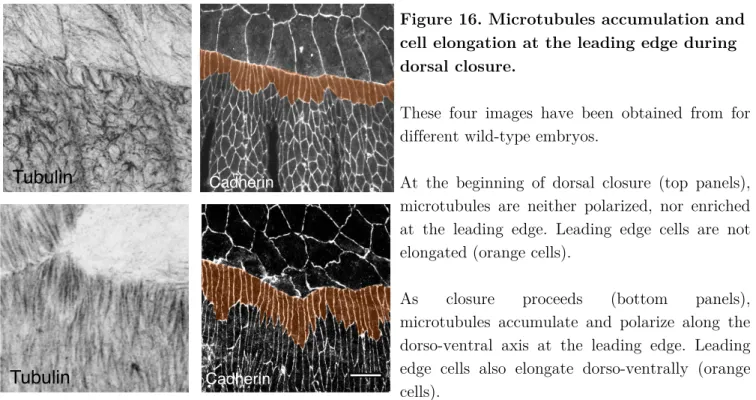
During dorsal closure, the leading edge cells are polarized and have a highly dynamic cytoskeleton. As closure proceeds, leading edge cells elongate along the dorso-ventral axis and display strong adherens junctions (Kaltschmidt et al., 2002; Ducuing et al., 2015). They accumulate a dense apical microtubule network that is orientated dorso-ventrally (Kaltschmidt et al., 2002; Jankovics and Brunner, 2006) (Figure 16).
In addition, leading edge cells produce a trans-cellular actin cable that circles the amnioserosa (Young et al., 1993; Jacinto et al., 2000; Jacinto et al., 2002). Leading edge cells also produce actin-based short protrusions called filopodia that are crucial for the zipping (Jankovics and Brunner, 2006; Millard and Martin, 2008).
Two major developmental pathways control dorsal closure: the stress response pathway JNK acts upstream and induces the Bone Morphogenetic Protein homologue Decapentaplegic (DPP) (Glise and Noselli, 1997; Hou et al., 1997; Kockel et al., 1997; Riesgo-Escovar and Hafen, 1997). Both JNK and DPP pathway are
Figure 16. Microtubules accumulation and cell elongation at the leading edge during dorsal closure.
These four images have been obtained from for different wild-type embryos.
At the beginning of dorsal closure (top panels), microtubules are neither polarized, nor enriched at the leading edge. Leading edge cells are not elongated (orange cells).
As closure proceeds (bottom panels),
microtubules accumulate and polarize along the dorso-ventral axis at the leading edge. Leading edge cells also elongate dorso-ventrally (orange cells).
crucial for dorsal closure since embryos where either JNK or DPP signalling is impaired fail to complete dorsal closure (Affolter et al., 1994; Glise et al., 1995).
There are three main driving forces of dorsal closure that will be detailed hereafter:
(1) Cell oscillation and delamination in the amnioserosa (Toyama et al., 2008; Solon et al., 2009; Muliyil et al., 2011).
(2) The actin cable that could either provide a contractile force or prevent the relaxation of leading edge cells (Young et al., 1993; Kiehart et al., 2000; Jacinto et al., 2002; Rodriguez-Diaz et al., 2008; Solon et al., 2009).
(3) The filopodia that make the zipping effective at each canthus (Jankovics and Brunner, 2006; Millard and Martin, 2008).
III.2 The amnioserosa
The amnioserosa is an extra-embryonic tissue composed of flat squamous cells that cover the yolk. During dorsal closure, the amnioserosa progressively disappears, hence participating to the progression of closure. Amnioserosa is specified during gastrulation by the transcription factor zerknullt (Doyle et al., 1986) and becomes sandwiched between the tail and the head of the embryo as germ-band extension proceeds.
Dorsal closure is divided into two phases (Gorfinkiel et al., 2009). During the first “slow” phase of dorsal closure, amnioserosa cells dynamically oscillate at the apical surface (Fernandez et al., 2007; Gorfinkiel et al., 2009; Solon et al., 2009; Blanchard et al., 2010; David et al., 2010; Sokolow et al., 2012) (Figure 17).
The second phase of dorsal closure is the “fast” phase (Gorfinkiel et al., 2009).
Amnioserosa cell oscillation is driven by transient relocalisation of actin and non-muscle myosin II (spaghetti squash, sqh) at the centre of the cells (Franke et al., 2005; Blanchard et al., 2010). During the first “slow” phase, the myosin-actin flow is therefore pulsed and is regulated by the PAR complex (David et al., 2010), but also by Ca2+ flux (Hunter et al., 2014). Indeed, blocking of the Ca2+ channels subunits Figure 17. Amnioserosa cell oscillations during dorsal closure.
Closeup of a shg ::GFP embryo during dorsal closure. The three first images are still images from a time-lapse movie showing the oscillation of one amnioserosa cell. The last image is a composite where the three first images have been colour-coded and super-imposed.
generates defects in actomyosin structures, and prevents amnioserosa cell contraction (Hunter et al., 2014). These amnioserosa cell oscillations drive the progressive constriction of their apical domain (Solon et al., 2009). In addition, laser ablation of a single amnioserosa cell leads to a decrease in or a stop of the oscillation of neighbouring amnioserosa cells, indicating that local tensions are crucial for these oscillations (Solon et al., 2009).
During the second, “fast” phase, amnioserosa cells stop oscillating, concomitantly with the formation of the actin cable. Myosin accumulates in a more sustained manner in the amnioserosa (Blanchard et al., 2010). Amnioserosa cells continue to reduce their apical surface. This contributes to the progression of the leading edge since ablation of in the amnioserosa leads to a transient ventral-ward retraction of the leading edge (Kiehart et al., 2000). The amnioserosa cells located at the periphery start to flatten first, followed by the next row of cells shortly after (Gorfinkiel et al., 2009) (Figure 18).
In addition, about 10 to 30% of the amnioserosa cells undergo apoptosis and delaminate in a stochastic fashion (Kiehart et al., 2000; Toyama et al., 2008). This occurs preferentially in the anterior part of the embryo during the “fast” phase (Muliyil et al., 2011). Enhancing or reducing apoptosis in the amnioserosa speeds or slows dorsal closure respectively, indicating that cell death in the amnioserosa tunes Figure 18. Amnioserosa during the slow and fast phases.
Still images of a shg ::GFP embryo during dorsal closure.
Left: amnioserosa during the slow phase. Right: Amnioserosa during the fast phase. Peripheral amnioserosa cells are flattened.
the speed of closure (Toyama et al., 2008; Muliyil et al., 2011). This could be the main driving force of dorsal closure, since the ablation of the canthi that breaks the continuity of the actin cable does not stop the progression of the leading edge (Wells et al., 2014) (Figure 19).
Recently, it has been shown that stochastic generation of Reactive Oxygen Species (ROS) in the amnioserosa is necessary and sufficient to trigger cell delamination by acting on actomyosin and mitochondrial architecture (Muliyil and Narasimha, 2014). However, we currently do not understand the genetic program that control amnioserosa cell death and what controls ROS regulation.
Altogether, amnioserosa is a major driving force during dorsal closure. During the initial first “slow phase”, pulses of actin and myosin in the centre of the cells drive rapid cell contraction and relaxation. This depends on the PAR complex and on the Ca2+ flux. While these cells oscillate, they progressively reduce their apical surface and volume. During the second “fast” phase, oscillations are reduced since myosin accumulates in the centre of cells in a sustained manner. Cells continue to reduce their apical surface and volume, while 10 to 30% undergo apoptosis due to ROS accumulation. In addition, the actin cable that circle the amnioserosa is also believed to be another major driving force during dorsal closure.
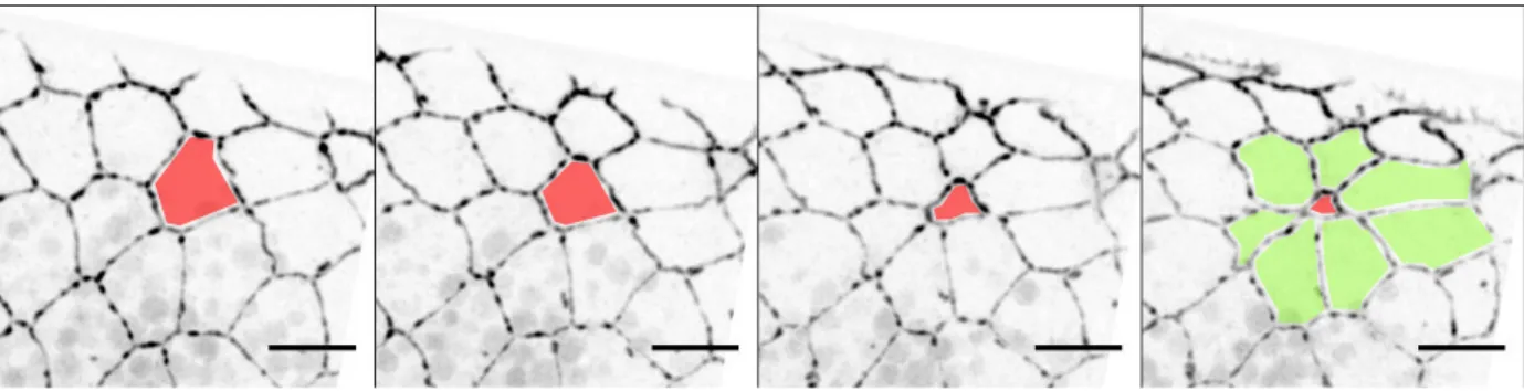
Figure 19. Amnioserosa cell delamination.
Still images of a shg ::GFP embryo during dorsal closure. The red cell progressively delaminates, leading to the formation of a so-called “rosette” (green cells). Scale bar: 10 µm.
III.3 The actin cable
The actin cable is a remarkable supra-cellular structure that is present during the developmental morphogenesis of many tissues such amnion sac closure in the chick embryo (Tipping and Wilson, 2011), ventral enclosure in C. elegans (Williams-Masson et al., 1997; Martin and Parkhurst, 2004) or during Drosophila dorsal closure (Young et al., 1993; Jacinto et al., 2000; Jacinto et al., 2002). The presence of an actin cable has first been described in the 90’s in cells around wounds in the chick embryo (Martin and Lewis, 1992). Shortly after, the presence of an actin cable has been also described in both vertebrate and invertebrate models of wound healing (Martin and Lewis, 1992; Brock et al., 1996; Davidson et al., 2002; Wood et al., 2002; Belacortu and Paricio, 2011). In the Drosophila embryo, at the beginning of dorsal closure, the leading edge cells that are in contact with the amnioserosa produce a dense trans-cellular actomyosin cable that can be easily visualized with a phalloidin staining that labels actin (Figure 20).
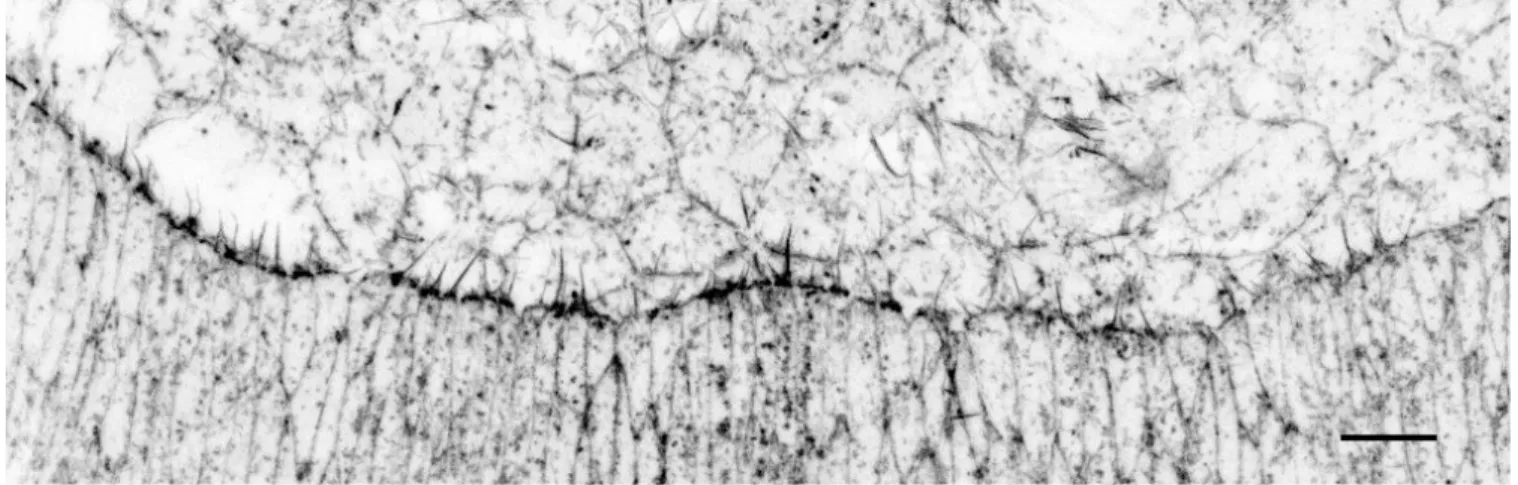
Figure 20. Actin cable during dorsal closure.
Closeup of a Stage 13/14 embryo marked with Phalloidin to label actin. Scale bar: 10 µm. Note the strong enrichment of actin at the amnioserosa / leading edge interface that corresponds to the actin cable. Filopodia produced by leading edge cells and pointing towards the amnioserosa are also visible.
The actin cable formed at stage 13 becomes stronger as closure proceeds. It accumulates filamentous actin and the non-muscle myosin II (spaghetti squash, sqh) (Young et al., 1993; Kiehart et al., 2000; Jacinto et al., 2002). In addition, the actin-capping molecule Enabled (Ena) is enriched at the actin cable during dorsal closure (Grevengoed et al., 2001; Gates et al., 2007), but also in tri-cellular junctions in the epidermis (Gates et al., 2007) and in groove cells (Vincent et al., 2008). Ena therefore constitutes an excellent marker to label the actin cable. In addition, the adhesion molecule Echinoid (Ed) is expressed in all the cells of the epidermis except in the junctions between the leading edge and the amnioserosa (Laplante and Nilson, 2006; Laplante and Nilson, 2011). This asymmetric distribution of Ed at the leading edge is crucial for actin cable formation since restoring a symmetric distribution of Ed by either depriving all cells from Ed or by ectopically expressing the Ed at the amnioserosa / leading edge interface results in similar actin cable defects (Laplante and Nilson, 2011) (Figure 21).
Figure 21. Ena, Ed and Baz expression pattern during dorsal closure.
(A-A’) closeup of a Stage 14 embryo marked with Ena and E-Cadherin. Scale bar: 10 µm. Ena is enriched in tri-cellular junction, in groove cells but most importantly at the level of the actin cable (Gates et al., 2007).
(B-C’) Closeup of embryos at the beginning (B-B’) or at the middle of dorsal closure (C-C’). Ed and Baz are progressively excluded from the leading edge / amnioserosa interface as closure proceeds. These images are taken from (Laplante and Nilson, 2011) and have been processed on ImageJ.
Baz! Baz! Ed! Ed! Ena$ Ena$/$E&Cadherin$ A A’ B B’ C C’
Ed also controls the correct localisation of the scaffolding protein and apical determinant Par3/Bazooka (Baz) (Laplante and Nilson, 2011). During development, Baz localizes to adherens junctions but is lost from the leading edge / amnioserosa interface as closure proceeds. Baz is important for acto-myosin contractility during amnioserosa cell apical constriction (David et al., 2010; David et al., 2013; Pickering et al., 2013) as well as for actin-based protrusions. Since Baz exclusion from the amnioserosa / leading edge interface is Ed-dependant, Baz might directly control actin cable formation, although the molecular mechanism is unknown (Laplante and Nilson, 2011).
The actin cable is therefore a striking feature of the highly specialized cytoskeleton of the leading edge cells during dorsal closure. There are to main questions that have been addressed extensively but still remain unsolved:
(1) Is the actin cable required or dispensable for dorsal closure? (2) What is the function of the actin cable during dorsal closure?
The requirement or dispensability of the actin cable during dorsal closure remains puzzling. Indeed, embryos where the cable is affected are showing a range of different phenotypes, probably because the missing components are not expressed solely at the leading edge but also in other tissues such as the amnioserosa. For instance, in embryos lacking zipper, the motor protein non-muscle myosin heavy chain, the actin cable is affected and dorsal closure often fails (Young et al., 1993). Alternatively, in embryos deficient for the non-receptor tyrosine kinase Abelson (Abl) where the actin cable formation is perturbed, a subset of Abl mutant embryos either close slowly or fail to complete dorsal closure, suggesting that the actin cable could be either dispensable or strictly required for dorsal closure (Grevengoed et al., 2001). Last, the asymmetric distribution of Ed at the leading edge is crucial for the actin cable formation (Laplante and Nilson, 2011). In embryos that are zygotically lacking Ed, dorsal closure is completed, although terminating with discontinuities and puckering
at the dorsal midline (Laplante and Nilson, 2006). This suggests that the cable would be dispensable for closure. In addition, laser ablation experiments showed that breaking the continuity of the cable does not prevents closure, although the continuity of the cable is restored shortly after the cut (Kiehart et al., 2000; Hutson et al., 2003; Rodriguez-Diaz et al., 2008). Altogether, the requirement of actin cable during dorsal closure is unclear. With the model we set up, we provide an unambiguous proof that the actin cable is dispensable for dorsal closure (Results section). Using our model, we then further investigated the effect of the absence of the acting cable during dorsal closure.
In Drosophila, two main models have been proposed to account for the function of the actin cable during dorsal closure. First, the actin cable has been proposed to operate as a contractile purse-string (Young et al., 1993; Kiehart et al., 2000; Jacinto et al., 2002; Rodriguez-Diaz et al., 2008). The name purse string refers to the analogy of the purse string procedure in medicine where a string surrounding a wound is pulled by the surgeon to artificially close the wound. In this model, the actin cable provides a contractile force to ensure the dorsal-ward migration of leading edge cells. This is supported by the observation that in embryos lacking the actin cable, dorsal closure often fails to complete (Young et al., 1993; Grevengoed et al., 2001). A prediction of this model is that leading edge cells should be under tensions, since the cable should be pulled along the antero-posterior axis. This has been confirmed by laser ablation experiments where cutting the actin cable leads to a retraction of the neighbouring leading edge cells (Kiehart et al., 2000; Hutson et al., 2003; Rodriguez-Diaz et al., 2008). However, laser ablation of the actin cable does not prevent dorsal closure, indicating that the actin cable is not the only driving force during dorsal closure (Kiehart et al., 2000). In addition, the purse string mechanism relies on the geometry of the system: the movement should be proportional to the curvature of the cable and no movement should proceed when the cable is straight. It has been