Recombinant hirudin: kinetic mechanism for the inhibition of
human thrombin
Stuart R.Stone1 and Jan Hofsteenge
Friednch Miescher-Institut, PO Box 2543, CH-4002 Basel, Switzerland 'To whom correspondence should be addressed
Recombinant hirudin variant-2(Lys47), was found to be a
competitive inhibitor of human a-thrombin with respect to peptidyl /Miitroanilide substrates. These results contrast with those of Degryse and coworkers that suggest that recombinant hirudin variant-2(Lys47) inhibited thrombin by a
non-competitive mechanism [Degryse et al. (1989) Protein Engng, 2,459-465]. 7-Thrombin, which can arise from a-thrombin by autolysis, was shown to have an affinity for recombinant hirudin variant-2(Lys47) that was four orders of magnitude
lower than that of a-thrombin. It was demonstrated that the apparent noncompetitive mechanism observed previously was probably caused by a contamination of the thrombin prepartion by 7-thrombin. Comparison of the inhibiprepartion of a-thrombin by recombinant hirudins variant-2(Lys47) and
variant-1, which differ from one another in eight out of 65 amino acids, indicated that the two variants have essentially the same kinetic parameters.
Key words: hirudin/kinetics/thrombin
Introduction
Hirudin has received attention recently as a potential therapeutic agent for the control of thrombosis (Wallis, 1988). It was originally isolated from the medicinal leech Hirudo medicinalis (Markwardt, 1970) and many different isoforms have been subsequently isolated and sequenced (Bagdy et ai, 1976; Dodt et ai, 1984, 1986; Tripier, 1988; Scharf et al., 1989). The major form of hirudin (termed variant 1) is a polypeptide of 65 amino acids which contains a single sulphotyrosyl residue. The possibility of using hirudin as a therapeutic agent has evolved with the development of systems for the recombinant expression of hirudin (Bergmann etai, 1986; Fortkamp et al., 1986; Harvey et al., 1986; Meyhack et al., 1987; Loison et al., 1988). The small size of hirudin has allowed the determination of its tertiary structure by two-dimensional NMR techniques (Clore et al., 1987; Folkers etai, 1989; Haruyama and Wiithrich, 1989). These studies indicate that hirudin is composed of a compact N-terminal domain (residues 3-49) held together by three disulphide bonds, and a disordered C-terminal tail (residues 50-65). The structures of complexes between human a-thrombin and two recombinant hirudins have also been determined recently (Rydel etai, 1990; Griitter etai, 1990). These structures indicate that hirudin inhibits thrombin by a novel mechanism. Hirudin binds with its N-terminal three residues in the active-site cleft of thrombin; the hirudin polypeptide chain runs in a direction opposite to that expected for a substrate. In addition, the primary specificity pocket of thrombin is not occupied by hirudin. The C-terminal tail of hirudin wraps around the putative fibrinogen secondary binding site.
The kinetics of inhibition of thrombin by hirudin have been studied by a number of groups and the results obtained are not entirely consistent between studies (Fenton etai, 1979; Walsmann and Markwardt, 1981; Stone and Hofsteenge, 1986; Braun etai, 1988a; Dodt etai, 1988, 1990; Degryse et ai, 1989). Although all studies indicate that hirudin is an extremely tight-binding inhibitor of thrombin, estimates of the dissociation constant of the thrombin-hirudin complex have varied by over three orders of magnitude (Walsmann and Markwardt, 1981; Stone and Hofsteenge, 1986). The discrepancies between the various values for the dissociation constants can be explained at least in part by the use of different reaction conditions and different forms of hirudin and thrombin. For example, the dissociation constant for the thrombin—hirudin complex has been shown to be strongly dependent on ionic strength (Stone and Hofsteenge, 1986; Stone etai, 1989). The post-translational modification of Tyr63 to form sulphotyrosine does not occur in any of the expression systems so far used and this lack of a sulphate group leads to a lower affinity of human a-thrombin for the recombinant molecule (Braun et al., 1988a; Dodt et ai, 1988, 1990). The affinity of human thrombin for hirudin is higher than that of bovine thrombin (Dodt et al., 1990). Proteolytically degraded forms of human thrombin have a lower affinity for hirudin (Landis et ai, 1978; Stone etai, 1987).
Different mechanisms of inhibition have been suggested also by different workers. While the results of Stone and Hofsteenge (1986) indicate that hirudin variant-1 inhibits thrombin by a competitive mechanism, Degryse et al. (1989) have suggested that a noncompetitive mechanism applies to recombinant hirudin variant-2 with the mutation Asn47—Lys (rHV2-Lys471). This
difference in the kinetic mechanism could be due to the different forms of hirudin used or to the different assay conditions in the two studies.
This paper attempts to determine the basis for the observed difference in kinetic mechanism. Moreover, we have determined the kinetic constants for rHV2-Lys47 and recombinant hirudin
variant-1 (rHVl) under identical conditions and, thus, have been able to evaluate to what extent the previous discrepancies in values of dissociation constants were due to differences in forms of hirudin used.
Materials and methods
Materials
The substances D-Phe-pipecolyl-Arg-p-nitroanilide (D-Phe-Pip-Arg-pNA; S-2238) and D-Val-Leu-Arg-/?-nitroanilide (D-Val-Leu-Arg-pNA; S-2266) were from Kabi (Molndal, Sweden) whereas tosyl-Gly-Pro-Lys-p-nitroanilide (tos-Gly-Pro-Arg-pNA; Chromozym-PL) was from BoehringeMannheim, FRG).
r-HV2-Lys47 was purchased from Pentapharm (Basel,
Switzerland) and rHVl was a gift from Ciba-Geigy (Basel, Switzerland). Human a-thrombin and 7-thrombin were prepared as described previously (Stone and Hofsteenge, 1986; Braun et al., 1988b) and were fully active as determined by active site titration with 4-methylumbelliferyl /j-guanidinobenzoate (Jameson
et al., 1973). Amino acid sequence analysis (Hewick et al., 1981) and polyacrylamide gel electrophoresis (Laemmli, 1970) indicated that the a-thrombin used was pure and free from degraded forms. In addition, human a-thrombin was obtained from Sigma (St Louis, MO). PAGE (Laemmli, 1970), followed by densiometric scanning of this thrombin preparation (4200 NIH units/mg; Catalogue no. T6759; Lot 114F-9461), indicated that it contained a substantial amount (>20%) of degraded forms of thrombin /3 and y.
Amidolytic assay of thrombin
Assays were performed at 37°C in buffers containing 0.1% poly(ethylene glycol) Mr 6000 as previously described (Stone
and Hofsteenge, 1986). Three different buffer systems were used. Buffer 1 consisted of 0.05 M Tris-HCl buffer, pH 7.8, containing 0.1 M NaCl. An ionic strength of 0.125 M can be calculated for this buffer (Ellis and Morrison, 1982). Buffer 2 was the buffer used by Degryse et al. (1989) and contained 0.05 M piperazineN',Nbis2ethanesulphonic acid (PIPES) -NaOH, pH 7.9, and 0.18 M NaCl with a calculated ionic strength of 0.327 M. Buffer 3 consisted of 0.05 M Tris-HCl, pH 7.8 and 0.3 M NaCl and had an ionic strength of 0.325 M. The type and concentration of substrate used and the concentration of thrombin varied between the experiments.
Data analysis
Determination of the concentrations of the hirudin variants. The concentration of active hirudin molecules was determined by titration of 1.0 nM thrombin in Buffer 1 containing 200 /*M D-Val-Leu-Arg-pNA. The dependence of the steady-state velocity on the amount of hirudin present in the assay could be described by equation (1) (Stone and Hofsteenge, 1986):
2£, CD
where vs is the observed state velocity, VQ is the
steady-state velocity in the absence of hirudin, Et is the total molar
enzyme concentration, Kv is the apparent inhibition constant, Ia
is /tl/ml of hirudin added and a is the molar concentration of 1.0 jd/ml of hirudin. Analysis of the data yielded values for a that could be used to calculate the concentrations of hirudin in the stock solutions.
Determination of kinetic parameters. The inhibition of thrombin by hirudin can be represented by the following scheme:
thrombin + hirudin' thrombin—hirudin
The dissociation constant (K{) for the thrombin —hirudin complex can be related to the association (k{) and dissociation (k2) rate constants by the expression K\ = k2lk\. The kinetic
parameters for the inhibition of human a-thrombin by rHV2-Lys47 and rHVl were determined under several different
experimental conditions. Under all conditions used, hirudin acted as a slow, tight-binding inhibitor of thrombin and the data were fitted to equation 4 of Stone and Hofsteenge (1986) by nonlinear regression. These analyses yielded values for the apparent dissociation constant (Kv) and apparent association rate constant
(kv). With the substrate tos-Gly-Pro-Lys-pNA, a background
rate of hydrolysis was observed in the absence of enzyme. This rate was dependent on the substrate concentration and equal to 0.035 ^M/min/mM substrate. The data were corrected for this background rate before analysis. The rate of hydrolysis of D-Phe-Pip-Arg-pNA in the absence of thrombin was negligible.
The dependence of the value of Kv on the concentration of
substrate for a competitive inhibitor is given by equation 2:
Kv = S/KJ (2)
where S is the substrate concentration and Km is its Michaelis
constant. This equation can be used to calculate K\ from the observed value of Kv.
Stimulation of the effect of contamination by y-thrombin. It has been shown previously that hirudin has a much lower affinity for human 7-thrombin than for a-thrombin (Landis et al., 1978; Stone et al., 1987). The effect of contamination by 7-thrombin was simulated according to equation 3:
V, = + vs. (3)
where vsa and vSy are the contributions to the observed velocity
(vs) made by the a- and 7-thrombin in the preparation
respectively. The values of vsa and vS7 are given by:
= TT l[*iy + A
-4ATIa.£J14 - - Ea)\ (4)
A
-where Ea and Ey are the concentrations of a- and 7-thrombin
respectively, and Kia' and K^ are the apparent dissociation
constants for hirudin with a- and 7-thrombin. The values Kw
and Kly. for these simulations can be calculated at different
substrate concentrations by using equation (2). Estimates for Kla, Kly and the Km values for a- and 7-thrombin with the
substrate are required for these calculations. The values of Km
to be used in these calculations were determined as described by Hofsteenge et al. (1986) to be 105 ± 9 /tM and 98 ± 2 ^M for the substrate tos-Gly-Pro-Lys-pNA with a- and 7-thrombin respectively. The value of the Kia was determined to be 2.3 pM
(Table I) under the conditions used by Degryse et al. (1989). The value of Kly was determined from the observed inhibition
of 7-thrombin by rHV2-Lys47 under the same conditions. The
data were fitted to the Dixon equation (Segel, 1975) and a value of 75 ± 3 nM for K]y was calculated after correcting for the
concentration of substrate according to equation (2).
Using the above constants, values for K\a' and Kly> were
calculated at a number of different substrate concentrations and inhibition data were simulated with different ratios of a- to 7-thrombin under the conditions similar to those used by Degryse et al. (1989): the total enzyme concentration was maintained at 4.5 nM and the concentration of rHV2-Lys47 was varied from
0 to 6 nM (10 points). For each substrate concentration, 10 sets of data were simulated according to equation 3. A normally distributed random error with a standard deviation of 2% of the theoretical value was introduced into the simulated data (Pollard, 1977). The 10 sets of simulated data were analysed according to equation (1) with Ex = Ea + Ey and 10 estimates for Kv were
obtained. The weighted mean estimate of Kr was calculated for
each substrate concentration. Results
The hirudin variant rHV2-Lys47 is a competitive inhibitor of
human a-thrombin
The kinetic mechanism of the inhibition of human a-thrombin by rHV2-Lys47 was studied under the conditions used by
50. 0 40. 0 . 30.0 _ 20. 0 . 10. 0 _ 0. 0
Table I. Kinetic parameters for the interaction of rHV2-Lys47 and rHV 1
with human a-thrombin
0.0 0 . 5 1.0 1.5
[tos-Gly-Pro-Lys-pNA] (inM)
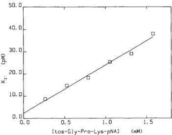
Fig. 1. Dependence of the apparent dissociation constant (Ky) for
rHV2-Lys on the concentration of the substrate tos-Gly-Pro-Lys-pNA. Assays were performed in PIPES-NaOH buffer (Buffer 2) and the data analysed as described in Materials and methods to yield estimates for Ky at the indicated substrate concentrations. These estimates were weighted according to the squared inverse of their standard errors and fitted to equation 2. The line drawn depicts the result of this fitting. For this analysis, the value of Km in equation (2) was fixed at the previously
determined value of 105 /iM.
Degryse et al. (1989), i.e. in PIPES-NaOH buffer (pH 7.9) with an ionic strength of 0.33 M (Buffer 2) with the substrate tos-Gly-Pro-Lys-pNA. Values for the apparent dissociation constant (Ky) were determined at a particular substrate concentration by varying the concentration of rHV2-Lys47 from 56 to 280 pM
with a fixed concentration of 200 pM a-thrombin. Under these conditions, rHV2-Lys47 acted as a slow, tight-binding inhibitor
(data not shown) and analysis of progress-curve data yielded values for Kv and for the apparent association rate constant (kv).
The values of Ky showed a linear dependence on the
concentration of the substrate as would be expected from equation (2) for a competitive inhibitor (Figure 1). A value of 105 ± 9 fiM was determined for the Km of tos-Gly-Pro-Lys-pNA from
initial-velocity studies. The value of Km in equation (2) was
fixed at this value and the data of Figure 1 fitted by weighted linear regression to this equation. The line drawn in Figure 1 represents the result of this analysis and it can be seen that the data fit well to the equation describing competitive inhibition. The value for Kt obtained from this analysis was 2.29 ±
0.05 pM (Table I).
The estimate of Jt|- did not vary greatly over the substrate range tested. Similar results were obtained previously with native hirudin variant-1 (Stone and Hofsteenge, 1986). The weighted average of the six determinations of ky was (3.6 ± 0.1) x 107/
M/s and it can be assumed that this value approximates the true value of the association rate constant fc| (Table I).
In order to confirm the competitive nature of the inhibition by rHV2-Lys47, the substrate-dependence of the value of Ky was
determined with another substrate. The results obtained with the substrate D-Phe-Pip-Arg-pNA are shown in Figure 2; a similar linear dependence was observed. The concentrations of a-thrombin and rHV2-Lys47 used to obtain the data presented in
Figure 2 were also different; the enzyme was present at a concentration of 20 pM while rHV2-Lys47 was varied from 61
to 306 pM. An estimate of 4.2 ± 0.2 /xM was determined from initial-velocity studies for the Km of D-Phe-Pip-Arg-pNA in the
PIPES-NaOH buffer used and the value of Km in equation (2)
was fixed at this value for the analysis of the data of Figure 2.
Experimental conditions Kinetic parameters
Buffer Substrate t, x 10" (/m/s) (pM) rHV2-Lys47 PIPES-NaOH" tos-Gly-Pro-Lys-pNA 3.61 ± 0.07 2.29 ± 0.05 (/ = 0.33 M) PIPES-NaOHb D-Phe-Pip-Arg-pNA 2.78 ± 0.03 2 35 ± 0.02 (/ = 0 33 M) Tris-HClc D-Phe-Pip-Arg-pNA 2.51 ± 0 . 1 1 2.47 ± 0 . 0 4 (/ = 0.33 M) Tris-HCl D-Phe-Pip-Arg-pNA 11.3 ± 0.64 0.364 ± 0.015 (/ = 0.125 M) rHVl Tris-HCl D-Phe-Pip-Arg-pNA 3.25 ± 0.16 0.991 ± 0.030 (/ = 0 33 M) Tris-HCld D-Phe-Pip-Arg-pNA 13.7 ± 0.3 0.231 ± 0.006 (/ = 0.125 M)
Values of Kt and kt were calculated from apparent values as described in the text. The Km values for D-Phe-Pip-Arg-pNA that were used in the
calculations were 3 6, 4.2 and 3 0 pM for Buffers 1, 2 and 3 respectively.
aThese values were calculated from the data of Figure 1.
*These values were calculated from the data of Figure 2.
cThese values represent the weighted mean of two determinations. dFrom Braun et al. (1988a).
150 100 . *.- 50 _ 50 100 150 [D-Pho-Pip-Arg-pNA] 200 250 (uM)
Fig. 2. Dependence of the apparent dissociation constant (Kv) for
rHV2-Lys4' on the concentration of the substrate D-Phe-Pip-Arg-pNA.
Assays were performed in PIPES-NaOH buffer (Buffer 2) and the data analysed as described in Materials and methods to yield estimates for Ky at different substrate concentrations. These estimates were fitted by weighted linear regression to equation 2 as described in the legend to Figure 1 with the value of Km set at 4.2 /tM The line drawn depicts the fit of the data to
equation (2).
This analysis yielded a value of 2.35 ± 0.02 pM for K{. The
good fit of the data to equation (2) shown in Figure 2 indicates that the competitive model, which this equation describes, is an appropriate one for the interaction of rHV2-Lys47 with
a-thrombin. Moreover, the good agreement between the values for K{ estimated from the data of Figures 1 and 2 confirms this
competitive model.
As was observed with the substrate tos-Gly-Pro-Lys-pNA, the value of lci * did not vary with the concentration of
D-Phe-Pip-Arg-pNA and the value for k\ given in Table I represents the weighted mean of the eight determinations of ky.
Comparison of the inhibitory properties of the hirudin variants rHVl and rHV2-Lys4?
For the comparison of the inhibitory properties of rHV2-Lys47
and rHVl, Tris-HCl buffers have been used. [PIPES-NaOH buffer was used in the above studies so that the results would
100 200 300 400 500 600
0 100 200 300 400 500 600 [tos-Gly-Pro-Lys-pNA] 0*M) Fig. 3. Dependence of the apparent dissociation constant {Kv) for
rHV2-Lys47 with different forms of thrombin on the concentration of the
substrate tos-Gly-Pro-Lys-pNA. Assays were performed in PIPES-NaOH buffer (Buffer 2) with thrombin from Sigma (A) or a-thrombin containing 4% 7-thrombin (B) and the data analysed according to equation (1) to yield estimates for Ky at the indicated substrate concentrations. For the data in (B) the estimates of Kv were weighted according to the squared inverse of their standard errors and fitted to equation (6). The line drawn depicts the result of this fitting with the value of Km fixed at 105 pM.
be directly comparable with those of Degryse et al. (1989). However, Tris-HCl buffers have a much better buffering capacity at the pH values used (Ellis and Morrison, 1982).] In Tris-HCl buffer with an ionic strength of 0.325 M (Buffer 3), a Kx value of 2.47 ± 0.04 pM was determined for rHV2-Lys47
(Table I). This value agrees well with those determined in the PIPES -NaOH buffer (Buffer 2) which has about the same ionic strength. Under the same conditions, a value of 0.99 ± 0.03 pM was determined for the K{ of rHVl (Table I). The association
rate constants (&,) for rHVl and rHV2-Lys47 were about equal
and, thus, the increase in the affinity observed with rHVl can be attributed to a lower rate of dissociation of rHV 1 from the hirudin—thrombin complex (Table I).
In Tris-HCl buffer with an ionic strength of 0.125 M (Buffer 1), the difference between the K{ values for rHVl and
rHV2-Lys47 was slightly less; values of 0.231 ± 0.006 and
0.364 ± 0.015 pM were observed for rHVl and rHV2-Lys47
respectively (Table I). Once again, the kl values for both forms
were about equal. The values of k} and K\ for both rHVl and
rHV2-Lys47 were lower at the lower ionic strength in
accordance with previous observations (Stone and Hofsteenge, 1986; Stone etal, 1989).
Effect of the presence of y-thrombin in preparations used for kinetics
The results shown in Figures 1 and 2 contrast with those obtained by Degryse et al. (1989), where a hyperbolic dependence of Kv
on substrate concentration was observed. Such a hyperbolic dependence suggests a noncompetitive mechanism for the inhibition of thrombin by hirudin. It seemed possible that the difference between the results obtained by Degryse et al. (1989) and those of the present study could be due to the different forms of thrombin used in the two studies. In order to test this possibility, K\< values were determined under the conditions used by Degryse etal. (1989) with commercial human a-thrombin from the same source; i.e. Buffer 2 and tos-Gly-Pro-Lys-pNA with 4.5 nM human thrombin from Sigma and 0—6 nM rHV2-Lys47. The substrate dependence of the Kv value
determined under these conditions is shown in Figure 3(A). The values of Kv determined using this source of thrombin were
much higher than the values at corresponding substrate concentrations shown in Figure 1. Moreover, the almost negligible substrate dependence of the Kv values suggests
Fig. 4. Comparison of the binding of rHV2-Lys47 and D-Phe-Pro-ArgCH2 to the active-site of thrombin. The binding of rHV2-Lys47 (thick lines: from Rydel
uncompetitive inhibition. The results of Figure 3 also differ somewhat from those obtained by Degryse et al. (1989) suggesting that there may be some batch-to-batch variation in the kinetic behaviour of commercial thrombin preparations. Human /3- and 7-thrombin can arise by autolytic degradation of a-thrombin (Fenton et al., 1977a,b) and it has been shown previously that 13- and 7-thrombin have a much lower affinity for hirudin than a-thrombin (Landis et al., 1978; Stone et al.,
1987). PAGE of the Sigma thrombin used in the present study indicated that it contained >20% degraded thrombin. The effect of contamination with 7-thrombin was determined experimentally by adding 7-thrombin to pure a-thrombin. The results obtained when a-thrombin was contaminated with 4% 7-thrombin are shown in Figure 3(B). These results are similar to those obtained by Degryse et al. (1989); the hyperbolic dependence of Kv on
the substrate concentration suggests noncompetitive inhibition. Equation (6) describes the dependence of Kv on substrate
concentration for a noncompetitive inhibitor:
_ Km + S
Kv ~ ( 6KJKa S!KU
where Kih and Kn represent the dissociation constant of the
inhibitor from the free enzyme and the enzyme-substrate complex respectively. Weighted nonlinear regression analysis of the data of Figure 3(B) according to this equation yielded values of 32 ± 1 and 86 ± 4 pM for Ka and Ku respectively. Degryse
et al. (1989) analysed their data according to equation (6) and obtained values of 14 and 129 pM for KK and Ki{ respectively.
An alternative explanation for the different kinetic behaviour observed with commercial thrombin is that it contains proteases other than thrombin that are not inhibited by hirudin. Hirudin demonstrates an absolute specificity for thrombin. All other proteases so far examined are not inhibited even with micromolar concentrations of hirudin (Wallis, 1988), while even the degraded forms of thrombin will be completely inhibited by micromolar concentrations. Under the conditions of the assay, rHVl has K\ values of 49 pM and 85 nM with /3- and 7-thrombin respectively (S.R.Stone and J.Hofsteenge, unpublished results). The prepara-tion of thrombin from Sigma used in this study was completely inhibited by 1.6 /*M rHVl and, thus, a contamination by other proteases is not the cause of the anomalous kinetic behaviour observed with this preparation of thrombin.
The effect of contamination with degraded forms of thrombin, specifically 7-thrombin, was further investigated by using data simulation techniques. The Kx value for rHV2-Lys47 with
7-thrombin required for these simulations was determined to be 75 ± 3 nM which is 104-fold higher than that observed with
a-thrombin. Using this value, data were simulated for various percentage contaminations with 7-thrombin as described in Materials and methods. Ten sets of data were simulated for several substrate concentrations and these data were analysed according to equation 1. The results obtained depended markedly on the percentage contamination by 7-thrombin, the total enzyme concentration and the range of hirudin concentrations used. The deviation of the results from those expected for a competitive inhibitor was smaller with lower contaminations of 7-thrombin, with lower total enzyme concentrations and with ranges of inhibitor concentrations that did not greatly exceed the concentration of a-thrombin. Higher levels of contamination ( > 10%) together with high enzyme concentrations ( > 1 nM) and inhibitor to enzyme ratios (>2) led to data that suggested uncompetitive inhibition and were similar to those shown in Figure 3(A). Low contaminations of 7-thrombin (2-5%) yielded
data that were consistent with noncompetitive inhibition. The data of Degryse et al. (1989) were best simulated by assuming a contamination of 2% 7-thrombin. Simulations of the effect of a contamination with /3-thrombin, assuming a K^ value of 50 pM for rHV2-Lys47 with this form (see above), indicated that
preparations with a relatively large contamination (10%) of /3-thrombin should still yield data consistent with competitive inhibition but the observed values of the Kv would be increased.
Discussion
The results presented here indicate that rHV2-Lys47 is a
competitive inhibitor of human a-thrombin. The competitive nature of the inhibition was demonstrated with two substrates (Figures 1 and 2). Similar results have been obtained previously for native hirudin variant-1 (Stone and Hofsteenge, 1986). These results, however, contrast with those of Degryse et al. (1989); these workers suggested a noncompetitive mechanism for the inhibition of human a-thrombin by rHV2-Lys47. The
discrep-ancy between the two studies is due probably to the different thrombin preparations used, as indicated by the data presented in Figure 3. The data obtained in the present study with a commercial thrombin preparation were also not consistent with competitive inhibition (Figure 3A). The kinetic behaviour of the commercial preparation is probably due to the presence of degraded forms of thrombin (j3 and 7). It was shown experi-mentally that a slight contamination of a-thrombin by 7-thrombin (4%) can yield data suggesting noncompetitive inhibition (Figure 3B). 7-Thrombin is a degraded form of a-thrombin that can arise through autolysis (Fenton et al., 1977a,b). This form of thrombin has a much reduced activity with fibrinogen (Lewis etal, 1987) and its affinity for hirudin is likewise greatly diminished (Landis et al., 1978; Stone et al., 1987). Simulations of the effect of contaminating 7-thrombin on the determined Kv
value indicate that a contamination as low as 2 % could yield data consistent with a noncompetitive mechanism. Larger contamina-tions would yield data that suggest uncompetitive inhibition. These results illustrate the importance of using pure a-thrombin preparations for the determination of the kinetics of inhibition with hirudin.
The recently determined crystal structures of thrombin—hirudin complexes also indicate that hirudin inhibits thrombin by a competitive mechanism (Grutter et al., 1990; Rydel et al., 1990). The binding of rHV2-Lys47 (bold structure) and the
substrate-like inactivator D-Phe-Pro-Arg-CH2 (dotted structure) to the
active site of thrombin are contrasted in Figure 4. From this figure, it is obvious that Del and Tyr3 of hirudin occupy the S2
and S3 sites of the substrate (nomenclature of Schechter and
Berger, 1967) and, thus, the binding of a tripeptidyl substrate and hirudin would be mutually exclusive. In other words, the structure illustrated in Figure 4 indicates that hirudin must be a competitive inhibitor with respect to tripeptidyl p-nitroanilide substrates as has been observed in the present study.
Although hirudin is a competitive inhibitor with respect to tripeptidyl p-nitroanilide substrates, it also utilizes binding sites on thrombin that are distant from the active site. The X-ray crystal structures of thrombin-hirudin complexes show that the C-terminal tail of hirudin makes numerous electrostatic and hydrophobic contacts with a surface groove of thrombin (Grutter et al., 1990; Rydel et al., 1990). It had been shown previously that fragments of hirudin consisting of the C-terminal tail were capable of binding to thrombin (Mao et al., 1988; Krstenansky and Mao, 1987; Maraganore et al., 1989). Indeed, hirudin itself is capable of binding to thrombin when its active site is occupied
by a tripeptidyl inhibitor (Stone etai, 1987). The affinity of thrombin for the C-terminaJ fragments of hirudin is, however, about seven orders of magnitude lower than its affinity for hirudin (Krstenansky and Mao, 1987; Braun et al., 1988a). Moreover, the binding of the C-terminal fragment of hirudin to thrombin does not inhibit the activity of thrombin with tripeptidyl substrates. Indeed, the binding of C-terminal fragments of hirudin to thrombin stimulates its activity with respect to small substrates (Dennis etal, 1990; Naski etal, 1990).
The data presented in Table I indicate that rHVl and rHV2-Lys47 have basically the same kinetic parameters for the
inhibition of human a-thrombin; rHVl has a slightly higher affinity. The primary structures of rHV2-Lys47 and rHVl differ
from each other in eight of the 65 residues in the polypeptide chain. Four of the differences between the two hirudins occur in residues that do not make any contacts with thrombin (Asn33, Lys35, Gly36 and Asn53 of rHV2-Lys47; Rydel et al., 1990).
The small difference in affinity between the two forms may be due to differences in interactions between thrombin and one or more of the other altered residues (Del, Thr2, Lys24 and Glu49 of rHV2-Lys47). Considering the similarity of the kinetic
parameters for rHVl and rHV2-Lys47, whose sequences are as
dissimilar as any two hirudin variants (Scharf et al., 1989), it seems unlikely that the previously observed large variation in dissociation constants is due to differences in the hirudin forms used. Differences in reaction conditions, thrombin preparations and/or methods of data analysis seem more likely to be the cause of these discrepancies.
Acknowledgements
We thank members of our laboratory and Dr W.Bode for their helpful discussions and Drs A.Tulinsky and W.Bode for providing Figure 4.
References
Bagdy.D., Barabas.E., Graf.L., Peterson.T.E. and Magnusson.S. (1976) Methods
Enzymol., 45, 669-678.
Bergmann.C, Dodt.J., Kohler.S., Fink.E. and Gassen,H.G. (1986) Biol. Chem.
Hoppe-Seyler, 367, 731-740.
Bode.W.. Mayr.I., Baumann.U., Huber.R , Stone.S.R. and Hofsteenge.J. (1989)
EMBOJ., 8, 3467-3475.
Braun.P.J., Dennis.S., Hofsteenge.J. and Stone.S.R. (1988a) Biochemistry, 27, 6517-6522.
Braun.P.J., Hofsteenge,J., Chang,J.-Y. and Stone.S.R. (1988b) Thromb. Res., 50, 273-283.
Clore.G.M., Sukumaran.D.K., Nilges.M., Zarbock.J. and Gronenborn.A.M. (1987) EMBOJ., 6, 529-537.
Degryse.E., Acker,M., Defreyn.G., Bernat.A., Maffrand.J.P., Roitsch.C. and Courtney.M. (1989) Protein Engng, 2, 459-465.
Dennis,S., Wallace.A.. Hofsteenge.J. and Stone.S.R. (1990) Eur J. Biochem.,
188, 6 1 - 6 6 .
DodtJ., Miiller.H., Seemuller.U. and Chang,J.-Y (1984) FEBS Lett., 165, 180-183.
Dodt.J., Machleidt.N., Seemuller.U., Maschler.R. and Fritz.H. (1986) Biol.
Chem. Hoppe-Seyler, 367, 803-811
Dodt.J., Kohler.S. and Baicr.A. (1988) FEBS Lett., 229, 87-90.
Dodt,J., Kohler.S , Schmitz.T. and Wilhelm.B. (1990) 7. Biol. Chem., 265, 713-714.
Ellis.K.J. and Morrison.J.F. (1982) Methods Enzymol., 87, 405-426. Fenton,J.W.,lI, Fasco.M.J., Stackrow.A.B., Aronson.D.L., Young,A M. and
Finlayson.J.S. (1977a) J. Biol. Chem., 252, 3587-3598.
Fenton,J.W.,II, Landis,B.H., Walz.D.A. and Finlayson.J.S. (1977b) In Lundblad.R.L., Fenton,J.W.,II and Mann.K.G. (eds), Chemistry and Biology
of Thrombin. Ann Arbor Science Publishers, Ann Arbor, pp. 4 3 - 7 0 .
Fenton,J.W.,II, Landis.B.H., Walz,D.A.. Bing.D.H.. Feinmann.R.D., Zabinski,M P., Sonder.S.A., Berliner.L.J. and Finlayson.J.S. (1979) In Bing.D.H. (ed ), The Chemistry and Physiology of Human Plasma Proteins. Pergamon, New York, pp. 151-183.
Folkers.P.J.M., Clore.G.M., Driscoll.P.C, Dodt.J., Kohler.S. and Gronenborn.A. (1989) Biochemistry. 28, 2601-2617
Fortkamp.E., Rieger.M., Heisterberg-Moutses,G., Schweitzer.S. and Sommer.R. (1986) DNA, 5, 511-517.
Griitter.M.G., PnestleJ.P., Rahuel.J., Grossenbacher.H., Bodc.W., HofsteengeJ and Stone.S.R (1990) EMBOJ.. 9, 2361-2365
Haruyama.H and Wuthrich.K (1989) Biochemistry, 28, 4301 -4312. Harvey,R.P., Degryse.E., Stefani.L., Schambcr.F., Cazenave.J.-P..
Courtney.M., Tolstoshev.P. and Lecocq,J.-P. (1986) Proc Natl Acad. Set
USA, 83, 1084-1088.
Hewick,R.M., Hunkapillar.M.W., Hood.L.E. and Dreyer.W J. (1987)/ Biol.
Chem., 256. 7990-7997
Hofsteenge,J., Taguchi.H. and Stone.S.R. (1986) Biochem J.. 237, 243-251. Jameson.G W., Roberts,D.V., Adams.R.W., Kyle.W.S.A. and Elmore.D.T.
(1973) Biochem J., 131. 101-117
Krstenansky,J.L. and Mao.S J.T. (1987) FEBS Lett.. 211, 10-16. Laemmli.U.K. (1970) Nature, 227, 680-685.
Landis.B.H., Zabinski.M.P., Lafleur.G.J.M., Bing.D.H. and Fenton.J.W..11 (1978) Fed. Proc, Fed. Am. Sot: Exp. Biol, 37. 1445.
Lewis.S.D., Lorand.L., Fenton.J.W ,11 and Shafer.J.A. (1987) Biochemistry,
26, 7597-7603.
Loison.G , Findel.A., Bernard,S., Nguyen-Juilleret.M., Marquet.M., Riehl-Bellon.N., Carvallo.D., Guerra-Santos.L., Brown,S.W.. Courtney.M.. Roitsch.C and Lemoine.Y. (1988) Biotechnology, 6, 7 2 - 7 7 .
Mao,S.J T., Yates.M.T., Owen.T.J. and Krstenansky.J.L. (1988) Biochemistry,
27, 8170-8173.
Maraganore.J.M., Chao.B., Joseph,M.L., Jablonski.J. and Ramachandran.K L. (1989)7. Biol Chem., 264, 8692-8698.
Markwardt.F. (1970) Methods Enzymol., 19, 924-932.
Meyhack.B., HeimJ., Rink.H., Zimmermann.W. and Maerki.W. (1987) Viroml).
Res., Suppi, 7, 33.
Naski.M C . Fenton.J.W.,II, Maraganore.J.M., Olson.S.T. and Shafer.J.A. (1990)7. Biol Chem., 265, 13484-13489.
Pollard.J.H. (1977) A Handbook of Numerical and Statistical Techniques Cambridge University Press, Cambridge, pp. 239-240.
Rydel.T.J., Ravichrandran.K.G., Tullinsky.R., Bode.W., Huber.R., Roitsch.C. and Fenton.J.W.,II (1990) Science, 245, 277-280.
Scharf.M., Engels.J. and Tripier.D. (1989) FEBS Lett., 255. 105-110. Schechter.I. and Berger.A. (1967) Biochem. Biophvs. Res. Commun., 27,
157-162.
Segel.l.H. (1975) Enzyme Kinetics. Wiley. New York, pp 196-198. Stone.S.R. and Hofsteenge.J. (1986) Biochemistry, 25, 4622-4628. Stone.S.R.. Braun.P.J and Hofsteenge.J. (1987) Biochemistry, 26. 4617-4624. Stone.S.R . Dennis.S. and Hofsteenge.J. (1989) Biochemistry, 28. 6857-6863 Tripier.D. (1988) Folia Haematol , 115. 3 0 - 3 5 .
Wallis.R.B. (1988) Trends Pharmacol. Sa , 9, 425-427. Walsmann.P. and Markwardt.F. (1981) Pharmazie. 36. 633-660