HAL Id: tel-02890180
https://tel.archives-ouvertes.fr/tel-02890180
Submitted on 6 Jul 2020
HAL is a multi-disciplinary open access
archive for the deposit and dissemination of sci-entific research documents, whether they are pub-lished or not. The documents may come from teaching and research institutions in France or abroad, or from public or private research centers.
L’archive ouverte pluridisciplinaire HAL, est destinée au dépôt et à la diffusion de documents scientifiques de niveau recherche, publiés ou non, émanant des établissements d’enseignement et de recherche français ou étrangers, des laboratoires publics ou privés.
Régulation épigénétique de la production de
mycotoxines chez Fusarium graminearum
Zhenhui Chen
To cite this version:
Zhenhui Chen. Régulation épigénétique de la production de mycotoxines chez Fusarium graminearum. Ingénierie des aliments. Université de Bordeaux, 2019. Français. �NNT : 2019BORD0600�. �tel-02890180�
THÈSE PRÉSENTÉE POUR OBTENIR LE GRADE DE
DOCTEUR DE
L’UNIVERSITÉ DE BORDEAUX
ÉCOLE DOCTORALE Sciences de la Vie et de la Santé SPÉCIALITÉ Génétique
Par Zhenhui CHEN
Régulation épigénétique de la production de mycotoxines
chez Fusarium graminearum
Sous la direction de : Nadia PONTS
Soutenue le 29 Novembre 2019
Membres du jury :
M. GALLUSCI Philippe Professeur Université de Bordeaux Président Mme. MALAGNAC Fabienne Professeure Université Paris Saclay Rapporteure M. LE TRIONNAIRE Gaël Chargé de Recherche INRA Rapporteur Mme PONTS Nadia Chargée de Recherche INRA Directrice de Thèse Mme. FOULONGNE-ORIOL Marie Chargée de Recherche INRA Invitée
Acknowledgements
Upon the completion of this thesis, I am grateful to those who have offered me encouragements and supports during my PhD study.
First and foremost, the profound gratitude should go to my supervisor, Nadia Ponts. In China, there is an old saying: a teacher is the one who could propagate the doctrine, impart professional knowledge, and resolve doubts. Nadia did it. She has given me great guidance and encouragement throughout the process of selecting my research topic, designing and doing experiments, developing and solving questions, and writing the thesis. When I got puzzled, her valuable suggestions always make me enlightened. She also encouraged me to put forward my own ideas and try to think as a researcher. I really enjoy discussing scientific questions with her. Besides, I want to give my thanks for her thoughtful kindness in life. When I was sick, when I was unhappy, when I was stressed, she was always there to help me. Her support and comfort gave me much strength. Sometimes, I even think she knows me better than myself. I want to tell her again, it is lucky to be her student.
Secondly, I want to express my heartfelt thanks to all the colleagues who have helped and taught me during the study. I am particularly grateful to Enric Zehraoui, Christine Ducos and Laetitia Pinson-Gadais. Enric was the one who worked with me as soon as when I arrived in our lab. He showed me the principle and manipulation of the creation of mutants with incomparable patience, and helped me to adapt to our lab. Christine is a warm-hearted and cute colleague, who taught me how to exact DNA & RNA, how to do qPCR. Together with Fabien Dumetz, she also helped me to do PCR for Sanger sequencing when I was writing, which really lightened my pressure. Laetitia is a specialist of fungal morphology, she helped me to identify species when I have difficulties in recognizing. We also shared one office for almost 3 years, as soon as I need help, she would come immediately no matter how busy she was.
Moreover, I would like to extend my gratitude to Sylvain Chereau, Vessela Atanasova-Penichon and Marie-Noelle Verdal-Bonnin, the biochemists, who explained and taught me the extraction and analysis of mycotoxins; Magalie Moinard, who showed me the methods to induce sexual reproduction of Fusarium graminearum; Marie Foulongne Oriol, we worked together to prepare the bank for whole genome sequencing; Thierry Gibard, our informatician, who solved many problems related with my computer; Corine Grimaldi and Marie-France Neveux, our secretaries, who helped me to deal with a lot of paperwork. I also want to thank all the other members and the trainees in our lab, especially Aurelie Etier, Manon Yager, Anastasia Lamaison, who helped me work on the side project. And my sweet hearts, Saranyaphat Boonmee, Yasmine Chakroun, Saoussen El Ghoul and Diane Dibi who gave me a lot of warmth.
Also, I owe many thanks to my intimate Chinese friends in Bordeaux, Junhua Kong, Lina Wang, Zaicheng Zhang, Yulin Cai, Jiaojiao Wang, Zhengyan Shen, Wencan He. Before we arrived here, we did not know each other at all. But during the three years, we get along like a family. As I am the youngest ‘sister’ in the family, they always give me more care and tolerate my caprice, which touched me again and again. We share happiness, talk our dreams, complain annoyance together and also encourage each other. I will appreciate the precious and special friendship forever.
Special thanks would go to my beloved family for their loving considerations and great confidence in me all through these years and their supporting without a word of complaint. I have the best parents, sister and brother in the world. I miss them so much.
Finally, I want to say thanks to my dear motherland, People's Republic of China. I love her deeply and I am proud of being a Chinese.
Abbreviations
5mC 5-Methylcytosine 6mA N6-methyladenine ADON AcetylDeoxynivalenol AFB1 Aflatoxin B1 ANIV AcetylNivalenolANP32E Acidic leucine-rich nuclear phosphoprotein 32 family member E
ATP Adenosine Triphosphate
AUR Aurofusarin
bp Base pair
CCR Carbon Catabolite Repression
cDNA Complementary Deoxyribonucleic acid
CHD Chromodomain Helicase Deoxyribonucleic acid-binding
ChIP-seq Chromatin Immunoprecipitation coupled to Deep Sequencing
CM Complete Medium
CMC Carboxymethyl Cellulose
Comp. Complementation
COMPASS Complex of Proteins Associated with Set1
DNA Deoxyribonucleic acid
DNase Deoxyribonuclease
DNMT DNA methyltransferases
dNTP deoxyNucleoside Triphosphate
DON Deoxynivalenol
DSB DNA double–strand breaks
EFSA European Food Safety Authority
FAO Food and Agriculture Organization of United Nations
FB1 Fumonisin B1
FHB Fusarium Head Blight
FLC Flowering Locus C
Gcn5 General control protein5
GCR Genetic Compensation Response
gDNA genomic Deoxyribonucleic acid
GER Gibberella Ear Rot
HDAC Histone Deacetylase
HFD Histone-fold Domain
HMT Histone methyltransferases
HOG High Osmolarity Glycerol
HP1 Heterochromatin Protein 1
HPLC-DAD High Performance Liquid Chromatography coupled to a Diode Array Detector
HR Homologous Recombination
HXKYme(1, 2, 3) (1 mono-, 2 di-, 3 tri-) methylation of lysine (Y) on Histone (X)
I156 Fusarium graminearum Strain INRA156
I171 Fusarium graminearum Strain INRA171
I349 Fusarium graminearum Strain INRA349
I605 Fusarium graminearum Strain INRA605
I812 Fusarium graminearum Strain INRA812, identical to the
reference strain PH-1 FGSC9075
INO80 Swr1-related Inositol requiring 80
ISWI Imitation SWI
KMT Lysine Methyltransferase
me5C methylated carbon 5 of the pyrimidine ring on cytosine
MIP Methylation induced premeiotically
mRNA Messenger Ribonucleic acid
MS Synthetic Medium
NCP Nucleosome Core Particle
NHEJ Non-Homologous End Joining
NIV Nivalenol
NMD Nonsense-mediated mRNA decay
NMR Nitrogen Metabolite Repression
OE Overexpression
OSR Oxidative Stress Response
PCR Polymerase Chain Reaction
PDA Potato Dextrose Agar
PRC Polycomb Repressive Complex
PTC Premature Termination Codons
PTM Post-Translational Modification
qPCR quantitative Polymerase Chain Reaction
RNA Ribonucleic acid
RNAPII Ribonucleic acid polymerase II
RNase Ribonuclease
ROS Reactive Oxygen Species
SAGA Spt-Ada-Gcn5 Acetyltransferase
SAM S-adenyl methionine
SM Secondary Metabolites
SNP Single-nucleotide Polymorphism
SRCAP Snf2-related CREBBP activator protein
SRI Set2 Rpb1 interacting
SUMO Small Ubiquitin-like MOdifier
SWI/SNF Switch/Sucrose Non-fermentable
TBP TATA-binding protein
TCT Trichothecene
TCTA Type A Trichothecene
TCTB Type B Trichothecene
TE Transposable Elements
Tip60 (KAT5) Histone acetyltransferase KAT5
Tri cluster Trichothecene biosynthetic gene (Tri) cluster
TSS Transcriptional Start Site
TWEEN Polysorbate
Vel Velvet-like
WGS Whole Genome Sequencing
Table of Contents
Chapter 1: Introduction ... 1
Foreword ... 3
1.1 The cereal killer: Fusarium graminearum ... 5
1.2 Potential roles of natural products of filamentous fungi with a focus on mycotoxins ... 12
1.3 Regulations of SMs biosynthesis by environmental factors ... 21
1.4 Dynamics of chromatin structure and SMs production ... 26
Review Paper ... 39
Summary ... 83
Chapter 2: Results Part 1: Main Project ... 85
Synopsis ... 87
Additional results ... 89
Research Paper ... 93
Chapter 3: Results Part 2: Side Project ... 127
Foreword ... 129
3.1 Materials and Methods ... 130
3.1.1 Fungal strains ... 130
3.1.2 Extraction of total RNA ... 130
3.1.3 Preparation of first-strand cDNA ... 131
3.1.4 Quantitative PCR ... 131
3.2 Results ... 135
3.2.1 Radial growth ... 135
3.2.2 Growth restart tests ... 136
3.2.3 Asexual reproduction: conidiation ... 136
3.2.4 Sexual reproduction ... 138
3.2.5 DON/15-ADON production ... 140
3.3 Discussion ... 143
3.4 Supplemental data ... 145
Chapter 4: Discussion ... 147
4.1 H2A.Z is essential in F. graminearum ... 150
4.2 Suppressions induced by deletion of H2A.Z ... 152
4.2.1 Suppressions by histone H3 and Swr1 genes ... 153
4.2.2 Suppressions by H2A.Z-related PTMs ... 154
4.2.3 Suppressions by transcription factor ... 156
4.2.4 Predict novel gene functions ... 157
4.2.5 Genetic suppression may overlap with dosage suppression network ... 157
4.3 H2A.Z overexpression has no impact on phenotype ... 157
4.4 Roles of heterochromatin markers depend on genetic backgrounds ... 158
4.5 Conclusions and perspectives ... 160
Chapter 1
Introduction
Foreword
This introduction is composed of five sections.
In Section 1.1, a general introduction was done for the filamentous fungus Fusarium
graminearum, including its taxonomic status, morphological characteristics and importance
in agriculture.
In Section 1.2, we summarized the potential roles of secondary metabolites (SMs) of filamentous fungi with a focus on mycotoxins, especially trichothecenes, which are mainly produced by F. graminearum and Fusarium culmorum on wheat.
In Section 1.3, we explained how environmental factors, such as nutrient source, pH, H2O2
and light, impact the production of SM in filamentous fungi.
Recent studies shown that chromatin structure changes also play key role on the regulation of SM biosynthesis by filamentous fungi. Therefore, in Section 1.4, we described the dynamic structure of chromatin in eukaryotes and how it controls gene expression in filamentous fungi.
As our project mainly focus on the histone variant H2A.Z, a Review Paper is presented to summarize the already known and potential functions of H2A.Z, with a focus on fungi.
Finally, the hypothesis and objectives pursued during my PhD work are presented in the
1.1 The cereal killer: Fusarium graminearum
1.1.1 What are filamentous fungi
The kingdom of Fungi, is a very diverse group of eukaryotic organisms, distinct from plants and animals. As an estimate, there may be as many as 3.8 million species of fungi worldwide (Hawksworth and Lücking 2017). The vast majority of these little-understood organisms are ‘filamentous fungi’, named because they are composed of a web of filaments called ‘hyphae’. Hyphae are multicellular structures with branches growing at the tip and extend in different dimensions to form a network of threads known as mycelium. Aerial mycelium allow vegetative (non-sexual) reproduction by producing spores, or conidia, which are specialized structures with a protective coat that shields them from harsh environmental conditions such as drying out and high temperatures (Figure 1). Additionally, some highly specialized sexual reproductive or protective structures could also be formed by many species under specific conditions, such as ascospores. With the help of wind, rain or insects, spores spread to new habitats and start to produce new hyphae (Reinhard and Meritxell 2017; “About Microbiology – Fungi”).
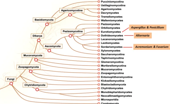
Due to the ability of filamentous fungi to grow on almost any substrate and under harsh conditions, they are ubiquitous in various environments (More et al. 2010) with Aspergillus,
Fusarium, Penicillium, Alternaria, and Acremonium genera being the most widely distributed
and reported genera belong to them (Figure 2) (Pitt and Hocking 2009).
1.1.2 The genus Fusarium
The genus Fusarium is classified as belonging to the Class Sordariomycetes of Phylum Ascomycota (Figure 2) (van Diepeningen et al. 2014). Up to now, around 1,000 species have been identified (Nelson, Toussoun, and Marasas 1983; Moretti 2009). Morphologies inter- species is highly variable and can be strongly influenced by environmental factors. Usually,
Fusarium species are fast-growing at 25°C in culture medium (Guarro 2013). They grow with
a velvety or cottony, flat surface whose color may be white, yellow, pink, purple, salmon or grey , with tan, red, violet or brown on the reverse (Nucci and Anaissie 2007).
Several Fusarium species undergo both asexual and sexual reproductions, even though only fewer than 20 % fusaria have a known sexual cycle (Ma et al. 2013). Typically, Fusarium
A
B
Figure 1. Fungal mycelium. (A) Representation of mycelium formation and distribution; (B)
Rhizopus nigricans growing on bread left in a moist plastic bag for seven days. Tangled
mycelium is visible (white filaments) as well as sporangia bearing spores (brownish/black spheres) (“About Microbiology – Fungi”)
Figure 2. Phylogenetic tree of Fungi kingdom. Among them, Aspergillus and Penicillium belong to class
Eurotiomycetes, Alternaria belongs to class Dothideomycetes, Fusarium and Acremonium belong to class Sordariomycetes. Those are the most widely distributed and reported genera of filamentous fungi (Grigoriev et
al. 2011; 2014).
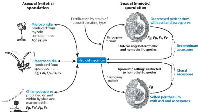
produces three types of spores during the asexual stage, including macroconidia, microconidia and chlamydospores as represented in Figure 3 (Tupaki-Sreepurna and Kindo 2018). Macroconidia can be produced on monophialides and polyphialides in the aerial mycelium, but also on short monophialides in specialized structures called sporodochia.They are hyaline, formed holoblastically and singly, banana / canoe-shaped, multicellular / multiseptate with a foot cell at the base (Nelson, Dignani, and Anaissie 1994). Microconidia are produced in the aerial mycelium in clumps or chains on both monophialides and polyphialides. Under unfavorable conditions, such as dry or hot seasons, the thick-walled large resting spores called chlamydospores are produced to ensure the survival of fungi (Moretti 2009). During sexual reproduction, ascospores are produced in groups of eight in an ascus contained within a flask-shaped structure known as perithecia. Warm, moist condition promotes the maturation of perithecia and the liberation of ascospores from them. Homothallic species are capable of self-fertilization, while heterothallic species can only produce ascospores through outcross (Ma et al. 2013; Prussin II et al. 2014). Spore types and morphology may vary between species, which contributes to distinguish species of Fusarium (Leslie and Summerell 2006).
Figure 3. Generalized life cycle of Fusarium. During sexual reproduction, ascospores are produced in ascus
contained within the flask-shaped perithecium through outcross (heterothallic species) or self-fertilization (homothallic species). Abbreviations: Fg, F. graminearum; Fol, F. oxysporum f. sp. lycopersici; Fp, F.
pseudograminearum; Fs, F. ‘solani’ f. sp. pisi; Fv, F. verticillioides (Ma et al. 2013).
1.1.3 F. graminearum-specific features
Even though many Fusarium species appear to be harmless, several of them are among the known most important plant-pathogenic fungi, causing diseases in a large number of crops, such as wheat, rice, bean, soybean, and can also be harmful to humans and animals (Nucci and Anaissie 2007; Moretti 2009). One example is the plant pathogen F. graminearum
(Figure 4). It is the primary causal agent of Fusarium Head Blight (FHB) in barley and wheat,
which is considered as one of the most devastating plant disease in the world (Windels 2000; Gilbert and Haber 2013). F. graminearum is a haploid ascomycete contains four chromosomes. Its whole genome (strain PH-1) has been sequenced with a total size of 48.4 Mb and a set of 14164 predicted genes (King et al. 2015). It is able to undergo asexual reproduction by producing macroconidia and chlamydospores or sexual reproduction by producing ascospores. Macroconidia are slender, thick-walled, 5 to 6 septate and sickle-shaped to almost straight, while ascospores are fusoid-sickle-shaped with 1-3 septate. F.
graminearum is homothallic and thus during the teleomorph stage, perithecia (Figure 4B)
can be formed without the need of out-cross, which makes it differs from other filamentous fungi (Leslie and Summerell 2006).
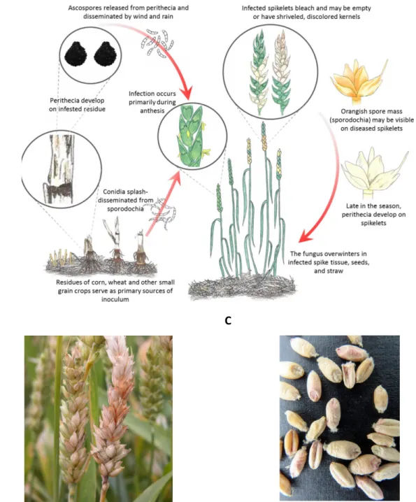
Infected plant debris on which the fungus overwinters as saprophytic mycelia is thought to be the primary inoculum for infection (Goswami and Kistler 2004). The development and maturation of F. graminearum conidia and perithecia occurs concurrently with the anthesis stage of cereal crops during spring, when the warm and moist weather is favorable for them (Markell and Francl 2003). Deposition of the sticky spores (primarily ascospores) by wind, rain or insects on or inside flowering spikes of the host plants initiates infection (Bushnell et
al. 2003). Instead of direct penetration through the epidermis, fungal hyphae develop on the
exterior surfaces of florets and glumes, allowing them to grow toward stomata and other susceptible sites within the inflorescence including anthers and stigmas. Through the vascular tissue in the rachis and rachilla, the fungus spreads from spikelet to spikelet (Figure
5A) (Bushnell et al. 2003; Yang et al. 2013).
Typical symptoms of disease are brown, dark purple to black necrotic lesions formed on the exterior surface of wheat florets and glumes as well as the bleaching of inflorescence tissue
(Figure 5B and 5C). In barley, symptoms are not as visible as in wheat. Infected spikelets may
show a browning or water-soaked appearance, while the kernels show a tan to dark brown discoloration. In both wheat and barley, pink to salmon-orange spores of the fungus could be
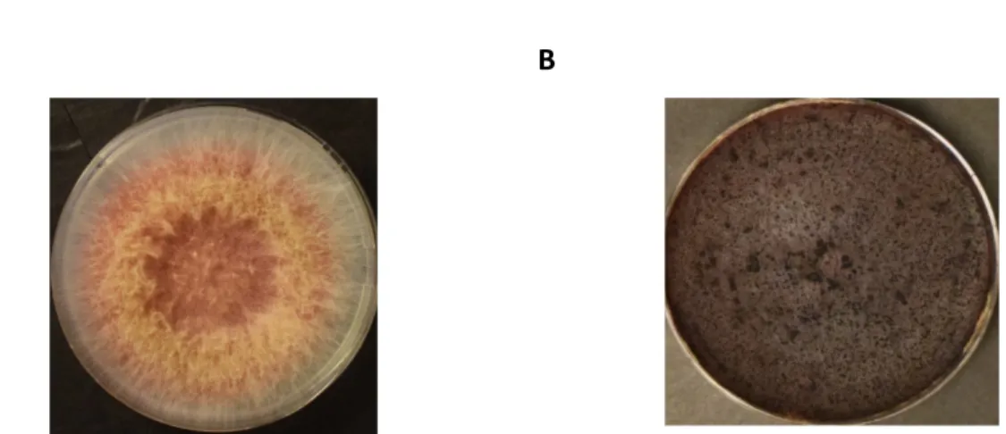
A B
Figure 4. Morphological features of F. graminearum (strain INRA812, PH-1). (A) 100 spores were incubated on
A
B C
Figure 5. FHB disease cycle and symptoms. (A) Disease cycle of FHB; (B) Wheat head infection with bleached
spikelets (“Fusarium Head Blight (Scab) of Wheat”) (C) Infected wheat kernels showing pink and white discolorations (“LSU Small Grains Data”).
observed on infected spikelets, glumes and kernels during prolonged wet periods (Goswami and Kistler 2004). In addition to the losses of yield and quality, during the infection stages, F.
graminearum possesses great potential to produce significant levels of mycotoxins such as
trichothecenes (TCT) and zearalenones (ZEA) (See details in Section 1.2), which are toxic to human and other animals (McMullen, Jones, and Gallenberg 1997).
Deoxynivalenol (DON), a type B trichothecene (TCTB) produced by F. graminearum is essential virulence factor associated with disease progression. In wheat, it not only leads to the inhibition of protein synthesis and mitochondrial function of host cells, but also apoptosis. It seems that DON production is tightly linked with fungal conidia and ascospore formation (Gunupuru, Perochon, and Doohan 2017). In addition, DON could be used by
Fusarium to disturb the plant defense pathway at several critical time points of infection to
assure successful colonization and symptom development (Calvo et al. 2002; Audenaert et al. 2013).
1.2 Potential roles of natural products of filamentous fungi with a focus on
mycotoxins
SMs are low molecular weight molecules that are not essential for organism growth and
development, but they play roles in important functions such as protection, competition, and species interactions. Filamentous fungi are an invaluable source of natural SMs which are difficult to mimic through chemical synthesis (Baral, Akhgari, and Metsä-Ketelä 2018). Among them, particular interest is given to antimicrobials because of their functions in pharmaceutical industries to treat bacterial infections (Aiken et al. 2014). For example, the β-lactam antibiotics such as penicillins and cephalosporins, produced by certain species of
Penicillium and Acremonium, are the most widely applied antibiotics in the world (Hamad
2010). Lovastatin and mevastatin are another compounds with clinical importance used as cholesterol-lowering agents which are primarily derived from Aspergillus terreus and
Penicillium citrinum, respectively (Manzoni and Rollini 2002). Meanwhile, filamentous fungi
have also been exploited for wastewater treatment based on their ability to produce various enzymes (Sankaran et al. 2010).
In addition to these metabolites with potential benefits, filamentous fungi also produce undesirable compounds such as mycotoxins. The term ‘mycotoxin’ originated from the Greek word for fungus ‘mykes’ and the Latin word ‘toxicum’ meaning poison (Turner, Subrahmanyam, and Piletsky 2009). They are defined as highly toxic SMs produced by saprophytic or endophytic molds, which infect a broad range of crops during their growth in the field or during storage (Niu 2010; Gruber-Dorninger, Jenkins, and Schatzmayr 2019). According to Food and Agriculture Organization of United Nations (FAO) surveys, about half of crops in the world may be contaminated with mycotoxins (FAO and Collette 2011).
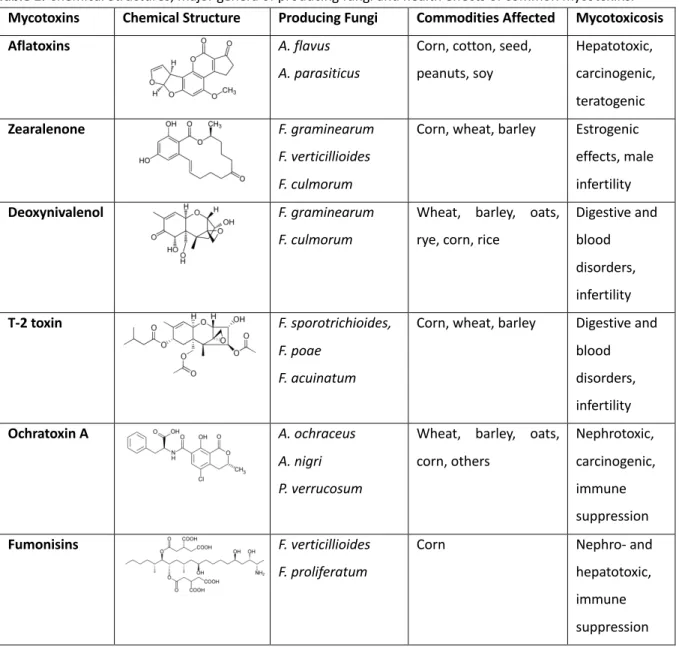
Mycotoxins are of low molecular weight (<700 Da), with highly variable structure. Between 300 and 400 mycotoxins have been isolated and identified (Berthiller et al. 2007), those with most concern are aflatoxins (e.g., Aflatoxin B1, or AFB1), fumonisins, ZEA, TCTB (e.g., DON), type A trichothecene, or TCTA (e.g., T-2 toxin), and ochratoxin (OTA) (Gruber-Dorninger, Jenkins, and Schatzmayr 2019; Alberti, Foster, and Bailey 2017; Hussein and Brasel 2001). Most of these toxins are released by several species of fungi and each strain can produce more than one mycotoxin as listed in Table 1 (Alshannaq and Yu 2017; Weidenbörner 2007). Furthermore, each plant may be infected by different species at the same time, thus leading
to the interaction between mycotoxins and the possible occurrence of synergistic effects (Kubena et al. 1997; Grenier and Oswald 2011). Once ingested by humans and other animals, mycotoxins can cause diseases or toxic syndromes that may lead to death (Bennett and Klich 2003b). The main toxic effects include carcinogenicity, genotoxicity, nephrotoxicity, hepatotoxicity, estrogenicity, reproductive and digestive disorders, immunosuppression and dermal effects (Table 1) (Bryden 2012; Grenier and Oswald 2011)
Table 1. Chemical Structures, major genera of producing fungi and health effects of common mycotoxins.
Mycotoxins Chemical Structure Producing Fungi Commodities Affected Mycotoxicosis
Aflatoxins A. flavus
A. parasiticus
Corn, cotton, seed, peanuts, soy Hepatotoxic, carcinogenic, teratogenic Zearalenone F. graminearum F. verticillioides F. culmorum
Corn, wheat, barley Estrogenic effects, male infertility
Deoxynivalenol F. graminearum
F. culmorum
Wheat, barley, oats, rye, corn, rice
Digestive and blood disorders, infertility T-2 toxin F. sporotrichioides, F. poae F. acuinatum
Corn, wheat, barley Digestive and blood disorders, infertility Ochratoxin A A. ochraceus A. nigri P. verrucosum
Wheat, barley, oats, corn, others Nephrotoxic, carcinogenic, immune suppression Fumonisins F. verticillioides F. proliferatum
Corn Nephro- and
hepatotoxic, immune suppression
As mycotoxins are invisible, tasteless and chemically stable, they are difficult to avoid. To minimize their harmful effects on both humans and animals, strict regulations have been established worldwide to control the maximum amount of ingestion (Niu 2010). For example,
in China, in formula feeds for piglets and young poultry, concentration of fumonisin B1 (FB1) should not exceed 10 ug/kg, while the concentration of DON should be lower than 1mg/kg (“Regulations” 2019) Cereals such as corn, wheat, barley are main sources for food by-product and feed by-production. Therefore, in addition to direct health risks, contamination of cereals with mycotoxins also cause economic losses at all levels including crop and animal production, processing and distribution (Wu 2007; Robens and Cardwell 2003). Thus, it is of great importance to control the production of mycotoxins by filamentous fungi.
1.2.1 Fumonisins
Fumonisins, a group of mycotoxins that were first discovered in 1988 (Bennett and Klich 2003b) and are mainly produced by Fusarium verticillioides and Fusarium proliferatum. They occur worldwide primarily in maize, causing the so-called Gibberella Ear Rot (GER), especially when cultivated in warm regions (Glenn 2007; Voss, Smith, and Haschek 2007). For example, in 2014, fumonisins were detected in more than 98% of corn product samples collected from Shandong Province in China (F. Li et al. 2015). They can also contaminate sorghum, wheat, barley, soybean, figs, black tea, and medicinal plants (Alshannaq and Yu 2017).
As illustrated in table 1, fumonisins are structurally different from most other mycotoxins and totally soluble in organic solvent (Blackwell et al. 1996). Among them, FB1 is the most commonly found and toxic. Fumonisins affect animals in different ways by disrupting sphingolipid metabolism, which are important components of cellular membrane and neural tubes (Dutton 1996; Marasas 1995; E. Wang et al. 1991). In humans, they may be cancer promoters (Eric W. Sydenham et al. 1991). In China, northeast Italy and South Africa, studies showed that the occurrence of FB1 is correlated with the occurrence of a higher incidence of esophageal cancer (Peraica et al. 1999; H. Wang et al. 2000; Persson et al. 2012). Meanwhile, they lead to leukoencephalomalacia in equines (Marasas et al. 1988) and rabbits (Bucci, Hansen, and LaBorde 1996), and cause hepatotoxic and carcinogenic effects as well as apoptosis in the liver of rats (Gelderblom et al. 1996; Pozzi et al. 2001).
1.2.2 Zearalenone
ZEA and derivatives are macrocyclic β-resorcyclic acid lactones which are biosynthesized through polyketide pathway by F. graminearum, F. culmorum mostly, as well as other
they are able to infect wheat, barley, rice, maize, and other crops during both pre- and post-harvested stages, resulting in the contamination of human foods and animal feed worldwide (Zinedine et al. 2007).
Once ingested by human, swine or other animals, ZEA is absorbed rapidly by the gastrointestinal tract and bio-transformed into α- and β-zearalenol, which are its major biologically active reductive metabolites. The binding affinity of both ZEA and its metabolites to estrogen receptors makes them become highly estrogenic, especially α-zearalenol (Bertrand Grenier and Applegate 2013; Zinedine et al. 2007; Binder and Krska 2012). It is reported that their hormonal action is higher than that of most other naturally occurring non-steroidal estrogens, such as soy and clover isoflavones (Bennett and Klich 2003). In pigs, hyperestrogenic syndrome appears with an dietary concentration of ZEA as low as 1 ppm; higher dosage can lead to disrupted conception, abortion and other reproductive problems (Kurtz and Mirocha 1978).
1.2.3 Trichothecenes
TCT constitute a family of more than 200 sesquiterpenoid metabolites. Each TCT contain a common 12,13-epoxytrichothene skeleton and a double bond at the 9, 10 carbon positions with various side chain substitutions of oxygen containing functional groups at possible sites on carbons 3, 4, 7, 8, and 15 (Protection Against Trichothecene Mycotoxins 1983). According to the types of these functional groups, TCT family can be divided into four groups: type A, B, C, and D. TCTA have a hydrogen or ester at the C-8 position such as T-2 toxin, while TCTB contains a ketone at the same position. Type C trichothecenes have an extra carbon 7, carbon 8 epoxide group such as crotocin. Concerning the type D trichothecenes, additional rings with diverse functional groups between carbon 4 and carbon 15 appear, for example, the roridin A and satratoxin H (McCormick et al. 2011).
Among the four types of TCTs, TCTA and TCTB are the most common and well-studied (Bennett and Klich 2003a). Exposure to these toxins induce feed refusal, immunological problems, vomiting, skin dermatitis, and hemorrhagic lesions (Ueno 1985; Pestka 2008). They are also phytotoxic and act as virulence factors in wheat head scab (Proctor, Hohn, and McCormick 1995).
1.2.4 The trichothecene biosynthetic pathway
My project focuses on TCTB. They are mainly produced by F. graminearum and F. culmorum. TCTB can be classified into two chemotypes: chemotype I, including DON and its acetylated derivatives 3-acetylDeoxynivalenol (3-ADON) and 15-acetylDeoxynivalenol (15-ADON); chemotype II, including nivalenol (NIV) and its acetylated derivative 4-acetylNivalenol (4-ANIV) (syn. fusarenone-X) (E. W. Sydenham et al. 1991).
Most genes involved in the TCT biosynthetic pathway have been identified. They are located at three different loci on different chromosomes (Figure 6): a 35-kb long trichothecene biosynthetic gene cluster (Tri cluster) containing 12 genes that control the biosynthesis on chromosome 2 (Brown et al. 2004); a two-gene Tri1-Tri16 cluster on chromosome 1; and a single gene Tri101 locus that on chromosome 4 (Kimura, Matsumoto, et al. 1998; W Peplow
et al. 2003). Besides, a Tri15 gene is also located outside the core cluster on chromosome 3
and involves in the negative control of trichothecene biosynthesis (Alexander et al. 2004).
Figure 6. Gene clusters involved in trichothecene biosynthetic pathway in Fusarium, including a Tri cluster
containing 12 genes, a two-gene Tri1-Tri16 cluster and a single gene Tri101.
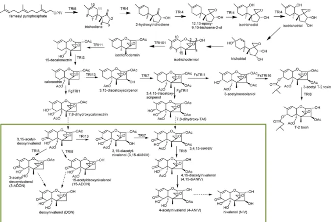
The pathway begins with the cyclization of farnesyl pyrophosphate, a primary common metabolic intermediate of different secondary metabolism, to form trichodiene (Figure 7). The reaction is mediated by the terpene cyclase trichodiene synthase which is encoded by
Tri5 (T. M. Hohn and Vanmiddlesworth 1986; T. M. Hohn and Beremand 1989). Trichodiene
undergoes six steps of oxygenations catalyzed by a cytochrome P450 monooxygenase encoded by Tri4 (Thomas M. Hohn, Desjardins, and McCormick 1995) to add four oxygens at C-2, C-3, C-11, and the C-12, C-13-epoxide to synthesize the intermediate isotrichotriol (McCormick, Alexander, and Proctor 2006b). Isotrichotriol is converted to isotrichodermol by a non-enzymatic isomerization and cyclization (McCormick et al. 1990). This step is followed
by a reaction to form isotrichodermin with the help of acetyltransferase encoded by Tri101, which is involved in the self-production of the trichothecenes-producing organisms (Kimura, Kaneko, et al. 1998). Under the control of Tri11, a hydroxyl group is then added to C-15 and subsequently acetylated to form calonectrin, a central metabolite (Alexander, Hohn, and McCormick 1998; McCormick, Hohn, and Desjardins 1996). These 10 steps are common for the production of TCTB and T-2 toxin.
Afterwards, the pathway divides into three directions. In DON producers (e.g., F.
graminearum), calonectrin is converted to either 3-ADON, 15-ADON or DON by the
catalyzation of products encoded by Tri1 and Tri8. Owing to polymorphisms in the sequences of the esterase-encoding gene Tri8, formation of 3-ADON or 15-ADON occurs in a strain specific manner in F. graminearum (Alexander et al. 2011). In NIV and T-2 toxin producers, a hydroxyl group is added at C-4 of calonectrin (controlled by Tri13) and subsequently acetylated (under the control of Tri7) to produce 3, 4, 15-triacetoxyscirpenol (Lee et al. 2002). In F. graminearum, this intermediate is converted to NIV or 4-ANIV by the metabolization of enzymes encoded by Tri1 and Tri8. In Fusarium sporotrichioides, another hydroxyl group is added to C-8 controlled by Tri1, followed by the addition of an isovaleryl moiety controlled by Tri16. Finally, the acetyl group is removed from the C-3 position by an esterase controlled by Tri8 to generate T-2 toxin (McCormick, Alexander, and Proctor 2006a). In addition to these genes involved directly in the pathway, there are several specific regulators. Deletion mutants of the positive transcription factors Tri6 and Tri10 in F.
graminearum were created in order to understand genome-wide regulatory control of toxin
synthesis (Seong et al. 2009). Results showed that transcript levels for over 200 genes, including those related to housekeeping functions, secondary metabolism, and pathogenesis, were altered in the Tri6 deleted mutant. Therefore, Tri6 is a global regulator rather than specific to the TCT pathway. They also found that Tri6 negatively regulated the expression of
Figure 7. Proposed biosynthetic pathway of trichothecene in Fusarium. Genes encoding enzymatic step are
Figure 8. Overview of metabolic pathways indicating the relationship between the TCTB biosynthesis and other
Notably, a predicted transcription factor Tri14, which is also located in the “Tri cluster”, is required for high virulence and DON production on wheat but not for DON synthesis in vitro in Fusarium species (Brown et al. 2002; Dyer et al. 2005). Finally, Tri15, a gene that represents a fourth TCT locus, is predicted to encode a Cys (2) - His (2) zinc finger protein. It is reported in F. sporotrichioides to act as a negative regulator of at least some of the TCT biosynthesis. However, in F. graminearum, Tri15 is found to be unlinked to the main TCT biosynthetic gene cluster (Alexander et al. 2004).
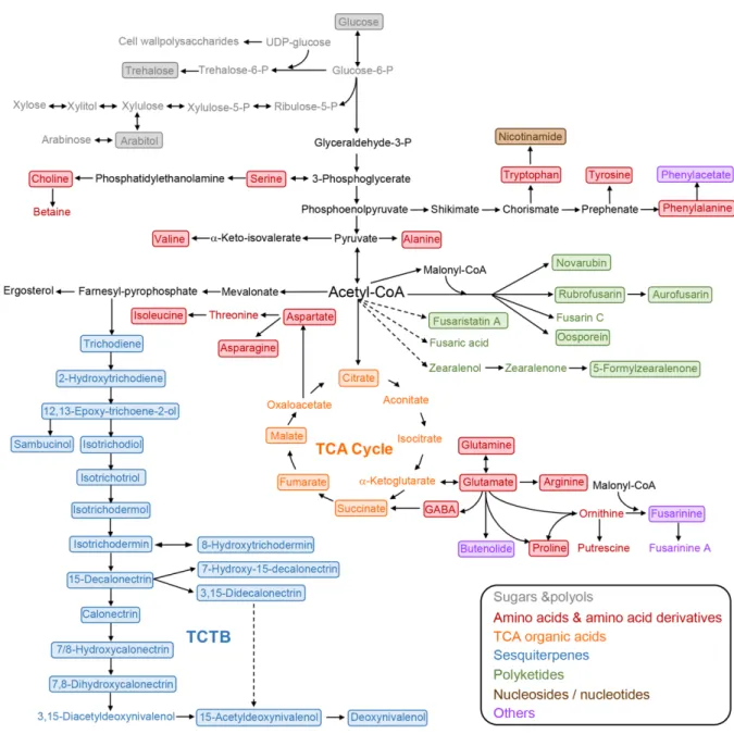
Recently, analyses combined with 1H nuclear magnetic resonance and liquid
chromatography-quadrupole time of flight-mass spectrometry were performed to elucidate the association of TCTB biosynthesis with other central and specialized processes in F.
graminearum (Atanasova-Penichon et al. 2018). Results demonstrated that the inhibition of
TCTB production induced by caffeic acid exposure was associated with significant changes in the secondary and primary metabolism of F. graminearum including the accumulation of several polyketides, alteration in tricarboxylic acid cycle, and modifications in the metabolism of several amino acids and sugars, even though the fungal growth was not affected. The authors found that, as shown in Figure 8, the TCTB biosynthetic pathway TCTB is closely linked with other primary or secondary metabolic pathways, suggesting that TCTB plays a multifaceted role in the life cycle of F. graminearum (Atanasova-Penichon et al. 2018).
1.3 Regulations of SMs biosynthesis by environmental factors
1.3.1 Carbon and nitrogen source
In many fungi, signals or substrates from the environment play an important role in controlling the production of mycotoxins, especially carbon resources. When a more readily available carbon source is present in the medium, certain enzymes required for less-favored carbon source will be switched off to help microorganisms to precisely adapt their physiology to the environment. This mechanism is called carbon catabolite repression, or CCR. It has been studied extensively in Aspergillus species, and is mediated by the transcription factor CreAp, a C2H2 DNA-binding protein (Katoh et al. 2007; Ries et al. 2016). In A. nidulans,
expression of isopenicillin-N-synthetase gene is repressed in glucose-containing medium and penicillin biosynthesis is inhibited (Espeso and Peñalva 1992). In order to see the effects of different types of carbon resources on TCT production in F. graminearum, nine strains of F.
graminearum were cultivated in media containing 12 various carbon sources (Jiao,
Kawakami, and Nakajima 2008). Significantly higher levels of TCT (DON and 3ADON) production were commonly observed in the presence of sucrose, 1-kestose and nystose for all of the strains tested. Meanwhile, Tri4 and Tri5 expressions were upregulated in sucrose-containing medium, but not with glucose. To further investigate whether trichothecene biosynthesis was subject to CCR, glucose was added into the sucrose-containing medium. However, TCT accumulation was not repressed, indicating that there may be other factors involved in the regulation of trichothecene production rather than CCR (Jiao, Kawakami, and Nakajima 2008).
Nitrogen is an essential requirement for fungal growth, and fungi are able to use a wide variety of compounds as nitrogen sources. Utilization of different nitrogen source is selective. Generally, favored nitrogen sources, such as glutamine and ammonium, are used preferentially (Wong, Hynes, and Davis 2008). The global regulation of nitrogen source utilization has been widely investigated in A. nidulans, a process known as nitrogen metabolite repression (NMR). Up to now, major transcription factors that have been identified underlying this regulation are GATA factors. These factors share a common DNA binding motif (Cys-X2-Cys-X17-Cys-X2-Cys) that recognizes a core 5’-GATA-3’ sequence (Scazzocchio 2000). In A. nidulans, AreA protein is a positively acting GATA factor. The AreAp deleted mutants lose the ability to use any nitrogen source other than glutamine and ammonium (Arst and Cove 1973; Marzluf 1997). The AreB protein contains an N-terminal
GATA zinc finger and a C-terminal leucine zipper domain, and is generally regarded as the negative counterpart to AreAp even though the function of AreBp seems to be more complex (Conlon et al. 2001; Dzikowska et al. 2003). Orthologues of AreAp or AreBp have been identified in F. graminearum, F. fujikuroi, A. flavus, and Penicillium chrysogenum. They were shown to be involved in various secondary metabolism pathways including DON, bikaverin, aflatoxin and penicillium biosynthesis (Haas and Marzluf 1995; Ehrlich and Cotty 2002; Mihlan et al. 2003). It was further reported that AreAp regulates the production of mycotoxins directly or indirectly, independently from nitrogen status, and plays a role in the utilization of certain amino acids in F. graminearum (Giese, Sondergaard, and Sørensen 2013). Besides, a study identified particular amine compounds (e.g., ornithine, agmatine, and putrescine) that could support, in defined media, levels of DON production and Tri5 expression equivalent to those observed during infection of F. graminearum. All these compounds belong to the arginine-polyamine biosynthetic pathway in plants, which means that metabolites produced in this pathway may promote TCT production (Gardiner, Kazan, and Manners 2009a). These authors also performed a global analysis of fungal gene expression using the Affymetrix Fusarium GeneChip during culture under DON-inducing conditions (with agmatine) compared with non-inducing conditions. Results indicated that agmatine differentially regulates a large number of fungal genes, including both known and previously uncharacterized putative SM biosynthetic gene clusters. Further study found three of the differentially regulated genes were under the control of Tri6 transcriptional regulator (Gardiner, Kazan, and Manners 2009b). All in all, even though nitrogen source selection and DON production in F. graminearum are linked, further studies should be carried out to investigate and address the underlying mechanisms.
1.3.2 pH
pH is a critical host-related environmental signal that can limit or enhance growth, reproduction, toxin production, and virulence of phytopathogenic fungi (Caracuel et al. 2003; Sánchez-Rangel et al. 2018). Studies showed that filamentous fungi have the ability to sense pH and respond with regulatory system (Peñalva and Arst 2002). For example, A. nidulans adapts to pH changes by adjusting the secretion of enzymes with optimal activity accordingly, which is mediated by a mechanism that tailors genes expression to ambient pH (Caracuel et al. 2003). It involves a highly conserved zinc finger transcription factor PacCp
that is activated by at least six components encoded by palA, palB, palC, palF, palH, palI (Nahas, F. Terenzi, and Rossi 1982; Tilburn et al. 1995). Once active, PacCp is translocated to the nucleus where it can recognize the consensus DNA site 5’-GCCAAG-3’ and consequently up-regulate the transcription of alkaline-pH responsive genes, and down-regulate acid responsive genes (Tilburn et al. 1995; Espeso and Peñalva 1996).
Homologues of A.nidulans PacCp have been identified in various filamentous ascomycetes, which were found involved in the production of penicillin, cephalosporin C and fumonisin in
P. chrysogenum (Suárez and Peñalva 1996), Acremonium chrysogenum (Schmitt, Kempken,
and Kück 2001) and F. verticillioides (Flaherty et al. 2003), respectively. In F. graminearum and F. culmorum, neither the toxin nor Tri transcripts were detected when the pH was maintained neutral (Merhej et al. 2010). However, shifting from neutral to acidic pH by mycelium transfer induced Tri genes expression and toxin accumulation, especially Tri5 and
Tri101. Meanwhile, the induction of toxin production seems not to depend on strains or
chemotype (Merhej et al. 2010). By constructing Pac1p (ortholog of PacCp) deleted mutant of F. graminearum, further study revealed that Pac1p negatively regulates Tri gene expression and toxin production, as the mutant exhibited an over-expression of Tri genes and an increase in the toxin production under acidic pH but showed a reduced development under neutral and alkaline pH. In fact, in F. graminearum, promoters of various Tri genes including Tri6 and Tri10 contain the sequence “GCCARG”, which corresponds to the consensus binding sequence for PacCp in A. nidulans. Therefore, the repression observed here could be exerted either directly by binding to the promoters of various Tri genes or through the regulation of a common regulator of Tri genes such as Tri6 which is quickly and strongly repressed upon pH change (Jawad Merhej, Richard-Forget, and Barreau 2011). Finally, the combination of low pH and supplementation with amines results in significantly enhanced expression of the Tri5 gene and increased DON production during axenic growth of
F. graminearum. It was hypothesized that nitrogen source may, at least partially, act through
triggering an acidification of the extracellular environment (Gardiner et al. 2009).
1.3.3 H2O2
During plant-pathogen interactions, various ways have been developed by the hosts to prevent infections. The earliest and most widely studied defense event is that plants will release reactive oxygen species (ROS) such as H2O2 as an early response to pathogens. In
response to such an oxidative burst, pathogens could induce mechanisms, broadly called oxidative stress response (OSR), to scavenge elevated ROS levels (Shetty et al. 2008; Heller and Tudzynski 2011). It was found that, in F. graminearum, daily exogenous supplementation of H2O2 enhanced DON accumulation (Ponts et al. 2006). On the other hand, adding catalase
to the culture medium leads to the degradation of H2O2, and meanwhile, a strong decrease
in the accumulation of DON and 15-ADON. The induction or suppression of DON production by adding H2O2 or catalase were accompanied by a up or down regulation of Tri genes
expression, respectively, in particularly Tri4, Tri5, and Tri12 (Ponts et al. 2007). In addition, induction of trichothecene by H2O2 could be chemotype-dependent. Indeed, accumulation of
toxins were less affected by adding H2O2 in NIV-producing strains compared to
DON-producting strains, which may relate to a H2O2-metabolizong capacity in NIV isolates (Ponts
et al. 2009). However, an opposite result was observed for the oxidative stress caused by diamide: the global level of TCTB production was drastically increased by the treatment with diamide regardless of the chemotype, suggesting the intervention of different response mechanisms (Ponts et al. 2009).
Yap1p is a well-known transcription factor activated by oxidative stress in the yeast
Saccharomyces cerevisiae (Moye-Rowley 2003; Rodrigues-Pousada, Menezes, and Pimentel
2010). Briefly, upon exposure to oxidative stress, Yap1p could activate the transcription of target detoxification genes, such as cta1 and ctt1, coding respectively for peroxisomal and cytosolic catalase (Yan, Lee, and Davis 1998; Jamieson 1998). In the Fgap1p (ortholog of yeast Yap1p) deleted mutant of F. graminearum, activation of OSR gene expression and toxin accumulation in response to oxidative stress was no longer observed (Montibus et al. 2013). This result indicated that the TCTB pathway is linked to the metabolism of H2O2 in F.
graminearum with Fgap1p playing a central role. Conversely, antioxidant compounds were
shown to inhibit toxin production in F. graminearum.
1.3.4 Light
Fungi use light as a source of information rather than a source of energy like plants do. They respond to illumination in various ways and develop considerable adaptations in their metabolic pathways upon growth in light or after perception of a light pulse (Tisch and Schmoll 2010). Most of current knowledge derives from the model A. nidulans. Formation of sexual fruiting bodies and production of certain SMs occur preferentially in darkness and are
coordinately inhibited by light as an external signal. In contrast, formation of asexual spores is promoted by light (Bayram et al. 2008; Sarikaya Bayram et al. 2010). This regulation is known to be mediated by the VeA-VelB-LaeA complex. VeAp is encoded by velvet identified in 1965 (Käfer 1965). VelBp interacts with the N-terminus of VeAp. They can enter the nucleus in darkness and act as activation of sexual development and inhibition of asexual development (Calvo 2008). Illumination reduces the cellular amounts of VeAp and VelBp. In nucleus, they assemble with the non-velvet protein LaeA, a master epigenetic regulator of secondary metabolism. Therefore, the VeA-VelB-LaeA complex regulates not only light-responding development but also SM production including sterigmatocystin, penicillin and many other compounds (Tisch and Schmoll 2010). Besides, VeAp could interact with the phytochrome-like red light receptor FphAp, therefore forming a complex which also comprises LreAp and LreBp, the A. nidulans orthologs of the N. crassa photoreceptors WC-1 (for white collar) and WC-2 (Purschwitz et al. 2008; Purschwitz, Müller, and Fischer 2009). The velvet and LaeA proteins are conserved in various genera of fungi. Homologues of VeA act as positive regulators of genes involved in biosynthesis of cephalosporin C in A.
chrysogenum (Dreyer et al. 2007), or production of the deleterious mycotoxins fumonisin,
fusarin C, TCT or DON, beauvericin in the plant pathogens F. verticillioides, F. fujikuroi, F.
graminearum and F. oxysporum, respectively (Myung et al. 2009; Wiemann et al. 2010; Jiang et al. 2011; Jawad Merhej et al. 2011; López-Berges et al. 2013).
1.4 Dynamics of chromatin structure and SMs production
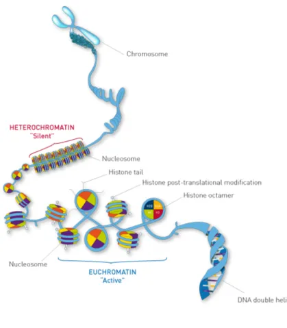
The complex between DNA and proteins in eukaryotes called chromatin, whose basic unit is nucleosome. A nucleosome core particle (NCP) consists of two copies of each of the core histones H2A, H2B, H3, and H4, to form the histone octamer around which approximately 146 bp of DNA wrap in 1.67 left-handed super helical turns (Hanson et al. 2003; Bargaje et al. 2012). The steps of nucleosome assembly are as following: first, a tetrasome consisting of an (H3·H4)2 tetramer is formed on the DNA followed by the addition of two H2A-H2B dimers.
(Krude 1995; Mazurkiewicz, Kepert, and Rippe 2006). Finally, NCPs are held together by stretches of linker DNA to form a 10-nm chromatin fiber, in association with linker histones such as H1 and its isoforms (Alberts et al. 2008). The chromatin can then be further condensed by coiling into 30-nm fibers (Figure 9).
Figure 9. Typical structure of chromatin. Each nucleosome consists of eight histones (purple: H2B; orange: H2A;
green: H4; red: H3) with DNA wrapped around, which helps its packaging into a compact form that fits inside the cell nucleus. Changes in chromatin structure are associated with DNA replication and transcription (“Chromatin Immunoprecipitation Sequencing (ChIP-Seq) Analyisis & Services | Diagenode”).
According to the level of condensation, chromatin can be divided into two types: euchromatin and heterochromatin. During the interphase of cell life cycle, most chromatin is
relatively decondensed – it is called euchromatin – especially in genomic regions containing genes actively transcribed (Cooper 2000; International Human Genome Sequencing Consortium 2004). In contrast, there is about 10% of interphase chromatin that is in a super condensed state called heterochromatin, which is transcriptionally inactive and typically contains highly repeated DNA sequences, such as those present at centromeres and telomeres. Heterochromatin is enriched during mitosis, to facility their distribution to daughter cells. Such condensed chromatin can no longer be used as a template for RNA synthesis, thus transcription stops during mitosis (Volpe et al. 2002; Ou et al. 2017).
Chromatin is a highly dynamic architecture intimately linked to the control of gene expression in eukaryotes. This property mainly results from several parameters: (1) ATP-dependent chromatin remodeling complexes that allow reposition (slide, twist or loop) of nucleosomes, eviction of histones away from DNA or exchange of histone variants (Lusser and Kadonaga 2003a; Wang, Allis, and Chi 2007); (2) Histone PTMs which leads to structure and function alteration (Fan et al. 2015); and (3) DNA methylation which affects gene expression (Tost 2010).
1.4.1 Chromatin remodeling
Ø Protein complexes that remodel chromatin
The process ‘chromatin remodeling’ is mediated by several chromatin remodeling complexes that utilize energy derived from adenosine triphosphate (ATP) hydrolysis to alter nucleosome structure or conformation and, thereby, regulate the access of transcription factors to their cognate DNA binding sites (Lusser and Kadonaga 2003b; Hota and Bruneau 2016). All chromatin remodeling complexes are made up of a single highly conserved ATPase, which belongs to SNF2 family, and multiple associated subunits. Generally, ATPase subunit binds and hydrolyzes ATP, while the associated subunits modulate the catalytic activity of the ATPase subunit and provide specificity to genome binding (Eisen, Sweder, and Hanawalt 1995). Based on the sequence similarity between their ATPase domains and the presence of other subunits, chromatin remodeling complexes can be divided into four major subfamilies including Switch/Sucrose Non-fermentable (SWI/SNF), Imitation SWI (ISWI), Chromodomain Helicase Deoxyribonucleic acid-binding (CHD), and Swr1-related Inositol requiring 80 (INO80) (Becker and Hörz 2002; Bao and Shen 2007). As showed in Figure 10, for each subfamily, the ATPase domain comprises an N-terminal DEXDc and a C-terminal HELICc subdomain,
separated by an insert region (Xu, Kanagaratham, and Radzioch 2013). The SWI/SNF family contains an HSA domain for actin binding, and a bromodomain which recognizes and binds to the acetylated histone tails. SWI/SNF regulates transcription, mitotic exit, and activation of weak replication origins. In vitro, it binds hyperacetylated nucleosomes, mobilizes nucleosomes, and acts as a directional DNA translocase. The ISWI family is characterized by SANT and SLIDE domains, which are important for histone binding. It is involved in transcription, DNA replication, and chromatin assembly. The CHD family contains two N-terminal chromodomains that play roles in the remodeling of chromatin structure and transcriptional regulation of genes. The INO80 family, similar with the SWI/SNF family, also has an HSA domain, however the insert region between the DEXDc and HELICc subdomains is three times longer than that of other three families. INO80 family is involved in the regulation of transcription, DNA repair and exchange of histone variant H2A.Z (Lusser and Kadonaga 2003a; Bao and Shen 2007; Hota and Bruneau 2016).
Figure 10. Domain architecture of SNF2 family of proteins. Domain organization of the catalytic subunits of
SWI/SNF, ISWI, CHD and INO80/SWR subfamilies of chromatin remodelers are shown. All of these subunits are SNF2 family proteins. They all contain an ATPase domain, which consists of a DEXDc and HELICc domains, with each subfamily possessing additional domains. For example, in SWI/SNF family, there is an HSA domain (for actin binding) and a bromodomain (recognizes and binds to the acetylated histone tails); in ISWI family, there are SANT and SLIDE domains (important for histone binding); in N-terminal of CHD family, there are two chromodomains (chromatin remodeling and gene transcriptional regulation); the SWI/SNF family, also has an HSA domain.
Ø Histone Variants
In most eukaryotes, canonical histones including H2A, H2B, H3, H4 are encoded by multiple genes. They are only expressed during the S-phase of cell cycle after DNA replication (Talbert and Henikoff 2014). In metazoans, histone mRNAs are generally intronless and are not
polyadenylated. Instead, they have special stem-loop structures to trigger translation (Marzluff and Koreski 2017). However, introns exist in histone mRNAs of Basidiomycetes and filamentous Ascomycetes including F. graminearum (Nishida and Yun 2011). The individual paralogous (non-allelic) genes of a histone family may also encode related but distinct protein isoforms, commonly referred to as “histone variants” (Talbert et al. 2012). Most of them are encoded by a single gene containing one or more introns expressed throughout the cell cycle, and can thus be incorporated into nucleosomes during the whole cell cycles (Kaygun and Marzluff 2005; Talbert and Henikoff 2014; Maze et al. 2014). Up to now, histone variants of all canonical histones have been recognized, with the exception of H4 (M Arnaudo, Molden, and A Garcia 2011).
Table 2. List of histones including linker histone, canonical histones and histone variant in F. graminearum
(strain PH-1) and their encoding genes.
Name Gene ID
Linker histone H1 FGRAMPH1_01G20501
Canonical histone H3 FGRAMPH1_01G14931
H4 FGRAMPH1_01G17029
H2B FGRAMPH1_01G26111
H2A FGRAMPH1_01G26109
Histone variant H2A.Z FGRAMPH1_01G03973
H3.3 FGRAMPH1_01G06247
H2A family encompasses the largest number of variants, including H2A.X, H2A.Bdb, H2A.Z and macroH2A. As shown in Table 2, H2A.Z is the only H2A histone variant found in F.
graminearum and it is my focus during my PhD. The details of histone variant H2A.Z will be
discussed in the Review Paper. H2A.X constitutes 2–25% of mammalian histone H2A depending on the organism and cell type (Dickey et al. 2009). It has the most similar sequence with H2A but with a divergent C-terminal tail. H2A.X can undergo both replication dependent and independent transcription as the mRNA of H2A.X can have either a polyA tail or a stem loop structure (Mannironi, Bonner, and Hatch 1989). It can be acetylated, ubiquitinated and phosphorylated (M Arnaudo, Molden, and A Garcia 2011). H2A.Bbd is the least studied histone variant. When compared to canonical H2A, the protein sequence of H2A.Bbd is shorter and arginine enriched. It is reported that nucleosomes containing H2A.Bbd contain 130 bp of DNA instead of the traditional 146 bp, meaning that the
incorporation of H2A.Bbd affect chromatin structure (Doyen et al. 2006). MacroH2A is an unusual histone variant with a bulky C-terminal non-histone domain (about 30kDa) which distinguishes it from all other histones (Sun and Bernstein 2019). Mass spectrometry analysis revealed that macroH2A can be ubiquitinated, phosphorylated and acetylated (M Arnaudo, Molden, and A Garcia 2011).
H3 has three main variants: H3.1 and H3.2 (known as the “canonical” histone H3), H3.3, and CenH3. These variants are surprising similar in sequence. For example, in mammals, H3.1 only differs from H3.2 by a change in Cysteine 96 to Serine and H3.3 differs from H3.1 by only five residues. Although the changes in amino acid sequence are subtle, they have large differences in their expression, localization in chromatin, and modification state. For example, compared to H3.1 and H3.2, H3.3 is usually localized to heterochromatin and enriched for histone modifications that are associated with gene activation (Hake et al. 2005; Schulmeister, Schmid, and Thompson 2007; M Arnaudo, Molden, and A Garcia 2011; Shi, Wen, and Shi 2017).
The variant of histone H2B mainly include H2B.1, H2B.W, subH2B and H2B.Z, among which H2B.Z is an apicomplexan specific variant that is known to interact with H2A.Z (Draizen et al. 2016). In fact, due to sequence similarity of the H2B gene family, variants of H2B are rarely encountered at least in mammals, apicomplexa and sea urchins and have not been studied as deeply as other histone variants. Variants of H1 are common, but similar with H2B, much less is known of their functional specialization (Takami, Takeda, and Nakayama 1995; Hoeijmakers
et al. 2013).
These histone variants are involved in diverse biological processes (summarized in Figure 11) including transcription, chromosome segregation, DNA repair and recombination, chromatin remodeling, ADP-ribosylation, germline-specific DNA packaging and activation, and even extra-nuclear acrosomal function (Maze et al. 2014; Henikoff and Smith 2015).
Figure 11. Canonical histones and their variants (Sarma and Reinberg 2005). The major core histones contain a
conserved histone-fold domain (HFD). In addition, they contain N- and C-terminal tails that harbor sites for various post-translational modifications. For simplicity, only well-established sites for lysine methylation (red flags) and serine phosphorylation (green circles) are shown (other types of modifications, such as ubiquitylation, are not shown). The proposed functions of the variants are listed. Among them, H2A.Z is the only histone variant found in F. graminearum.
1.4.2 Histone post-translational modifications (PTMs)
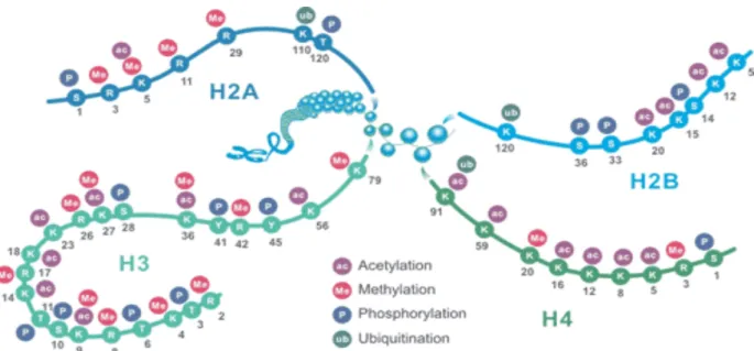
Histone tails are extensive marked by covalent PTMs, including methylation, phosphorylation, acetylation, ubiquitylation and sumoylation. These PTMs impact gene expression by altering chromatin state or recruiting histone modifiers (Fan et al. 2015; Audia and Campbell 2016). Histone H3 are the most modified histone, followed by H4. In most species, histone H3 is primarily acetylated at lysines 9, 14, 18, 23, and 56, methylated at arginine 2 and lysines 4, 9, 27, 36, and 79, and phosphorylated at serine 10 and 28 as well as threonine 3 and 11. Histone H4 is primarily acetylated at lysines 5, 8, 12 and 16, methylated at arginine 3 and lysine 20, and phosphorylated at serine 1 (Figure 12) (“Histone Modifications” 2013). In filamentous fungi, a well-characterized system has been established in the model fungus N. crassa to elucidate epigenetic phenomena. More and more studies focus on the roles of chromatin change in the regulation of SMs have been carried out in
we discuss histone methylation and acetylation in filamentous fungi, with a focus on
Fusarium species.
Figure 12. Schematic representation of common histone modification sites. K: lysine; R: arginine; S: serine; Y:
tyrosine; T: threonine (“Four Common Histone Modifications-CUSABIO”). 1.4.2.1 Histone methylation
Histone methylation is defined as the transfer of one, two, or three methyl groups from S-adenosyl-L-methionine to lysine or arginine residues of histone proteins by histone methyltransferases (HMTs). The methyl group can be removed by histone demethylases (HDACs). In nuclei, the occurrence of histone methylation is related with the activation or silencing of specific genes within the DNA complexed with the modified histone (Greer and Shi 2012). For example, H3K9 methylation is the mark of heterochromatin, which contributes to the transcriptionally inactive state of chromatin. Normally, H3K9me3 is necessary for the recruitment of heterochromatin protein 1 (HP1) by creating a binding site. HP1 is involved in the establishment and maintenance of heterochromatin (Lomberk, Wallrath, and Urrutia 2006).
In both N. crassa and F. graminearum, deletion of HP1 did not show any cytological or morphological defect. However, in N. crassa, there was a strong up regulation of several SM gene clusters (Smith et al. 2011). In F. graminearum, both up and down regulation of SM genes could be detected. For example, expression of genes required for the production of the pigment aurofusarin (AUR) was greatly enhanced while gene expression and metabolites
involved in the DON pathway was inhibited. However, the heterochromatic mark H3K9me3 was both enriched in these two gene clusters in HP1 deleted mutants (Reyes-Dominguez et
al. 2012). Dim5p, or Kmt1p, a lysine histone methytransferase (HMT) enzyme, is responsible
for the methylation of H3K9 in eukaryotes. In Botrytis cinerea, Dim5p is required for the regulation of its development and virulence (Zhang et al. 2016). In F. verticillioides, mutants lacking Dim5p showed significant defects in fungal development and pathogenicity but an increased tolerance to osmotic stress (Gu et al. 2017).
H3K27 methylation is a histone modification known to be linked with gene silencing. In F.
graminearum. Anti-H3K27me3 Chromatin Immunoprecipitation coupled to Deep Sequencing
(ChIP-seq) experiment showed that extensive segments were enriched with H3K27me3, covering a third of the genome. Removal of the mark by mutation of the methyltransferase subunit (Kmt6p) of the Polycomb Repressive Complex 2 (PRC2) resulted in activation of more than 1,500 genes, i.e., ~11% of the genome, predominantly genes involved in the production or detoxification of SMs or predicted to play a role in pathogenicity (Connolly, Smith, and Freitag 2013). In F. fujikuroi, Kmt6p appears to be essential (Studt, Rösler, et al. 2016). Knock down of Kmt6p reduced H3K27me3 levels at usually decorated gene loci and induced four otherwise silent putative SM gene clusters accompanied by the accumulation of novel metabolites.
H3K36me is closely associated with euchromatic region of eukaryotic genomes (Ho et al. 2014). In filamentous fungi, this mark can be deposited at specific loci by two genes, Set2 and Ash1. Similar to S. cerevisiae, F. fujikuroi Set2p may also interact directly with the elongation form of RNA polymerase II via its conserved Set2 Rpb1 interacting (SRI) domain (Janevska et al. 2018). Meanwhile, it is reported that Set2p is most likely responsible for H3K36 methylation at euchromatic regions of the genome, while Ash1p methylates H3K36 at the subtelomeric region of all chromosomes (Janevska et al. 2018). In spite of showing the roles of H3K36me involved in vegetative growth, sporulation, SM biosynthesis and virulence in F. fujikuroi, this study also found that Ash1p deleted mutant exhibit an enhanced level of H3K27me3, an increased instability of subtelomeric regions and losses of the accessory chromosome XII, indicating that Ash1p may be involved in DNA repair process. Simultaneously, another study carried out in N. crassa also demonstrated that Ash1p-marked