Digital Reconstructions of Fossil Morphologies,
Nama Group, Namibia
by
Wesley Andres Watters
Submitted to the Department of Earth, Atmospheric, and Planetary Sciences
in partial fulfillment of the requirements for the degree of
Master of Science in Earth & Planetary Science
at the
MASSACHUSETTS INSTITUTE OF TECHNOLOGY
-January 2000
@
Massachusetts Institut~eof Technology 2000. All rights reserved.
A> IAuthor ...
Department of Earth, tmospheric, and Planetary Sciences
January 18, 2000
C ertified by ...-
.
Pro/ John.
Grotzinger
Professor of Geology
Thesis Supervisor
Accepted by .
... ...Prof. Ronald Prinn
Chairman, Department of EAPS
MASSACHUSETTS INSTITUTEDigital Reconstructions of Fossil Morphologies,
Nama Group, Namibia
by
Wesley Andres Watters
Submitted to the Department of Earth, Atmospheric, and Planetary Sciences on January 18, 2000, in partial fulfillment of the
requirements for the degree of
Master of Science in Earth & Planetary Science
Abstract
Previously undescribed fossils of weakly calcified metazoans were recently discovered in the terminal Proterozoic Nama Group of central and southern Namibia (Grotzinger et al., 1995), in sediments that contain the terminal Proterozoic index fossil Cloudina. The new fossils are closely associated with thrombolites and stromatolites that form laterally continuous biostromes, isolated patch reefs, and isolated pinnacle reefs. Because these fossils are preserved as calcitic void-fill in a calcite matrix, individual specimens cannot be freed by conventional techniques. Rocks containing the fossils are ground and digitally photographed at thickness intervals of 25 pm. A battery of image processing techniques is used to obtain the contour outlines of the fossils in serial cross sections. A Delaunay triangulation method is then used to reconstruct the morphology from tetrahedral components which connect the contours in adjacent layers. It is found that most of the fossils resemble a single morphology with some well-defined characters that vary slightly among individual specimens. This fossil morphology is described in this thesis as Namacalathus hermanastes. A mathematical description of the morphology is used to obtain a database of randomly-oriented synthetic cross sections. This database reproduces the vast majority of cross sections observed in outcrop. In addition, the most common orientation, the mean size, and other population statistics are measured for Namacalathus fossils within an individual rock sample.
Thesis Supervisor: Prof. John P. Grotzinger Title: Professor of Geology
Contents
Acknowledgements 4
Introduction 5
Chapter 1 Calcified Metazoans of the Nama Group 7
Chapter 2 The Serial Imaging Method 33
Chapter 3 A Mathematical Description of the Morphology 55
References 69
Appendix A Population Statistics 75
Appendix B Database of Synthetic Cross Sections 79
Acknowledgements
I am profoundly grateful to John P. Grotzinger for his generosity, for his support and guidance, and
most of all for his friendship. I have learned and I have grown in profound ways because of my interactions with John, because of my work on this project, and because of my visit to Namibia in 1997. I am also greatly indebted to my parents, Allan and Cecilia Farfin Watters, for their encouragement and support. Special thanks go to Rob and Marianne Field and Roy Magson for access to their farms in central Namibia, Zebra River and Donkergange, and for their hospitality. I wish to thank Roger Swart and NAMCOR for providing the Landsat TM image in Figure 1-3A, and the Geological Survey of Namibia for providing a field vehicle and logistical support. This research is supported by NSF grant EAR-9628257 and the NASA Astrobiology Institute.
Introduction
Siliciclastic successions of terminal Proterozoic age contain moderately diverse, commonly problem-atic fossils of soft-bodied organisms, collectively known as the Ediacaran biota. Many rocks that host Ediacaran assemblages also contain a limited diversity of metazoan trace fossils. In contrast-and in strong contrast to Cambrian and younger successions-terminal Proterozoic limestones have been thought to yield only limited evidence of animal life. A major exception to this pattern is provided
by Cloudina, a calcified fossil known almost exclusively from uppermost Proterozoic carbonate rocks.
Until now it has been unclear whether Cloudina should be regarded as the exception that proves the rule or as a hint that more diverse animals inhabited carbonate platforms before the dawn of the Cambrian.
This thesis presents evidence for a paleoecologically distinctive assemblage of calcified metazoans in thrombolite-stromatolite reefs and associated facies of the terminal Proterozoic Nama Group, Namibia, where Cloudina was originally discovered. Particularly abundant are cm-scale goblet-shaped fossils described in this thesis as Namacalathus hermanastes gen. et sp. nov. and interpreted as lightly mineralized, attached benthos comparable to simple cnidarians in body plan. These fossils are characterized by a slender stem open at both ends, attached to a broadly spheroidal cup marked by a circular opening with a downturned lip and six (or seven) side holes interpreted as diagenetic reflections of underlying biological structure. Namacalathus lived atop the rough topography created by ecologically complex microbial-algal carpets; they appear to have been sessile benthos attached either to the biohermal substrate or to seaweeds that grew on the reef surface. The phylogenetic affinities of Namacalathus are uncertain, although preserved morphology is consistent with a cnidarian-like body plan. In general aspect, these fossils resemble some of the unmineralized taxa found in contemporaneous sandstones and shales, but do not appear to be closely related to the well-skeletonized bilaterian animals that radiated in younger oceans.
The three-dimensional morphology of the reef-associated fossils was reconstructed by computer. Analog methods for reconstructing the morphology of stromatolites and fossil invertebrates are widely used (e.g., Vanyo and Awramik 1982), while digital reconstruction techniques are still un-common. For one example, Schmidtling (1995) presented a digital reconstruction of the internal morphology of blastoids. The reconstruction techniques described in this study were developed
independently of these methods.
The reconstructions in the present study are based upon digital images of sections taken at
25 pm intervals through numerous specimens. This process involves a number of image processing
techniques, used for obtaining the contour outlines of fossils in cross-sectional images. The resulting "tomographic" models are then used to construct a mathematical description of the morphology, which is specified using a pair of two-dimensional vector-valued functions of a vertical position pa-rameter. Several characters that are observed to vary between individual tomographic specimens are assigned parameters in the mathematical model. In this way it is possible to construct "idealiza-tions" of individual tomographic reconstructions. Two mathematical models are used to generate a database of synthetic cross sections that is nearly comprehensive. This database contains the vast majority of cross sections observed in outcrop, and can be used to identify assemblages of
Namaca-lathus in the field. Moreover, this database is used to identify NamacaNamaca-lathus specimens in the stacks
of cross-sectional images, in order to collect statistics about the percentage of fossils by type, and also regarding the orientation, size, and shape of Namacalathus specimens from a single rock sample. This thesis is organized as follows. Chapter 1 provides a description of the geological and pale-oecological context in which the Nama specimens are found. This chapter also presents the tomo-graphic and mathematical models of the Namacalathus morphology, as well a systematic description. Chapter 2 presents the methods used to obtain tomographic reconstructions of individual fossils, and Chapter 3 contains a detailed discussion of how the mathematical model was developed. Ap-pendix A contains a discussion of statistics regarding the types of fossils found in typical rocks, as well as their distribution in size, shape, and orientation. Appendix B contains the aforementioned database of synthetic cross sections. Finally, Appendix C contains the documentation and source code for programs developed and used to obtain both kinds of models.
Chapter 1
Calcified Metazoans
of the Nama Group
Geological Setting of Nama Reefs'
The geology of southern Namibia, including the distribution of the Nama Group, is shown in Figure
1-1. The Nama Group has been interpreted as a foreland basin fill (Germs 1983; Gresse and Germs 1993) related to convergence along the western and northern margins of the Kalahari craton and
overthrusting in the Gariep and Damara orogens (Miller 1983). The general stratigraphy of the Nama Group (Figure 1-2) was outlined by Martin (1965) and developed in a series of papers by Germs (1972, 1974, 1983). Regional isopachs and facies distributions define two subbasins; the Witputs subbasin, located in southern Namibia, thickens toward the Gariep orogenic belt, while the more northerly Zaris subbasin thickens northward toward the Damara orogenic belt. The Osis Arch represents a site of depositional thinning of all Nama units that separates the two subbasins (Figure 1-1).
In general, the Nama Group consists of a number of marine shelf siliciclastic and carbonate sequences (Kuibis and Schwarzrand Subgroups) overlain by alluvial to shallow marine molasse (Fish River Subgroup) that documents unroofing of the Damara/Gariep hinterlands. Near the subbasin axes, thicknesses are on the order of 2-3 km, thinning to less than 1 km farther onto the craton toward the Osis Arch (Germs 1974; Germs 1983). Thrombolite-stromatolite reefs are well developed in the Kuibis Subgroup of the northern, Zaris subbasin, and in the Huns platform of the southern, Witputs subbasin (Figure 1-2).
Geochronologic constraints (Grotzinger et al. 1995) are provided by U-Pb zircon ages on several
1
This chapter is adapted from Grotzinger, J.P., Watters, W.A., and Knoll, A.H., Calcified Metazoans in Thrombolite-Stomatolite Reefs of the Terminal Proterozoic Nama Group, Namibia. Submitted to Paleobiology, Dec 1999.
Tertiary Cover
D
P-Tr Karoo
Nama Group
L
Fish River Subgroup
Schwarzrand Subgroup
Kuibis Subgroup
Damara-Gariep Orogen
Basement
100 km
Ae'w Osis Arch
Figure 1-1: Geological map of southern Namibia, showing the distribution of major sedimentary and tectonic elements, including the Nama Group, discussed in the text.
units within the Nama Group (Figure 1-2). An ash bed within the northern Nama basin yields an age of 548.8+/-1 Ma for the middle Kuibis Subgroup (Grotzinger et al. 1995). In southern exposures of the Nama, the overlying Schwarzrand Subgroup contains ash beds that yield, in ascending order, ages of 545.1 Ma, 543.3 Ma and 539.4 Ma (all +/- 1 Ma; Grotzinger et al. 1995). The
Precambrian-Cambrian boundary in Namibia is bracketed by the 543.3 Ma and 539.4 Ma ages, although this also includes a significant unconformity; on a global basis the Precambrian-Cambrian boundary is currently regarded to be on the order of 543 Ma.
Zaris Subbasin - Kuibis Platform. The basal unit of the Nama Group, the Kuibis Subgroup is
regionally widespread, consisting of a thin, basal, transgressive sandstone that grades upward into a carbonate platform 150 to 500 meters thick (Figures 1-2, 1-3A). In the Zaris subbasin, Kuibis rocks form a well-developed carbonate ramp that thickens from the Osis arch northward to the Naukluft Mountains (Germs 1983; Saylor et al. 1998; Smith 1998). This change in thickness is accompanied by a gradation from shallow water facies in the south to deeper-water, basinal facies near the Naukluft Mountains (Germs 1983). Germs (1972b, 1974, 1983) subdivided the Kuibis platform into two members (Omkyk and Hoogland; Figure 1-2) that correspond approximately to stratigraphic sequences (Grotzinger et al. 1995; Saylor et al. 1995, 1998; Smith 1998). The surfaces defining the member boundaries, however, represent flooding events rather than sequence boundaries
Zaris Subbasin
0. N 0. 0 ) .0 U) E 00SANDSTONE
SHALE
LIMESTONE BASEMENTI
200
m
*1
S S *S0 4
13
6 C(PDB)
E =U) *0 L. N L_ cc Cl) U) 0.a I-a 30 539.4 Ma 4-43.3 .'- = MaaAk
I-C
RANGE OF CALCIFIED FOSSILSE
RANGE OF EDIACARN FOSSILS THROMBOLITE ,-v- SEQUENCE BOUNDARYBUILDUP
Figure 1-2: Generalized stratigraphy of the Nama Group for the Zaris (north) and Witputs (south) subbasins. Showing major lithostratigraphic, chemostratigraphic, biostratigraphic, and sequence stratigraphic attributes. Note the positions of thrombolite reef complexes above sequence bound-aries.
Witputs Subbasin
-404
1C(PDB)
*'
*
Ie
(Smith 1998). Detailed mapping of these sequences (Smith 1998) shows that the reefs described here, including the Driedoornvlagte bioherm identified by Germs (1972a, 1974) all nucleated within the Transgressive Systems to early Highstand Systems Tract (TST to HST) at the base of their respective sequences (Figure 1-2). This implies that the most favorable conditions for reef growth occurred during times of increased accommodation and lowered sediment flux on the platform.
In updip sections, biostromes form continuous sheets (within the Zebra River farm) which break up into patch-reef bioherms toward and within the Donkergange farm (Figures 1-3B, C). Further downdip, large pinnacle reefs are developed at Driedoornvlagte in a position of maximum accommo-dation within the depositional profile (Figure 1-3A).
Witputs Subbasin - Huns Platform. The Huns Member consists of a thick section (up to 500
meters) of platform carbonates in the middle of the Nama Group, within the Witputs subbasin of southern Namibia (Figure 1-2). Germs (1972b, 1974, 1983) first recognized pinnacle reefs on the Swartkloofberg farm; subsequent work (Grotzinger et al. 1995; Saylor and Grotzinger 1996; Saylor et al. 1998) has shown that these reefs are associated with drowning of the platform. Fifty to 70 meters wide at their base and up to 50 meters high (Figure 1-4), Huns reefs are blanketed by shales deposited at or below wave base.
Facies and Diagenesis
The thrombolite biostrome facies consists of massive thrombolites, stratiform thrombolites and stro-matolitic thrombolite-forming columns, branching columns, heads and domes of decimeter-scale dimensions and relief (Figure 1-5A). In general, the cores of domes and columns are characterized
by a thrombolitic texture, whereas the margins become progressively more stromatolitic in nature,
in the sense that they exhibit crude lamination (Figure 1-5A; equivalent to the stromatolitic throm-bolites of Kennard and James, 1986). In the Kuibis reefs, columns are consistently elongated with an azimuth of 270" - 3100. Calcified macrofossils occur within thrombolite domes and columns,
and the intrachannel fill between domes and columns consists of trough cross-bedded, fossiliferous packstone and grainstone containing simple tubes, more complex cups and goblets, and their bio-clastic detritus (Figure 1-5A). Figure 1-6 shows the close relationship between fossil content and thrombolitic texture.
The thrombolite biostrome facies characteristically developed as broad, laterally continuous reef complexes that became discontinuous down depositional dip to form isolated bioherms and pinnacle reefs. Energy conditions were high, as demonstrated by the trough cross-bedded intrachannel grain-stone, the elongation of the stromatolitic thrombolite columns, and the association of this facies with the grainstone facies. The consistent orientation of the thrombolite elongation is comparable to that of other platforms where wave action is inferred to have been responsible for the elongation
Figure 1-3: Reefs of the Zaris subbasin. (A) Landsat TM image showing pinnacle reef (Driedoornvlagte bioherm) on the Driedoornvlagte farm, Zaris subbasin. The reef is developed on platform carbonates (dark blue) and is overlain by deep-water shales (orange-red). The platform carbonates unconformably overlie much older quartzites (mauve-gray colors at top of photograph). Color in the TM image indicates composition-the reef has a gray-lavender color that reflects high dolomite content, whereas the dark blue color of shallow-water platform carbonates reflects dominance of limestone. Rocks have a structural dip of about 400 toward the south. (B) Laterally continuous biostrome at the top of the Omkyk Member within Donkergange passes laterally into discrete patch reefs, each indicated by an "R." Reefs are overlain by deepwater shales, which in turn pass upward into a grainstone shoal complex. (C) Close-up of a representative patch reef showing stratigraphic position immediately above a sequence boundary, which separates reef facies from underlying thick-bedded grainstone facies. The reef passes laterally into relatively thin-bedded shales and fine calcarenites. Bedding thickness decreases immediately above the sequence boundary, indicating a regime of increasing accommodation.
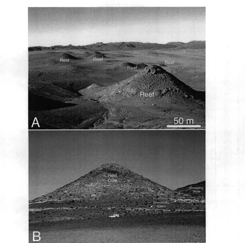
Figure 1-4: Thrombolite-stromatolite pinnacle reefs at top of Huns platform, Witputs subbasin, southern Namibia. Reefs are formed of a core of coalesced thrombolite mounds, overgrown by a stromatolitic mantle.
(A) Overview of multiple pinnacle reefs formed at top of Huns platform. Reefs have been exhumed by erosion
of overlying shales. (B) Cross section of representative pinnacle reef showing its foundation of stromatolite sheet facies and remnants of overlying shales which have been eroded to expose reefs. A sequence boundary separates the stromatolite sheet- and pinnacle reef facies; thus, the pinnacle reefs formed during a time of increasing accommodation space within a Transgressive System Tract.
Figure 1-5: Representative photographs of microbialites and associated textures. (A) Stromatolitic throm-bolite columns forming a laterally continuous biostrome at the top of the Omkyk Member, Zaris subbasin. The core of each column has a predominantly thrombolitic texture, whereas margins have better-laminated stromatolitic thrombolite texture. Columns are strongly elongate normal to the trend of the Omkyk carbon-ate ramp, indicating persistent high-velocity, wave-genercarbon-ated flows. Arrows point to goblet- and tube-shaped fossils that are abundant within the intercolumn fill sequences, but also occur within the thrombolitic tex-ture. (B) Well-preserved thrombolite texture in a pinnacle reef at the top of the Huns platform, Witputs subbasin. Reefal framework is created by dark-colored mesoclots; these create shelter pores that are par-tially filled by light-colored geopetal sediment and are ultimately filled by fibrous marine and later blocky burial cements. Inset shows fabric details; T, thrombolite mesoclot; GS, geopetal sediment; SP, shelter porosity filled with cement. Coin is approximately 1.2 cm in diameter. Inset shows enlargement of region near center of photograph. (C) Neptunian dike transecting thrombolite mound, Driedoornvlagte bioherm, Zaris subbasin. Dike is infilled with botryoidal fibrous calcite cement, interpreted to have replaced primary aragonite. Cements fill fractures within the reef, providing evidence for early lithification of the reef. Coin is approximately 1 cm in diameter. Inset shows enlargement of region below coin.
13
A
B
mudstone
stromatolite
4% n=176 n=125 13% pkst/grst 25% microbial 71% 87%thrombolite
Figure 1-6: Proportional distribution of calcified fossils within broadly-defined lithologic facies of the Nama Group (A) and within microbial facies of the Nama Group (B). Results show that fossils are very strongly correlated with microbial facies in general, and with thrombolitic facies in particular. Fossils are less abundant in packstones and grainstones, which are composed principally of intraclasts and peloids, and are rare in mudstones. The simplest interpretation of this distribution is that the fossil organisms were benthic, with a strong preference for microbially colonized substrates. In addition, the preference for thrombolitic over stromatolitic substrates suggests further paleoecologic control on their distribution. See text for discussion.
of stromatolites (Hoffman 1969; Cecile and Campbell 1978; Grotzinger 1986; Bartley et al. 1998), analogous to the development of "spur and groove" structure in modern coral reefs. This interpreta-tion supports other evidence that the Kuibis ramp was a wave- and storm-dominated system, where the dominant currents were induced by strong wave-generated flows.
The internal texture of the thrombolite reefs is characterized by mesoclots that range from a few millimeters to a few centimeters in diameter (Figures 1-5A and B, 1-7). Nama mesoclots are restricted to simple ovoid, globose, and colliform morphologies and do not include the complex digitate or dendriform fabrics found in younger thrombolites (e.g. Kennard and James 1986; Riding et al. 1995). Nama mesoclots formed an open framework that resulted in development of abundant cavities (Figures 1-5B, 1-7). Mesoclots typically were overgrown by a synsedimentary crust of fibrous marine cement that now consists of calcite but is interpreted to have replaced original aragonite (Figure 1-7). Marine cement crusts were subsequently covered by geopetal sediment, which partially filled framework pores (Figures 1-5B, 1-7). Remaining porosity was occluded by a dolomite rim cement, followed by infilling with blocky calcite spar (Figure 1-7). Recrystallization commonly makes interpretation of growth processes difficult, but sporadically distributed patches with relatively good fabric preservation show evidence that coccoidal microorganisms, expressed as poorly preserved spheroids in mesoclot cores, contributed to reef accretion (see Figure 1-7).
Early lithification of thrombolites and stromatolites in Nama pinnacle reefs resulted in the devel-opment of penetrative networks of neptunian dikes (Figure 1-5C). These are best expressed in the
Figure 1-7: Thin section photomicrograph of thrombolite fabrics within a pinnacle reef at top of Huns carbonate platform, Witputs subbasin. Thrombolite mesoclots (Throm) are encrusted with a fringe of fibrous calcite (Fibrous Marine Cement) interpreted to have replaced original aragonite (note square crystal terminations). This was followed by deposition of a layer of crystal silt (Geopetal Sediment). Occlusion of remaining shelter porosity began with precipitation of an isopachous fringe of Dolomite Rim Cement, followed by a final infill of blocky calcite spar. Note that mesoclots are composed of distinct aggregates of smaller clots. These smallest fabric elements most likely are formed by the early cementation of coccoid microorganisms. Note the accumulation of Geopetal Sediment (crystal silt) which smoothes out the rugged relief created by clots and encrusting Fibrous Marine Cements.
Huns reefs and Driedoornvlagte bioherm, where fractures that cut sharply across reefs are filled with fibrous marine cements, interpreted as calcite-replaced aragonite (Figure 1-5C). Neptunian dikes are formed only in the larger reefs, where gravitationally-induced failure of the reef was likely. A related effect includes failure of the sides of the pinnacle reefs, which resulted in down slope transport of slide breccias. These breccias are composed of angular blocks up to 1 meter in diameter with thrombolite and stromatolite textures. Voids between breccia blocks are filled with massive lime or dolomite mudstones, or fibrous marine cements.
Calcified Metazoans of the Nama Group
Within thombolitic facies, fossils occur abundantly both in the domal and columnar structures that make up individual reefs and in trough cross-bedded intrachannel fill. The fossils-millimeter to centimeter-scale calcareous cups, goblets, and tubes-are particularly abundant in thrombolites and thrombolitic stromatolites, evidently preferring the firm substrate and bathymetric elevation that these structures offered (Figure 1-6)). Intrachannel fill consists of skeletal packstone and grainstone containing goblets, cups, cylindrical tubes, Cloudina, and their bioclastic detritus.
Morphology. Figure 1-8 shows a number of calcified goblet-shaped fossils. Two dimensional slices provided by outcrops, polished slabs, and thin sections reveal a moderate degree of assemblage variability, ranging from clearly defined goblet-shaped forms (Figure 1-8E), originally illustrated by Grotzinger et al. (1995), to rounded cups without "stems," to bulbous structures showing apparent hexagonal symmetry in cross section. Exposed specimens reflect some combination of intraspecific, interspecific, and taphonomic variation, but the relative contributions of these factors cannot be eval-uated in the absence of information about the three-dimensional morphology of constituent fossils. Because the fossils are preserved as calcitic void-fill in a calcite matrix, individual specimens cannot be freed by conventional techniques-posing a challenge for morphological and, hence, systematic interpretation. Our response has been to develop a computer-based, "tomographic" reconstruction technique to determine their true three-dimensional geometry. This technique is described in de-tail in Chapter 2. In brief, appropriate samples were selected, the position of each sample and its contained fossils calibrated with respect to an external reference system, 25 microns ground from the polished sample surface, and the ground surface photographed using a digital camera. In the present study, the last two steps were iterated several hundred times for samples taken from reefs at Vioolsdrif (Witputs subbasin) and Donkergange (Zaris subbasin). Contours of the fossils in each cross-sectional image were obtained using a battery of image processing techniques, including a bi-nary thresholding procedure. Ultimately, the three-dimensional surface of our models is determined
by the shape of these contours, placed adjacent to one another in the third dimension. In the
layer, ultimately allowing the complete surface to be described by a set of tetrahedrons. Multiple views of one complete and eight partial tomographic reconstructions are shown in Figure 1-9.
All complete or nearly complete reconstructed specimens have a stem and an outward flaring
cup. The cup, a few millimeters to 2.5 cm in maximum dimension, has a broad circular opening at the top with a lip that curves inward. The cup is perforated by six or seven holes of similar size and shape, and has a matching number of regularly arranged side walls. Cups taper to a shallow base from which a hollow cylindrical stem extends. Some specimens contain an additional hole near the
cup base. Stems are commonly longer than maximum cup dimension and are open at both ends. In constructing a model morphology from reconstructed individuals, we took note of variation in the following parameters: (1) the radial profile of the cup, (2) the aspect ratio of the cup (i.e., maximum diameter to total height), (3) the size and shape of the cup's inner lip, (4) the relative size of the cup's circular opening, (5) the curvature of cup walls (wall curvature varies with height, in some cases flattening midway up the cup wall and in others remaining rounded), (6) the size, shape, and position of holes (e.g., also called "windows," which are normally oval or circular, located at the top, middle or base of the cup, and with or without an inward-turning lip), (7) the thickness of walls, (8) the number of sides and holes (i.e., six or seven), (9) the length of the stem, (10) the size of the cup, and (11) the trajectory and radial profile of the stem.
In developing a mathematical model for the morphology of these fossils, only the features listed above were assumed. Individual tomographic reconstructions were used to obtain values for all 11 parameters, enabling us to generate individual models. Figures 1-10A and 1-10C show sample cross sections of tomographic reconstructions that were used to measure profiles of the cup's radius and inward lip, the cup's aspect ratio, and the number of holes and side wall sections. The corresponding mathematical models are shown in Figures 1-10B and 1-10D. The specimen shown in Figures 1-10A and 1-10B was collected from a locality in the Zaris subbasin; the specimen in 1-10C and 1-10D is from the Witputs subbasin. The two models are primarily distinguished by (1) the number of holes (6 in the case of 1-10A-B, 7 in the case of 1-10C-D), (2) the radial profile of the cup (reaching maximum diameter near the top of the cup for 1-10C-D and near the middle in the case of 1-10A-B),
(3) the wall curvature (nearly flat at the cup's mid-height in 1-10C-D while still slightly round in
1-10A-B), and (4) the size of the inward-turning lip that lines the cup's large circular opening. A complete description of the mathematical model is supplied in Chapter 3.
A database of randomly-oriented synthetic cross sections was obtained from the models depicted
in Figure 1-10, and is shown in Appendix B. These synthetic cross sections reproduce the vast majority of shapes observed in rocks, a subset of which is depicted in Figure 1-11. Because we can account for most observed variation by a combination of random sections through the morphologies reconstructed in Figure 1-10 and simple taphonomic alterations (e.g., physical distortion) observed in our samples, we interpret most of the variation seen in outcrop as representing a single species
Figure 1-8: Assemblage of calcified Namacalathus fossils within reefal and related thrombolitic horizons within the Nama Group. (A) Namacalathus packstone illustrating a range of cross sectional morphologies. (B) Single large Namacalathus, over 2 cm in diameter. (C) Cross sections of two Namacalathus fossils. The first defines a well-developed hexagon, while the second, which came to rest inside the first, is composed of three segments. (D) Another specimen showing hexagonal geometry. (E) Original goblet-shaped fossil described by Grotzinger et al. (1995). Note adjacent cup-shaped fossils, which also represent cross sections of Namacalathus. All specimens in A-E can be explained as random sections through a single three-dimensional morphology (see text).
Figure 1-9: Tomographic reconstructions of the calcified fossil Namacalathus hermanastes. First described as having a "goblet" shape [Grotzinger, 1995 #1028], the fossil has a well-defined stem which is attached to an outward flaring cup perforated by 6 or 7 incurved holes, with an upper circular opening lined by an inward-turning lip. Partial specimens A through H are shown in inferred growth position (top) and also viewed from above (bottom). Note basal stem (or what is left of it) attached to a cup (A, C, D, E, F, G, I). The cup has 6 or 7 holes, and these are apparent with a regular symmetry in the majority of cases. Note also the single, circular opening (or part of it) in the inferred tops of each specimen. Complete specimen I is shown from several perspectives.
Figure 1-10: Mathematical models, based on 11 geometric parameters that capture the essential morphology of Namacalathus hermanastes (B and D) and cross sections of tomographic models upon which these are based and which were used to obtain parameter values (A and C). (A) From model B in Figure 1-9: a cross section showing regular holes sliced horizontally (relative to growth position) through center of cup (top), vertically through growth axis (upper-middle), and wire-frame views of circular opening at inferred top (lower-middle) and of the cup in profile (bottom). (B) Mathematical model informed by the images in
A. (C) The same cross sections and wire-frame diagrams for Model I in Figure 1-9. (D) Mathematical model
based upon the images in C.
population. Figure 1-12 illustrates the variation in cup size (maximum diameter) and shape (aspect ratio = maximum diameter/height) for a sample population of reconstructed specimens from a single rock sample (see Appendix A for a detailed discussion of these results).
Mineralization. Cloudina was only lightly mineralized, apparently by the precipitation of calcite in an organic template (Grant 1990). The same is true of the new taxon, Namacalathus hermanastes, reported here. Like Cloudina, Namacalathus had thin walls (little more than 100 Pm thick in best preserved specimens) that deformed flexibly during compaction (Figure 1-13). Petrographically, most fossils are preserved as casts of void-filling calcite, with rare evidence of thin, organic-rich wall structures (Figure 1-13D-F). Such fossils might be interpreted as casts and molds of unminer-alized organisms, were it not for fragmental remains that were almost certainly minerunminer-alized before fracture and dispersal. Original carbonate mineralogy is difficult to determine because of extensive dissolution or replacement, but common overgrowth of shells by euhedral calcite crystals supports the interpretation of a calcite precursor for Namacalathus. Overgrowths show strong preference for the outer walls of these fossils; inner walls tend to be smooth (Figure 1-13G). Overgrowths are not observed on the simple tubes or on Cloudina.
Evidence of calcite mineralization is intriguing because field and petrographic studies show that aragonite was favored as the common marine cement in Nama environments, accreting ooids, filling neptunian dikes that cross-cut pinnacle reefs, growing among thrombolitic mesoclots in reefs, and lining primary void space in grainstones (Grant 1990; JPG unpublished data). This implies that Nama animals had already evolved the physiological capacity to regulate mineral precipitation.
Paleoecology. Namacalathus populations are closely associated with thrombolitic textures, but
they do not appear to have participated in reefal frame construction or sediment baffling. The "stem and cup" morphology of the Nama fossils suggests that they were benthic organisms tethered to rather than nestling on or within their substrate; however, most specimens are oriented horizontally and not in life position (Figure 1-12A).
Namacalathus may have been attached directly to reef surfaces or anchored by an unmineralized
basal holdfast to locally accumulating sediments. It is also possible, however, that Namacalathus individuals were attached to seaweeds that colonized accreting microbialites-much as living stau-romedusae of the scyphozoan Halyclystus auricula attach to algae and sea grasses by means of discs at the ends of their exumbrellar stalks (Ruppert and Barnes 1994; AHK personal observation). Invertebrate epibionts of seagrasses are well known from Cretaceous and Tertiary rocks, although the plants, themselves, are rarely preserved (Brasier, 1975). More to the point, diverse and abun-dant macroscopic algae occur in terminal Proterozoic successions of South China (Chen et al., 1994; Steiner 1994; Xiao et al. 1998), and thallose algae grow profusely on subtidal microbialite surfaces today in places like the Bahama Banks (Feldmann and McKenzie 1998) and Shark Bay, Australia
Figure 1-11: Diverse cross sections of Namacalathus hermanastes might be interpreted as different taxa (upper photos); however, virtual slices through the mathematical models shown in Figure 1-10 produce a diverse array of synthetic cross sections (lower images) that can be used to estimate species richness. Most of the diverse cross-sections of calcified fossils in Nama rocks can be explained in terms of a single morphology with some minor variations. The cross sections shown in the upper photos were obtained from 40 specimens, only two of which were used to generate tomographic reconstructions (Figure 1-9E and F).
A
n = 51 27% oblique4%
vertical
69% horizontal 10% unidentifiable 90% NamacalathiB
n = 79D
n=48 20 ... ... ... . ... ... ... 10 0 0 0.4 0.8 1.2 centimetersC
n =87 14% Cloudina 9% unidentifiable 77% NamacalathusE
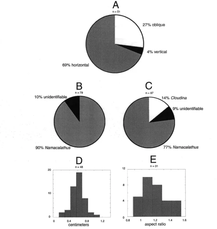
12 =31 8 4 0. 1.2 aspect ratioFigure 1-12: Statistics on the orientation, diversity, and geometry of calcified fossils in the Nama Group, from two samples. (A) Percentage of Namacalathus fossils in three categories of orientation. (B) Percentage of fossils by type. (C) Percentage of fossils by type found in a sample from the Witputs subbasin. (D) Size distribution of Namacalathus cups. (E) Distribution in aspect ratio (maximum cup diameter to cup height). See Appendix A for a detailed discussion of these results.
Figure 1-13: Taphonomic aspects of Namacalathus. Most of the photographs indicate the relatively flexible nature of the wall, suggesting only light mineralization at time of death. (A)-(C) Deformational features include: folds and wrinkles in walls and stems; squashed and flattened cups; cups without stems, presumably because of post-mortem breakage; wall ruptures, holes, and impressions caused by adjacent objects; and dis-integration of walls. (D) Cross section through a goblet-shaped fossil, illustrating replacement by void-filling calcite. Arrow points to section enlarged in Figure 1-13E. (E) Void-filling calcite indicates decay/dissolution of primary wall. Note crystal size enlargement toward center of fossil wall. (F) Rare preservation of primary shell structure (arrow) showing calcite-impregnated, dark organic matrix. In most specimens, this material has decayed/dissolved, with secondary void space filled by void-filling calcite (as in Figure 1-13D, E). (G) Wall of cup-shaped fossil. Note that the inner part of wall is smooth (white arrow), while the outer part is overgrown by a relatively thick layer of replacive calcite. Note crystal terminations extending out into and replacing matrix. Black arrows point to replaced intraclasts in original matrix. This suggests that nucleation and orientation of mineral overgrowths were controlled by primary crystal orientation within the fossil wall.
(AHK and JPG, personal observations). Whatever their mode of attachment, Namacalathus far outnumbered Cloudina and other calcified metazoans in Nama reef communities (Figure 1-12B, C).
Paleobiological Interpretation. Among metazoans, goblet morphology might be expected to
occur among the Porifera, the Cnidaria, or early stem branches of the Bilateria (before the evolution of strongly bilateral symmetry). Namacalathus could, thus, be viewed as the remains of simple asconid sponges with thin body walls and a relatively large spongocoel. Such an interpretation would be consistent with reports of Ediacaran impression (Gehling and Rigby 1996), biomarker (McCaffrey et al. 1994) and spicule (Brasier et al. 1997) fossils that suggest a broader importance of this group in terminal Proterozoic communities, and, more generally, with the phylogenetic expectation that sponges should have appeared early (Nielsen et al. 1996). On the other hand, the walls of the Nama fossils contain no evidence of pores or spicules. The former could, of course, have been obliterated during diagenesis and the latter is not conclusive insofar as not all sponges synthesize mineralized spicules. The observations are made to underscore the point that Nama goblets contain no features beyond overall shape that suggest affinities with sponges. The symmetric distribution of side holes is not easily reconciled with poriferan functional biology.
Chalice- or goblet-like morphologies can be found among the Cnidaria-for example, in hydrozoan polyps, the asexual scyphopolyps of some scyphozoans, and sessile scyphozoan stauromedusae in which exumbrellar surfaces are elongated into stalks (Ruppert and Barnes 1994). The side holes ("windows") characteristic of Namacalathus fossils can be reconciled with a cnidarian body plan, especially if these holes are interpreted as diagenetic features whose persistent occurrence and regular arrangement reflect underlying biological structure. In a number of specimens, sediment within goblets is distinct in color or texture from externally encompassing sediments. The boundary between sediment types is commonly just as sharp across side holes as it is across preserved walls (see Figure
1-11), suggesting that wall material once existed where holes are found today. In contrast, mixing
of internal and external sediments suggests that the gap at the top of the cups was open at time of burial.
Features potentially capable of governing the positions of lateral holes include (unmineralized) tentacles at some distance from the mouth (Brusca and Brusca 1990), planuloid buds arising from surfaces of goblet-shaped scyphopolyps (e.g., Cassiopea; Lesh-Laurie and Suchy 1991, Figure 59), or radially distributed biological structures such as septa and gonads whose decay contributed to localized dissolution of wall carbonate. Viewed in this way, Namacalathus can be interpreted in terms of cnidarian structure and function: sessile organisms with a mouth that opened into a large gastrovascular cavity.
Other interpretations are less attractive. Stem groups to the bilaterian (or ctenophore
+
bila-terian; Ax 1989; Eernisse et al. 1992; Nielsen et al. 1996) clade might have had goblet-likemor-phologies, but the large internal cavity characteristic of Nama specimens isn't easily incorporated into such a view.
Calcification occurs within the red, green, and, to a limited extent, brown algae, but no known living or fossil forms (Wray 1977; Fritsch 1965; Graham and Wilcox 2000) are similar in morphology to the Nama fossils. Namacalathus does not display the internal cellular structure that is ubiq-uitous among calcified rhodophytes; nor is the goblet morphology of this fossil approximated by known red algae. Calcifying green algae in the orders Codiales and Dasycladales precipitate arag-onite intercellularly within thalli, so that preserved skeletal carbonates include hollow molds of the complex filaments that underpin thallus morphology; no such internal structure marks the Nama fossils. Some Paleozoic dasyclads had a bulbous morphology, but their external profile reflects inter-locking elements that arise from a central stalk rather than a continuous sheet of cellular material.
Namacalathus is unlikely to be an alga.
Still less attractive are comparisons with broadly vase-shaped protists such as tintinnids, allogro-mid foraminifera, and testate amoebae. Known living and fossil species have volumes as much as three orders of magnitude less than those of the Nama fossils (Lee et al. 1985).
In addition to comparisons with living organisms, one can ask how Namacalathus compares with other fossils. The cup-like structure of N. hermanastes is at least broadly reminiscent of the small, radially symmetrical impressions found abundantly in some Ediacaran assemblages (e.g., Narbonne and Hofmann 1987; Sokolov 1997). It calls to mind, as well, specimens of the problematic calcareous fossil Khatsaktia and rare corallimorphs from Lower Cambrian carbonates in Siberia and elsewhere (Debrenne et al. 1990). The hexagonal symmetry of many Namacalathus specimens further recalls the triradiate symmetry characteristic of some Ediacaran organisms (e.g., Tribrachidium and Albumares; Fedonkin 1990) and the calcareous anabaritids, tubular fossils found in uppermost Proterozoic and Lower Cambrian successions (Bengtson et al. 1990; Qian and Bengtson 1989). The weakly calcified Nama fossils do not, in general, appear to be phylogenetic precursors of the diverse, skeletonized bilaterian animals found in Cambrian and younger rocks.
Evidence of Further Diversity. Germs (1972a) described two species of Cloudina, C. reimkeae and C. hartmannae. Namacalathus hermanastes adds to the diversity of calcified Nama animals, but
does not exhaust it. Approximately 10% of the forms observed on thrombolitic surfaces cannot be ascribed to Namacalathus or Cloudina, and we believe that these represent one or more additional, if poorly characterized, taxa. Calcified Nama fossils certainly include additional populations of simple tubes. These fossils, which commonly show a gentle curvature, are typically 1-2 millimeters wide. Maximum lengths are harder to estimate because the fossils are almost always broken, but segments up to 3 cm long have been observed. Shell walls are very thin and appear to reflect carbonate impregnation of an organic matrix. At least some of the tubes are closed at the base (Figure
1-14D), differentiating them from Namacalathus stems. Smooth walls, as seen in petrographic thin section, further differentiate these tubes from Cloudina (see discussion, below). Monospecific tube assemblages occur in micritic carbonates that accumulated on level bottoms on the Nama platform. Like other Nama fossils, the simple tubes are problematic. Functional considerations suggest that their makers were benthic organisms that lived erect on the seafloor, perhaps filter feeders analogous to if not necessarily homologous with modern fan or feather-duster polychaete (annelid) worms (Brusca and Brusca 1990). Calcareous polychaete tubes are known from limestones as old as Ordovician (Steele-Petrovich and Bolton 1998), and unmineralized polychaetes occur in the Burgess Shale and its Lower Cambrian counterparts (Conway-Morris 1998).
Exceptionally preserved populations allow fresh observations of C. riemkeae, the smaller and lesser known of the two Cloudina species in Nama rocks. The highest concentrations of this taxon occur in association with a flooding surface developed atop the Driedoornvlagte bioherm. Like the larger C. hartmannae, C. riemkeae has been reconstructed as a series of stacked cones (see Grant
1990; Bengtson and Zhao 1992); however, primary marine cements that line a continuous inner
surface emphasize that the "cones" are funnel-like flanges formed episodically during the growth of a cylindrical tube. The outer part of each flange diverges from the tube wall, whereas the inner part merges with the bases of earlier formed flanges (Figure 1-14A-C). Several workers (Conway Morris et al. 1990; Germs 1972a; Glaessner 1976) have noted points of structural similarity between Cloudina and tube-forming annelids such as the serpulid Merceriella (Fauvel 1923). The morphologies illus-trated in Figure 1-14 also resemble the (unmineralized) tubes of some modern pogonophorans (Ivanov
1963), as well as diagenetically mineralized vestimentiferan worm tubes recovered from Cretaceous
vent deposits in California (Little et al. 1999). (Pogonophorans and vestimentiferans are consid-ered to nest phylogenetically within the Annelida; McHugh 1997.) Alternatively, C. riemkeae might be compared to the episodically accreting (unmineralized) sheaths of budding scyphozoan polyps (Werner, 1967).'Whatever its affinities to living organisms, C. riemkeae does resemble Saarina, or-ganically preserved tubes from the Vendian of the Russian Platform that are comparably organized (Sokolov 1997). Some Early Cambrian anabaritids also have well developed flanges (Bengtson et al.
1990; Qian and Bengtson 1989).
The Nama Group, thus, contains at least five taxa of calcified metazoans: C. hartmannae and C.
riemkeae (Germs, 1972a; Grant 1990), N. hermanastes, simple closed tubes, and the uncharacterized
forms in reef assemblages. More taxa may be recognized in continuing work; for example, fragmental remains in skeletal packstones hint at additional diversity. In absolute terms, observed diversity is modest, although only about a dozen species of canonical Ediacaran fossils have been reported from Nama sandstones (Richter 1955; Pflug, 1970a, 1970b, 1972; Germs 1972b, 1972c; Runnegar 1992; Narbonne et al. 1997).
Figure 1-14: Other calcified fossils in the Nama Group. (A-C) Thin section photomicrographs of
Cloudina riemkeae showing unusually good preservation of shell structure. In all three cases, note
the straight inner wall, highlighted best in B and C, but also visible in A. A and B show external wall structure indicating en echelon stacking of flanges, which have been largely recrystallized in C.
(D) Tube-shaped fossils comprising a distinct population of shapes, distinct from both Cloudina and Namacalathus. (E) Tomographic reconstruction of a tube-shaped fossil, showing its closed base and
flared top.
28
-Additional species of calcified metazoans have been described from terminal Proterozoic beds in Brazil (Hahn and Pflug 1985), and small mineralized fossils (foraminiferans?) have been claimed to occur in correlative units in Uruguay (Gaucher and Sprechmann 1999). Phosphatized tubes in the terminal Proterozoic Doushantuo Formatiuon, China, may or may not be of animal origin (Li et al. 1997). Regardless of uncertainties surrounding the Uruguayan and Chinese structures, calci-fied fossils comprise at least five percent of known Ediacaran diversity-not very different from the proportional representation of mineralized species in the Middle Cambrian Burgess Shale (Conway Morris 1998). The confirmation that a number of Proterozoic animals were able to precipitate min-eralized skeletons lends support to hypotheses that relate the subsequent Cambrian diversification of well-skeletonized animals to increasing levels of predation rather than physiological innovation
per se (Bengtson 1994).
Despite expanding a distinct taphonomic window on early animal evolution, calcified Nama fos-sils do not reveal Proterozoic roots for the skeletonized bilaterians that radiated across carbonate platforms in the Paleozoic Era. On the contrary, like the Ediacaran assemblages preserved in con-temporaneous siliciclastic rocks, calcified Nama assemblages appear to document a distinctively Proterozoic fauna.
Systematic Paleontology
Genus NAMACALATHUS n. gen.
Type species. Namacalathus hermanastes n. sp.
Diagnosis. Centimeter-scale, chalice- or goblet-shaped fossils consisting of a calcareous wall less than 1 mm thick; a basal stem open at either end connects to a broadly spheroidal cup perforated
by 6 or 7 holes with slightly incurved margins distributed regularly around the cup periphery and
separated by lateral walls; the cup contains an upper circular opening lined by an incurved lip. Etymology. From the Nama Group and the Greek kalathos, denoting a lily- or vase-shaped basket with a narrow base or, in latinized form, a wine goblet.
NAMACALATHUS HERMANASTES n. sp.
Diagnosis. species of Namacalathus distinguished by cups 2 to 25 mm in maximum dimension, with aspect ratio (maximum cup diameter/cup height) of 0.8 to 1.5.
Description. Goblet-shaped calcified fossils; walls flexible, ca. 100 microns thick (original wall
open at both ends, 1-2 mm wide and up to 30 mm long, attached to spheroidal cup; cup with maximum dimension 2-25 mm, broad circular opening at top with inward-curving lip, perforated
by 6-7 slightly incurved holes of similar size and shape distributed regularly about cup periphery.
Specimens preserved principally by void-filling calcite, with rare preservation of primary, organic-rich wall.
Etymology. From the Greek, herma, meaning sunken rock or reef, and nastes, meaning inhabitant. Material. More than 1000 specimens from biohermal carbonates of the Kuibus and Schwarzrand subgroups, terminal Proterozoic Nama Group, Namibia.
Type specimen. Our understanding of Namacalathus hermanastes derives principally from virtual fossils modeled from serially ground surfaces. Systematic practice, however, requires that real fossils be designated as types. Accordingly, the specimen illustrated in the lower right corner of Figure 1-8E is designated as holotype for the species. The type specimen is to be reposited in the paleontological collections of the Namibian Geological Survey. Representative specimens are also housed in the Paleobotanical Collections of the Harvard University Herbaria (HUHPC #62989).
Type locality. Reefal biostrome developed at the top of the Omkyk Member, Zaris Formation, Kuibis Subgroup, exposed along the Zebra River near the boundary between Donkergange and Zebra River farms, south of Bullsport, Namibia.
Thrombolite-Stromatolite-Metazoan Reefs
Thrombolite/invertebrate associations characterize Early Cambrian and some younger Paleozoic reefs (Kruse et al., 1995; Riding and Zhuravlev, 1995; Soja, 1994). Nama bioherms show that the close ecological relationship between invertebrate and microbial populations was established before the end of the Proterozoic Eon, prompting questions about the origins of this distinctive reef association.
Microbial mat populations have contributed to stromatolitic reef accretion since the Archean (Hofmann et al., 1999; Grotzinger and Knoll 1999), but bioherms characterized by thrombolitic tex-tures are rare before the latest Proterozoic Eon (Aitken 1967). Based in part on this stratigraphic distribution, Walter and Heys (1984) interpreted thrombolitic clots as ordinary stromatolitic lami-nae disrupted by burrowing. Kennard and James (1986), however, subsequently demonstrated that thombolitic textures are primary, confirming the original inference of Aitken (1967). Kennard and James argued specifically that the diagnostic clotted fabric of thrombolites is generated by in situ calcification of coccoid-dominated microbial communities. Coccoid cyanobacteria form rough, irreg-ular mats that may, upon calcification, yield a clotted texture with only crude lamination (Hofmann,
1975), and recent work supports this interpretation for some Neoproterozoic thrombolites (Turner et
al., 1997). Such an interpretation, however, leaves open an important question. Mats built by coc-coidal cyanobacteria are common in Paleo- and Mesoproterozoic tidal flats (Golubic and Hofmann,
1976; Sergeev et al., 1995), but thrombolitic fabrics are essentially absent. How can this discrepancy
be explained?
Feldmann and McKenzie (1998) have recently proposed that ancient thrombolites reflect the participation of higher algae in mat-building consortia. This inference is based on actualistic ob-servations on the Bahama Banks, where cyanobacteria-dominated mat communities accrete well-developed laminae in intertidal environments, whereas eukaryotic communities produce poorly lam-inated, thrombolitic textures in open marine subtidal environments. Coccoid spores thought to be produced by dasycladalean green algae play a particularly important role in fabric generation.
The observations of Feldmann and McKenzie (1998) are consistent with paleoenvironmental research that locates Paleozoic thrombolites in subtidal environments (Aitken, 1967; Bova and Read,
1987; Pratt and James, 1986). They are also congruent with biostratigraphic data that bracket the
expansion of thrombolites between the initial diversification of green algae and the appearance of well-skeletonized dasyclads (Wray 1977; Knoll 1992).
Observations of Nama thrombolites are also consistent with predictions of the Feldmann and McKenzie (1998) model. Nama thrombolites formed in open marine, wave-swept environments, of-ten associated with grainstone shoals. Well-laminated stromatolites are rare in the Nama Group, but so are intertidal facies. The best developed Nama stromatolites form columns within a sheet-like biostromal unit at the top of the Omkyk Member (Figures 1-3B, C)-a unit interpreted to have formed in a very shallow subtidal environment. Significantly, thrombolitic textures are most abun-dant and best developed in pinnacle reefs at the top of the Huns platform and at Driedoornvlagte. These reefs are composed of large mounds, which are rarely elongate; where elongation is developed, it is defined by a low length-to-width ratio, consistent with low current velocities in deeper water subtidal settings. Thrombolite textures developed in this environment are similar to those found in
Paleozoic rocks (compare Figure 1-5B with Figure 2B of Kennard and James 1986).
As noted above, Namacalathus and other Nama metazoans lived in specific association with thrombolite-forming communities, but they did not participate in any substantial way in reef build-ing, either as frame-builders or sediment bafflers. With this in mind, it is straightforward to visualize a paleoecologic reconstruction in which the calcified organisms were loosely attached to the accreting thrombolitic substrate, much as calcisponges and green algae are in modern Bahamian thrombo-lites. Alternatively, Namacalathus may have been an epibiont attached to seaweeds that colonized the thrombolitic substrate. Upon death or dislocation of the algal components, calcified organisms would have collapsed to the sea floor as detrital particles to be swept into the depressions between thrombolite columns and mounds, or occasionally to be trapped in random positions within
ac-creting mats. This interpretation also provides a mechanistic basis for understanding the locally great abundance of detached, horizontally oriented fossils in thin (a few cm) beds of thrombolitic laminites that form spatially extensive sheets within deeper subtidal facies. Before the radiation of metazoan macrograzers, broad expanses of the terminal Proterozoic seafloor may have been covered
by microbial-algal carpets, with calcified metazoans tethered to a (tiered?) algal lawn. Animals
assumed a constructional role in bioherm accretion only with the evolution of heavily skeletonized, attached epibenthos.
Conclusions
Nama bioherms document the emergence of a distinctive reef association. Animals that lived in the reef environment include some of the first organisms able to form skeletons by the enzymatic precip-itation of CaCO3 on an organic template. In important ecological and physiological respects, then,
Nama bioherms and the fossils they contain foreshadow Paleozoic biology. At the same time, how-ever, the distinctive and problematic morphologies of Namacalathus hermanastes and other calcified Nama fossils underscore the phylogenetic differences between terminal Proterozoic and Paleozoic skeleton formers. At present the significance of these similarities and differences is not completely known, but continuing study of the vast and beautifully exposed carbonate accumulations of the Nama Group promises to clarify our understanding of terminal Proterozoic biology and geobiology.
Chapter 2
The Serial Imaging Method
Three methods were tried for obtaining the morphology of the Nama fossil specimens. First, an unsuccessful attempt was made to dissolve the calcite matrix using an acid solution. Second, a Nuclear Magnetic Resonance (NMR) scanning device was used to image the distribution of hydrogen atoms in a water-saturated sample. There was, however, only a slight difference between the amount of water absorbed by the fossil and matrix materials. Ultimately, a serial imaging method was adopted, whereby the morphology is reconstructed from cross sections of the fossils, imaged with a digital camera at 25.4 pm intervals. This process is described in detail in the present chapter.
Obtaining Serial Images
In overview, the process whereby serial images are obtained occurs as follows. First, a small square slab of sufficient thickness containing suitable fossils is cut out from a rock. The surface of this sample is then photographed by a digital camera to obtain an image. Then, a thin layer measuring 25.4 Pm in thickness is removed from the surface using a surface-grinding apparatus. The photographing and grinding is repeated in this way until the sample is ground away, producing a set of several hundred images obtained at 25.4 pm intervals.
What follows is a discussion of how the fossils are prepared for processing. First, rocks are chosen according to their suitability for analysis. The fossils reside in layered limestone slabs that are normally less than two inches thick and at least five inches in diameter. Suitable rocks have a relatively high concentration of fossils that are well-preserved. In particular, the fossils are 5 mm apart on the average, and not usually in contact with each other. Therefore, these were not deformed from contact with neighboring fossils, as occurs frequently in rocks where the fossils are densely packed. Shapes with a clear radial or bilateral symmetry are assumed to represent the original, undeformed morphologies, and therefore these are sought for study. Fossils are selected with the finest structure preserved-such as with thin walls and with minimal evidence of recrystallization. A