1
Tirés à part : H. BUSCAIL, LVEEM, 8 rue J.B. Fabre, CS 10219, 43009 Le Puy en Velay, France
INFLUENCE OF WATER VAPOUR ON 316L OXIDATION AT HIGH TEMPERATURE -
IN SITU X-RAY DIFFRACTION
Henri BUSCAILa, Raphaël ROLLANDa, Sébastien PERRIERa
a
Université de Clermont Auvergne, LVEEM, 8 rue J.B. Fabre, 43009 Le Puy-en-Velay, France
Abstract – Water vapour mixed with air (10 vol.% H2O) has little effect on the AISI 316L stainless
steel oxidation under isothermal conditions at 800 and 900 °C. The oxide scale is composed of Cr2O3 and Mn1.5Cr1.5O4 located at the external interface. Results show that the presence of water
lowers the specimen’s mass gain. At 1000 °C, the effect of water vapour on the scale structure is deleterious. In situ X-ray diffraction permits to show that the breakaway is due to iron oxidation starting after about 30 hours. This leads to an external Fe2O3 scale growth and an internal
multilayered FeCr2O4 scale formation.
Résumé – L’air contenant 10 vol.% H2O a peu d’effet sur l’oxydation de l’alliage AISI 316L à 800
et 900°C. La couche d’oxyde est alors constituée de Cr2O3 et de Mn1.5Cr1.5O4 qui est situé à
l’interface externe. Les résultats montrent que la présence de vapeur d’eau diminue la vitesse de prise de masse. A 1000 °C la vapeur d’eau a un effet désastreux sur la structure de la couche d’oxyde. La diffraction des rayons X in situ à 1000°C sous air humide a permis de montrer que l’oxydation du fer débute après environ 30 heures d’oxydation. Ceci conduit à la formation d’une couche externe de Fe2O3 et une couche interne stratifiée de FeCr2O4.
1. INTRODUCTION
The AISI 316L austenitic stainless steel is widely used in industrial applications due to its good creep properties and oxidation resistance provided by the high alloy chromium content [1-8]. Several studies proposed that, at high temperature, chromia-forming alloys such as AISI 316L SS exhibit an oxide growth leading mainly to Cr2O3 and Mn1.5Cr1.5O4 formation [9-11]. It has also been
demonstrated that yttria addition leads to an increased oxidation resistance at 800°C [12]. The role of molybdenum on the AISI 316L oxidation mechanism has been already discussed. It has been demonstrated that molybdenum plays a similar protective role as the one provided by silicon. In dry air, it hinders iron oxidation and leads to a better keying of the chromia scale in the alloy [13-15]. Water vapour is present in many industrial gases. It is a constituent of combustion gases and can affect high temperature corrosion of heat exchangers or engines. Many investigations have been conducted to increase solid oxide fuel cells performance and increase the corrosion resistance of metallic interconnects [16, 17].
literature [18-21]. Several studies were conducted on chromia forming alloys [22-26]. Generally, water vapour has a major effect on the growth rate of oxide scales. Increasing the amount of water vapour in the atmosphere reduces the time to the onset of breakaway oxidation [27].Galerie et al. have explained that the breakaway on a Fe-15%Cr alloy was induced by rapid growth of hematite at the metal/chromia interface, at 800-1000 °C, in Ar-15 vol.% H2O [23]. But in many cases, the
chromia scale failure was related to the formation of the CrO2(OH)2 and Cr(OH)3 volatile
hydroxides [28-30]. Evaporation can lead to the alloy chromium depletion resulting in the formation of an iron-rich non-protective scale. Young proposed that water vapour affects chromia scale growth similarly to what is observed with reactive metal elements [31]. Both segregate to oxide grain boundaries. Grain boundary cation diffusion is then largely suppressed increasing the oxygen internal diffusion and refining the oxide grain structure. On the other hand, he also explained that in short-term laboratory experiments at temperatures above 800 °C, chromium volatilization could usually be neglected, even in O2/H2O gas mixtures. Refining of the oxide grain structure under wet
conditions has also been observed by Jacob et al. after pure chromium oxidation [32].Recently, Othman et al. proposed that water vapour has no significant effect if a large chromium reservoir induces the growth of a continuous Cr2O3-scale layer [33]. The ability of Fe–Cr alloys to form a
protective oxide scale was related to the chromium concentration at the alloy surface. When the concentration of chromium is too low, iron-rich oxides form, inducing breakaway oxidation. Rahmel et al. suggested that an H2/H2O gas mixture can be formed in voids within the scale then
facilitating rapid inward transport of oxygen [18]. From the literature review it can be stated that owing to its relatively high chromium content and alloying elements, AISI 316L is oxidation resistant at temperatures up to 1000 °C in dry environments and in isothermal conditions.
The aim of the present work is to determine the effect of water vapour on the oxidation mechanism of the AISI 316L alloy compared with dry air experiments. In situ X-ray diffraction with flowing moist air (10 vol.% H2O) will provide useful information on the oxidation process at
operating temperatures. It will be determined if a change in the structural composition of the layer occurs during the oxidation test and if any oxide phase transitions occur during cooling to room temperature.
2. EXPERIMENTAL
The material used in the present work is an AISI 316L austenitic stainless steel. The alloy composition is given in table I. The specimens, provided by APERAM Alloys Imphy, were polished on SiC paper up to 800 grade, then washed with ethanol and finally dried prior to isothermal oxidation at 800, 900 and 1000°C. The specimens are 1.5 mm thick and show a total surface area of about 6 cm2.
The kinetic results obtained under isothermal conditions were recorded during 96 h using a Setaram TG-DTA 92-1600 microthermobalance. The in situ characterization of the oxide scales was carried out in a high temperature MRI chamber adapted on an X-ray Philips X' PERT MPD diffractometer (Copper radiation, λKα= 0.154 nm). The diffraction patterns are registered every hour at 1000 °C in 10 vol.% H2O flowing air.
X-ray diffraction (XRD) conditions were: Θ−2Θ scan, step 0.02°, range from 10 to 80°, 3 s counting time. The morphology of the external interface as well as the cross sections was observed using a Scanning Electron Microscope (SEM) JEOL JSM-7600F. The analysis of the scale was performed with a LINK energy dispersive X-ray spectroscopy (EDXS). The EDXS point analyses were performed with an electron probe focused to 1 µm spot. The water vapour experiments were performed in air containing 10 vol.% H2O. The flowing air (8 L/h flow rate) is saturated through
3
Tirés à part : H. BUSCAIL, LVEEM, 8 rue J.B. Fabre, CS 10219, 43009 Le Puy en Velay, France boiling water. Then, the water bath humidifier maintained at 50 °C controls the water vapour ratio by condensation of the excess water vapour. All the connecting tubes are maintained at 100°C in order to avoid any water condensation out of the furnaces. A schematic drawing of the rig has been provided in a previous paper [34].Similar gas humidifiers were used either for TGA and wet in situ DRX (MRI chamber). Dry air testing was performed in flowing air at atmospheric pressure. The weight change of the samples was determined after each oxidation test on a balance with an accuracy of 0.01 mg.
Table I. Composition of the AISI 316L SS (weight %)
AISI 316L Fe Cr Ni Mo Mn Si C
Content Balance 17.7 10.9 2.16 1.57 0.55 0.023
3. RESULTS
3.1. Isothermal kinetics
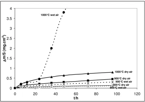
The mass gain curves per unit area are given in figure 1. All isothermal thermogravimetric analyses were carried out during 96 h at 800, 900 and 1000 °C in ambient air and wet air (10 vol.% H2O). A parabolic regime was observed throughout the 96 h oxidation tests except when testing at
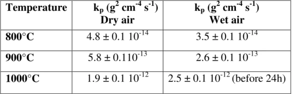
1000 °C in wet air. The kpvalues are given in table II. At 800 °C, the oxidation rates are similar under dry and wet air. At 900 °C, the oxidation rate is slightly higher in dry air than in wet air. At 1000 °C, a parabolic regime is followed in dry air whereas in wet air, 10 vol.% water vapour induces an important increase of the oxidation rate after 30 hours oxidation. At 1000 °C, the oxidation rate recorded before the breakaway regime is similar in wet air and dry air. An apparent activation energy (Ea) was calculated from the slope of the Arrhenius plots of the kp values, Ea =
208 ± 30 kJ mol-1 under dry conditions and Ea = 241 ± 30 kJ mol-1 in wet air.
Table II . kp values obtained on the AISI 316L SS after dry air and wet air (10 vol.% H2O)
oxidation between 800 at 1000 °C. Temperature kp (g2 cm-4 s-1) Dry air kp (g2 cm-4 s-1) Wet air 800°C 4.8 ± 0.1 10-14 3.5 ± 0.1 10-14 900°C 5.8 ± 0.110-13 2.6 ± 0.1 10-13 1000°C 1.9 ± 0.1 10-12 2.5 ± 0.1 10-12 (before 24h) 3.2. Scale morphology
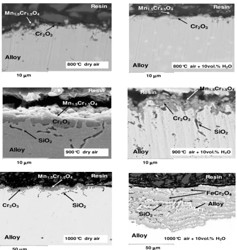
SEM examinations were carried out on the specimen cross section (figure 2) in order to identify the elements constituting the oxide scale. After 96 h oxidation in dry and wet air at 800, 900 and in dry air at 1000 °C, the scale is adherent. EDXS analyses performed on the cross section show that manganese chromite is located at the scale/gas interface. The middle part of the oxide scale is mainly composed of chromia. Some internal silicon oxidation is observed along the alloy grain boundaries at temperatures higher than 800 °C. No influence of water vapour is observed on
spalled off during cooling to room temperature (30 °C/minute) and is not observed on the cross section. The inner scale is multilayered and shows alternatively FeCr2O4 scales and non-oxidized
nickel-rich metallic layers. EDXS analyses performed in the metallic substrate, under the oxide scale, show that in the upper 2 µm depth the chromium content is 14.2 ± 0.3 wt.%. The 17.7 wt.% bulk content is reached at 15 µm deep inside the alloy. The alloy chromium ratio close to the interface is the same whatever the temperature and gaseous environment. It then appears that water vapour does not lead to important chromium depletion inside the metallic substrate.
Figure 1. Mass gain curves obtained after 96h isothermal oxidation of AISI 316L SS under dry air
and wet air (10 vol.% H2O) at 800, 900 °C, and 1000°C.
3.3. X-Ray Diffraction results after isothermal oxidation
XRD patterns obtained from the AISI 316L SS sample after 96 h isothermal oxidation at 800, 900, 1000 °C in dry air reveal the presence of Mn1.5Cr1.5O4 (JCPDS 33-0892) and Cr2O3
(JCPDS 38-1479) after oxidation at all temperatures. The relative intensity of the alloys peaks is smaller at 1000 °C due to the higher oxide thickness. After wet air oxidation, Mn1.5Cr1.5O4 and
Cr2O3 are present on the alloy surface after oxidation at 800 and 900°C. At 1000°C, XRD analysis
of the remaining oxide only shows the presence of FeCr2O4 (JCPDS 34-0140) because the external
Fe2O3 scale totally spalled off during cooling at 30°C/minute. Fe2O3 scale pieces have found fallen
down out of the crucible.
3.4. In situ X-Ray Diffraction results in wet air at 1000°C
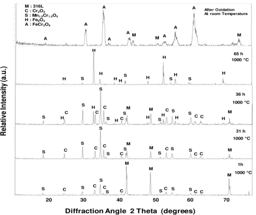
The oxide scale formed on AISI 316L was analysed by in situ XRD on the metallic substrate at 1000°C, in wet air (10 vol.% H2O) during 65 hours. Figure 3 shows the presence of the
Mn1.5Cr1.5O4 (JCPDS 33-0892) and Cr2O3 (JCPDS 38-1479) during the 30 first hours of oxidation.
It appears that both oxides nucleate simultaneously on the specimen surface during the first hour of oxidation and remain present together on the surface. No other oxides were formed during the 30 first hour oxidation. After 36 hours oxidation, hematite Fe2O3 (JCPDS 24-0027) is detected on the
0 0.5 1 1.5 2 2.5 3 3.5 4 0 20 40 60 80 100 120 t/h ∆∆∆∆ m /S ( m g .c m -2 ) 1000°C wet air 900°C wet air 800°C wet air 1000°C dry air 900°C dry air 800°C dry air
5
Tirés à part : H. BUSCAIL, LVEEM, 8 rue J.B. Fabre, CS 10219, 43009 Le Puy en Velay, France alloy surface and Cr2O3 peaks are still observed. After 65 hours oxidation, the hematite scale is
thick and the FeCr2O4 inner oxide is hardly detected. After cooling to room temperature, figure 3
shows that the XRD pattern obtained does not show the presence of hematite anymore. FeCr2O4 and
the alloy are the only phases observed on the diffraction pattern due to the complete hematite scale spallation during cooling. In situ XRD results at 1000°C, in dry air, were already presented in a previous paper [13]. It was shown that Mn1.5Cr1.5O4 and Cr2O3 were detected on the alloy surface all
along the oxidation test in dry air and after cooling to room temperature.
4. DISCUSSION
Kinetic results have been obtained on AISI 316L between 800 at 1000 °C under isothermal conditions (figure 1). The parabolic behaviour was always followed in this temperature range under dry conditions and under wet conditions at 800 and 900 °C. This permitted the calculation of the parabolic rate constants kp at each temperature (table II). The parabolic rate constant at 1000 °C in
moist air was calculated from the kinetic curve obtained before 24 hours oxidation. At 1000 °C, in dry air, the kp value is calculated from the data obtained during 96 h oxidation. An apparent
activation energy (Ea) was calculated from the slope of the Arrhenius plots of the kp values, Ea =
208 ± 30 kJ mol-1 under dry conditions and Ea = 241 ± 30 kJ mol-1 in wet air. These values are close
to the one generally proposed in the literature when Cr2O3 is formed on the alloys in this
temperature range [32, 35].
Figure 2. SEM cross-section on the AISI 316L SS oxidized at 800, 900, 1000°C in dry air or wet
air (10 vol.% H2O) for 96 h.
900°C dry air 50 µµµµm 10 µµµµm Resin Resin Resin Cr2O3 Cr2O3 Cr2O3 Alloy Alloy Alloy SiO2 SiO2 Mn1.5Cr1.5O4 Mn1.5Cr1.5O4 Mn1.5Cr1.5O4 10 µµµmµ Resin Resin Cr2O3 FeCr2O4 Alloy SiO2 Alloy Alloy Mn1.5Cr1.5O4 10 µµµµm 50 µµµµm 10 µµµµm Resin Mn1.5Cr1.5O4 Cr2O3 SiO2 Alloy
800°C dry air 800°C air + 10vol.% H
2O
900°C dry air 900°C air + 10vol.% H2O
1000°C air + 10vol.% H2O
observation (figure 2) showing that the scale was mainly composed of a continuous chromia scale, acting as a diffusion barrier. According to SEM and XRD analyses, the good kinetic behaviour at 800 and 900°C, in dry and moist air, appears to be mainly due to the absence of iron containing oxides in the scale. Figure 1 also shows that water vapour slightly decreases the oxidation rate relative to those registered in dry conditions and the apparent activation energy is higher in wet air. As proposed by other authors it could be due to weight losses related to the formation of the volatile hydroxide CrO2(OH)2 [28-30]. At 1000 °C, the scale remains protective during the oxidation test in
dry air, (figure 1 and figure 2). Mn1.5Cr1.5O4 and Cr2O3 are present all along the oxidation test. In
wet air, oxidation at 1000 °C firstly leads to Mn1.5Cr1.5O4 and Cr2O3 formation. Nevertheless,
breakaway corrosion occurred after 30 hours oxidation. In situ X-ray diffraction permit to analyse the oxides formed on AISI 316L during isothermal oxidation at 1000 °C in moist air (figure 3). The breakaway oxidation is due to the iron oxidation starting after 30 hours oxidation. This leads to an external hematite scale growth and an internal multilayered FeCr2O4 scale formation.
Figure 3. In situ XRD patterns obtained during the oxidation of the 316L alloy at 1000°C under wet
air (10 vol.% H2O).
Some authors have observed that water vapour can induce a breakaway during high temperature oxidation of iron-based chromia-forming alloys [20, 23].Then, it was observed that it depends upon the oxidation time, testing conditions and the chromium alloy content [23]. As discussed above, chromium volatilization by formation oxy-hydroxides CrO2(OH)2 can be
envisaged [36, 37]. Some authors also envisaged the transport of H2O into the oxide scale by the
diffusion of hydrogen as H+, OH- or H2O in the oxide scale [38, 39]. In this study, manganese
chromite and chromia were observed during the initial oxidation stage at 1000°C under wet air. Nevertheless, after about 30 hours oxidation, the protective chromia scale fails and breakaway appeared. It is proposed that water dissociation occurred on the specimen surface [23, 40]. This
R el at iv e In te n si ty (a .u .)
Diffraction Angle 2 Theta (degrees)
After Oxidation At room Temperature 20 30 40 50 60 70 M : 316L C : Cr2O3 S : Mn1.5Cr1.5O4 H : Fe2O3 A : FeCr2O4 65 h 36 h 31 h 1h A A A A A A A A H H H H H H H H S H S S S H H H H H H H C C C C C C M M M S S S S S S S S S S S S S S S S C C C C C C C C M M M M M M S S S S S S S C C C C C C C 1000 °C 1000 °C 1000 °C 1000 °C M M M
7
Tirés à part : H. BUSCAIL, LVEEM, 8 rue J.B. Fabre, CS 10219, 43009 Le Puy en Velay, France dissociation is more important at 1000 °C than at lower temperature because water dissociation is endothermic. Then, the diffusion of hydrogen-containing species could occur. Transport of H2O
into the oxide scales is performed by the incorporation of hydrogen in the form of H+ or H2O [38,
39, 41]. It is assumed that protons (H+) from water vapour might dissolve in the oxides and diffuse at grain boundaries. Essuman et al. proposed a model to explain the internal oxidation of chromium alloys [20, 21]. Ardigo et al. performed gold and deuterium marking experiments and have evidenced that Fe3O4 and Cr2O3 scales are permeable to hydrogen [42]. The low H+ and OH- radii
permit their diffusion through the oxide scale. Then, the increase of protons in the scale changes the oxide defect chemistry and could be responsible for the change in the growth mechanism. It can then be considered that the reaction of H+ (and/or H2O) with the metallic substrate under the
chromia scale form H2 andwüstite [36, 37].Thermodynamic data indicate that, at 1000 °C, wüstite
is more stable than H2O. Mikkelsen et al. also explained that addition of water vapour favours the
formation of wüstite [43]. When FeO and chromia are present together they react to form FeCr2O4
wich explains that wüstite does not accumulate at the internal interface and is not detected by XRD [44]. As observed by in situ XRD (figure 3), iron ions also diffuse through the scale to form Fe2O3
at the external interface, then inducing the breakaway oxidation.
5. CONCLUSION
In situ X-ray diffraction was used to identify the oxides formed on the AISI 316L stainless steel during isothermal oxidation at 1000 °C in moist air (10 vol.% H2O). Our results show the
presence of the Mn1.5Cr1.5O4 and Cr2O3 chromia growing simultaneously on the specimen surface
before 30 hours oxidation. After about 30 hours oxidation, Fe2O3 is growing on the specimen
surface at the external interface and internal corrosion leads to a multilayered FeCr2O4 scale.
Hematite, located at the external interface, spalled off easily during cooling to room temperature. Water vapour clearly promots iron oxidation at 1000°C. At 800 and 900°C, 10 vol.% H2O in air has
no detrimental effect on the AISI 316L stainless steel isothermal oxidation. The oxide scale is always composed of Cr2O3 with a small amount of Mn1,5Cr1,5O4 at the external interface. The lower
oxidation rate can be related to chromia evaporation in wet air.
6. REFERENCES
[1] J.E. Indacochea, J.L. Smith, K.R. Litko, E.J. Karell, A.G. Raraz, Oxid. Met. (2001) 55 1-15. [2] A.L. Johnson, D. Parsons, J. Manzerova, D.L. Perry, D. Koury, B. Hosterman, J.W. Farley, J.
Nuc. Mat. 328 (2004) 88-96.
[3] J. Pitter, F. Cerny, J. Cizner, J. Suchanek, D. Tischler, Surf. Coat. Tech. 200 (2005) 73-76. [4] A.A. Syed, T.A. Denoirjean, P. Fauchais, J.C. Labbe, Surf. Coat. Tech. 20 (2006) 4368-4382. [5] C. Vernault, J. Mendez, Ann. Chim. Sci. Mat. 24 (1999) 351-362.
[6] X. Wang, L.F. Wang, M.L. Zhu, J.S. Zhang, M.K. Lei, Trans. Nonferrous Met. Soc. China 16 (2006) s676-s680.
[7] Seung-Goo Kim, Ming-Zhi Hong, Sung Pil Yoon, Jonghee Han, Suk Woo Nam, Tae Hoon Lim, Seong-Ahn Hong, J. Sol-Gel Sci. Tech. 28 (2003) 297-306.
[8] A.V.C. Sobral, M.P. Hierro, F.J. Pérez, W. Ristow Jr., C.V. Franco, Mater. Corros. 51 (2000) 791-796.
[9] Raghuvir Singh, Narendra B. Dahotre, J. Mat. Sci. 40 (2005) 5619-5626. [10] P.N.S. Stokes, F.H. Stott, G.C. Wood, Mat. Sci. Engi. A121 (1989) 549-554.
[11] A. Bautista, F. Velasco, M. Campos, M.E. Rabanal, J.M. Torralba, Oxid. Met. 59 (2003) 373-393.
Phys., 111 (2008) 491-496.
[14] H. Buscail, S. El Messki, F. Riffard, S. Perrier, R. Cueff, C. Issartel, J. Mat. Sci. 43 (2008) 6960-6966.
[15] H. Buscail, S. El Messki, F. Riffard, S. Perrier, C. Issartel, Oxid. Met. 75 (2011) 27-39. [16] L. Antoni, Mater. Sci. Forum 461–464 (2004) 1073-1090.
[17] S. Fontana, S. Chevalier, G. Caboche, J. Power Sources 193 (2009) 136-145. [18] A. Rahmel, J. Tobolski, Corros. Sci. 5 (1965) 333-346.
[19] H. Buscail, S. Heinze, P. Dufour, J.P. Larpin, Oxid. Met. 47 (1997) 445-464.
[20] E. Essuman, G.H. Meier, J. Zurek, M. Hänsel, L. Singheiser, W.J. Quadakkers, Scripta Materialia, 57 (2007) 845-848.
[21] E. Essuman, G.H. Meier, J. Zurek, M. Hänsel, T. Norby, L. Singheiser, W.J. Quadakkers, Corros. Sci. 50 (2008) 1753-1760.
[22] D.L. Douglass, P. Kofstad, A. Rahmel, G.C. Wood, Oxid. Met. 45 (1996) 529-621.
[23] A. Galerie, S. Henry, Y. Wouters, M. Mermoux, J.P. Petit, L. Antoni, Mater. High Temp. 22 (2005) 105-112.
[24] G.R. Holcomb, D.E. Alman, Scripta Materialia 54 (2006) 1821-1825.
[25] Y. Wouters, G. Bamba, A. Galerie, M. Mermoux, J.P. Petit, Mater. Sci. Forum 461–464 (2004) 839-846.
[26] H. Asteman, J.E. Svensson, L.G. Johansson, Oxid. Met. 57 (2002) 193-216. [27] Shen Jianian, Zhou Longjiang, Li Tiefan, Oxid. Met. 48 (1997) 347-356. [28] E.J. Opila, Mater. Sci. Forum 461-464 (2004) 765-774.
[29] X. Peng, J. Yan, Y. Zhou, F. Wang, Acta Materialia 53 (2005) 5079-5088. [30] M. Schütze, D. Renusch, M. Schorr, Mater. High Temp. 22 (2005) 113-120. [31] D.J. Young, Mater. Sci. Forum 595-598 (2008) 1189-1197.
[32] Y.P. Jacob, V.A.C. Haanappel, M.F. Stroosnijder, H. Buscail, P. Fielitz, G. Borchardt, Corros. Sci. 44 (2002) 2027-2039.
[33] N.K. Othman, N. Othman, J. Zhang, D.J. Young, Corros. Sci. 51 (2009) 3039-3049.
[34] R. Rolland, C. Issartel, S. Perrier, H. Buscail, Corros. Engi., Sci. Technol. 46 (2011) 634-641 [35] J.H. Chen, P.M. Rogers, J.A. Little, Oxid. Met. 47 (1997) 381-410.
[36] N.K. Othman, N. Othman, J. Zhang, D.J. Young, Corros. Sci. 51 (2009) 3039-3049. [37] N.K. Othman, J. Zhang, D.J. Young, Corros. Sci. 52 (2010) 2827-2836.
[38] A. Galerie, Y. Wouters, M. Caillet, Mater. Sci. Forum, 369-370 (2001) 231-238. [39] T. Norby, Amer. Ceram. Soc. 23 (1987) 107-123.
[40] C. Issartel, H. Buscail, Y. Wang, R. Rolland, M. Vilasi, L. Aranda, Oxid. Met. 76 (2011) 127-147.
[41] T. Norby, J. de Phys. IV 3 (1993) 99-106.
[42] M.R. Ardigo, I. Popa, S. Chevalier, S. Weber, O. Heintz, M. Vilasi, Oxid. Met. 79 (2013) 495– 505.
[43] L. Mikkelsen, S. Linderot, Mater. Sci. Engi. A361 (2003)198-212.