HAL Id: hal-01343311
https://hal.sorbonne-universite.fr/hal-01343311
Submitted on 8 Jul 2016
HAL is a multi-disciplinary open access archive for the deposit and dissemination of sci-entific research documents, whether they are pub-lished or not. The documents may come from teaching and research institutions in France or abroad, or from public or private research centers.
L’archive ouverte pluridisciplinaire HAL, est destinée au dépôt et à la diffusion de documents scientifiques de niveau recherche, publiés ou non, émanant des établissements d’enseignement et de recherche français ou étrangers, des laboratoires publics ou privés.
pancreatitis
Perrine Bortolotti, Fabienne Saulnier, Delphine Colling, Alban Redheuil,
Sebastien Preau
To cite this version:
Perrine Bortolotti, Fabienne Saulnier, Delphine Colling, Alban Redheuil, Sebastien Preau. New tools for optimizing fluid resuscitation in acute pancreatitis . World Journal of Gastroenterology, Baishideng Publishing Group Co. Limited, 2014, 20 (43), pp.16113-16122. �10.3748/wjg.v20.i43.16113�. �hal-01343311�
Perrine Bortolotti, Fabienne Saulnier, Delphine Colling, Se-bastien Preau, Critical Care Center, University Hospital of Lille, 59037 Lille, France
Perrine Bortolotti, Fabienne Saulnier, Delphine Colling, Se-bastien Preau, Laboratoire de Physiologie, EA 4484, Lille Nord de France University, 59037 Lille, France
Alban Redheuil, Cardiovascular Imaging, Pitié Salpêtrière Ho-spital Institute of Cardiology, 75013 Paris, France
Alban Redheuil, Sorbonne Universités, Laboratoire d’Imagerie Biomédicale, 75005 Paris, France
Alban Redheuil, Université Pierre et Marie Curie-Paris 6, 75005 Paris, France
Alban Redheuil, ICAN Imaging Core Lab, 75005 Paris, France Author contributions: Bortolotti P and Preau S contributed equally to this work by performing literature search, writing and drafting the manuscript; Saulnier F, Colling D and Redheuil A carefully reviewed the submitted version.
Correspondence to: Sebastien Preau, MD, PhD, Critical Care Center, University Hospital of Lille, Avenue du Professeur Emile Laine, 59037 Lille, France. [email protected]
Telephone: +33-20-444084 Fax: +33-20-445094 Received: March 2, 2014 Revised: May 2, 2014 Accepted: June 12, 2014
Published online: November 21, 2014
Abstract
Acute pancreatitis (AP) is a frequent disease with de-grees of increasing severity responsible for high mor-bidity. Despite continuous improvement in care, mortal-ity remains significant. Because hypovolemia, together with microcirculatory dysfunction lead to poor outcome, fluid therapy remains a cornerstone of the supportive treatment. However, poor clinical evidence actually support the aggressive fluid therapy recommended in recent guidelines since available data are controversial. Fluid management remains unclear and leads to cur-rent heterogeneous practice. Diffecur-rent strategies may help to improve fluid resuscitation in AP. On one hand, integration of fluid therapy in a global hemodynamic re-suscitation has been demonstrated to improve outcome
in surgical or septic patients. Tailored fluid administra-tion after early identificaadministra-tion of patients with high-risk of poor outcome presenting inadequate tissue oxy-genation is a major part of this strategy. On the other hand, new decision parameters have been developed recently to improve safety and efficiency of fluid thera-py in critically ill patients. In this review, we propose a personalized strategy integrating these new concepts in the early fluid management of AP. This new approach paves the way to a wide range of clinical studies in the field of AP.
© 2014 Baishideng Publishing Group Inc. All rights reserved.
Key words: Pancreatitis; Fluid; Passive leg raising;
Pre-load; Central venous pressure
Core tip: Fluid therapy is a cornerstone of the early
supportive treatment of acute pancreatitis. However, poor clinical evidence actually support the aggressive fluid therapy recommended in recent guidelines since available data are controversial. In this review, based on our experience of fluid management in the critically ill patients, we propose a tailored fluid administration relying on the individual benefit to risk balance, as a part of a global goal-directed hemodynamic strategy.
Bortolotti P, Saulnier F, Colling D, Redheuil A, Preau S. New tools for optimizing fluid resuscitation in acute pancreatitis. World
J Gastroenterol 2014; 20(43): 16113-16122 Available from:
URL: http://www.wjgnet.com/1007-9327/full/v20/i43/16113.htm DOI: http://dx.doi.org/10.3748/wjg.v20.i43.16113
INTRODUCTION
The incidence of acute pancreatitis (AP), currently rang-ing from 13 to 45/100000 per year, increases steadily[1],
making AP the first gastro-intestinal cause of hospi-TOPIC HIGHLIGHT DOI: 10.3748/wjg.v20.i43.16113 © 2014 Baishideng Publishing Group Inc. All rights reserved.
New tools for optimizing fluid resuscitation in acute
pancreatitis
WJG 20th Anniversary Special Issues (18): Pancreatitis
talization in the United States. Persistent organ failure occurring in the first few days is the main determinant of severity and defines severe AP[2]. Despite early
man-agement, in-hospital mortality of these patients, around 30%, remains high[3].
Due to numerous mechanisms, hypovolemia is a well-recognized risk factor of poor outcome in patients with AP[4]. During severe AP, an uncontrolled inflammatory
response alters endothelial functions leading to vasodila-tion, capillary leakage and edema. Together with vomit-ing, ascite or ileus, this vascular dysfunction promotes hypovolemia and acute circulatory failure. Circulatory dysfunction leads to tissue hypoperfusion, ischemia and subsequently to self-sustaining disease with persistent pancreatic injury, extra-pancreatic tissue damage and or-gan failures[5].
Despite better knowledge of its pathophysiology[6,7],
treatment of AP remains mostly supportive[8]. Rapid fluid
perfusions, so called fluid loading or volume expansion are a cornerstone of AP management. Fluid loading allows rapid correction of hypovolemia, and efficient prevention of circulatory dysfunction[9]. Nevertheless,
if appropriate fluid resuscitation prevents worsening of pancreas injury and development of organ failures, it may lead to poor outcome when excessive or insufficient[10-14].
Because of potential adverse effects, fluid resuscitation should therefore be cautiously administered in accor-dance with relevant evidence.
OPTIMIZING FLUID RESUSCITATION
IN ACUTE PANCREATITIS: WHAT IS
RECOMMENDED? WHAT IS CURRENTLY
DONE?
When taking care of patients suffering from AP, it is strongly recommended to immediately assess hemody-namic status and begin resuscitative measures[15]. Early and
aggressive fluid resuscitation is usually recommended and seems to reduce morbidity and mortality[1,15-19].
Early resuscitation refers mostly to fluid loading within the first 24 h of management[2,9,20]. Aggressive
re-suscitation is a liberal strategy of fluid administration to reach predetermined endpoints. In the latest guidelines, aggressive fluid therapy is defined as the administration of 250-500 mL per hour to all patients, except for those suffering from cardiovascular, renal and other comorbid conditions. Moreover, in case of suspicion of severe volume depletion, additional fluids are recommended. Proposed endpoints for guiding fluid therapy are mostly based on clinical parameters [arterial blood pressure, heart rate (HR) and urinary output (UO)], blood urea nitrogen (BUN)[3,15], hematocrit changes at 12-24 h
af-ter admission, and optionally central venous pressure (CVP)[4,9,21]. Finally, based on these endpoints,
reassess-ment of fluid requirereassess-ment is advised every 6 h within the first 24 to 48 h.
Nevertheless, there is poor consistent evidence to
support such fluid strategy[5]. Recommendations are
based on moderate levels of evidence, since studies are mostly observational with conflicting results[6,9]. As a
re-sult, current practice shows great heterogeneity, with vari-ous attitudes regarding fluid administration and chosen endpoints. In a recent New Zealand survey, physicians declared using aggressive fluid therapy in AP with organ failure. More than 70% of physicians estimated giving more than 4 L of fluids in patients with severe AP dur-ing the first 24 h after hospital admission. In theory, fluid administration as recommended might lead to an amount of about 6-12 L of fluids during the first 24 h[7,9,15].
How-ever, aggressive fluid therapy as routinely performed corresponds to an average of 4.5 L of fluid over the first 24 h[8,9], against 3.5 L for non-aggressive therapy. In the
same survey, fluid loading was mostly guided by UO, HR, blood pressure, hematocrit, BUN and lactate, even if the latter is not mentioned in the recommendations.
This explains the current controversy in the litera-ture about necessary fluid volume, adequate timing and endpoints to achieve[5,8,9]. Moreover, some studies rather
support restrictive strategies and report a positive im-pact on mortality[5,10,16]. Indeed, aggressive fluid loading
may be detrimental, not only for patients suffering from AP[2,11,12,22] but more generally when any significant fluid
therapy is needed[3,5-7,10,13-15,23-25]. The failure to clearly
demonstrate the superiority of one fluid strategy over another may come from the great variability of individual response to volume expansion and the specific hemo-dynamic status of each patient at a given time. Conse-quently, aggressive therapy may be appropriate for some patients and deleterious for others.
New methods allowing better hemodynamic and fluid management have been developed over the last 15 years. These strategies aim to restore specific hemodynamic pa-rameters with an individualized management named “early goal-directed therapy”, in which fluid expansion takes a major part. The first step of this method is to clearly determine the specific population to which it should be applied. The second step is to assess tissue perfusion and oxygenation goals to be achieved. The last step is to choose the appropriate therapy in order to reach these predetermined goals. Fluid management then becomes part of a global hemodynamic strategy that has proved to be valuable in high-risk surgical patients and severe sep-sis[16,26]. Understanding how hemodynamic criteria can be
used to guide fluid therapy in these patients would help improving care and research in the field of AP[1,17,27].
GLOBAL HEMODYNAMIC
RESUSCITATION: THE EARLY
GOAL-DIRECTED THERAPY
Early goal-directed therapy (EGDT) is an aggressive, time-sensitive and individualized approach of global hemodynamic management. It is started within the very first hours after admission, before the occurrence of
per-sistent organ failure that it aims to prevent[13,27].
This strategy arises from the finding that early ag-gressive therapy in acute diseases such as stroke, trauma or acute myocardial infarction improves mortality and outcomes[27]. EGDT has been conceived for optimizing
treatment when tissue oxygenation is impaired by hemo-dynamic failure. It is a multifaceted strategy aiming to adjust oxygen delivery to oxygen consumption[13,14]. The
concept of a global hemodynamic strategy guided by oxygen transport variables was first proposed in 1983 for high-risk surgical patients[28]. EGDT as a time-sensitive
method has been initially applied to patients suffering from severe sepsis and septic shock[27], then in all patients
with elevated lactate level, regardless of etiology[13]. It
also has been proposed for perioperative management of patients undergoing major surgery, like cardiovascular or gastro-intestinal surgeries[29-33]. In these populations,
EGDT is a now widely performed strategy that reduces morbidity, mortality and healthcare resource consump-tion[26,27,34]. Although no human trial evaluated such
strat-egy in AP, most patients suffering from AP share similar pathophysiology, risk factors and severity with patients in whom this approach has been studied. Thus, even though clinical studies are needed to allow transposition to AP, EGDT may be suitable for this severe disease in the course of which many rapid hemodynamic changes can happen[35,36].
Immediate identification on admission of patients requiring EGDT based on the evaluation of the patient severity and potential outcome constitutes the very first step of the strategy. In severe sepsis and septic shock, EGDT is performed when patients present persistent hypotension with systolic blood pressure < 90 mmHg after a volume expansion of 20-30 mL/kg over a 30-min period or hyperlactatemia > 4 mmol/L[14]. In their study,
Jansen et al[13] performed EGDT for every patient with
lactatemia > 3 mmol/L on admission to the ICU. When included, patients were stratified into four groups: sepsis, neurologic, cardiac arrest and other nonsepsis, which ac-counted for 38% of the inclusions. Even though the
au-thors did not mention whether some AP were included, these patients frequently meet these inclusion criteria.
Twenty percent of patients will develop moderately severe to severe AP[37], characterized by the presence of
either local or systemic complication, or organ failures. The resolution of organ failures in the first two days defines moderately severe AP. This group has prolonged hospitalizations and requires ICU care in 50% of cases, but maintains a mortality rate similar to the mild AP group[38]. Persistent organ failure is the main determinant
of severity in AP and defines severe AP. Eighty percent of patients with severe AP will stay in the ICU. As pa-tients with severe AP are at high risk of poor outcome, patients with high risk of severe AP would be considered at risk of poor outcome too. Despite the lack of reli-able markers for early prediction of AP severity, several indices have been proposed[15]. Thus, along with
refrac-tory hypotension and elevated lactatemia, established risk factors for severe AP might be good candidates for early detection of patients at risk of poor outcome (Table 1). Nevertheless, further studies are needed to determine the most suitable parameters for early identification of at risk-patients in whom EGDT would be needed in this setting.
For those pre-selected patients, optimization of pa-rameters reflecting tissue perfusion and oxygenation remains the major goal to achieve during severe sepsis and high-risk surgery. Thus, essential determinants or es-timates of oxygen delivery are assessed step by step and corrected if needed.
In order to monitor and optimize microcirculatory function, HR and mean arterial pressure (MAP) are mainly used. As tachycardia remains a clinical sign of circula-tory failure therapeutic strategy aims to lower HR under 100 beats/min. MAP, reflecting effective organ perfusion pressure, has to be maintained above 65 mmHg[27].
Microcirculatory function, finally ensuring tissue per-fusion, can be estimated by lactate level and UO[27,39,40].
Lactate level increases when aerobic cellular respiration is impaired and switched towards anaerobic metabolism.
Table 1 Diagnostic criteria for acute pancreatitis with high risk of poor outcome
Criteria for high risk of poor outcome Hospitalization setting
Organ or system dysfunction
Severe AP:
Persistent organ or system dysfunction (> 48 h)
Intensive care Cardio-vascular: SAP < 90 mmHg despite 20-30 mL/kg fluid loading Respiratory: PaO2 < 60 mmHg
Renal: Creatinine ≥ 2 mg/dL or UO < 0.5 mL/kg of body weight/h for 1 h, despite
20-30 mL/kg fluid loading
Hematological: Platelet count < 80000/mm3 or decrease > 50% of initial platelet count
Metabolic: pH ≤ 7.30 or base deficit ≥ 5.0 mmol/L in association with lactate > 3
mmol/L Gastro-intestinal: Gastro-intestinal bleeding (> 500 mL/24 h)
Neurological: Altered mental status Risk factors for severe AP:
Organ or system dysfunction (< 48 h) Lactate > 3 mmol/L
Persistent SIRS1 (> 24 h)
Pancreatic necrosis
Pleural effusion or pulmonary infiltrates BUN > 20 mg/dL or rising BUN Hematocrit > 40% or rising hematocrit Age > 55 yr or comorbid disease or obesity
Intermediate or intensive care
1SIRS is defined by the presence of ≥ 3 of the following criteria: Pulse > 90 beats/min, Respirations > 20/min or PaCO
2 < 32 mmHg, Temperature > 38 ℃
or < 36 ℃, WBC count > 12000 or < 4000 cells /mm3 or > 10% immature neutrophils. AP: Acute pancreatitis; BUN: Blood urea nitrogen; PaCO 2: Partial
pressure of carbon dioxide in arterial blood; PaO2: Partial pressure of oxygen in arterial blood; SAP: Systolic arterial pressure; SIRS: Systemic inflammatory
fluid prescription whether fluid infusion would improve the patient’s hemodynamics and organ perfusion, with minimal risk of adverse effect. Three situations can be encountered. The first one is a patient with undisputed need for volume expansion, presenting obvious hypo-volemia with a clearly identified etiology. For instance patients with severe AP or sepsis at the very beginning of the treatment are very likely hypovolemic and usu-ally receive 20-30 mL/kg of fluids within the first 60-90 min. In this case, the benefit to risk balance is obvious. The second situation is obvious fluid overload such as a patient with congestive heart failure and acute pulmo-nary edema for whom volume expansion would clearly be deleterious. The last situation concerns patients with hemodynamic impairment for whom volume expansion represents a major therapeutic option, but with uncertain benefit to risk balance. This remains the most frequently encountered case for which specific tools have been cre-ated. Indeed, when only based on clinical parameters (e.g.,
mottling, HR, blood pressure or UO), barely one half of the critically ill patients will respond positively to fluid loading[45]. Because of the potential adverse effects of
inappropriate fluid perfusions[10-14,46], tools intended to
as-sess and predict the effects of fluid loading may be help-ful to guide fluid therapy and improve patients outcome.
When applied in practice, EGDT leads to differ-ences in patients’ fluid management. Rivers and al. found that a greater amount of fluid was given to the EGDT group compared with the standard group in the first 6 h (4981 mL vs 3499 mL; P < 0.001), even though the total
amount of fluid over the first 72 h was similar (13443 mL vs 13358 mL; P = 0.73). As a result, a 30% decrease
in hematocrit associated with a larger amount of transfu-sion of red blood cells was observed in the EGDT group compared with standard care in the first 6 h[27]. As the
beneficial effect of this strategy is based on the adapta-tion of hemodynamic management on tissue oxygen-ation, there is still a lack of evidence concerning the best tools to use for guiding fluid resuscitation.
Assessing fluid responsiveness: fluid challenge
Fluid challenge (FC) intends to assess a patient’s fluid responsiveness during a volume expansion test. First described by Weil and Henning[47], FC is a titrated
ad-ministration of 50-200 mL of fluid over a 10 min in-terval, with a concomitant close monitoring of patient’s cardiovascular response. Fluid responsiveness is defined by a fluid-induced increase in stroke volume (SV), or in CO as the product of SV by HR. A positive response is considered when fluid loading leads to an increase in SV ≥ 10%-15%[45]. Indeed, if optimization of systemic
hemodynamics and tissue perfusion remains the ultimate goal of fluid therapy, increase in SV is considered as a prerequisite to achieve it[48]. FC is the reference standard
method to distinguish responders from non-responders to fluid loading[34]. Current international guidelines
rec-ommend 250-1000 mL of crystalloids or 250-500 mL of colloids over 15-30 min, repeated after reassessment until endpoints are achieved[26,34,49].
UO, in roughly reflecting glomerular perfusion, provides valuable information on general tissue perfusion. Both are good clues to evaluate tissue perfusion even if not entirely specific. For instance, lactate levels can possibly increase in rare metabolic diseases or when liver failure occurs. UO can be altered during organic renal failure, independently of hemodynamic disorders[41]. Similarly,
mottling score, reflecting skin hypoperfusion can also be helpful to estimate global tissue perfusion[42,43]. EGDT
aims to normalize lactate level and Jansen and al. targeted a 20%-decrease every two hours[13]. Therapeutic
interven-tion also aims to maintain UO over 0.5 mL/kg per hour and make mottling disappear.
The balance between oxygen delivery (DO2) and
sys-temic oxygen consumption (VO2) is approached by
mea-surement of central venous oxygen saturation (ScvO2).
Its measurement can be easily performed on a blood sample taken from a central venous catheter inserted in the superior vena cava territory. SvO2 depends on global
oxygen transport and tissue oxygen extraction and con-sumption as can be seen in the modified Fick equation: SvO2 ≈ SaO2 - [VO2 /(CO × Hb × 1.34)] where SaO2
represents arterial oxygen saturation, CO cardiac output and Hb hemoglobin[44]. Each parameter described
previ-ously should be optimized to reach an ScVO2 level >
70%, associated with a normal lactate level. Importantly, when ScVO2 is superior to 70% but lactate level remains
high, the presence of microcirculatory dysfunction with oxygen extraction impairment leading to persistent tissue hypoxia despite adequate oxygen transport should be sus-pected.
To carry out this step-by-step strategy, patients should be closely monitored. Together with standard monitoring of vital signs, specific devices including central venous catheters and urinary catheters have to be implemented when patients meet severity criteria. EGDT is then imple-mented during the first 6-8 h of the patient’s manage-ment. Previously described endpoints should be closely and regularly checked to assess treatment efficiency. For instance, Rivers et al[14] checked endpoints every 30 min.
Jansen et al[13] measured blood lactate level together with
other chosen endpoints every two hours.
Global hemodynamic goals are achieved by numerous treatments (e.g., fluids, red blood cell transfusion, oxygen,
ventilation, analgesics, sedatives, antipyretics, vasocon-strictors, vasodilators and cardiac treatments) depending on the presence of hypovolemia, anemia, low SaO2,
va-soplegia and cardiac dysfunction. In this global approach, fluid therapy plays an early and major role (Figure 1). A rigorous management of fluid loading is essential to succeed in reaching endpoints and requires simple but adequate guiding tools.
MANAGEMENT OF FLUID
RESUSCITATION: A CORNERSTONE OF
THE EARLY GOAL-DIRECTED THERAPY
The clinician’s major concerns are to assess for eachWhen performed in anesthesiology, where invasive monitoring techniques such as trans-esophageal Doppler, esophageal echocardiography or thermodilution enable continuous assessment of CO, fluid infusion is contin-ued as long as CO increases[50,51]. However, continuous
CO measurement is often not available for non-surgical patients. In that case, noninvasive measurement of SV before and after FC with transthoracic echocardiography is a relevant parameter to estimate fluid responsiveness[52].
If SV monitoring cannot be performed, blood pres-sure derived indexes may help to predict fluid respon-siveness. Indeed, fluid-induced changes in arterial pulse pressure (PP) are correlated to some extent to changes in SV[53,54]. Monnet et al[54] found that a fluid-induced increase
in invasive PP over 17% attested of fluid responsiveness with a sensitivity of 65% and a specificity of 85%. Lakhal
et al[53] showed that an increase beyond 23% for invasive
PP, or 35% for noninvasive PP reliably predicted fluid re-sponsiveness. On the opposite, fluid responsiveness was
unlikely under 5% of PP change. Nonetheless, the large range of inconclusive results (i.e., 5%-17% of changes in
PP) represents a major limit of this method.
In parallel, dynamic analysis of CVP can be moni-tored as an indicator of safety limits[13,47,55]. CVP is
com-monly used as an estimation of cardiac preload at the bedside. Preload is defined as the load in cardiac cham-bers present before isovolumetric ventricular contraction has started. It represents the stress exerted on ventricular walls in end diastole. Venous return is a major
determi-nant of preload and is mostly dependent on volemia. Thus, hypovolemia decreases preload whereas volume expansion increases it. Described by Frank and Starling, there is up to a certain limit a positive relationship be-tween end-diastolic ventricular load and systolic SV, called preload-dependence[45]. In that case, fluid administration
leads to a large increase in SV while CVP remains stable or presents only a minimal increase. Preload-dependence is thus associated with a positive response to volume
ex-Figure 1 Suggested algorithm for fluid management in acute pancreatitis. AP: Acute pancreatitis; MAP: Mean arterial pressure; HR: Heart rate; ScvO2: Central venous oxygen saturation; UO: Urinary output; SV: Stroke volume; PP: Arterial pulse pressure; PLR: Passive leg raising; CVP: Central venous pressure; FC: Fluid challenge. HH HH
AP with high risk of poor outcome? YES
Inadequate tissue oxygenation? MAP < 65 mmHg HR > 100 bpm ScVO2 < 70 % Lactate > 3 mmol/L UO < 0.5 mL/kg per hour Mottling YES
High risk of fluid overload? Acute lung injury Renale failure Cardiac dysfunction
NO YES
Fluid challenge
(250-500 mL of crystalloid or colloid over 15-30 min, AND fluid responsiveness assessment)
FC efficient but unsufficient in SV or PP during FC CVP < 2 mmHg during FC Inadequate tissue oxygenation
FC efficient and sufficient Adequate tissue oxygenation
FC unefficient No in SV or PP during FC in CVP > 5 mmHg during FC Inadequate tissue oxygenation
Consider other supportive therapies Red Blood Cell transfusion Oxygen Mechanical ventilation Analgesics, sedatives Antipyretics Vasoconstrictors Vasodilators Cardiac treatments
Benefit/risk of fluid challenge? Benefit > Risk in SV or PP during PLR CVP < 4 mmHg
Risk > benefit No in SV or PP during PLR
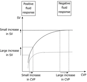
pansion. However, beyond a certain individual threshold, an increase in preload does not increase SV anymore, which corresponds to a preload-independence state. For those patients, fluid administration leads to poor SV improvement but consistent increase in CVP with high risk of fluid overload (Figure 2). Subsequently, volume expansion-induced changes in CVP have been proposed as a safety limit of FC[47,55]. As long as changes in CVP
re-main below 2 mmHg FC is continued until hemodynamic endpoints are fulfilled. For an increase in CVP ranging from 2-5 mmHg, fluid infusion should be stopped for a while then restarted. Over a 5 mmHg increase, FC should be stopped. The time interval to assess filling pressures and fluid responsiveness was every 10 min in the initial description. However, with the availability of continuous vital signs monitoring, the intervals may be extended to 30 min.
FC allows a prompt correction of fluid deficit, with a shorter duration of hypovolemia and organ hypoperfu-sion, compared with a protracted fluid infusion strategy over 12 h or more[55]. FC only requires a central venous
catheter to control safety limits, together with conven-tional monitoring of vital signs and CO if available (Figure 1). Nevertheless, this strategy, although approved by ex-perts and routinely used in intensive care has never been confirmed by a prospective controlled trial[55]. In addition,
despite close monitoring, the effect of fluid infusion is retrospectively assessed, and the repetition of FC might lead to fluid overload. Such risk remains a major concern for patients with AP, as they present an increased risk of acute lung injury[56]. Therefore, fluid responsiveness should
ideally be estimated before fluid is administered to avoid ineffective or deleterious fluid administration for patients
with unclear benefit to risk balance, such as those who de-velop pulmonary, cardiac or renal dysfunction[11,12,57]. New
parameters aiming to predict fluid responsiveness have been developed to this end.
Predicting fluid-responsiveness: preload and preload-dependence
The ultimate goal of tools aiming to predict fluid-respon-siveness is to find where individual ventricular hemody-namic status is located on the Franck-Starling curve (Figure 2). In other terms, indexes predicting fluid responsiveness are assessing cardiac preload-dependence[58].
Based on aforementioned physiological concepts, one could postulate that low preload values are more likely to be associated with preload-dependence and conversely for high preload values. However, several studies show that this assertion is not true. When CVP or pulmonary artery occlusion pressure (PAOP) are used as estimates of cardiac preload, they usually fail to predict fluid re-sponsiveness[45,59]. This can easily be understood because
Franck-Starling curve is specific to each patient[45]. Thus,
there is no way to know whether a single absolute CVP or PAOP level corresponds to a preload-dependence or -independence zone[60] (Figure 2). Even for extreme
values of CVP or PAOP, there is no reliable threshold that can be used in current practice to predict a positive or negative response to volume expansion[45,59]. However,
preload evaluation, and particularly CVP measurements are still recommended in hemodynamic management algorithms for several reasons[34]. First, it is easy to assess,
only requiring a central venous catheter. Second, as de-tailed above dynamic analysis of CVP is still valuable in evaluating FC response. Eventually, CVP values standing below 4 mmHg, even if not predictive of fluid respon-siveness, ensure safe fluid loading with little risk of over-filling[34,61] (Figure 1).
Consequently, indexes predicting fluid-responsiveness focus on preload-dependence rather than preload assess-ment[58]. Passive leg raising (PLR) maneuver is an easy
maneuver that mimics volume expansion by shifting venous blood from the lower limbs and the splanchnic vessels toward the intrathoracic vessels[62]. Thus, PLR
leads to a rapid and reversible increase in cardiac preload and subsequently in SV in case of preload dependence. To be efficient, PLR maneuver has to be performed as follows[63]: the patient’s baseline position is lying down on
a bed, half-sitting in semirecumbent position, with a 45° angle between trunk and lower limbs, which are horizon-tal. Then, a 45° bascule of the bed should be done, so that the trunk becomes horizontal and the lower limbs rise up. Impact of the maneuver appears within the first minute, while the hemodynamic measurements are re-corded. PLR mimics an approximate 300-450 mL FC[63,64].
A close correlation is observed between changes in SV measured with TTE or esophageal Doppler, after PLR and after a 500 mL of fluid loading in critically ill patients with sepsis or acute pancreatitis[64-67]. When considering a
recent meta-analysis enrolling 9 clinical studies that
evalu-Small increase in CVP SV Large increase in CVP CVP Small increase in SV Large increase in SV Positive fluid response Negative fluid response
Figure 2 Schematic representation of central venous pressure/stroke volume of normal (solid line) and failing heart (dotted line). When the heart
is fluid responsive, a fluid challenge induces a large increase in stroke volume (SV) and a small increase in central venous pressure (CVP). When the heart is fluid unresponsive, a fluid challenge induces a small increase in SV and a large increase in CVP. In contrast, there is no reliable threshold of CVP that can be used in current practice to predict a positive or negative response to fluid load-ing. This threshold depends mostly on the cardiac function at the time of fluid infusion.
ated the accuracy of PLR to predict fluid responsive-ness, a PLR-induced change in SV superior to 8%-15% predicted fluid responsiveness with a sensitivity of 89% and specificity of 91%[68]. When considering PP as a
sur-rogate of SV, a PLR-induced change in PP > 9%-12% predicts fluid responsiveness with sensitivity of 60% and a specificity of 86%[68] (Figure 1). The main limit of
the PLR technique is the presence of intra-abdominal hypertension. Indeed, in ventilated critically ill patients, Mahjoub et al[69] showed that PLR failed to predict fluid
responsiveness when intra-abdominal pressure exceeded 16 mmHg due to false negatives. As demonstrated by Kitano et al[70], Takata et al[71] when intra-abdominal
pres-sure exceeds right atrial prespres-sure, the inferior vena cava collapses and impairs venous return. The PLR-induced change in cardiac preload is decreased making the PLR maneuver inefficient[69-71]. As intra-abdominal
hyperten-sion is a common complication of AP, intra-abdominal pressure should be measured before using PLR.
Other indexes based on heart-lung interactions have also been developed. However, they are only validated for mechanically ventilated patients under strict conditions of sedation, ventilation and cardiac rhythm[72]. Because
the proportion of patients requiring mechanical ventila-tion during AP remains very low, with specific multidisci-plinary management in intensive care[56], these parameters
are not discussed in this review. In spontaneously breath-ing patients, respiratory variations in inferior vena cava diameter or PP are still in development[73-75]. As existing
data were not confirmed in large population studies, and as most of them didn’t include patients with AP, the use of such parameters in spontaneously breathing patients with AP seems hazardous and yet to be validated.
The impact of fluid therapy based on preload-depen-dence parameters has been evaluated in studies involving surgical patients[76-78]. When compared with liberal or
based fluid administration, the use of preload-dependence parameters drives to a decrease in lactate level, perioperative complications and time to discharge. Interestingly, this strategy leads either to a greater[77,78] or
to a lesser[76] amount of fluid compared with the control
group. These results suggest that the efficiency of such strategy comes from volume expansion adjustment to patient’s needs rather than from the total amount of fluid administered. Patients involved in these studies were me-chanically ventilated and no similar trial exists in sponta-neously breathing patients. Moreover, there is a great lack of data in patients with AP. Nevertheless, a recent study performed on anesthetized pigs with experimental AP compared a fluid therapy based on preload-dependence indexes to a CVP-based strategy. The fluid therapy guid-ed by preload-dependence parameters increasguid-ed survival (29.4% vs 11.8%; P < 0.05) by preventing
microcircula-tion dysfuncmicrocircula-tion, pancreatic damages and pulmonary edema. These results are concordant with human findings described just before, and confirm the inability of CVP to guide fluid therapy[79]. These encouraging data might
open the way to further research in humans with AP.
CONCLUSION
Adopting an individualized early goal-directed strategy seems very promising to optimize fluid resuscitation in patients with AP. However, since AP has specific patho-physiology, evolution, complications and outcome, fur-ther studies are required to provide a suitable algorithm. The first step will be to define parameters allowing early identification of patients needing EGDT, notably those at risk to develop severe or necrotizing AP. Among pa-rameters previously described in the literature, elevated lactate level and refractory hypotension could be good candidates. The second step is to clearly define ultimate goals of hemodynamic resuscitation reflecting tissue perfusion and oxygenation. If those are not achieved, EGDT should immediately be implemented and car-ried on until adequate systemic perfusion is restored. Close reassessment of initial endpoints has to be per-formed every 30 min to readjust treatment without delay. Because volume expansion plays a major role in this strategy, fluids should be administered early. Inadequate fluid replacement can occur when guided on clinical pa-rameters alone, static preload assessment with CVP or worse, blindly. A safe and practical way to perform fluid loading remains FC, with simultaneous assessment of fluid-responsiveness and control for risk of overload. However, for patients with high risk of fluid overload, predicting fluid-responsiveness before volume expansion may reduce the number of FC and improve patient out-comes. PLR is an accurate validated maneuver to predict fluid-responsiveness. It can widely be used provided the absence of intra-abdominal hypertension. These consid-erations open the way to a wide range of clinical studies aiming to adapt and validate such strategies in the specific population of patients with AP.
REFERENCES
1 Yadav D, Lowenfels AB. The epidemiology of pancreatitis
and pancreatic cancer. Gastroenterology 2013; 144: 1252-1261 [PMID: 23622135 DOI: 10.1053/j.gastro.2013.01.068]
2 Banks PA, Bollen TL, Dervenis C, Gooszen HG, Johnson
CD, Sarr MG, Tsiotos GG, Vege SS. Classification of acute pancreatitis--2012: revision of the Atlanta classification and definitions by international consensus. Gut 2013; 62: 102-111 [PMID: 23100216 DOI: 10.1136/gutjnl-2012-302779]
3 Petrov MS, Shanbhag S, Chakraborty M, Phillips AR,
Wind-sor JA. Organ failure and infection of pancreatic necrosis as determinants of mortality in patients with acute pancreatitis.
Gastroenterology 2010; 139: 813-820 [PMID: 20540942 DOI:
10.1053/j.gastro.2010.06.010]
4 Brown A, Baillargeon JD, Hughes MD, Banks PA. Can fluid
resuscitation prevent pancreatic necrosis in severe acute pancreatitis? Pancreatology 2002; 2: 104-107 [PMID: 12123089 DOI: 10.1159/000055899]
5 Trikudanathan G, Navaneethan U, Vege SS. Current
contro-versies in fluid resuscitation in acute pancreatitis: a system-atic review. Pancreas 2012; 41: 827-834 [PMID: 22781906 DOI: 10.1097/MPA.0b013e31824c1598]
6 Frossard JL, Steer ML, Pastor CM. Acute pancreatitis. Lancet
2008; 371: 143-152 [PMID: 18191686]
micro-circulation in acute pancreatitis. Br J Surg 2006; 93: 518-530 [PMID: 16607683 DOI: 10.1002/bjs.5316]
8 McKay CJ, Imrie CW. The continuing challenge of early
mortality in acute pancreatitis. Br J Surg 2004; 91: 1243-1244 [PMID: 15382103 DOI: 10.1002/bjs.4750]
9 Haydock MD, Mittal A, Wilms HR, Phillips A, Petrov MS,
Windsor JA. Fluid therapy in acute pancreatitis: anybody’ s guess. Ann Surg 2013; 257: 182-188 [PMID: 23207241 DOI: 10.1097/SLA.0b013e31827773ff]
10 Boyd JH, Forbes J, Nakada TA, Walley KR, Russell JA. Fluid resuscitation in septic shock: a positive fluid balance and el-evated central venous pressure are associated with increased mortality. Crit Care Med 2011; 39: 259-265 [PMID: 20975548 DOI: 10.1097/CCM.0b013e3181feeb15]
11 Payen D, de Pont AC, Sakr Y, Spies C, Reinhart K, Vincent JL. A positive fluid balance is associated with a worse out-come in patients with acute renal failure. Crit Care 2008; 12: R74 [PMID: 18533029 DOI: 10.1186/cc6916]
12 Wiedemann HP, Wheeler AP, Bernard GR, Thompson BT, Hayden D, deBoisblanc B, Connors AF, Hite RD, Harabin AL. Comparison of two fluid-management strategies in acute lung injury. N Engl J Med 2006; 354: 2564-2575 [PMID: 16714767 DOI: 10.1056/NEJMoa062200]
13 Jansen TC, van Bommel J, Schoonderbeek FJ, Sleeswijk Viss-er SJ, van dViss-er KloostViss-er JM, Lima AP, Willemsen SP, BakkViss-er J. Early lactate-guided therapy in intensive care unit patients: a multicenter, open-label, randomized controlled trial. Am J
Respir Crit Care Med 2010; 182: 752-761 [PMID: 20463176 DOI:
10.1164/rccm.200912-1918OC]
14 Rivers E, Nguyen B, Havstad S, Ressler J, Muzzin A, Knoblich B, Peterson E, Tomlanovich M. Early goal-directed therapy in the treatment of severe sepsis and septic shock. N Engl J
Med 2001; 345: 1368-1377 [PMID: 11794169 DOI:
10.1056/NEJ-Moa010307]
15 Tenner S, Baillie J, DeWitt J, Vege SS. American College of Gastroenterology guideline: management of acute pancreati-tis. Am J Gastroenterol 2013; 108: 1400-1416 [PMID: 23896955 DOI: 10.1038/ajg.2013.218]
16 Warndorf MG, Kurtzman JT, Bartel MJ, Cox M, Mackenzie T, Robinson S, Burchard PR, Gordon SR, Gardner TB. Early fluid resuscitation reduces morbidity among patients with acute pancreatitis. Clin Gastroenterol Hepatol 2011; 9: 705-709 [PMID: 21554987 DOI: 10.1016/j.cgh.2011.03.032]
17 Wall I, Badalov N, Baradarian R, Iswara K, Li JJ, Tenner S. Decreased mortality in acute pancreatitis related to early ag-gressive hydration. Pancreas 2011; 40: 547-550 [PMID: 21499208 DOI: 10.1097/MPA.0b013e318215368d]
18 Peery AF, Dellon ES, Lund J, Crockett SD, McGowan CE, Bulsiewicz WJ, Gangarosa LM, Thiny MT, Stizenberg K, Mor-gan DR, Ringel Y, Kim HP, Dibonaventura MD, Carroll CF, Allen JK, Cook SF, Sandler RS, Kappelman MD, Shaheen NJ. Burden of gastrointestinal disease in the United States: 2012 update. Gastroenterology 2012; 143: 1179-1187.e1-e3 [PMID: 22885331 DOI: 10.1053/j.gastro.2012.08.002]
19 Baillargeon JD, Orav J, Ramagopal V, Tenner SM, Banks PA. Hemoconcentration as an early risk factor for necrotiz-ing pancreatitis. Am J Gastroenterol 1998; 93: 2130-2134 [PMID: 9820385 DOI: 10.1111/j.1572-0241.1998.00608.x]
20 Solanki NS, Barreto SG. Fluid therapy in acute pancreati-tis. A systematic review of literature. JOP 2011; 12: 205-208 [PMID: 21386654]
21 Banks PA, Freeman ML. Practice guidelines in acute pan-creatitis. Am J Gastroenterol 2006; 101: 2379-2400 [PMID: 17032204 DOI: 10.1111/j.1572-0241.2006.00856.x]
22 Mao EQ, Fei J, Peng YB, Huang J, Tang YQ, Zhang SD. Rapid hemodilution is associated with increased sepsis and mortality among patients with severe acute pancreatitis.
Chin Med J (Engl) 2010; 123: 1639-1644 [PMID: 20819621]
23 Vincent JL, Sakr Y, Sprung CL, Ranieri VM, Reinhart K, Gerlach H, Moreno R, Carlet J, Le Gall JR, Payen D.
Sep-sis in European intensive care units: results of the SOAP study. Crit Care Med 2006; 34: 344-353 [PMID: 16424713 DOI: 10.1097/01.CCM.0000194725.48928.3A]
24 Prowle JR, Echeverri JE, Ligabo EV, Ronco C, Bellomo R. Fluid balance and acute kidney injury. Nat Rev Nephrol 2010;
6: 107-115 [PMID: 20027192 DOI: 10.1038/nrneph.2009.213]
25 Silva JM, de Oliveira AM, Nogueira FA, Vianna PM, Pereira Filho MC, Dias LF, Maia VP, Neucamp Cde S, Amendola CP, Carmona MJ, Malbouisson LM. The effect of excess fluid bal-ance on the mortality rate of surgical patients: a multicenter prospective study. Crit Care 2013; 17: R288 [PMID: 24326085 DOI: 10.1186/cc13151]
26 Dellinger RP, Levy MM, Rhodes A, Annane D, Gerlach H, Opal SM, Sevransky JE, Sprung CL, Douglas IS, Jaeschke R, Osborn TM, Nunnally ME, Townsend SR, Reinhart K, Kleinpell RM, Angus DC, Deutschman CS, Machado FR, Rubenfeld GD, Webb S, Beale RJ, Vincent JL, Moreno R. Sur-viving Sepsis Campaign: international guidelines for man-agement of severe sepsis and septic shock, 2012. Intensive
Care Med 2013; 39: 165-228 [PMID: 23361625 DOI: 10.1007/
s00134-012-2769-8]
27 Rivers EP, Coba V, Whitmill M. Early goal-directed therapy in severe sepsis and septic shock: a contemporary review of the literature. Curr Opin Anaesthesiol 2008; 21: 128-140 [PMID: 18443478 DOI: 10.1097/ACO.0b013e3282f4db7a]
28 Shoemaker WC, Appel P, Bland R. Use of physiologic moni-toring to predict outcome and to assist in clinical decisions in critically ill postoperative patients. Am J Surg 1983; 146: 43-50 [PMID: 6346913]
29 Giglio MT, Marucci M, Testini M, Brienza N. Goal-directed haemodynamic therapy and gastrointestinal complications in major surgery: a meta-analysis of randomized controlled trials. Br J Anaesth 2009; 103: 637-646 [PMID: 19837807 DOI: 10.1093/bja/aep279]
30 Pearse R, Dawson D, Fawcett J, Rhodes A, Grounds RM, Bennett ED. Early goal-directed therapy after major surgery reduces complications and duration of hospital stay. A ran-domised, controlled trial [ISRCTN38797445]. Crit Care 2005; 9: R687-R693 [PMID: 16356219 DOI: 10.1186/cc3887]
31 Arulkumaran N, Corredor C, Hamilton MA, Ball J, Grounds RM, Rhodes A, Cecconi M. Cardiac complications associated with goal-directed therapy in high-risk surgical patients: a meta-analysis. Br J Anaesth 2014; 112: 648-659 [PMID: 24413429 DOI: 10.1093/bja/aet466]
32 Aya HD, Cecconi M, Hamilton M, Rhodes A. Goal-directed therapy in cardiac surgery: a systematic review and meta-analysis. Br J Anaesth 2013; 110: 510-517 [PMID: 23447502 DOI: 10.1093/bja/aet020]
33 Yadav D, Garg PK. Spectrum of perforation peritonitis in delhi: 77 cases experience. Indian J Surg 2013; 75: 133-137 [PMID: 24426408]
34 Antonelli M, Levy M, Andrews PJ, Chastre J, Hudson LD, Manthous C, Meduri GU, Moreno RP, Putensen C, Stewart T, Torres A. Hemodynamic monitoring in shock and implica-tions for management. International Consensus Conference, Paris, France, 27-28 April 2006. Intensive Care Med 2007; 33: 575-590 [PMID: 17285286 DOI: 10.1007/s00134-007-0531-4] 35 Buter A, Imrie CW, Carter CR, Evans S, McKay CJ. Dynamic
nature of early organ dysfunction determines outcome in acute pancreatitis. Br J Surg 2002; 89: 298-302 [PMID: 11872053 DOI: 10.1046/j.0007-1323.2001.02025.x]
36 de-Madaria E, Martínez J, Pérez-Mateo M. The dynamic na-ture of fluid resuscitation in acute pancreatitis. Clin
Gastroen-terol Hepatol 2012; 10: 95-6; author reply 96 [PMID: 21888883
DOI: 10.1016/j.cgh.2011.08.020]
37 Tonsi AF, Bacchion M, Crippa S, Malleo G, Bassi C. Acute pancreatitis at the beginning of the 21st century: the state of the art. World J Gastroenterol 2009; 15: 2945-2959 [PMID: 19554647 DOI: 10.3748/wjg.15.2945]
Clain JE, Petersen BT, Baron TH, Farnell MB, Sarr MG. Low mortality and high morbidity in severe acute pancreatitis without organ failure: a case for revising the Atlanta clas-sification to include “moderately severe acute pancreatitis”.
Am J Gastroenterol 2009; 104: 710-715 [PMID: 19262525 DOI:
10.1038/ajg.2008.77]
39 Mégarbane B. Severe lactic acidosis except for shock states.
Réanimation 2013; 22: 435-445 [DOI: 10.1007/s13546-013-0654-2]
40 Hernandez G, Boerma EC, Dubin A, Bruhn A, Koopmans M, Edul VK, Ruiz C, Castro R, Pozo MO, Pedreros C, Veas E, Fuentealba A, Kattan E, Rovegno M, Ince C. Severe abnor-malities in microvascular perfused vessel density are asso-ciated to organ dysfunctions and mortality and can be pre-dicted by hyperlactatemia and norepinephrine requirements in septic shock patients. J Crit Care 2013; 28: 538.e9-538.14 [PMID: 23566729 DOI: 10.1016/j.jcrc.2012.11.022]
41 Andersen LW, Mackenhauer J, Roberts JC, Berg KM, Coc-chi MN, Donnino MW. Etiology and therapeutic approach to elevated lactate levels. Mayo Clin Proc 2013; 88: 1127-1140 [PMID: 24079682 DOI: 10.1016/j.mayocp.2013.06.012] 42 Ait-Oufella H, Bourcier S, Alves M, Galbois A, Baudel
JL, Margetis D, Bige N, Offenstadt G, Maury E, Guidet B. Alteration of skin perfusion in mottling area during septic shock. Ann Intensive Care 2013; 3: 31 [PMID: 24040941 DOI: 10.1186/2110-5820-3-31]
43 Ait-Oufella H, Lemoinne S, Boelle PY, Galbois A, Baudel JL, Lemant J, Joffre J, Margetis D, Guidet B, Maury E, Offenstadt G. Mottling score predicts survival in septic shock. Intensive
Care Med 2011; 37: 801-807 [PMID: 21373821 DOI: 10.1007/
s00134-011-2163-y]
44 Teboul JL, Hamzaoui O, Monnet X. SvO2 to monitor re-suscitation of septic patients: let’s just understand the basic physiology. Crit Care 2011; 15: 1005 [PMID: 22078239 DOI: 10.1186/cc10491]
45 Michard F, Teboul JL. Predicting fluid responsiveness in ICU patients: a critical analysis of the evidence. Chest 2002; 121: 2000-2008 [PMID: 12065368 DOI: 10.1378/chest.121.6.2000] 46 Maitland K, Kiguli S, Opoka RO, Engoru C, Olupot-Olupot
P, Akech SO, Nyeko R, Mtove G, Reyburn H, Lang T, Brent B, Evans JA, Tibenderana JK, Crawley J, Russell EC, Levin M, Babiker AG, Gibb DM. Mortality after fluid bolus in Af-rican children with severe infection. N Engl J Med 2011; 364: 2483-2495 [PMID: 21615299 DOI: 10.1056/NEJMoa1101549] 47 Weil MH, Henning RJ. New concepts in the diagnosis and
fluid treatment of circulatory shock. Thirteenth annual Bec-ton, Dickinson and Company Oscar Schwidetsky Memorial Lecture. Anesth Analg 1979; 58: 124-132 [PMID: 571235] 48 Teboul JL, Asfar P, Bernardin G, Cariou A, Chemla D.
Indi-cateurs du remplissage vasculaire au cours de l’insuffisance circulatoire. Réanimation 2004; 13: 255-263
49 Pottecher T, Calvat S, Dupont H, Durand-Gasselin J, Ger-beaux P. Haemodynamic management of severe sepsis: rec-ommendations of the French Intensive Care Societies (SFAR/ SRLF) Consensus Conference, 13 October 2005, Paris, France.
Crit Care 2006; 10: 311 [PMID: 16941754 DOI: 10.1186/cc4965]
50 Vallet B, Blanloeil Y, Cholley B, Orliaguet G, Pierre S, Tav-ernier B. Guidelines for perioperative haemodynamic opti-mization. Ann Fr Anesth Reanim 2013; 32: e151-e158 [PMID: 24126197 DOI: 10.1016/j.annfar.2013.09.010]
51 Phan TD, Ismail H, Heriot AG, Ho KM. Improving periop-erative outcomes: fluid optimization with the esophageal Doppler monitor, a metaanalysis and review. J Am Coll Surg 2008; 207: 935-941 [PMID: 19183542 DOI: 10.1016/j.jamcollsu rg.2008.08.007]
52 Lewis JF, Kuo LC, Nelson JG, Limacher MC, Quinones MA. Pulsed Doppler echocardiographic determination of stroke volume and cardiac output: clinical validation of two new methods using the apical window. Circulation 1984; 70: 425-431 [PMID: 6744546]
53 Lakhal K, Ehrmann S, Perrotin D, Wolff M, Boulain T. Fluid
challenge: tracking changes in cardiac output with blood pressure monitoring (invasive or non-invasive). Intensive
Care Med 2013; 39: 1953-1962 [PMID: 24061631 DOI: 10.1007/
s00134-013-3086-6]
54 Monnet X, Letierce A, Hamzaoui O, Chemla D, Anguel N, Osman D, Richard C, Teboul JL. Arterial pressure al-lows monitoring the changes in cardiac output induced by volume expansion but not by norepinephrine. Crit Care
Med 2011; 39: 1394-1399 [PMID: 21336124 DOI: 10.1097/
CCM.0b013e31820edcf0]
55 Vincent JL, Weil MH. Fluid challenge revisited. Crit Care
Med 2006; 34: 1333-1337 [PMID: 16557164 DOI: 10.1097/01.
CCM.0000214677.76535.A5]
56 Mole DJ, Olabi B, Robinson V, Garden OJ, Parks RW. Inci-dence of individual organ dysfunction in fatal acute pancre-atitis: analysis of 1024 death records. HPB (Oxford) 2009; 11: 166-170 [PMID: 19590643 DOI: 10.1111/j.1477-2574.2009.00038. x]
57 Charpentier J, Luyt CE, Fulla Y, Vinsonneau C, Cariou A, Grabar S, Dhainaut JF, Mira JP, Chiche JD. Brain natriuretic peptide: A marker of myocardial dysfunction and prognosis during severe sepsis. Crit Care Med 2004; 32: 660-665 [PMID: 15090944 DOI: 10.1097/01.CCM.0000114827.93410.D8] 58 Cherpanath TG, Geerts BF, Lagrand WK, Schultz MJ,
Groeneveld AB. Basic concepts of fluid responsiveness. Neth
Heart J 2013; 21: 530-536 [PMID: 24170232]
59 Kumar A, Anel R, Bunnell E, Habet K, Zanotti S, Marshall S, Neumann A, Ali A, Cheang M, Kavinsky C, Parrillo JE. Pulmonary artery occlusion pressure and central venous pressure fail to predict ventricular filling volume, cardiac performance, or the response to volume infusion in normal subjects. Crit Care Med 2004; 32: 691-699 [PMID: 15090949 DOI: 10.1097/01.CCM.0000114996.68110.C9]
60 Tavernier B, Makhotine O, Lebuffe G, Dupont J, Scherpereel P. Systolic pressure variation as a guide to fluid therapy in patients with sepsis-induced hypotension. Anesthesiology 1998; 89: 1313-1321 [PMID: 9856704]
61 Marik PE, Cavallazzi R. Does the central venous pressure predict fluid responsiveness? An updated meta-analysis and a plea for some common sense. Crit Care Med 2013; 41: 1774-1781 [PMID: 23774337 DOI: 10.1097/CCM.0b013e31828a25fd] 62 Rutlen DL, Wackers FJ, Zaret BL. Radionuclide assessment
of peripheral intravascular capacity: a technique to measure intravascular volume changes in the capacitance circulation in man. Circulation 1981; 64: 146-152 [PMID: 6786793] 63 Jabot J, Teboul JL, Richard C, Monnet X. Passive leg
rais-ing for predictrais-ing fluid responsiveness: importance of the postural change. Intensive Care Med 2009; 35: 85-90 [PMID: 18795254 DOI: 10.1007/s00134-008-1293-3]
64 Préau S, Saulnier F, Dewavrin F, Durocher A, Chagnon JL. Passive leg raising is predictive of fluid responsiveness in spontaneously breathing patients with severe sepsis or acute pancreatitis. Crit Care Med 2010; 38: 819-825 [PMID: 20016380 DOI: 10.1097/CCM.0b013e3181c8fe7a]
65 Monnet X, Rienzo M, Osman D, Anguel N, Richard C, Pin-sky MR, Teboul JL. Passive leg raising predicts fluid respon-siveness in the critically ill. Crit Care Med 2006; 34: 1402-1407 [PMID: 16540963 DOI: 10.1097/01.CCM.0000215453.11735.06] 66 Lamia B, Ochagavia A, Monnet X, Chemla D, Richard C, Teboul JL. Echocardiographic prediction of volume respon-siveness in critically ill patients with spontaneously breath-ing activity. Intensive Care Med 2007; 33: 1125-1132 [PMID: 17508199 DOI: 10.1007/s00134-007-0646-7]
67 Maizel J, Airapetian N, Lorne E, Tribouilloy C, Massy Z, Slama M. Diagnosis of central hypovolemia by using passive leg raising. Intensive Care Med 2007; 33: 1133-1138 [PMID: 17508202 DOI: 10.1007/s00134-007-0642-y]
68 Cavallaro F, Sandroni C, Marano C, La Torre G, Mannocci A, De Waure C, Bello G, Maviglia R, Antonelli M. Diagnostic accuracy of passive leg raising for prediction of fluid
respon-siveness in adults: systematic review and meta-analysis of clinical studies. Intensive Care Med 2010; 36: 1475-1483 [PMID: 20502865 DOI: 10.1007/s00134-010-1929-y]
69 Mahjoub Y, Touzeau J, Airapetian N, Lorne E, Hijazi M, Zogheib E, Tinturier F, Slama M, Dupont H. The passive leg-raising maneuver cannot accurately predict fluid respon-siveness in patients with intra-abdominal hypertension. Crit
Care Med 2010; 38: 1824-1829 [PMID: 20639753 DOI: 10.1097/
CCM.0b013e3181eb3c21]
70 Kitano Y, Takata M, Sasaki N, Zhang Q, Yamamoto S, Mi-yasaka K. Influence of increased abdominal pressure on steady-state cardiac performance. J Appl Physiol (1985) 1999;
86: 1651-1656 [PMID: 10233131]
71 Takata M, Robotham JL. Effects of inspiratory diaphrag-matic descent on inferior vena caval venous return. J Appl
Physiol (1985) 1992; 72: 597-607 [PMID: 1559938]
72 Marik PE, Lemson J. Fluid responsiveness: an evolution of our understanding. Br J Anaesth 2014; 112: 617-620 [PMID: 24535603 DOI: 10.1093/bja/aet590]
73 Muller L, Bobbia X, Toumi M, Louart G, Molinari N, Ragon-net B, Quintard H, Leone M, Zoric L, Lefrant JY. Respiratory variations of inferior vena cava diameter to predict fluid re-sponsiveness in spontaneously breathing patients with acute circulatory failure: need for a cautious use. Crit Care 2012; 16: R188 [PMID: 23043910 DOI: 10.1186/cc11672]
74 Préau S, Dewavrin F, Soland V, Bortolotti P, Colling D, Cha-gnon JL, Durocher A, Saulnier F. Hemodynamic changes dur-ing a deep inspiration maneuver predict fluid responsiveness
in spontaneously breathing patients. Cardiol Res Pract 2012;
2012: 191807 [PMID: 22195286 DOI: 10.1155/2012/191807]
75 Monge García MI, Gil Cano A, Díaz Monrové JC. Arterial pressure changes during the Valsalva maneuver to predict fluid responsiveness in spontaneously breathing patients.
Intensive Care Med 2009; 35: 77-84 [PMID: 18830578 DOI:
10.1007/s00134-008-1294-2]
76 Forget P, Lois F, de Kock M. Goal-directed fluid manage-ment based on the pulse oximeter-derived pleth variability index reduces lactate levels and improves fluid manage-ment. Anesth Analg 2010; 111: 910-914 [PMID: 20705785 DOI: 10.1213/ANE.0b013e3181eb624f]
77 Lopes MR, Oliveira MA, Pereira VO, Lemos IP, Auler JO, Michard F. Goal-directed fluid management based on pulse pressure variation monitoring during high-risk surgery: a pilot randomized controlled trial. Crit Care 2007; 11: R100 [PMID: 17822565 DOI: 10.1186/cc6117]
78 Benes J, Chytra I, Altmann P, Hluchy M, Kasal E, Svitak R, Pradl R, Stepan M. Intraoperative fluid optimization using stroke volume variation in high risk surgical patients: results of prospective randomized study. Crit Care 2010; 14: R118 [PMID: 20553586 DOI: 10.1186/cc9070]
79 Trepte CJ, Bachmann KA, Stork JH, Friedheim TJ, Hinsch A, Goepfert MS, Mann O, Izbicki JR, Goetz AE, Reuter DA. The impact of early goal-directed fluid management on sur-vival in an experimental model of severe acute pancreatitis.
Intensive Care Med 2013; 39: 717-726 [PMID: 23287870 DOI:
10.1007/s00134-012-2775-x]
P- Reviewer: Capolongo G, Pellicano R S- Editor: Qi Y L- Editor: A E- Editor: Wang CH
8226 Regency Drive, Pleasanton, CA 94588, USA
Telephone: +1-925-223-8242
Fax: +1-925-223-8243
E-mail: [email protected]
Help Desk: http://www.wjgnet.com/esps/helpdesk.aspx
http://www.wjgnet.com
I S S N 1 0 0 7 - 9 3 2 7
9 7 7 1 0 07 9 3 2 0 45 4 3