HEAD AND NECK
Diffusion-weighted MR imaging including bi-exponential
fitting for the detection of recurrent or residual tumour
after (chemo)radiotherapy for laryngeal and hypopharyngeal
cancers
Dechen W. Tshering Vogel&Peter Zbaeren&
Andreas Geretschlaeger&Peter Vermathen&
Frederik De Keyzer&Harriet C. Thoeny
Received: 8 May 2012 / Revised: 25 June 2012 / Accepted: 2 July 2012 / Published online: 4 August 2012 # European Society of Radiology 2012
Abstract
Objectives To assess whether diffusion-weighted magnetic resonance imaging (DW-MRI) including bi-exponential fit-ting helps to detect residual/recurrent tumours after (chemo)radiotherapy of laryngeal and hypopharyngeal carcinoma.
Methods Forty-six patients with newly-developed/worsen-ing symptoms after (chemo)radiotherapy for laryngeal/ hypopharyngeal cancers were prospectively imaged using conventional MRI and axial DW-MRI. Qualitative (visual assessment) and quantitative analysis (mono-exponentially: total apparent diffusion coefficient [ADCT], and
bi-exponentially: perfusion fraction [FP] and true diffusion
coefficient [ADCD]) were performed. Diffusion parameters
of tumour versus post-therapeutic changes were compared, with final diagnosis based on histopathology and follow-up. Mann-Whitney U test was used for statistical analysis. Results Qualitative DW-MRI combined with morphological images allowed the detection of tumour with a sensitivity of 94% and specificity 100%. ADCTand ADCDvalues were
lower in tumour with values 120±49×10−5mm2/s and 113± 50 × 10−5 mm2/s, respectively, compared with post-therapeutic changes with values 182 ± 41 × 10−5 mm2/s (P < 0.0002) and 160 ± 47 × 10−5 mm2/s (P < 0.003), respectively. FP values were significantly lower in
tumours than in non-tumours (13 ± 9% versus 31 ± 16%, P < 0.0002), with FP being the best quantitative
parame-ter for differentiation between post-therapeutic changes and recurrence.
Conclusions DW-MRI in combination with conventional MRI substantially improves detection and exclusion of tu-mour in patients with laryngeal and hypopharyngeal cancers after treatment with (chemo)radiotherapy on both qualitative and quantitative analysis, with FPbeing the best quantitative
parameter in this context. Key Points
• DW-MRI is increasingly used to detect tumour recurrence. • DW-MRI allows accurate post-treatment recurrence
detec-tion in laryngeal or hypopharyngeal cancer
• ADC values in recurrent tumour are lower than in benign tissue alterations
• Both qualitative and quantitative DW-MRI approaches allow detection of recurrence
• DW-MRI can easily be added to daily clinical routine imaging
D. W. Tshering Vogel
:
P. Vermathen:
H. C. Thoeny (*) Department of Diagnostic, Interventional and Paediatric Radiology, Inselspital, University of Bern,Freiburgstrasse 10, 3010 Bern, Switzerland e-mail: [email protected] P. Zbaeren
Department of Oto-Rhino-Laryngology, Head and Neck Surgery, Inselspital, University of Bern,
Bern, Switzerland A. Geretschlaeger
Department of Radiation Oncology, Inselspital, University of Bern,
Bern, Switzerland F. De Keyzer
Department of Radiology, University Hospitals Leuven, Leuven, Belgium
Keywords (Chemo)radiotherapy . Cancer . Diffusion-weighted MRI . Apparent diffusion coefficient . Perfusion fraction
Introduction
Patients with cancer of the larynx and hypopharynx are increasingly treated with (chemo)radiotherapy, aiming to preserve organ function and improve quality of life [1,2]. However, it is well known that (chemo)radiotherapy, in addition to its anti-tumoral effects, also results in local oedema, inflammation, fibrosis and, less commonly, necro-sis [2–4]. These secondary changes pose a problem, not only for the patient’s quality of life, but also for the post-treatment assessment of anti-tumour efficacy. Although de-tection of residual/recurrent tumour against this background is very challenging and a well-known diagnostic dilemma for both clinicians and radiologists [1, 4–8], early tumour assessment can increase the chance of curative salvage surgery, sometimes allowing preservation of laryngeal func-tion [4, 5]. Clinical evaluation of possible post-treatment recurrence is difficult owing to oedema obstructing evalua-tion of the larynx, and owing to the swelling and induraevalua-tion of the soft tissues preventing adequate palpation. Moreover, imaging evaluation is also hampered, as enhancing benign focal mass-like lesions can mimic residual/recurrent tumour [3], leading to possible false-positive diagnosis and unnec-essary surgery [9]. Adversely, some residual or recurrent tumours do not enhance after contrast medium administra-tion, further confounding the issue [10].
The most reliable method of detecting recurrent tumour was considered to be direct laryngoscopy under general anaesthesia [5], but this technique is limited as it can miss recurrent tumour directly under the mucosa or wrongly assess the multifocal nature of the tumour. While a positive biopsy is conclusive, a negative biopsy is not sufficient to exclude tumour [2,6]. A non-invasive imaging test allowing accurate and comprehensive post-treatment lesion character-isation could therefore be used to select those patients who might benefit from a laryngoscopy, thereby limiting unnec-essary biopsies and patient morbidity, and may even indicate the most appropriate biopsy location.
Diffusion-weighted magnetic resonance imaging (DW-MRI) has only recently been introduced in the evaluation of head and neck cancers [11]. It is based on the relative mobility of water molecules in different tissues and provides information about the microenvi-ronment such as cell density, cell integrity and vascu-larity. Initial results suggest that this non-invasive test may be more specific than FDG-PET in the detection of tumour recurrence or persistence after treatment of var-ious head and neck tumours [7, 12].
The present prospective study is aimed at assessing the ability of qualitative and quantitative DW-MRI, including bi-exponential fitting based on the intravoxel incoherent motion theory [13,14], to detect residual/recurrent tumour against the background of treatment-induced changes in a well-defined group of patients with laryngeal and hypophar-yngeal tumours after (chemo)radiotherapy.
Methods and materials
Approval
The local ethics committee approved the study protocol and written informed consent was obtained from all participants.
Patients
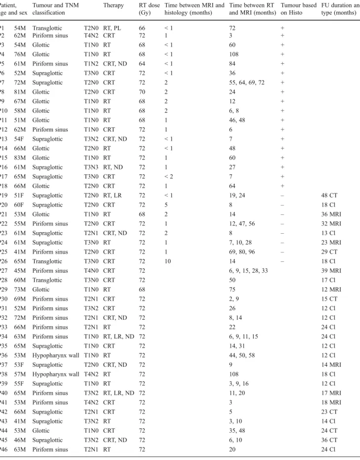
Fifty consecutive patients with laryngeal or hypophar-yngeal cancers, presenting with newly-developed or worsening symptoms after (chemo)radiotherapy, between May 2007 and December 2010, prospectively underwent MRI of the neck to exclude residual/recurrent tumour. (Chemo)radiotherapy was performed with total doses of 66–72 Gy (5×2 Gy/week). Patients’ symptoms included pain, stridor, increasing hoarseness, and breathing and swallowing difficulties. Four patients were excluded owing to susceptibility artefacts compromising image quality, leaving a group of 46 patients (41 men, 5 women, median age 61 years, range 41–83 years) with laryngeal (n030) or hypopharyngeal cancer (n016). Of the 30 patients with laryngeal cancers, 11 were staged as T1, 10 as T2 and 9 as T3. Of the 16 patients with hypopharyngeal cancer, 4 were staged as T1 disease, 6 as T2, 2 as T3 and 4 as T4. All patients were treated with fractionated radiotherapy: alone (n017), or after surgery of the larynx (n04), or alongside chemotherapy with either methotrexate or cisplatin (n025). Nine patients also had a neck dissection. All patient and tumour characteristics are presented in Table 1.
MRI
MRI was performed on a 1.5-T unit (Sonata; Siemens, Erlangen, Germany) using a dedicated neck coil. The con-ventional MRI included an axial T1-weighted turbospin-echo (TSE) sequence (slice thickness, 3 mm; 24 slices; intersection gap, 0.6 mm; repetition time/echo time [TR/ TE], 624 ms/12 ms; matrix, 512 × 256; field of view [FoV], 280×170 mm) and an axial T2-weighted TSE se-quence (slice thickness, 3 mm; 24 slices; intersection gap, 0.6 mm; TR/TE, 3,630 ms/76 ms; matrix, 512×256; FoV, 280 × 170 mm), covering the larynx. After gadolinium
Table 1 Patient and tumour characteristics Patient,
age and sex
Tumour and TNM classification
Therapy RT dose (Gy)
Time between MRI and histology (months)
Time between RT and MRI (months)
Tumour based on Histo FU duration and type (months) P1 54M Transglottic T2N0 RT, PL 66 < 1 72 + P2 62M Piriform sinus T4N2 CRT 72 1 3 + P3 54M Glottic T1N0 RT 68 < 1 60 + P4 76M Glottic T1N0 RT 68 < 1 108 + P5 61M Piriform sinus T1N2 CRT, ND 64 < 1 84 + P6 52M Supraglottic T3N0 CRT 72 < 1 36 + P7 72M Supraglottic T2N0 CRT 72 2 55, 64, 69, 72 + P8 81M Glottic T2N0 CRT 70 2 24 + P9 67M Glottic T1N0 RT 68 2 12 + P10 58M Glottic T1N0 RT 68 2 6, 8 + P11 51M Glottic T1N0 RT 68 1 46, 48 + P12 62M Piriform sinus T1N0 CRT 72 1 6 + P13 54F Supraglottic T3N2 CRT, ND 72 < 1 7 + P14 66M Glottic T2N0 RT 72 < 1 48 + P15 83M Glottic T1N0 RT 72 1 60 + P16 61M Supraglottic T3N3 RT, ND 72 1 27 + P17 65M Supraglottic T3N0 CRT 72 < 2 7 + P18 66M Glottic T2N0 CRT 72 1 64 + P19 51F Supraglottic T2N0 RT, LR 72 < 1 19, 24 – 48 CT P20 60F Supraglottic T2N0 CRT 72 5 8 – 18 Cl P21 53M Glottic T1N0 RT 68 2 14 – 36 MRI
P22 55M Piriform sinus T2N0 CRT 72 1 12, 47, 56 – 32 MRI
P23 61M Supraglottic T2N1 CRT, ND 72 2 8 – 13 Cl
P24 61M Supraglottic T3N0 RT 72 1 7, 10, 28 – 23 MRI
P25 41M Piriform sinus T2N0 CRT 72 1 69, 80, 96 – 29 CT
P26 65M Transglottic T3N0 CRT 72 10 14 – 18 Cl
P27 45M Piriform sinus T4N0 CRT 72 6, 9, 15, 28, 33 39 MRI
P28 60M Transglottic T3N0 CRT 72 50 17 Cl P29 73M Glottic T1N0 RT 68 75 12 MRI P30 69M Piriform sinus T2N1 CRT 72 2, 9 15 CT P31 52M Piriform sinus T3N2 CRT 72 26 12 Cl P32 72M Piriform sinus T2N1 CRT, ND 72 8, 14 12 Cl P33 66M Piriform sinus T2N1 RT 72 22 24 Cl P34 63M Piriform sinus T1N0 RT, LR, ND 72 6, 9, 11, 15 24 Cl P35 65M Supraglottic T1N0 CRT 72 14, 31 12 Cl P36 53M Hypopharynx wall T1N0 RT 72 44, 50, 58 12 Cl P37 53F Supraglottic T2N0 CRT, ND 72 9 14 MRI P38 57M Hypopharynx wall T4N2 RT 72 108 18 Cl P39 55F Supraglottic T1N0 RT 72 3, 9, 16 12 Cl
P40 65M Piriform sinus T3N2 RT, LR, ND 72 11, 20 17 MRI
P41 53M Piriform sinus T4N2 CRT 72 3 18 MRI
P42 66M Supraglottic T2N1 CRT 72 5 23 CT
P43 41M Supraglottic T3N2 RT 72 3, 10 14 Cl
P44 53M Glottic T1N0 CRT 72 35, 48 24 CT
P45 46M Supraglottic T3N2 CRT, ND 72 6, 10 36 CT
P46 63M Piriform sinus T2N1 RT 72 20 24 Cl
MRI magnetic resonance imaging, RT radiotherapy, Histo histopathology, FU follow-up, M male, PL partial laryngectomy, + positive, CRT (chemo)radiotherapy, F female, ND neck dissection, LR laser resection,− negative, CT computed tomography, Cl clinical
injection, T1-weighted fat-saturated sequences were per-formed in the axial plane (using identical parameters as pre-contrast medium administration) and in the coronal or sagittal plane (19 slices; slice thickness, 3 mm; intersection gap, 0.75 mm; TR/TE, 630 ms/18 ms; matrix, 512×256; field of view, 320×250 mm). Echo-planar DW-MRI was performed in the axial plane, using the above-mentioned slice geometry, and 4 signals acquired, TR/TE, 3,500 ms/69 ms; matrix, 128×104; FoV, 290×290 mm; bandwidth 2,056 Hz per pixel. Six diffusion gradient b values (0, 50, 100, 500, 750 and 1,000 s/mm2) were applied in three orthogonal directions, minimising the effects of diffusion anisotropy.
Image analysis
Conventional MR images were evaluated on an EasyVision picture archiving and communication system workstation (Philips Medical Systems, Netherlands). Baseline computed tomography (CT) and MRI, as well as repeat examinations after treatment, were available for all patients. CT and MRI examinations were evaluated by two experienced radiolog-ists in consensus (first and last authors, 7 and 12 years of experience in head and neck radiology, respectively). Changes at the tumour site, tissue swelling (focal or diffuse), size changes and enhancement pattern were assessed. Focal areas of enhancement increasing in size were suspicious for tumour, whereas size decrease indicating treatment response and absence of growth on follow-up imaging was consid-ered compatible with post-therapeutic changes, although small residual tumour foci could not be excluded.
The qualitative DW-MRI analysis was performed in con-junction with the conventional images as the poor spatial
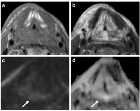
resolution of DW-MRI precludes interpretation on its own. DW-MR images at a b value of 1,000 s/mm2 (b-1,000 images) and the corresponding apparent diffusion coeffi-cient (ADC) maps were matched to and evaluated with the morphological images. The primary tumour site was evalu-ated. Hyperintense signal on the b-1,000 image compared with the surrounding tissue with corresponding low signal intensity in the matching ADC map was considered positive for tumour (Fig.1). High signal intensity on b-1,000 images with corresponding high signal on the matching ADC map was considered to represent T2 shine-through and therefore no tumour (Fig. 2). Absence of hyperintensity on the b-1,000 image was also considered negative for tumour (Fig.3).
Quantitative DW-MRI analysis was performed on a pixel-by-pixel basis in two ways as previously described [13,14], yielding total ADC (ADCT) by mono-exponential fitting,
and the perfusion fraction (FP) and true diffusion coefficient
(ADCD) by bi-exponential fitting. Using mono-exponential
fitting, the ADCT value is calculated from all the data
obtained, ignoring different contributions of diffusion and perfusion, which is most commonly used in the literature. Acquiring multiple b values allows separating diffusion and micro-perfusion contributions by bi-exponential fitting. This yields FP, which represents the contribution of
microcircu-lation of blood and movement in pre-defined structures to the signal decay and the diffusion component ADCD, which
mainly reflects true diffusion [14].
Elliptical regions of interest (ROIs) were placed on areas of diffusion impediment taking care to exclude areas of obvious necrosis. If no areas showed diffusion restriction on the b-1000 images, ROIs were placed at or near the
Fig. 1 MR images of an 81-year-old man with hoarseness after radiotherapy for a laryn-geal tumour. Axial T1-weighted (a) and T2-weighted images (b) showing heterogeneous signal at the right thyroid cartilage and surrounding soft tissues. A fo-cal bright area is visible on the axial b-1,000 image (c) with hypointense signal on the corresponding ADC map (d) indicating focal diffusion im-pediment, implying tumour (arrows)
original site of the tumour on the basis of the morphological images regardless of the signal intensity on the DW-MR images. The ROI size varied according to the size of the suspicious lesion (mean size 124±53 mm3).
Histopathology
Imaging results were compared with histopathology and follow-up. Eighteen patients had histopathologically proven tumour (six laryngectomy specimens and 12 biopsies). Twenty-eight patients did not have a tumour with at least
1 year follow-up after the MR study, and negative histology (biopsy) was also available in 8 of these patients (Table1).
Statistical analysis
Statistical analysis was performed with a Mann–Whitney U test to compare patients with recurrent tumour with those with benign alterations. A P value of less than 0.05 was considered statistically significant. Box-whisker plots were used to look at group-wise differences in quantitative diffu-sion parameters, and optimal cut-off values were determined
Fig. 2 MR images of a 61-year-old man with hoarseness after radiotherapy for a laryn-geal tumour. Axial T1-weighted (a) and T2-weighted images (b) showing diffuse and asymmet-ric swelling of the aryepiglottic folds and hypopharynx in this patient treated for cancer of the right piriform sinus. The b-1,000 image (c) shows hyper-intense signal, with hyperinten-sity in the corresponding ADC map (d) representing T2 shine-through, correlating with post-therapeutic changes (arrows)
Fig. 3 MR images of a 65-year-old man with hoarseness after radiotherapy for a laryngeal tu-mour. Axial fat-saturated T1-weighted images (a) after con-trast medium injection showing cricoid cartilage destruction on the right side (arrow) in this pa-tient treated for a transglottic tu-mour. The patient also had a tracheostoma (star). The 6-month follow-up axial fat-saturated T1-weighted image af-ter contrast medium injection (b) shows increasing destruction of the cricoid cartilage on the right (arrow). The b-1,000 image (c) and ADC map (d) at that time did not show diffusion restric-tion. Biopsy and follow-up at 18 months were negative
using receiver operating characteristics (ROC) curves, at-tributing equal weight to sensitivity and specificity. Statisti-cal analysis was performed with SPSS, version 12.0.1 (SPSS, Chicago, IL, USA) and Excel 2002 (Microsoft, Red-mond, WA, USA).
Results
Conventional MR images showed various degrees of post-therapeutic anatomical distortion with most patients show-ing an asymmetrical swellshow-ing. A focal swellshow-ing and en-hancement was seen in 13 out of 18 patients with histologically proven tumour and in 12 out of 28 patients without tumour (sensitivity 72%, specificity 57% and accu-racy 63%).
By adding the qualitative DW-MRI analysis, it was pos-sible to exclude tumour in all patients without tumour and to detect recurrence/residual tumour in 17 out of 18 patients with tumour. DW-MRI did not show diffusion restriction in one patient with recurrent tumour, resulting in one false-negative study (sensitivity 94%, specificity 100% and accu-racy 98%).
All patients diagnosed with tumour underwent a bi-opsy within 2 months of the MRI. Only six patients without a suspected tumour underwent a biopsy within 2 months of the DW-MRI study, one patient after
5 months and one after 10 months, based on clinical grounds. The biopsies in patients without suspicion of tumour were performed early in the study when the confidence level in DW-MRI was low. With increasing experience and higher confidence in DW-MRI, unneces-sary biopsies were avoided in 20 patients without tu-mour. All these patients were closely followed and did not present with recurrent tumour for at least 1 year after the MR study.
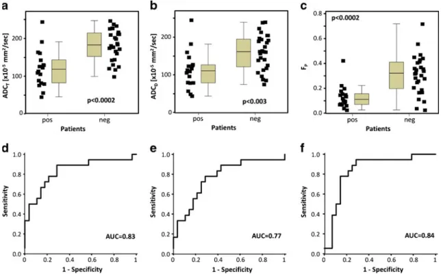
The results of the quantitative analysis with the ADCT, ADCD and FP from mono- and bi-exponential
fitting are presented in Fig. 4. ADCT values were lower
for patients with tumour (mean 120 ± 49 × 10−5 mm2/s) compared with those without tumour (mean 182 ± 41 × 10−5 mm2/s; P < 0.0002). ROC analysis provided an optimal threshold for ADCT of 130 × 10−5 mm2/s
result-ing in a sensitivity of 67%, specificity of 86% and accuracy of 78%. Similarly, ADCD values were
signifi-cantly lower for patients with tumour (mean 113 ± 50 × 10−5 mm2/s) than for those without tumour (mean 160 ± 47 × 10−5 mm2/s; P < 0.003). For the ADCD, a threshold
value of 130 × 10−5 mm2/s results in a sensitivity of 78%, specificity of 71% and accuracy of 74%. FP
values were significantly reduced in tumours compared with non-tumours (13 ± 9% versus 31 ±16%, P < 0.0002). For FP a threshold value of 23% results in a sensitivity
of 94%, specificity of 71% and accuracy of 80%.
Fig. 4 Box-whisker charts showing the distribution of values of the quantitative parameters ADCT(a), ADCD(b) and FP(c) in patients
with (pos) and without (neg) tumour. Corresponding ROC curves for
the qualitative parameters showing the area under the curve (AUC) for ADCT(d), ADCD(e) and FP(f)
Discussion
The present prospective study in a well-defined group of patients with laryngeal or hypopharyngeal cancer treated with (chemo)radiotherapy shows that MRI including DW-MRI allows detection or exclusion of tumour with high accuracy. Based on qualitative analysis using visual assess-ment of high b-value images and the respective ADC maps, tumour could be correctly excluded in all patients without recurrence, while a recurrent tumour was missed in only one patient. These findings are important in daily clinical routine as a DW-MRI sequence can easily be added to any MR protocol without an undue time penalty. Thus, DW-MRI overcomes the diagnostic dilemma of recurrence versus post-therapeutic changes allowing correct tumour detection against a background of treatment-induced soft tissue alter-ation. Correlation with morphological images in order to accurately localise the findings and reader experience in DW-MRI are additional prerequisites for successful inter-pretation of this functional imaging technique. However, in some cases image quality of DW-MRI can be reduced due to susceptibility artefacts impeding image interpretation.
Several studies have already shown the benefit of DW-MRI in detecting residual or recurrent tumour in head and neck squamous cell cancer, but included tumours of various anatomical regions and focused on quantitative image inter-pretation. Our results using visual analysis and the ADCT
are in agreement with a short communication [7] on DW-MRI in four patients with laryngeal cancer after radiothera-py, where lower ADC in tumour recurrence than in benign post-therapeutic alterations has been reported. The quantita-tive values in our study were also very similar to a study on 32 patients with various head and neck tumours [10] that found ADC values for residual or recurrent tumour of 117× 10−5 mm2/s and for post-treatment changes of 207 × 10−5 mm2/s, even though we only included patients with laryngeal and hypopharyngeal cancers. The authors of that study found an identical optimal ADC cut-off, and reported a broad range of overlap, comparable to the current study. However, in that study the overlap of ADC values might also be due to sampling error of the reference standard biopsy, whereas in our study we provided a follow-up of at least 1 year in all negative biopsies.
Another study reported a sensitivity of 95%, specificity of 96% and accuracy of 95.5%, again using the same ADC threshold of 130×10−5 mm2/s, in differentiating between residual or recurrent tumour and post-therapeutic changes [12] in 26 patients with various head and neck cancers. The b values used and the resulting ADC values (111 and 185× 10−5mm2/s for tumour and benign alterations, respectively) were nearly identical to ours. Thus, we corroborate these findings in a larger patient population but including only a well-defined group of patients with laryngeal and
hypopharyngeal tumours. We did find a larger overlap be-tween benign and malignant outcomes, most likely due to the more difficult imaging site of the larynx (susceptibility and movement artefacts), compared with other locations in the head and neck, the overall smaller tumour size in our study (15 of the 46 patients were classified as T1, compared with 7 out of 26 in the above-mentioned study), and their inclusion of lymph nodes in the analysis.
The overlap in diffusion values could be caused by partial volume effects due to tiny structures, falsely lowered ADC in fibrosis and falsely increased ADC in diffuse necrotic parts of recurrent tumour. Furthermore, the ADC and FP
values depend on the ROI placement during visual analysis, and are therefore linked to observer experience. The com-plex structure of the larynx with many different tissues, including mucosa, cartilage (ossified or non-ossified), mus-cle, fat and air in close proximity can also lead to suscepti-bility effects and impaired image contrast, further hampering correct ADC measurements.
In order to improve quantitative image interpretation, we also provided information on bi-exponential fitting (ADCD
and FP) which, to the best of our knowledge, has not been
reported in the literature for the head and neck. However, these parameters have been described in the evaluation of the kidney [13,14], liver [15] and pancreas [16], and the FP
even proved the best parameter to differentiate between normal pancreatic tissue and pancreatic tumour [16]. The current dataset shows that the FPwas significantly lower in
patients with tumours than in those with post-therapeutic changes, possibly because of the presence of leaky ineffi-cient tumour capillaries resulting in a decrease in the fast moving blood pool. The relative diffusion pool is increased because of slowly moving or stagnant blood into capillaries, but diffusion is not increased because of the presence of microvessel membranes [17].
In our prospective study, applied in a well-defined group of 46 patients with laryngeal and hypopharyngeal tumours, conventional MRI with qualitative DW-MRI was highly accurate for the detection of residual/recurrent tumour, even more than the quantitative assessment, which suffered from extensive overlap, limiting its use in the individual patient. However, the subjective nature of visual analysis and the required level of reader expertise (two experienced head and neck radiologists in consensus in this study) make the extrapolation of qualitative data to other centres or appli-cations more difficult. Therefore, both qualitative and quantitative analyses have their place in this setting. Moreover, there have been very promising reports on the usefulness of the ADC values in the prediction of out-come and monitoring of treatment of head and neck squamous cell carcinoma during (chemo)radiotherapy [18–20], which could be even more difficult using visual analysis only.
Our study has several limitations. Firstly, the functional images were evaluated in conjunction with the morphological images and compared with baseline images, as is usual in clinical practice but induces an element of bias because of the size changes compared with baseline. However, lesions with mass effect are not necessarily always tumour and diffuse swelling can obscure tumour. Secondly, the time interval between the end of (chemo)radiotherapy and imaging was variable as patients in our clinic are not routinely imaged after therapy, but instead are followed clinically every 2 months, with imaging only being performed when patients are symp-tomatic. This could cause a selection bias, but as we still ended up with more patients without tumour, we believe that this effect is minimal. The absence of surveillance imaging can cause a delay in detection of tumours and therefore its imple-mentation into routine follow-up would be beneficial for the patients. In some patients, the time period between the end of radiotherapy and DW-MRI was relatively long and it can be argued that these patients had second tumours and not recur-rent or residual tumours. However, the aim of the study was to differentiate between tumour and post-therapeutic changes, thus, we still included them in the study.
In conclusion, MRI that includes a diffusion-weighted sequence allows detection or exclusion of recurrence in patients treated for laryngeal or hypopharyngeal cancer with a high likelihood, even though four patients had to be excluded from the study due to susceptibility artefacts com-promising image quality and its interpretation. This func-tional imaging technique can easily be added to daily clinical routine provided that good image quality and reader experience are combined with correct correlation with mor-phological images. Although the results of qualitative image interpretation are excellent and quantitative analysis shows significantly lower ADC values and the perfusion fraction FP being the best parameter for differentiation between
recurrent tumour compared with post-therapeutic changes, larger scale studies should be performed in order to improve its application in individual patients.
Acknowledgements Harriet C. Thoeny was supported by a research grant of Carigest SA, representative of an anonymous donor and Majores Foundation, Liechtenstein.
References
1. Zbären P, Weidner S, Thoeny HC (2008) Laryngeal and hypophar-yngeal carcinomas after (chemo)radiotherapy: a diagnostic dilem-ma. Curr Opin Otolaryngol Head Neck Surg 16:147–153 2. Becker M, Burkhardt K, Allal AS, Dulguerov P, Ratib O, Becker
CD (2009) Prä- und posttherapeutische Larynxbildgebung. Radi-ologe 49:43–58
3. Nömayr A, Lell M, Sweeney R, Bautz W, Lukas P (2001) MRI appearance of radiation-induced changes of normal cervical tis-sues. Eur Radiol 11:1807–1817
4. Brouwer J, Hooft L, Hoekstra OS et al (2008) Systematic review: accuracy of imaging tests in the diagnosis of recurrent laryngeal carcinoma after radiotherapy. Head Neck 30:889– 897
5. Brouwer J, Bodar EJ, De Bree R et al (2004) Detecting recurrent laryngeal carcinoma after radiotherapy: room for improvement. Eur Arch Oto Rhino Laryngol 261:417–422
6. de Bree R, van der Putten L, Brouwer J, Castelijns JA, Hoekstra OS, Leemans CR (2009) Detection of locoregional recurrent head and neck cancer after (chemo)radiotherapy using modern imaging. Oral Oncol 45:368–393
7. Vandecaveye V, De Keyzer F, Vander Poorten V et al (2006) Evaluation of the larynx for tumor recurrence by diffusion-weighted MRI after radiotherapy: initial experience in four cases. Br J Radiol 79:681–687
8. Zbären P, Christe A, Caversaccio MD, Stauffer E, Thoeny HC (2007) Pretherapeutic staging of recurrent laryngeal carcinoma: clinical findings and imaging studies compared with histopatholo-gy. Otolaryngol Head Neck Surg 137:487–491
9. Lell M, Baum U, Greess H et al (2000) Head and neck tumors: imaging recurrent tumor and post-therapeutic changes with CT and MRI. Eur J Radiol 33:239–247
10. Abdel Razek AA, Kandeel AY, Soliman N et al (2007) Role of diffusion-weighted echo-planar MR imaging in differentiation of residual or recurrent head and neck tumors and posttreatment changes. AJNR Am J Neuroradiol 28:1146–1152
11. Thoeny HC (2011) Diffusion-weighted MRI in head and neck radiology: applications in oncology. Canc Imag 10:209–214 12. Vandecaveye V, De Keyzer F, Nuyts S et al (2007) Detection of
head and neck squamous cell carcinoma with diffusion weighted MRI after (chemo)radiotherapy: correlation between radiologic and histopathologic findings. Int J Radiat Oncol Biol Phys 67:960–971
13. Thoeny HC, Zumstein D, Simon-Zoula S et al (2006) Func-tional evaluation of transplanted kidneys with diffusion-weighted and BOLD MR imaging: initial experience. Radiol-ogy 241:812–821
14. Thoeny HC, Binser T, Roth B, Kessler TM, Vermathen P (2009) Noninvasive assessment of acute ureteral obstruction with diffusion-weighted MR imaging: a prospective study. Radiology 252:721–728
15. Yamada I, Aung W, Himeno Y, Nakagawa T, Shibuya H (1999) Diffusion coefficients in abdominal organs and hepatic lesions: evaluation with intravoxel incoherent motion echo-planar MR imaging. Radiology 210:617–623
16. Lemke A, Laun FB, Klauss M et al (2009) Differentiation of pancreas carcinoma from healthy pancreatic tissue using multiple b-values: comparison of apparent diffusion coefficient and intra-voxel incoherent motion derived parameters. Invest Radiol 44:769–775
17. Lewin M, Fartoux L, Vignaud A, Arrivé L, Menu Y, Rosmorduc O (2011) The diffusion-weighted imaging perfusion fraction f is a potential marker of sorafenib treatment in advanced hepatocellular carcinoma: a pilot study. Eur Radiol 21:281– 290
18. King AD, Mo FK, Yu KH et al (2010) Squamous cell carci-noma of the head and neck: diffusion-weighted MR imaging for prediction and monitoring of treatment response. Eur Radiol 20:2213–2220
19. Vandecaveye V, Dirix P, De Keyzer F et al (2012) Diffusion-weighted magnetic resonance imaging early after chemoradiother-apy to monitor treatment response in head-and-neck squamous cell carcinoma. Int J Radiat Oncol Biol Phys 82:1098–1107
20. Thoeny HC, Ross BD (2010) Predicting and monitoring cancer treatment response with diffusion-weighted MRI. J Magn Reson Imaging 32:2–16