1996 Oxford University Press Nucleic Acids Research, 1996, Vol. 24, No. 1 189–196
The PROSITE database, its status in 1995
Amos Bairoch*
,
Philipp Bucher
1and Kay Hofmann
1Department of Medical Biochemistry, University of Geneva, 1 rue Michel Servet, 1211 Geneva 4, Switzerland and 1Biocomputing Group, Swiss Institute for Experimental Cancer Research (ISREC), 1066 Epalinges s/Lausanne, Switzerland
Received October 3, 1995; Revised and Accepted October 13, 1995
ABSTRACT
The PROSITE database consists of biologically signifi-cant patterns and profiles formulated in such a way that with appropriate computational tools it can help to determine to which known family of proteins (if any) a new sequence belongs or which known domain(s) it contains.
INTRODUCTION
PROSITE (1,2) is a method of determining the function of uncharacterized proteins translated from genomic or cDNA sequences. It consists of a database of biologically significant patterns and profiles formulated in such a way that with appropriate computational tools it can rapidly and reliably determine to which known family of proteins (if any) the new sequence belongs or which known domain(s) it contains.
In some cases the sequence of an unknown protein is too distantly related to any protein of known structure to detect its resemblance by overall sequence alignment, but relationships can be revealed by the occurrence in its sequence of a particular cluster of residue types, which is variously known as a pattern, motif, signature or fingerprint. These motifs arise because specific region(s) of a protein which may be important, for example for their binding properties or for their enzymatic activity, are conserved in both structure and sequence. These structural requirements impose very tight constraints on the evolution of these small but important portions of a protein sequence. The use of protein sequence patterns or profiles to determine the function of proteins is becoming very rapidly one of the essential tools of sequence analysis. This reality has been recognized by many authors (3,4). Based on these observations, we decided in 1988 to actively pursue the development of a database of regular expression-like patterns which could be used to search against sequences of unknown function.
However, while sequence patterns are very useful, there are a number of protein families, as well as functional or structural domains, that cannot be detected using patterns, due to their extreme sequence divergence. Typical examples of important functional domains which are weakly conserved are the globin, the immunoglobulin, the SH2 and the SH3 domains. In such domains there are only a few sequence positions which are well conserved. Any attempt to build a consensus pattern for such
regions will either fail to pick up a significant proportion of the protein sequences that contain such a region (false negatives) or will pick up too many proteins that do not contain the region (false positives).
The use of techniques based on profiles or weight matrices (the two terms are used synonymously here) allows detection of such proteins or domains. A profile is a table of position-specific amino acid weights and gap costs. These numbers (also referred to as scores) are used to calculate a similarity score for any alignment between a profile and a sequence or parts of a profile and a sequence. An alignment with a similarity score higher than or equal to a given cut-off value constitutes a motif occurrence. As with patterns, there may be several matches to a profile in one sequence, but multiple occurrences in the same sequences must be disjoint (non-overlapping) according to a specific definition included in the profile. Another feature that distinguishes patterns from profiles is that the latter are usually not confined to small regions with high sequence similarity. Rather, they attempt to characterize a protein family or domain over its entire length.
We therefore started in 1994 to complement the approach based on patterns by gradually adding to PROSITE profile entries. The profile structure (5,6) used in PROSITE is similar to, but slightly more general than, that introduced by Gribskov and co-workers (7); additional parameters allow representation of other motif descriptors, including the currently popular hidden Markov models. Profiles can be constructed by a large variety of different techniques. The classical method developed by Gribskov and co-workers (8) requires a multiple sequence alignment as input and uses a symbol comparison table to convert residue frequency distributions into weights. The profiles included in PROSITE are generated by this procedure, applying recently described modifi-cations (9,10). In the future we intend to apply additional profile construction tools, including structure-based approaches and methods involving hidden Markov modelling.
LEADING CONCEPTS
The design of PROSITE follows four leading concepts.
Completeness
For such a compilation to be helpful in the determination of protein function it is important that it contains as many biologically meaningful patterns and profiles as possible.
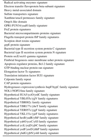
Table 1. List of patterns documentation entries which have been added to PROSITE since the last publication of the NAR database issue
C1q domain signature Death domain profile
Forkhead-associated (FHA) domain profile Src homology 2 (SH2) domains profile Src homology 3 (SH3) domains profile S-layer homology domain signature TPR repeat profile
WW domain signature and profile Prokaryotic dksA/traR C4-type zinc finger PHD finger profile
Copper-fist domain
Bacterial regulatory proteins, iclR family signature Bacterial regulatory proteins, marR family signature Sigma-70 factors ECF subfamily signature Ribosomal protein L10 signature Ribosomal protein L24 signature Ribosomal protein L31 signature Ribosomal protein L7Ae signature Ribosomal protein L13e signature Ribosomal protein L18e signature Ribosomal protein L24e signature Ribosomal protein L27e signature Ribosomal protein L31e signature Ribosomal protein L34e signatures Ribosomal protein L35Ae signature Ribosomal protein L37e signature Ribosomal protein S6 signature Homoserine dehydrogenase signature
Aspartate-semialdehyde dehydrogenase signature Pyridoxamine 5′-phosphate oxidase signature
Respiratory chain NADH dehydrogenase 20 kDa subunit signature Respiratory chain NADH dehydrogenase 24 kDa subunit signature NNMT/PNMT/TEMT family of methyltransferases signature Ribosomal RNA adenine dimethylases signature
Squalene and phytoene synthases signatures ROK family signature
Casein kinase II regulatory subunit signature Shikimate kinase signature
Prokaryotic diacylglycerol kinase signature Acetate and butyrate kinases family signatures RNA polymerases H 23 kDa subunits signature RNA polymerases N 8 kDa subunits signature RNA polymerases L 13–16 kDa subunits signature RNA polymerases RPB6 6 kDa subunits signature Lipolytic enzymes ‘G-D-S-L’ family, serine active site DNA/RNA non-specific endonucleases active site Thermonuclease family signature
Chitinases family 18 signature
Glycosyl hydrolases family 45 active site
ATP-dependent serine proteases, lon family, serine active site Interleukin-1β converting enzyme family active sites Hydroxymethylglutaryl-coenzyme A lyase active site DNA photolyases class 2 signatures
Adenylate cyclases class-I signatures
Ribulose-phosphate 3-epimerase family signatures PpiC-type peptidyl-prolyl cis-trans isomerase signature Terpene synthases signature
SAICAR synthetase signatures NAD-dependent DNA ligase signatures Transposases, IS30 family, signature
Molybdenum cofactor biosynthesis proteins signatures
Radical activating enzymes signature
Electron transfer flavoprotein beta-subunit signature Heavy metal-associated domain
Sulfate transporters signature
Xanthine/uracil permeases family signature OmpA-like domain
GPR1/FUN34/yaaH family signature FtsZ protein signatures
Bacterial microcompartiments proteins signature Flagella transport protein fliP family signatures Scorpion short toxins signature
grpE protein signature
Bacterial type II secretion system protein C signature Bacterial type II secretion system protein N signature Protein secE/sec61-gamma signature
Fimbrial biogenesis outer membrane usher protein signature Apoptosis regulator proteins, Bcl-2 family signature GTP-binding nuclear protein ran signature Elongation factor Ts signatures
Translation initiation factor SUI1 signature Calponin family repeat
CAP protein signatures
Hydrogenases expression/synthesis hupF/hypC family signature NOL1/NOP2/fmu family signature
Hypothetical SUA5/yciO/yrdC family signature Hypothetical YBL055c/yjjV family signatures Hypothetical YBR002c family signature Hypothetical YBR177c/yheT family signature Hypothetical YER057c/yjgF family signature Hypothetical YKL151c/yjeF family signatures Hypothetical hesB/yadR/yfhF family signature Hypothetical yabO/yceC/yfiI family signature Hypothetical yciL/yejD/yjbC family signature Hypothetical yedF/yeeD/yhhP family signature Hypothetical yhdG/yjbN/yohI family signature
High specificity
In the majority of cases we have chosen patterns or profiles that are specific enough that they do not detect too many unrelated sequences, yet they will detect most, if not all, sequences that clearly belong to the set in consideration.
Documentation
Each of the entries in PROSITE is fully documented. The documentation includes a concise description of the protein family that it is designed to detect, as well as a summary of the reasons leading to the development of the pattern or profile.
Periodic reviewing
It is important that each entry be periodically reviewed to ensure that it is still valid.
FORMAT AND DOCUMENT FILES
The core of the PROSITE database is composed of two ASCII (text) files. The first file (prosite.dat) is a computer readable file that contains all the information necessary for programs that make use of PROSITE to scan sequences for the occurrence of the patterns and/or profiles. This file also includes, for each of the entries described, statistics on the number of hits obtained while
191
Nucleic Acids Research, 1994, Vol. 22, No. 1Nucleic Acids Research, 1996, Vol. 24, No. 1 191
scanning for that pattern or profile in the SWISS-PROT protein sequence database (9). Cross-references to the corresponding SWISS-PROT entries are also present in the file. The second file (prosite.doc), which we call the textbook, contains textual information that documents each pattern.
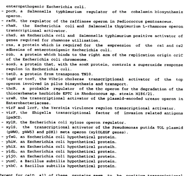
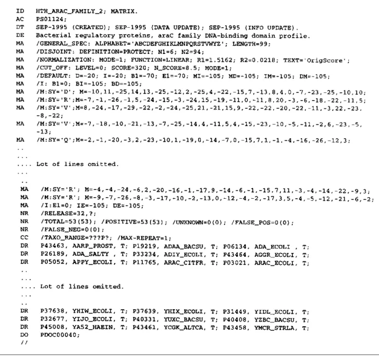
A sample textbook entry is shown in Figure 1a (see following pages). This particular entry is linked to two entries in the prosite.dat file, a pattern and a profile (Fig. 1b).
Several document files are also distributed with the database: prosuser.txt, the database user’s manual;
profile.txt, a detailed description of the syntax for the profiles; prosite.lis, a list of PROSITE documentation entries;
prosite.get, a document on how to obtain a local copy of PROSITE;
prosite.prg, a description of programs and electronic mail servers that make use of PROSITE;
pautindx.txt, an index of authors cited in the prosite.doc file.
CONTENT OF THE CURRENT RELEASE
Release 13 of PROSITE (October 1995) contains 883 docu-mentation entries describing 1156 different patterns, rules and profiles. The list of entries which have been added since publication of the previous article (2) describing PROSITE is provided in Table 1. The database requires ∼5 Mb disk storage space. The present distribution frequency is two releases per year. No restrictions are placed on use or redistribution of the data.
HOW TO OBTAIN A LOCAL COPY OF PROSITE
By CD-ROM
PROSITE is distributed on CD-ROM by the EMBL Outstation– the European Bioinformatics Institute (EBI) (12). For all enquiries regarding subscription to and distribution of PROSITE one should contact The EMBL Outstation–The European Bioin-formatics Institute, Hinxton Hall, Hinxton, Cambridge CB10 1RQ, UK (tel. +44 1223 494 400; fax +44 1223 494 468; email [email protected]).
By anonymous ftp
If you have access to a computer system linked to the Internet you can obtain PROSITE using ftp (File Transfer Protocol) from the following file servers:
EBI anonymous ftp server (ftp.ebi.ac.uk or 192.54.41.33); NCBI Repository, National Library of Medicine, NIH, Washington, DC (ncbi.nlm.nih.gov or 130.14.20.1);
ExPASy (Expert Protein Analysis System) server, University of Geneva, Switzerland, (expasy.hcuge.ch or 129.195.254.61);
National Institute of Genetics (Japan) ftp server (ftp.nig.ac.jp or 133.39.16.66).
By email through the EBI file server
PROSITE can be obtained from the EBI file server (13). Detailed instructions on how to make the best use of this service and, in particular, on how to obtain PROSITE can be obtained by query to the network address [email protected]
HELP
HELP PROSITE
HOW TO MAKE USE OF PROSITE
Computer programs
Many academic groups and commercial companies have devel-oped computer programs that make use of the pattern entries in PROSITE. The prosite.prg file contains a full list of these programs, their operating system specificity and characteristics, as well as information on how to obtain them.
To make use of profile entries we are distributing, with the PROSITE release the source code (in FORTRAN77) of two programs that should help software developers implement profile-specific routines in their application(s): pfscan loads a sequence from a file and scans it with all (or one) of the PROSITE profiles; pfsearch loads a profile from a file and scans for it in a SWISS-PROT database file.
Email servers
There are many email servers that are available to molecular biologists (14). At least three of these servers can be used in conjunction with PROSITE.
Name: EBI Mail-PROSITE Server.
Organization: European Bioinformatics Institute, Hinxton, UK. Description: Allows rapid comparison of a new protein sequence against all patterns stored in PROSITE.
Server email address: [email protected]. Address to report problems: [email protected]. Name: BLOCKS e-mail searcher.
Organization: Fred Hutchinson Center, Seattle, WA, USA. Description: Compares a protein or DNA sequence to the database of protein blocks. Blocks are short multiply aligned ungapped segments corresponding to the most highly conserved regions of proteins. The BLOCKS database (15) has been derived from PROSITE. This server can also be used to retrieve specifics blocks and PROSITE entries.
Server email address: [email protected]. Address to report problems: [email protected]. Name: MOTIF E-Mail Server on GenomeNet.
Organization: Supercomputer Laboratory, Kyoto Institute for Chemical Research, Japan.
Description: Allows rapid comparison of a new protein sequence against all patterns stored in PROSITE, as well as in the MotifDic library (16).
Server email address: [email protected].
Address to report problems: [email protected].
INTERACTIVE ACCESS TO PROSITE USING THE WORLD WIDE WEB
The most efficient and user friendly way to browse interactively in PROSITE, as well as to analyse a sequence for the occurrence of a pattern or a profile, is to use the World Wide Web (WWW) molecular biology server ExPASy (17). WWW is a global information retrieval system merging the power of worldwide networks, hypertext and multimedia. Through hypertext links it gives access to documents and information available on thou-sands of servers around the world. To access a WWW server one needs a WWW browser. Popular browsers available for most computer platforms include Mosaic, developed at the National Center for Supercomputing Applications (NCSA) of the Univer-sity of Illinois at Champaign (obtainable by anonymous ftp from
193
Nucleic Acids Research, 1994, Vol. 22, No. 1Nucleic Acids Research, 1996, Vol. 24, No. 1 193
195
Nucleic Acids Research, 1994, Vol. 22, No. 1Nucleic Acids Research, 1996, Vol. 24, No. 1 195
Figure 1. continued
ftp.ncsa.uiuc.edu), and Netscape Navigator, from Netscape Communications Corp. (available from ftp.netscape.com). Using a WWW browser one has access to all the hypertext documents stored on the ExPASy server, as well as many other WWW servers, and also can make use of many sequence analysis software tools.
The ExPASy server may be accessed through its Uniform Resource Locator (URL, the addressing system defined in WWW), which is http://expasy.hcuge.ch/. You can directly access the ’top page’ of the section of ExPASy that allows you to browse through the PROSITE documentation and data entries by opening the URL http://expasy.hcuge.ch/sprot/prosite.html.
To use the PROSITE patterns and profiles you can make use of the following software tools. ScanProsite allows either scanning of a protein sequence, from SWISS-PROT or provided by the
user, for the occurrence of patterns stored in PROSITE or scanning of the SWISS-PROT database, including weekly releases, for the occurrence of a pattern that can originate from PROSITE or be provided by the user. The URL for scanprosite is http://expasy.hcuge.ch/sprot/scnpsite.html. ProfileScan allows scanning of a protein sequence, from SWISS-PROT or provided by the user, for the occurrence of profiles stored in PROSITE. The URL for profilescan is
http://ulrec3.unil.ch/software/profilescan.html
REFERENCES
1 Bairoch,A. (1993) Nucleic Acids Res., 21, 3097–3103.
3 Doolittle,R.F. (1986) In Of URFs and ORFs: A Primer on How to Analyze
Derived Amino Acid Sequences. University Science Books, Mill Valley,
CA.
4 Lesk,A.M. (1988) In Lesk,A.M. (ed.), Computational Molecular Biology. Oxford University Press, Oxford, UK, pp. 17–26.
5 Bucher,P. and Bairoch,A. (1994) In Altman,R., Brutlag,D., Karp,P., Lathrop,R. and Searls,D. (eds), ISMB-94; Proceedings of the Second
International Conference on Intelligent Systems for Molecular Biology.
AAAI Press, Menlo Park, pp. 53–61.
6 Bucher,P., Karplus,K., Moeri,N. and Hofmann,K. (1995) Comput. Chem., in press.
7 Gribskov,M., McLachlan,A.D. and Eisenberg,D. (1987) Proc. Natl. Acad.
Sci. USA, 84, 4355–4358.
8 Gribskov,M., Luethy,R. and Eisenberg,D. (1990) Methods Enzymol., 183, 146–159.
9 Luethy,R., Xenarios,I. and Bucher,P. (1994) Protein Sci., 3, 139–146. 10 Thompson,.D., Higgins,D.G. and Gibson,T.J. (1994) Comput. Appl.
Biosci., 10, 19–29.
11 Bairoch,A. and Boeckmann,B. (1994) Nucleic Acids Res., 22, 3578–3580. 12 Emmert,D.B., Stoehr,P.J., Stoesser,G. and Cameron,G.N. (1994) Nucleic
Acids Res., 22, 3445–3449.
13 Stoehr,P.J. and Omond,R.A. (1989) Nucleic Acids Res., 17, 6763–6764. 14 Henikoff,S. (1993) Trends Biochem. Sci., 18, 267–268.
15 Henikoff,S. and Henikoff,J.G. (1991) Nucleic Acids Res., 19, 6565–6572. 16 Ogiwara,A., Uchiyama,I., Seto,Y. and Kanehisa,M. (1992) Protein Engng,
5, 479–488.
17 Appel,R.D., Bairoch,A. and Hochstrasser,D.F. (1994) Trends Biochem.