Sylvain R. Duc Bernard Mengiardi Christian W. A. Pfirrmann Juerg Hodler Marco Zanetti Received: 16 December 2005 Revised: 19 May 2006 Accepted: 31 July 2006
Published online: 17 October 2006 # Springer-Verlag 2006
Improved visualization of collateral ligaments
of the ankle: multiplanar reconstructions
based on standard 2D turbo spin-echo MR
images
Abstract The purpose of the study was to evaluate the visualization of the collateral ankle ligaments on multi-planar reconstructions (MPR) based on standard 2D turbo spin-echo im-ages. Coronal and axial T2-weighted turbo spin-echo and MPR angled parallel to the course of the ligaments of 15 asymptomatic and 15 symp-tomatic ankles were separately analyzed by two musculoskeletal radiologists. Image quality was assessed in the asymptomatic ankles qualitatively. In the symptomatic ankles interobserver agreement and reader confidence was determined for each ligament. On MPR the tibionavicular and calcaneofibular ligaments were more commonly demonstrated on a single image than on standard MR images (reader 1: 13 versus 0, P=0.002; reader 2: 14 versus 1, P=0.001 and reader 1: 13
versus 2, P=0.001; reader 2: 14 versus 0, P<0.001). The tibionavicular ligament was considered to be better delineated on MPR by reader 1 (12 versus 3, P=0.031). In the symp-tomatic ankles, reader confidence was greater with MPR for all
ligaments except for the tibiocalcanear ligament (both readers) and the anterior and posterior talofibular liga-ments (for reader 2). Interobserver agreement was increased with MPR for the tibionavicular ligament. Multiplanar reconstructions of 2D turbo spin-echo images improve the visualization of the tibionavicular and calcaneofibular ligaments and strengthen diagnostic confidence for these ligaments.
Keywords Ankle joint . Ligaments . Articular . Magnetic resonance imaging . Image reconstruction
Introduction
Multiplanar reconstruction (MPR) is routinely used to evaluate multi-detector CT examinations. In MR imaging, MPR has also been used mainly for vascular [1–3] and abdominal [4] imaging, typically based on 3D gradient echo sequences. With the exception of cartilage imaging, MPR has less commonly been used for the musculoskeletal system [5–11], although angled images may be useful for structures with an oblique course such as the calcaneofib-ular ligament [12]. 2D spin echo sequences are typically employed in the assessment of ligaments and tendons
because signal changes are more characteristic for patho-logic conditions on 2D spin echo sequences than on gradient echo sequences [13]. Additional 2D spin echo sequences in various imaging planes are acquired to address the complex course of ligaments and tendons in the foot and ankle. However, the number of such additional sequences is limited for practical reasons [12]. MPR using 2D spin echo sequences could solve this problem. So far, radiologists were reluctant to use 2D spin echo MPRs because they tend to be relatively thick (2–4 mm) and have an interslice gap. Rather unexpectedly, a number of 2D spin echo MPRs obtained during clinical routine work S. R. Duc (*) . B. Mengiardi .
C. W. A. Pfirrmann . J. Hodler . M. Zanetti
From the Departments of Radiology, University Hospital, Balgrist, Forchstrasse 340,
8008 Zurich, Switzerland e-mail: [email protected] Tel.: +41-44-3863308
proved to be of good quality when the reformation plane was only slightly angled in relation to the source images.
Thus, the purpose of our study was to evaluate the visualization of the collateral ankle ligaments on MPRs of 2D turbo spin-echo images.
Materials and methods
Volunteers
MR images of the ankle were obtained in 15 asymptomatic volunteers (8 men and 7 women; mean age: 25.9 years; range: 23–33 years). The study protocol was approved by the hospital’s institutional review board. Written informed consent was obtained from all volunteers.
MR imaging
The MR examination was performed on a 1.5-T scanner (Symphony, Siemens Medical Solutions, Erlangen, Germany) with a dedicated send-receive extremity coil. Axial and coronal T2-weighted turbo spin-echo (TSE) sequences were used. For the axial sequence the following parameters were used: TR: 3,900 ms; TE: 85 ms; number of averages: 2; turbo factor: 9; FOV: 124×180 mm; matrix: 512×176; number of images: 19; section thickness: 4 mm; gap: 0.4 mm; scan time: 2:43 min. For the coronal sequence the parameters were as follows: TR: 4,130 ms; TE: 86 ms; number of averages: 2; turbo factor: 7; FOV: 104×170 mm; matrix: 512×187; number of images: 25; section thickness: 3.5 mm; gap: 1.0 mm; scan time: 3:51 min.
Image analysis
The medial collateral ligament (tibionavicular, anterior tibiotalar, tibiocalcaneal, tibiospring and posterior tibiotalar ligament) and the lateral collateral ligament (anterior talofibular, posterior talofibular and calcaneofibular liga-ment) were evaluated. The ligaments were assessed independently by two experienced musculoskeletal radi-ologists [reader 1 (R1) and reader 2 (R2)] with 2 and 12 years of experience in musculoskeletal MR imaging. Commercially available PACS software (Cedara I-Read 5.2 P11, Cerner Image Devices Idstein, Germany) was used for the evaluations, including calculation of the MPRs.
The readers initially determined if the ligaments were visible on at least one of the two original sequences. They then determined if the ligaments were visualized through-out their entire course on a single image, if their borders were sharp or blurred, and if their signal intensity was normal (hypointense).
One week later MPRs were obtained for each ligament by the readers based on either the axial or the coronal 2D
TSE sequence, whichever was closer to the course of the ligament (Fig.1).
The reading time necessary for the evaluation of the primary 2D TSE images and for the reconstruction and evaluation of the MPRs was measured by the readers to the nearest second.
Image quality
The image quality of the MPRs was evaluated in relation to the source images by a single experienced musculoskeletal radiologist not involved in the assessments described above (18 years of experience with musculoskeletal MR imaging). The reader was blinded with regard to the type of image. A five-point grading system was employed for comparison of the MPRs to the standard images: grade 0: MPRs identical; grade 1: MPR minimally blurred, suitable for making a diagnosis without limitations; grade 2: MPR quality moderately blurred, no step off, diagnostic value acceptable; grade 3: MPR substantially blurred, some step-off artifacts, diagnostic value questionable; grade 4: MPR inadequate. The reformation angle for the MPR was measured by an independent fourth reader (2 years of experience in musculoskeletal radiology).
Patients
In a patient group (ten men and five women; mean age: 32.6 years; range: 18–32 years), the visibility of ligament lesions was analyzed. The patients were recruited con-secutively from a MR data base when a collateral ligament lesion either on the medial or lateral side was described in the prospective MR report. The MR imaging protocol was identical to the protocol used with the volunteers. For retrospective data evaluation, no specific institutional review board approval is necessary at our institution. Patient rights are protected by a law requiring patient information about the possibility of anonymous scientific review of their data and about the opportunity to reject such use of their data.
The medial collateral ligament (tibionavicular, anterior tibiotalar, tibiocalcaneal, tibiospring and posterior tibiotalar ligament) and the lateral collateral ligament (anterior talofibular, posterior talofibular and calcaneofibular liga-ment) were characterized independently by the same readers who read the asymptomatic volunteers using the same standard sequences as mentioned above. They classified the ligaments as normal (homogenous; well-delineated, hypointense; grade 0), structurally alterated (inhomogenous; hyperintense; caliber changes; grade 1) or absent (ligament discontinuity or non-visualization; grade 2). Diagnostic confidence was graded for each ligament on a scale between 0 (absolutely unconfident) and 10 (abso-lutely confident). Subsequently, MPRs of the pathological
ankles were evaluated using the same grading and confidence score. Surgical correlation was not available.
Statistical evaluation
The McNemar test was used to evaluate differences in qualitative results. Interobserver percentage agreement was calculated for the qualitative data. A paired t-test was used for comparison of reading times. Spearman’s rho test was
used to evaluate the relationship between the five-point image quality rating and the reformation angle. Interobserv-er agreement for ligament grading of the symptomatic ankles was evaluated by kappa statistics. The Wilcoxon signed rank test was used to test if confidence in the diagnosis was significantly different between standard images and MPRs. P values smaller than 0.05 were considered to be significant.
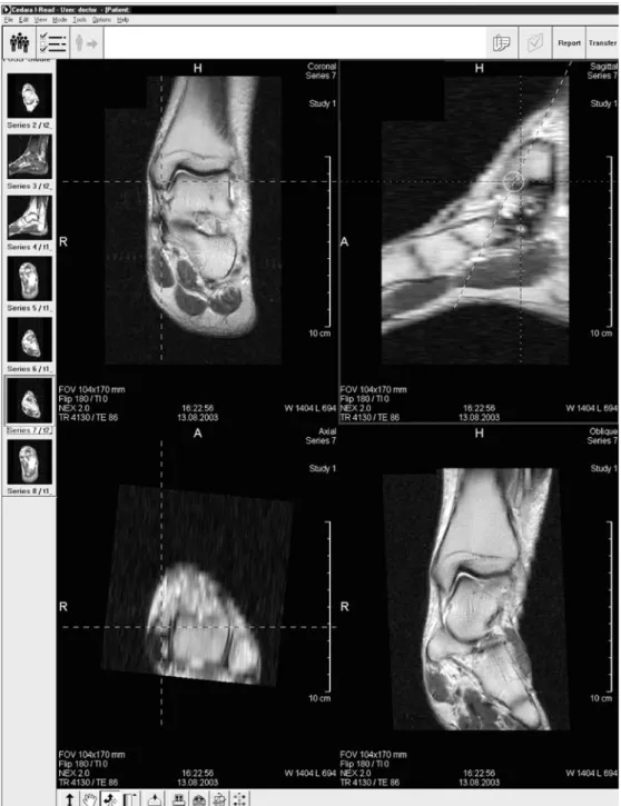
Fig. 1 Screenshot of MPR tool. Source image is shown on the upper left quadrant. The auto-matically provided planning planes are shown on the upper right and lower left quadrants. Note oblique reformation plane (dashed white and black line) on the upper right quadrant. The resulting MPR appears in the lower right quadrant
Results
Asymptomatic ankles
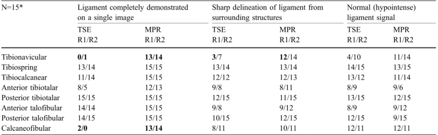
The results for the asymptomatic ankles are summarized in Table1. The statistically significant advantages using MPR are the following: On MPRs, the tibionavicular (Fig.2) and calcaneofibular (Fig. 3) ligaments were more commonly demonstrated on a single image than on standard MR images (reader 1: 13 versus 0, P=0.002; reader 2: 14 versus 1, P=0.001 and reader 1: 13 versus 2, P=0.001, reader 2: 14 versus 0, P<0.001). The tibionavicular ligament was considered to be significantly better delineated on multi-planar reconstructions by reader 1 (12 versus 3, P=0.031). For reader 2, delineation of the tibionavicular ligament was also better for MPRs (14 versus 7), but not statistically significant. For reader 1, the mean evaluation time was 237 (standard images) and 362 s (MPRs) (P<0.001). The corresponding values for reader 2 were 168 and 249 s (P<0.001). No significant differences were found for the remaining ligaments and for the ligament signal intensity.
MPR image quality was rated as grade 0 in 84, as 1 in 34, and as 2 on 2 MPRs. No grade 3 or 4 was found. The mean MPR angles were 39° (range: 18°–52°) for the tibionavicu-lar ligament, 10° (2°–24°) for the tibiospring ligament, 3° (range: 0°–10°) for the tibiocalcanear ligament, 7° (0°– 22°) for the anterior tibiotalar ligament, 1° (0°–6°) for the posterior tibiotalar ligament, 3° (0°–14°) for the anterior talofibular ligament, 1° (0°–8°) for the posterior talofibular ligament and 16° (0°–38°) for the calcaneofibular liga-ment. The grading of the image quality was not significantly correlated with the angulation of the MPR (correlation coefficient: 0.82, P=0.37).
Symptomatic ankles
The interobserver agreement for the symptomatic ankles using the standard sequences was 0.74 for the tibionavicu-lar (Fig. 4), 0.17 for the anterior tibiotalar, 0.87 for the tibiospring, 0.46 for the tibiocalcanear, 0.87 for the posterior tibiotalar, 0.44 for the anterior talofibular and 1.00 for the calcaneofibular ligament (Fig. 5). For MPR, interobserver agreement was 0.82 for the tibionavicular, 0.29 for the anterior tibiotalar, 0.70 for the tibiospring, 0.57 for the tibiocalcanear, 0.60 for the posterior tibiotalar, 0.42 for the anterior talofibular and 0.53 for the calcaneofibular ligament.
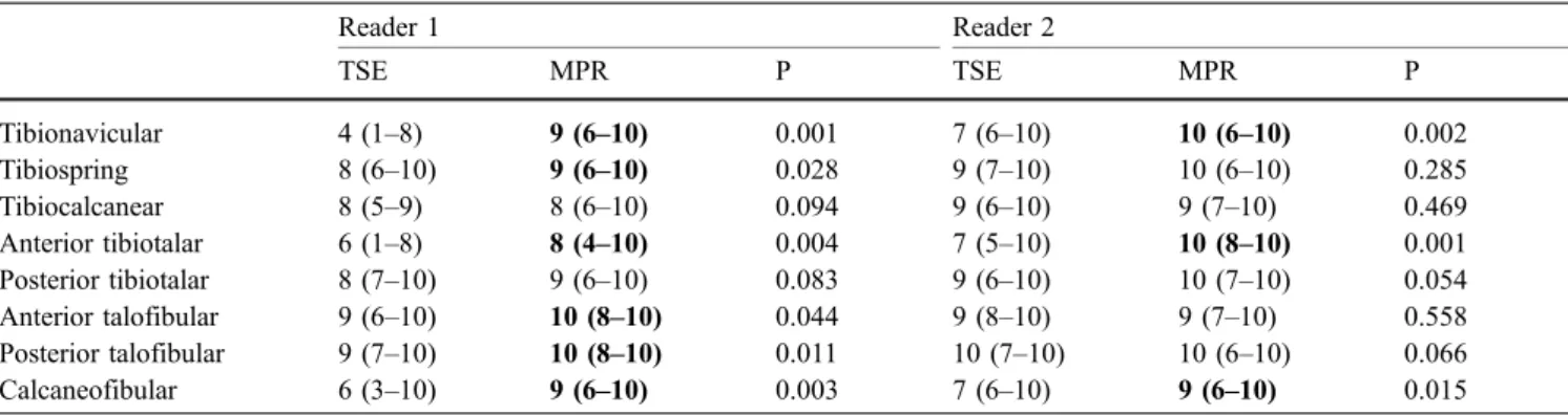
The results for the reader confidence are shown in Table2. The confidence was significantly better with MPR for the tibionavicular, anterior tibiotalar, tibiospring, ante-rior talofibular, posteante-rior talofibular and calcaneofibular for reader 1 and for the tibionavicular, anterior tibiotalar and calcaneofibular for reader 2.
Discussion
MPRs are typically based on thin continuous or over-lapping sections originating from spiral CT or 3D MR images. MPRs based on spin-echo sequences with their slice thickness of several millimeters are not part of routine protocols because reformations suffer from blurring and step offs. Such problems depend on the reformation angle, however, and may be negligible if angulations are sufficiently small. This fact is successfully implemented in advanced multi-detector row CT reconstruction algo-rithms. In the AMPR (adaptive multiple plane
reconstruc-Table 1 Qualitative evaluation of standard TSE images and MPR N=15* Ligament completely demonstrated
on a single image
Sharp delineation of ligament from surrounding structures
Normal (hypointense) ligament signal
TSE MPR TSE MPR TSE MPR
R1/R2 R1/R2 R1/R2 R1/R2 R1/R2 R1/R2 Tibionavicular 0/1 13/14 3/7 12/14 4/10 11/14 Tibiospring 13/14 15/15 13/14 13/14 14/15 13/15 Tibiocalcanear 11/14 15/15 12/12 12/13 13/12 11/14 Anterior tibiotalar 8/5 12/13 9/8 8/11 8/9 9/6 Posterior tibiotalar 15/15 15/15 12/15 11/15 13/15 12/15 Anterior talofibular 14/14 15/15 9/8 9/12 8/9 9/12 Posterior talofibular 14/15 15/15 10/15 12/15 12/15 9/15 Calcaneofibular 2/0 13/14 8/11 10/11 12/11 12/11 R1/R2: readers 1 and 2 TSE: turbo spin-echo images MPR: multiplanar reconstruction
Bold numbers represent the statistically significant differences between TSE and MPR.
The readers excluded ligaments that they thought were not visible. Reader 1 excluded five (TSE) and two (MPR) tibionavicular ligaments and three (TSE) and three (MPR) anterior tibiotalar ligaments. Reader 2 excluded two tibionavicular ligaments (TSE), five (TSE) and two (MPR) anterior tibiotalar ligaments and one (TSE) calcaneofibular ligament.
tion) [14, 15] approach, the imaging plane is not per-pendicular to the axis of the patient, but rather is tilted in order to match the spiral path of the focal spot of the CT tube. The transverse images expected by the radiologists are interpolated from the tilted original images [14, 15]. The techniques employed for this purpose are more advanced than the standard MPRs employed in our study.
In addition, the angles are smaller than in many images obtained in our investigation.
An already published paper about reformations based on 3D acquisitions [10] obtained a higher sensitivity in the diagnosis of calcaneofibular and anterior talofibular liga-ment tears in comparison to previously published standard 2D acquisitions [16], the specificity of 3D acquisitions Fig. 2 Visualization of the
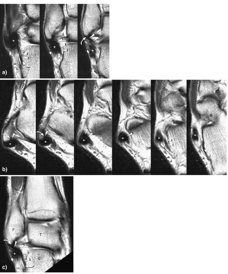
tibio-navicular ligament on coronal and axial 2D TSE and on MPR. Consecutive coronal (posterior to anterior) (a) and axial (proximal to distal) (b) TSE images demonstrate the oblique course of the tibionavicular ligament (arrowheads). The vicinity of the tibialis posterior (*), the flexor digitorum longus (+) tendons and the tibiospring ligament (white double arrows) render the evaluation of the tibionavicular ligament difficult. Curved arrows: bony insertion of the tibionavicular ligament; N: navicular bone, M: medial malleolus; T: talar bone; black double arrow: tibiocalcanear ligament. MPR (c) shows the entire course of the tibionavicu-lar ligament (arrowheads) on one single image without interference by the neighboring structures. Curved arrows: bony insertion; M: medial malleolus, N: navicular bone; T: talar bone
Fig. 3 Visualization of the calcaneofibular ligament on coronal and axial 2D TSE and on MPR. Consecutive coronal (posterior to anterior) (a) and axial (proximal to distal) (b) TSE images demonstrate the course of the calcaneofibular ligament (arrowheads). Difficult
delinea-tion of the proximal part of the ligament from the peroneal tendons (*). The MPR (c) shows the entire course of the calcaneofibular ligament and allows better delineation of its proximal portion. Curved arrows: bony insertion; F: tip of the fibula; T: talar bone; C: calcaneus
being slightly higher for calcaneofibular tears and clearly lower for anterior talofibular ligament tears. According to our results, the evaluation of thin obliquely running ligaments is facilitated by MPR. Such ligaments can more commonly be followed on a single image and are
better delineated from surrounding structures. Improved visualization of the calcaneofibular and tibionavicular ligaments is clinically relevant. The calcaneofibular liga-ment contributes to the ankle and subtalar stability in all foot positions [17,18]. In inversion-adduction injuries of Fig. 4 Abnormal tibionavicular
ligament. Consecutive coronal (posterior to anterior) (a) and axial (proximal to distal) (b) TSE image. The course of the tibionavicular ligament is difficult to follow. White arrow-heads: proximal ligament part; gray arrowheads: distal ligament part; curved arrows: bony inser-tion of the tibionavicular ligament; N: navicular bone; M: medial malleolus; T: talar bone. MPR (c) shows the entire course of the tibionavicular ligament (arrowheads) on one single image. The tear of the ligament is clearly visible (black circle). White arrowheads: proximal part of the ligament; gray arrowheads: distal part of the ligament; curved arrows: bony insertion; M: medial malleolus; N: navicular bone; T: Talar bone
Fig. 5 Abnormal calcaneofibu-lar ligament. Consecutive coro-nal (posterior to anterior) (a) and axial (proximal to distal) (b) TSE images show a torn calca-neofibular ligament. White arrowheads: proximal ligament part; gray arrowheads: distal ligament part; curved arrows: bony insertion; *: peroneal ten-dons; F: tip of the fibula; T: talar bone; C: calcaneus. The MPR (c) shows the entire course of the torn calcaneofibular ligament (white arrowheads: proximal ligament part; grey arrowheads: distal ligament part) including the proximal and distal bony insertion (curved arrows) and the localization of the tear (white circle). F: tip of the fibula; T: talar bone; C: calcaneus
the ankle, the calcaneofibular ligament is the second structure of the lateral ligament complex to be torn after the anterior talofibular ligament [19, 20]. When surgery is considered, the functional results are better with surgical repair of both the anterior talofibular and the calcaneofib-ular ligament than with an isolated repair of the anterior talofibular ligament [21]. Thus, the detection of calcaneo-fibular ligament tears is important. The tibionavicular ligament is the longest component of the medial collateral band complex [22]. It is also the broadest component of the superficial layer of this complex [23, 24]. On the other hand, it is the weakest component of the medial collateral band complex, together with the tibiocalcanear ligament [11,22]. Its biomechanical function is not fully understood, but it appears to be a secondary stabilizer against pronation [25]. In a clinical study, Hintermann et al. reported [26] that most of the injuries of the medial collateral ligament complex occurred at the proximal insertion of the tibiona-vicular and tibiospring ligaments, emphasizing the need of an adequate visualization of the entire course of the tibionavicular ligament.
MPRs based on standard 2D MR images obtained with a section thickness of a few millimeters typically suffer from artifacts such as blurring and step offs (insufficient information). Such problems were rare in our study population because the required angulations required relatively little interpolation between image voxels that are relatively close.
The additional time required for MPRs is relevant for routine work [5, 6]. In our setting, the time required for
reformation and image evaluation was approximately longer by half for MPR, but remained within an acceptable range (approximately 2 and 1 min for readers 1 and 2, respectively, for the evaluation of all eight ligaments included in this study).
The use of MPR increased the reader’s diagnostic confidence for the majority of the ligaments of the medial and lateral collateral complex and especially for the tibionavicular and calcaneofibular ligament with an oblique course to orthogonal imaging planes. These results point out that MPR may be useful for the evaluation of ankle ligaments in some cases when the standard orthogonal planes remain equivocal.
As limitations of the study, we acknowledge the low number of patients and the absence of surgical correlation. Surgical correlation of ankle ligament lesions is not easily available because conservative treatment is much more commonly performed than surgery. Moreover, an exact surgical description of normal and abnormal ligaments of the medial collateral band complex is difficult. Never-theless, we believe that the present study has value by demonstrating that the commonly used 2D spin-echo images can be used for MPRs for better visualization of some ligaments of the lateral and medial collateral ligament complex in selected cases.
In conclusion, MPRs of 2D turbo spin-echo images improve the visualization of the tibionavicular and calca-neofibular ligaments and strengthen the diagnostic con-fidence for these ligaments within a reasonable time. Table 2 Symptomatic ankles: reader confidence
Reader 1 Reader 2 TSE MPR P TSE MPR P Tibionavicular 4 (1–8) 9 (6–10) 0.001 7 (6–10) 10 (6–10) 0.002 Tibiospring 8 (6–10) 9 (6–10) 0.028 9 (7–10) 10 (6–10) 0.285 Tibiocalcanear 8 (5–9) 8 (6–10) 0.094 9 (6–10) 9 (7–10) 0.469 Anterior tibiotalar 6 (1–8) 8 (4–10) 0.004 7 (5–10) 10 (8–10) 0.001 Posterior tibiotalar 8 (7–10) 9 (6–10) 0.083 9 (6–10) 10 (7–10) 0.054 Anterior talofibular 9 (6–10) 10 (8–10) 0.044 9 (8–10) 9 (7–10) 0.558 Posterior talofibular 9 (7–10) 10 (8–10) 0.011 10 (7–10) 10 (6–10) 0.066 Calcaneofibular 6 (3–10) 9 (6–10) 0.003 7 (6–10) 9 (6–10) 0.015
TSE: turbo spin-echo images MPR: multiplanar reconstruction
The numbers indicate the median grading in confidence of the diagnosis (0= unconfident; 10= absolutely confident) The numbers in parentheses represent the range with minimal and the maximal confidence in the diagnosis. Bold numbers represent a significantly (P<0.05, Wilcoxon signed rank test) better diagnostic confidence with MPR
References
1. Schoenberg SO, Rieger J, Weber CH, Michaely HJ, Waggershauser T, Ittrich C, Dietrich O, Reiser MF (2005) High-spatial-resolution MR angiography of renal arteries with integrated parallel acquisitions: comparison with digital subtraction angiography and US. Radiology 235:687–698
2. Baskaran V, Pereles FS, Nemcek AA Jr, Carr JC, Miller FH, Ly J, Krupinski E, Finn JP (2002) Gadolinium-enhanced 3D MR angiography of renal artery stenosis: a pilot comparison of maxi-mum intensity projection, multiplanar reformatting, and 3D volume-rendering postprocessing algorithms. Acad Radiol 9:50–59
3. Adams WM, Laitt RD, Jackson A (2000) The role of MR angiography in the pretreatment assessment of intra-cranial aneurysms: a comparative study. AJNR Am J Neuroradiol 21:1618– 1628
4. Yamakawa K, Naganawa S, Maruyama K, Kato T, Fukatsu H, Ishigaki T (1999) Clinical evaluation of three-dimensional MR-cholangiopancreatog-raphy using three-dimensional Fourier transform fast asymmetric spin echo method (3DFT-FASE): usefulness of observation by multi-planar recon-struction. Radiat Med 17:15–19 5. Gay SB, Chen NC, Burch JJ, Gleason
TR, Sagman AM (1993) Multiplanar reconstruction in magnetic resonance evaluation of the knee. Comparison with film magnetic resonance interpre-tation. Invest Radiol 28:142–145 6. Wieslander SB, Rappeport ED, Lausten
GS, Thomsen HS (1998) Multiplanar reconstruction in MR imaging of the knee. Comparison with standard sagit-tal and coronal images. Acta Radiol 39:116–119
7. Wang C, Petursdottir S, Leifsdottir I, Rehnberg L, Ahlstrom H (1999) MRI multiplanar reconstruction in the as-sessment of congenital talipes equi-novarus. Pediatr Radiol 29:262–267 8. Smith DK (1993) Volar carpal
liga-ments of the wrist: normal appearance on multiplanar reconstructions of three-dimensional Fourier transform MR imaging. AJR Am J Roentgenol 161:353–357
9. Klein MA (1993) Reformatted three-dimensional Fourier transform gradi-ent-recalled echo MR imaging of the ankle: spectrum of normal and abnor-mal findings. AJR Am J Roentgenol 161:831–836
10. Verhaven EF, Shahabpour M,
Handelberg FW, Vaes PH, Opdecam PJ (1991) The accuracy of three-dimen-sional magnetic resonance imaging in the diagnosis of ruptures of the lateral ligaments of the ankle. Am J Sports Med 19:583–587
11. Klein MA (1994) MR imaging of the ankle: normal and abnormal findings in the medial collateral ligament. AJR Am J Roentgenol 162:377–383
12. Kreitner KF, Ferber A, Grebe P, Runkel M, Berger S, Thelen M (1999) Injuries of the lateral collateral ligaments of the ankle: assessment with MR imaging. Eur Radiol 9:519–524
13. Zanetti M, De Simoni C, Wetz HH, Zollinger H, Hodler J (1997) Magnetic resonance imaging of injuries to the ankle joint: can it predict clinical out-come? Skeletal Radiol 26:82–88 14. Kachelriess M, Schaller S, Kalender
WA (2000) Advanced single-slice re-binning in cone-beam spiral CT. Med Phys 27:754–772
15. Larson G, Ruth C, Crawford C (WO 98/44847) Nutating slice CT image reconstruction. U.S. patent application filed April 8, 1998
16. Chandnani VP, Harper MT, Ficke JR, Gagliardi JA, Rolling L, Christensen KP, Hansen MF (1994) Chronic ankle instability: evaluation with MR arthrography, MR imaging, and stress radiography. Radiology 192:189–194
17. Stephens MM, Sammarco GJ (1992) The stabilizing role of the lateral liga-ment complex around the ankle and subtalar joints. Foot Ankle 13:130–136 18. Baumhauer JF, O’Brien T (2002)
Sur-gical considerations in the treatment of ankle instability. J Athl Train 37:458– 462
19. Rosenberg ZS, Beltran J, Bencardino JT (2000) From the RSNA refresher courses. Radiological Society of North America. MR imaging of the ankle and foot. Radiographics 20(Spec No): S153–S179
20. Brand RL, Collins MD (1982) Opera-tive management of ligamentous inju-ries to the ankle. Clin Sports Med 1:117–130
21. Karlsson J, Bergsten T, Lansinger O, Peterson L (1988) Reconstruction of the lateral ligaments of the ankle for chronic lateral instability. J Bone Joint Surg Am 70:581–588
22. Siegler S, Block J, Schneck CD (1988) The mechanical characteristics of the collateral ligaments of the human ankle joint. Foot Ankle 8:234–242
23. Leardini A, O’Connor JJ, Catani F, Giannini S (2000) The role of the passive structures in the mobility and stability of the human ankle joint: a literature review. Foot Ankle Int 21:602–615
24. Pankovich AM, Shivaram MS (1979) Anatomical basis of variability in injuries of the medial malleolus and the deltoid ligament. I. Anatomical studies. Acta Orthop Scand 50:217–223 25. Boss AP, Hintermann B (2002)
Ana-tomical study of the medial ankle ligament complex. Foot Ankle Int 23:547–553
26. Hintermann B, Valderrabano V, Boss A, Trouillier HH, Dick W (2004) Medial ankle instability: an exploratory, prospective study of 52 cases. Am J Sports Med 32:183–190