CURRENT OPINION
View point: what should the future design of clinical
imaging studies be?
Oliver Gaemperli
1
*
, Thomas F. Lu¨scher
1
, and Jeroen J. Bax
2
1
Cardiovascular Center, University Hospital Zurich, Zurich, Switzerland; and2
Leiden University Medical Center, Leiden, The Netherlands Received 26 March 2013; revised 26 March 2013; accepted 27 March 2013; online publish-ahead-of-print 24 May 2013
The ischaemic cascade
Coronary artery disease (CAD) is primarily a structural disease
char-acterized by the build-up of atherosclerotic plaques in the coronary
arteries. With progressive impingement on coronary blood flow, a
given coronary stenosis will elicit functional consequences that are
described by the ‘ischaemic cascade’ and emerge progressively as
in-ducible changes of perfusion, wall motion, and electrocardiogram,
and finally manifest as chest pain (Figure
1
). Thus, definitions for
clin-ically significant CAD may vary considerably based on whether rather
anatomical vs. functional criteria are used (Table
1
).
Paradigms of cardiac imaging
Soon after F Mason Sones seminal first angiography on 30 October
1958 in the Cardiac Laboratories of the Cleveland Clinic, the
diagno-sis of CAD was largely based on angiographical documentation of
coronary stenoses. The term ‘significant CAD’ was defined by the
presence of coronary stenoses exceeding a certain threshold of
luminal diameter narrowing (50 or 70%).
1Over the last three
decades; however, several non-invasive techniques have been
devel-oped, validated, and widely accepted for the diagnosis of CAD. They
include stress-echocardiography (stress-echo), single photon
emis-sion computed tomography (SPECT), positron emisemis-sion
tomog-raphy (PET), cardiac magnetic resonance (CMR), and CT coronary
angiography (CTCA). Except for CTCA, the majority of non-invasive
techniques is aiming at detecting myocardial perfusion abnormalities
caused by coronary stenoses, by either assessing the regional
distri-bution of ‘contrast agents’ throughout the left ventricular
myocar-dium or changes in regional wall motion during conditions of stress
or hyperaemia. In the 1990s, fractional flow reserve (FFR) was
devel-oped as a way of assessing the functional significance of a given
cor-onary stenosis during invasive angiography using an intracorcor-onary
pressure wire. To establish its value as a functional test that would
predict myocardial ischaemia, FFR was validated in several studies
against the non-invasive functional tests that were more established
at that time, predominantly SPECT or stress-echo.
Confused at a higher level
It is one of the most fundamental—albeit unexpected—realities in
CAD that the agreement between anatomical (i.e. degree of
coron-ary stenosis) and functional (i.e. myocardial perfusion) aspects is
poor.
2–7In other words, a given diameter narrowing of 60% in a
cor-onary artery may be haemodynamically relevant in patient A, while in
patient B the same stenosis is not flow-limiting. The reason for this
variability is summarized in Figure
2
and relates to the fact that
myo-cardial blood flow is determined by many other factors besides
diam-eter stenosis which are poorly appraised by conventional
angiography. Hence, a perfect agreement between functional tests
of myocardial ischaemia and angiographical gold standards of
coron-ary artery structure is by nature impossible, since different aspects of
CAD are visualized. And yet, ironically, the majority of non-invasive
tests have been validated against conventional CA claiming the
super-iority of one or the other modality, if a higher diagnostic accuracy was
obtained against the gold standard of invasive angiography.
8–11From imaging to clinical impact
In search of an appropriate definition for CAD, one should consider
those nosological features that will have an impact on the patient’s
prognosis and therefore guide treatment. Several large-scale
follow-up studies have demonstrated that myocardial ischaemia as detected
by non-invasive functional testing is one of the strongest predictors
of outcome in stable CAD patients, regardless of whether it is
caused by a 50% or a 70% coronary artery stenosis.
12–14Accordingly,
the concept of ischaemia-guided coronary revascularization has
emerged.
15,16The FAME I trial has strengthened this concept by
demonstrating that an ischaemia (FFR)-guided revascularization
strategy improves patients’ outcomes compared with an
angiography-guided strategy.
17Finally, the FAME II trial has proved—for the first
time in a prospective randomized setting—the superiority of
re-vascularization over optimal medical treatment for patients with
flow-limiting coronary stenoses (FFR ,0.80).
18Accordingly, the
def-inition of clinically relevant CAD has shifted from significant stenoses
The opinions expressed in this article are not necessarily those of the Editors of the European Heart Journal or of the European Society of Cardiology.
*Corresponding author. Tel:+41 44 255 10 52, Fax: +41 44 255 4414, Email:[email protected]
Published on behalf of the European Society of Cardiology. All rights reserved.
&
The Author 2013. For permissions please email: [email protected]European Heart Journal (2013) 34, 2432–2435
doi:10.1093/eurheartj/eht170
(defined as % luminal narrowing) to haemodynamically relevant
sten-osis (as reflected by ischaemia on non-invasive imaging or a reduction
in the FFR value at angiography).
Coronary arteries and the
microcirculation
However, several areas of uncertainty remain: It is still unclear how
FFR compares with functional imaging techniques and some have
sug-gested considerable disagreement.
19,20Fractional flow reserve does
not account for alterations of microvascular or endothelial
vasoreac-tivity which may contribute to myocardial ischaemia under certain
conditions and influence prognosis (Table
1
).
21,22For instance, in
the presence of severe microvascular dysfunction, FFR values
across a given coronary stenosis following adenosine-induced
hyper-aemia may be blunted. This could result in an underestimation of
pathophysiological severity of epicardial coronary disease, while at
the same time myocardial blood flow could be markedly reduced
thereby contributing to myocardial ischaemia and impaired
progno-sis (Figure
3
).
22Conversely, relatively healthy subjects with marked
hyperaemic microvascular vasodilation may exhibit borderline FFR
values slightly ,0.80 across a given stenosis, despite preserved
hyperaemic myocardial blood flow. This is why early validation
studies of FFR performed in relatively ‘healthy’ subjects with
predom-inantly one-vessel disease found the ideal cut-off for predicting
Figure 1
The ischaemic cascade: this concept assumes that with an increasing mismatch between coronary blood flow and myocardial oxygen
demand, coronary stenoses will elicit functional consequences in the supplied myocardium, i.e. changes of perfusion, diastolic or systolic wall
motion, electrocardiogram, and finally manifest as chest pain.
. . . .
Table 1
Semantics of coronary artery disease and
optimal diagnostic testing
Definition of CAD Optimal diagnostic test Endothelial or
microvascular dysfunction
Absolute flow quantification with PET, physiological ICA studies (e.g. acetylcholine)
(Epicardial) coronary atherosclerosis
CTCA, ICA (with or without
intravascular imaging, i.e. IVUS, OCT) Ischaemic heart disease SPECT, PET, S-Echo, perfusion CMR, ICA
with FFR
CAD, coronary artery disease; PET, positron emission tomography; ICA, invasive coronary angiography; CTCA, CT coronary angiography; IVUS, intravascular ultrasound; OCT, optical coherence tomography; SPECT, single photon emission computed tomography; S-Echo, stress-echocardiography; CMR, cardiac magnetic resonance; FFR, fractional flow reserve.
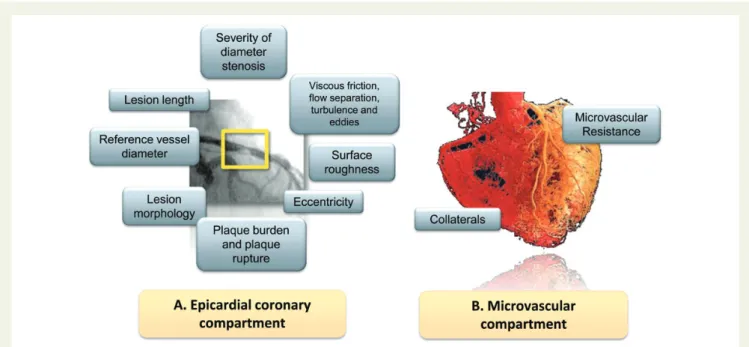
Figure 2
Myocardial blood flow is determined by a variety of factors that affect the epicardial coronary compartment (A) or the microvascular
compartment (B). The epicardial coronary compartment is generally affected by coronary atherosclerosis obstructing conductive blood flow (A).
However, besides the angiographical severity of diameter narrowing, several other factors that are not or only poorly captured by invasive
angiog-raphy can modulate the haemodynamic relevance of a given lesion. (B) However, under resting conditions, 90% of coronary vascular resistance
resides in the microcirculation (arterioles and pre-arterioles). Microvascular dilation in response to various stressors is crucial to match blood
flow with myocardial oxygen demands. This regulation of microvascular resistance can be disturbed in the presence of a variety of functional or
structural pathologies and thereby contribute to myocardial ischaemia.
Figure 3
Comparison of fractional flow reserve vs. Non-invasive imaging modalities. Fractional flow reserve assesses only the pressure gradient
across a lesion in the epicardial coronary arteries. However, ischaemia by myocardial perfusion imaging is an integrated measurement of blood flow
through the epicardial and microvascular compartement and, therefore, can also be affected by microvascular or endothelial dysfunction.
O. Gaemperli et al.
2434
myocardial ischaemia to be in the range of 0.72 – 0.75, i.e.
consider-ably lower than the currently accepted cut-off of 0.80.
20From pictures to outcome
All of the above have important implications for the design of future
imaging studies: at present, there is considerable discussion which of
the available imaging techniques is most accurate to diagnose CAD,
and a number of comparisons using CA as the gold standard have
been conducted and employed to prove the superiority of one
tech-nique over the other. However, given the changing paradigm of CAD,
this approach appears outdated. Additionally, one should also realize
that perfusion and systolic function as markers of ischaemia may
rep-resent different phenomena and may therefore not be directly
com-parable. Consequently, trials are needed that are designed to
demonstrate that non-invasive imaging can guide treatment and
sub-sequently improve patients’ outcome. Such trials may follow the
design of the FAME I and II trials and thereby continue the exemplary
path that FFR has set in invasive cardiology.
17,18Accordingly, efforts
across countries are currently joined to design appropriately sized
prospective randomized trials to test this hypothesis.
23In an era of
ever increasing pressure from financial reimbursement systems
such trials are eagerly needed to solidify the role of non-invasive
cardiac imaging in cardiology. By these means, clinical validation of
established or novel cardiac imaging techniques will centre around
the ultimate and most important gold standard in cardiology:
guid-ance of therapy aiming at improvement of patients’ outcome.
Conflict of interest: none declared.
References
1. Gould KL, Lipscomb K, Hamilton GW. Physiologic basis for assessing critical coronary stenosis. Instantaneous flow response and regional distribution during coronary hyperemia as measures of coronary flow reserve. Am J Cardiol 1974;33: 87 – 94.
2. Gaemperli O, Schepis T, Valenta I, Koepfli P, Husmann L, Scheffel H, Leschka S, Eberli FR, Luscher TF, Alkadhi H, Kaufmann PA. Functionally relevant coronary artery disease: comparison of 64-section CT angiography with myocardial perfusion SPECT. Radiology 2008;248:414 – 423.
3. Gould KL. Does coronary flow trump coronary anatomy? JACC Cardiovasc Imaging 2009;2:1009 – 1023.
4. Meijboom WB, Van Mieghem CA, van Pelt N, Weustink A, Pugliese F, Mollet NR, Boersma E, Regar E, van Geuns RJ, de Jaegere PJ, Serruys PW, Krestin GP, de Feyter PJ. Comprehensive assessment of coronary artery stenoses: computed tom-ography coronary angitom-ography versus conventional coronary angitom-ography and cor-relation with fractional flow reserve in patients with stable angina. J Am Coll Cardiol 2008;52:636 – 643.
5. Schuijf JD, Wijns W, Jukema JW, Atsma DE, de Roos A, Lamb HJ, Stokkel MP, Dibbets-Schneider P, Decramer I, De Bondt P, van der Wall EE, Vanhoenacker PK, Bax JJ. Relationship between noninvasive coronary angiography with multi-slice computed tomography and myocardial perfusion imaging. J Am Coll Cardiol 2006;48:2508 – 2514.
6. Tonino PA, Fearon WF, De Bruyne B, Oldroyd KG, Leesar MA, Ver Lee PN, Maccarthy PA, Van’t Veer M, Pijls NH. Angiographic versus functional severity of cor-onary artery stenoses in the FAME study fractional flow reserve versus angiography in multivessel evaluation. J Am Coll Cardiol 2010;55:2816 – 2821.
7. White CW, Wright CB, Doty DB, Hiratza LF, Eastham CL, Harrison DG, Marcus ML. Does visual interpretation of the coronary arteriogram predict the physiologic im-portance of a coronary stenosis? N Engl J Med 1984;310:819 – 824.
8. Jaarsma C, Leiner T, Bekkers SC, Crijns HJ, Wildberger JE, Nagel E, Nelemans PJ, Schalla S. Diagnostic performance of noninvasive myocardial perfusion imaging using single-photon emission computed tomography, cardiac magnetic resonance, and positron emission tomography imaging for the detection of obstructive coron-ary artery disease: a meta-analysis. J Am Coll Cardiol 2012;59:1719 – 1728. 9. Klocke FJ, Baird MG, Lorell BH, Bateman TM, Messer JV, Berman DS, O’Gara PT,
Carabello BA, Russell RO Jr, Cerqueira MD, St John Sutton MG, DeMaria AN, Udelson JE, Kennedy JW, Verani MS, Williams KA, Antman EM, Smith SC Jr, Alpert JS, Gregoratos G, Anderson JL, Hiratzka LF, Faxon DP, Hunt SA, Fuster V, Jacobs AK, Gibbons RJ, Russell RO. ACC/AHA/ASNC guidelines for the clinical use of cardiac radionuclide imaging—executive summary: a report of the American College of Cardiology/American Heart Association Task Force on Practice Guide-lines (ACC/AHA/ASNC Committee to Revise the 1995 GuideGuide-lines for the Clinical Use of Cardiac Radionuclide Imaging). J Am Coll Cardiol 2003;42:1318 – 1333. 10. Nandalur KR, Dwamena BA, Choudhri AF, Nandalur MR, Carlos RC. Diagnostic
per-formance of stress cardiac magnetic resonance imaging in the detection of coronary artery disease: a meta-analysis. J Am Coll Cardiol 2007;50:1343 – 1353.
11. Stein PD, Yaekoub AY, Matta F, Sostman HD. 64-slice CT for diagnosis of coronary artery disease: a systematic review. Am J Med 2008;121:715 – 725.
12. Iskander S, Iskandrian AE. Risk assessment using single-photon emission computed tomographic technetium-99m sestamibi imaging. J Am Coll Cardiol 1998;32:57 – 62. 13. Shaw LJ, Iskandrian AE. Prognostic value of gated myocardial perfusion SPECT. J Nucl
Cardiol 2004;11:171 – 185.
14. Hachamovitch R, Berman DS, Shaw LJ, Kiat H, Cohen I, Cabico JA, Friedman J, Diamond GA. Incremental prognostic value of myocardial perfusion single photon emission computed tomography for the prediction of cardiac death: differential stratification for risk of cardiac death and myocardial infarction. Circulation 1998; 97:535 – 543.
15. Shaw LJ, Berman DS, Maron DJ, Mancini GB, Hayes SW, Hartigan PM, Weintraub WS, O’Rourke RA, Dada M, Spertus JA, Chaitman BR, Friedman J, Slomka P, Heller GV, Germano G, Gosselin G, Berger P, Kostuk WJ, Schwartz RG, Knudtson M, Veledar E, Bates ER, McCallister B, Teo KK, Boden WE. Optimal medical therapy with or without percutaneous coronary inter-vention to reduce ischemic burden: results from the Clinical Outcomes Utilizing Revascularization and Aggressive Drug Evaluation (COURAGE) trial nuclear sub-study. Circulation 2008;117:1283 – 1291.
16. Hachamovitch R, Hayes SW, Friedman JD, Cohen I, Berman DS. Comparison of the short-term survival benefit associated with revascularization compared with medical therapy in patients with no prior coronary artery disease undergoing stress myocardial perfusion single photon emission computed tomography. Circula-tion 2003;107:2900 – 2907.
17. Tonino PA, De Bruyne B, Pijls NH, Siebert U, Ikeno F, van’t Veer M, Klauss V, Manoharan G, Engstrom T, Oldroyd KG, Ver Lee PN, MacCarthy PA, Fearon WF. Fractional flow reserve versus angiography for guiding percutaneous coronary inter-vention. N Engl J Med 2009;360:213 – 224.
18. De Bruyne B, Pijls NH, Kalesan B, Barbato E, Tonino PA, Piroth Z, Jagic N, Mobius-Winkler S, Rioufol G, Witt N, Kala P, MacCarthy P, Engstrom T, Oldroyd KG, Mavromatis K, Manoharan G, Verlee P, Frobert O, Curzen N, Johnson JB, Juni P, Fearon WF. Fractional flow reserve-guided PCI versus medical therapy in stable coronary disease. N Engl J Med 2012;367:991 – 1001.
19. Melikian N, De Bondt P, Tonino P, De Winter O, Wyffels E, Bartunek J, Heyndrickx GR, Fearon WF, Pijls NH, Wijns W, De Bruyne B. Fractional flow reserve and myocardial perfusion imaging in patients with angiographic multivessel coronary artery disease. JACC Cardiovasc Intervent 2010;3:307 – 314.
20. Johnson NP, Kirkeeide RL, Gould KL. Is discordance of coronary flow reserve and fractional flow reserve due to methodology or clinically relevant coronary patho-physiology? JACC Cardiovasc Imaging 2012;5:193 – 202.
21. Murthy VL, Naya M, Foster CR, Hainer J, Gaber M, Di Carli G, Blankstein R, Dorbala S, Sitek A, Pencina MJ, Di Carli MF. Improved cardiac risk assessment with noninvasive measures of coronary flow reserve. Circulation 2011;124: 2215 – 2224.
22. Gaemperli O, Kaufmann PA. Why quantify myocardial perfusion? Curr Cardiovasc Imaging Rep 2012;5:133 – 143.
23. The ISCHEMIA trial.https://www.ischemiatrial.org/(24 January 2013).