HAL Id: hal-02369299
https://hal.inria.fr/hal-02369299
Submitted on 20 Nov 2019
HAL is a multi-disciplinary open access
archive for the deposit and dissemination of
sci-entific research documents, whether they are
pub-lished or not. The documents may come from
teaching and research institutions in France or
L’archive ouverte pluridisciplinaire HAL, est
destinée au dépôt et à la diffusion de documents
scientifiques de niveau recherche, publiés ou non,
émanant des établissements d’enseignement et de
recherche français ou étrangers, des laboratoires
MNE: Software for Acquiring, Processing, and
Visualizing MEG/EEG Data
Lorenz Esch, Christoph Dinh, Eric Larson, Denis Engemann, Mainak Jas,
Sheraz Khan, Alexandre Gramfort, M. Hämäläinen
To cite this version:
Lorenz Esch, Christoph Dinh, Eric Larson, Denis Engemann, Mainak Jas, et al.. MNE: Software for
Acquiring, Processing, and Visualizing MEG/EEG Data. Magnetoencephalography, Springer
Inter-national Publishing, pp.355-371, 2019, �10.1007/978-3-030-00087-5_59�. �hal-02369299�
MNE: Software for Acquiring, Processing and Visualizing
MEG/EEG Data
Lorenz Escha,b,c, Christoph Dinhc,d, Eric Larsone, Denis Engemannf, Mainak Jasa, Sheraz
Khana,g,h, Alexandre Gramfortf, Matti S. H¨am¨al¨ainena,g,h
aAthinoula A. Martinos Center for Biomedical Imaging, Massachusetts General Hospital, Charlestown, MA, USA bInstitute of Biomedical Engineering and Informatics, Technische Universit¨at Ilmenau, Ilmenau, Germany
cBoston Children’s Hospital, Boston, MA, USA
dInstitute for Medical Engineering, Research Campus STIMULATE, Otto-von-Guericke University, Magdeburg, Germany eInstitute for Learning and Brain Sciences, University of Washington, Seattle WA, USA
fINRIA, CEA, Universit´e Paris-Saclay, Palaiseau, France gMassachusetts Institute of Technology, Cambridge, MA, USA
hHarvard Medical School, Boston, MA, USA
Abstract
The methods for acquiring, processing, and visualizing Magnetoencephalography (MEG) and Electroencephalography (EEG) data are rapidly evolving. Advancements in hardware and software development o↵er new opportunities for cognitive and clinical neuroscientists but at the same time introduce new challenges as well. In recent years the MEG/EEG community has developed a variety of software tools to overcome these challenges and cater to individual research needs. As part of this endeavour, the MNE software project, which includes MNE-C, MNE-Python, MNE-CPP, and MNE-MATLAB as its subprojects, o↵ers an efficient set of tools addressing certain common needs. Even more importantly, the MNE software family covers diverse use case scenarios. Here, we present the landscape of the MNE project and discuss how it will evolve to address the current and emerging needs of the MEG/EEG community.
Keywords: Magnetoencephalography (MEG), Electroencephalography (EEG), Software, Analysis tools, Open-Source, Real-time analysis, Signal processing, Machine Learning
1. MEG/EEG Data Analysis in Research and Clinical Settings
The field of neuroscience is rapidly expanding through interdisciplinary e↵orts and has enabled studies of the nervous system at several scales, starting from the molecular level and the study of single neurons in animals and extending to recording and manipulating large-scale human brain networks. Brain activity can be studied with a wide variety of temporal and spatial resolutions using diverse techniques. The research community thus has access to a growing amount of shared multi-modal as well as multi-scale brain data. MEG and EEG have a unique position in this endeavor as the only non-invasive means for studying electrophysiological activity. Both methods can track neuronal dynamics at millisecond resolution and, hence, capture behaviorally relevant fast changes inaccessible to hemodynamic methods, e.g., functional magnetic-resonance imaging (fMRI). While MEG can have a higher spatial resolution due to absence of the smearing e↵ects present in EEG, it is also more selective by favoring signals from cortical pyramidal neurons in the walls of the sulci (Baillet, 2017). MEG and EEG, therefore, have a complementary
nature, and it has been suggested that improved results can be obtained when combining the two methods (Sharon et al., 2007). The research in non-invasive electrophysiology can be systematized by considering at least two aspects.
First, how MEG and EEG are used depends substantially on the broader practical context of data acquisition. Common settings range from basic academic research to clinical studies aiming at diagnostics of individual patients to inform subsequent treatment choices. The interaction with data and the optimal software tools will, therefore, assume a di↵erent form for the researcher attempting to, e.g, decode low-level visual features from gamma band sensor level dynamics (Westner et al., 2018) than for a clinician employing MEG for pre-surgical assessment of an epileptic patient (De Ti`ege et al., 2012). In the first case, a researcher may want to compose an appropriate sequence of processing operations that will preserve the e↵ects of interest and lead to best decoding performance. On the other hand, in clinical practice, a fixed set of tools are required emphasizing interactive visualization for e↵ective identification of the salient epileptiform activity. For this purpose, a stand-alone medical software application with a graphical user interface (GUI) with limited customization options is likely to be preferred.
Second, the MEG and EEG communities follow the recent trend toward data-centric research in the biomedical sciences (Leonelli, 2016). This trend is characterized by the increasing volume of publicly available curated scientific data (Poldrack et al., 2017) and their reuse by teams who have not been involved in the acquisition of the data. Accordingly, new consortia keep emerging that curate large-scale MEG and EEG datasets (Niso et al., 2016; Zhang et al., 2018; Van Essen et al., 2013; Taylor et al., 2017). As a result, researchers from diverse backgrounds can now work on human electrophysiology data without having access to MEG and/or EEG acquisition infrastructure. A researcher who acquires MEG data in a semantic auditory processing experiment with a limited number of subjects would need a di↵erent set of tools than one who studies cognitive aging employing thousands of MEG recordings from a database (Taylor et al., 2017; Van Essen et al., 2013; Niso et al., 2016). The former would use a combination of GUIs for assessment of data quality, setting annotations, scripting for preprocessing and data analysis backed by reporting tools for quality assessment. The latter would almost solely rely on scripts, emphasize automated processing (Engemann and Gramfort, 2015; Jas et al., 2017) and utilize dedicated libraries for classical machine learning (Pedregosa et al., 2011), deep learning, and specialized forms of data visualization.
In the following, we will discuss how the research and software development activities in the MNE community have responded to the variety of needs. We will first briefly summarize the history of the MNE software (Esch et al., 2018; Gramfort et al., 2014, 2013a; Jas et al., 2018) and then cover the di↵erent MNE packages. We will detail how each of the MNE software packages responds to the needs of di↵erent types of MEG/EEG users. Subsequently, we will discuss emerging data analysis use cases that require novel innovations in software, or even some rethinking of the way MEG/EEG data are visualized and analyzed. With this perspective, we will discuss how the MNE software tools can already partially address these new needs and what could be a path forward.
2. MNE and its history
MNE is a software package that provides complete data analysis pipelines for MEG/EEG data processing. In comparison to other (MATLAB-based) software packages for MEG/EEG data processing, e.g., Brainstorm (Tadel et al., 2011), EEGLAB (Delorme and Makeig, 2004; Delorme et al., 2011), FieldTrip (Oostenveld et al., 2011), NutMeg (Dalal et al., 2011), and SPM
(Litvak et al., 2011), MNE comes in multiple flavors, i.e., MNE-C (historically referred simply to as MNE), MNE-CPP, MNE-MATLAB and MNE-Python, each addressing needs from di↵erent segments of the academic and clinical communities. While the original MNE-C was started by Matti H¨am¨al¨ainen at MGH in 2001 and made publicly available in 2006, the other MNE packages started later (MNE-CPP in 2010 and MNE-Python in 2011), incorporating the same core features as MNE-C, such as direct integration with the anatomical reconstruction provided by the FreeSurfer software (Fischl et al., 1999).
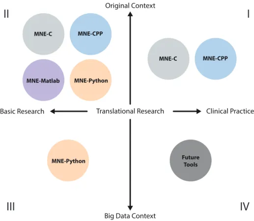
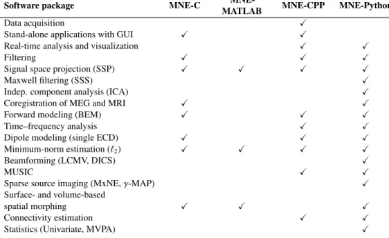
Figure 1 illustrates the landscape of di↵erent MNE flavors and their di↵erent roles. All MNE packages are currently engaged in the original context of MEG/EEG processing (second quadrant), where “original context” refers to well-established workflows ranging from the actual experimental design, data acquisition, processing, to analysis for basic research purposes. The first quadrant includes the MNE-C and MNE-CPP packages that use the C and CPP, a.k.a. C++, programming languages. Both are used in translational research bringing state-of-the-art methods to clinical applications and practice. Here, high-level graphical user interface controls provide tools for clinicians and researchers with minimal or non-existent programming background. The need for large scale data (big data) analysis is covered by MNE-Python, paving the way for more computer intensive data science and machine learning approaches (third quadrant). MNE-Python and its growing support for EEG and machine learning methods has recently enabled large-scale analysis of clinical EEG in neurology (Engemann et al., 2018). The future tools, discussed in greater detail in section 4, could specifically respond to the needs of clinical practice powered by data-driven methods and recycling of consortium data (quadrant four). Di↵erent features of the MNE packages are summarized in Table 1: All packages read and write data in the same file format, enabling users to use the tool that is best suited for each processing step. In this table, ECD stands for equivalent current dipole, LCMV for linearly constrained minimum-variance (Van Veen et al., 1997), DICS for dynamic imaging of coherent sources (Gross et al., 2001), MxNE for mixed-norm estimates (Gramfort et al., 2013b), MVPA for multivariate pattern analysis (often referred to as decoding) (KING et al., 2018), BEM for Boundary Element Method, MUSIC for MUltiple SIgnal Classification (Mosher and Leahy, 1999), and we refer to (Wipf and Nagarajan, 2009) for details on -MAP.
As illustrated in Figure 1, MNE-Python o↵ers a unique opportunity to connect the data science and machine learning communities with the MEG and EEG data processing challenges. This is presently possible thanks to the modern open source Python tools that are now available for advanced statistical computations and analysis of big data.
3. The scope and features of the MNE packages
3.1. O↵-line analysis with MNE-C GUIs and command line tools
The original MNE-C, conceived and written at the Martinos Center at Massachusetts General Hospital, consists of command line programs that can be used in shell scripts for automated processing and two GUI applications for interactive data processing and inspection. MNE-C supports band-pass, low-pass, and high-pass filtering. The GUI mne browse raw also allows previewing the filtered data, so one can investigate the impact of the filter on the signal, as well as the interactive creation of the projection operator for the Signal-Space Projection (SSP) method using a Singular Value Decomposition (SVD) of a selected portion of the data (Uusitalo and Ilmoniemi, 1997). The same software module can also be used for computing averages over multiple trials and for the estimation of the noise-covariance matrix.
Clinical Practice Translational Research
Basic Research
Original Context
Big Data Context
MNE-Python MNE-CPP MNE-Matlab MNE-C MNE-Python MNE-CPP MNE-C Future Tools
I
II
III
IV
Figure 1: The MNE landscape. The quadrants are populated by the four MNE packages based on their individual scope and key qualifications. The individual MNE packages have slightly di↵erent mission statements focusing on di↵erent user needs and research questions.
The other GUI mne analyze allows the interactive alignment of the MRI and MEG coordinate systems, the so-called coregistration step, as well as the interactive exploration of cortically constrained source estimates obtained by MNE, dSPM, and sLORETA inverse methods. The command line tools can be assembled in Unix shell scripts for non-interactive analysis. The usage of the MNE-C software is primarily described in the PDF manual: https://www.martinos. org/meg/manuals/MNE-manual-2.7.pdf.
3.2. O↵-line analysis with MNE-MATLAB
The MNE-MATLAB toolbox (compatible with MATLAB versions 7.0 or later) started from a desire of the MNE user community to go beyond the possibilities made available by the compiled MNE-C tools. It is a collection of m-files to facilitate interfacing with binary file formats of the MNE software and is redistributed as a part of several MATLAB-based MEG/EEG software packages (Brainstorm, FieldTrip, NutMeg, and SPM). The included functionality can be roughly divided into following four categories: (1) High-level reading and writing routines, which provide interfacing with binary file formats like .fif, .stc, .label, and .w files. (2) Signal processing routines, which implement the software gradient compensation and signal-space
Table 1: Overview of the features provided by the di↵erent MNE packages (X= supported).
Software package MNE-C MATLABMNE- MNE-CPP MNE-Python
Data acquisition X
Stand-alone applications with GUI X X
Real-time analysis and visualization X X
Filtering X X X
Signal space projection (SSP) X X X X
Maxwell filtering (SSS) X
Indep. component analysis (ICA) X
Coregistration of MEG and MRI X X
Forward modeling (BEM) X X X
Time–frequency analysis X X
Dipole modeling (single ECD) X X X
Minimum-norm estimation (`2) X X X X
Beamforming (LCMV, DICS) X
MUSIC X X
Sparse source imaging (MxNE, -MAP) X
Surface- and volume-based
spatial morphing X X X
Connectivity estimation X X
Statistics (Univariate, MVPA) X
projection. (3) Utility functions, which include auxiliary functions to reading and writing binary files, transforming data between coordinate systems, assembling inverse operators, proving coil definition for various sensor types, etc. (4) Examples demonstrating the use of the toolbox, which form a basis for user-specific processing routines. The MNE-MATLAB code is available online at https://github.com/mne-tools/mne-matlab.
3.3. O↵-line analysis and scripting with MNE-Python
The growing popularity of the Python stack for data science in academia and industry has prompted the development of MNE in Python as an alternative for the MNE-C tools. Since its inception, Python (Gramfort et al., 2014) has grown from replicating most of the MNE-C functionality to implementing many popular but also novel advanced analysis tools while fully supporting EEG analysis. MNE-Python was built to support analysis of multiple imaging modalities beyond MEG/EEG and now has some support for stereotactic electroencephalography (sEEG), functional near-infrared spectroscopy (fNIRS), and electrocorticography (ECoG) data as well.
Figure 2 highlights some of the key features of MNE-Python. It implements input/output (IO) routines for reading a variety of MEG/EEG file formats (http://martinos.org/mne/ stable/manual/io.html), advanced preprocessing tools such as the Signal Space Separation (SSS) algorithm, and XDAWN (Taulu and Kajola, 2005; Rivet et al., 2009), a dedicated decoding module for MEG/EEG (King et al., 2018), Bayesian and sparse source imaging methods (Gramfort et al., 2013b; Wipf and Nagarajan, 2009), beamforming, and scanning methods such as LCMV, DICS or RAP-MUSIC (Van Veen et al., 1997; Mosher and Leahy, 1999; Gross et al., 2001), statistics (Maris and Oostenveld, 2007; Kriegeskorte et al., 2008), real-time analysis of data (http: //martinos.org/mne/stable/auto_examples/#real-time-m-eeg-acquisition), and
Reporting and visualization for quality control
Different modalities: MEG, EEG, ECOG Visualization Statistics / Machine Learning
Automated preprocessing (ICA) Source localization
Figure 2: MNE-Python: processing and visualizing electrophysiology data
quality assurance and reporting tools (Jas et al., 2018). The decoding module is built to fa-cilitate the use of the popular scikit-learn (Pedregosa et al., 2011) software, which provides simple and e↵ective programmatic access to a wide array of classical machine learning algorithms and procedures in Python. Like scikit-learn, MNE-Python is distributed under a permissive Berke-ley Software Development (BSD) license and readily supports academic as well as commercial reusage.
The code is available online at https://github.com/mne-tools/mne-python. More than 80 people have so far contributed to the MNE-Python source code. Online documentation is available at http://martinos.org/mne/stable/documentation.html with examples continuously updated thanks to sphinx and sphinx-gallery (https://sphinx-gallery. readthedocs.io/en/latest/) packages.
3.4. Acquisition and real-time analysis with MNE-CPP
MNE-CPP provides a cross-platform framework which allows the development of software applications for real-time MEG/EEG data acquisition, processing and visualization. The project is open-source BSD licensed (3-clause). It can be used to develop stand-alone applications on Windows, MacOS, and Linux. MNE-CPP builds upon two external dependencies: The Qt framework (QtProject, 2018) for GUI programming and the Eigen library (Guennebaud et al., 2018) for linear algebra. All MNE-CPP tools are designed to function in o↵-line as well as in real-time scenarios. In addition to giving experienced C++ developers the opportunity to create their own applications, MNE-CPP o↵ers pre-developed stand-alone applications with GUIs. Currently, three applications are being developed: MNE Scan (Esch et al., 2018), MNE Browse, and MNE Analyze. All three are available as pre-built binaries for Windows, MacOS, and Linux. MNE Scan is a plug-in based tool that can be used to acquire data from MEG/EEG devices and store the received data to a file and/or provide real-time data streams. Acquisition and processing tasks are developed as individual units. The workflow follows a pipeline approach where the user can select and connect the acquisition plug-in and subsequent real-time processing plug-ins. The acquisition plug-ins o↵er connections to MEG (Elekta Neuromag, BabyMEG) and EEG (TMSI Refa, EEGoSports, gTec USB, BrainAmp, and Natus) devices. It is also possible to stream in
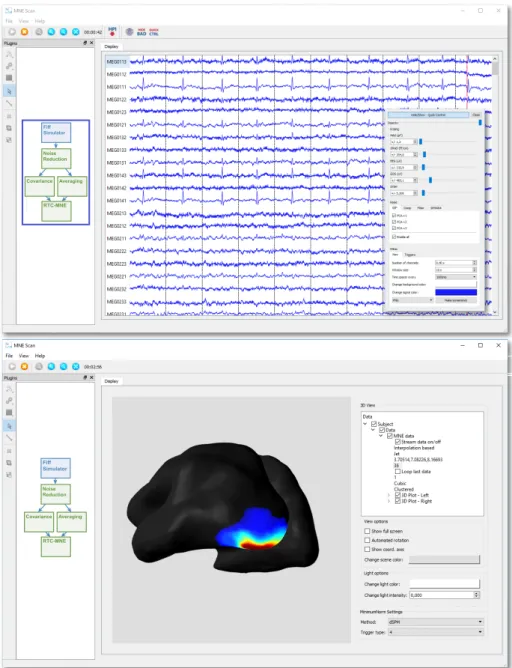
recorded data from a file to imitate an ongoing measurement session. This is especially useful when debugging and testing new plug-ins. The processing plug-ins include real-time capable implementations for temporal filtering, SSP, SPHARA (Graichen et al., 2015), software gradients, averaging, source localization, connectivity estimation, and a Brain Computer Interface (BCI). A clinically oriented use case for MNE Scan is its use in the BabyMEG system. The BabyMEG (Okada et al., 2016) is a 375-channel, whole-head pediatric MEG instrument. It is used in a clinical environment at Boston Children’s Hospital for both patients and healthy neonates, infants, and preschool children up to three years of age. Another use case of MNE Scan is the computation and visualization of source estimates in real-time (Dinh et al., 2015, 2018) via a dedicated plug-in. The estimated source activity is visualized on a cortical surface reconstructed with the Freesurfer software (Fischl et al., 1999). Fig. 3 shows a snapshot of MNE Scan during real-time source localization setup. MNE Scan can also be used in neurofeedback research and applications. For example, the SSVEP BCI plug-in provides a visual reactive BCI to control a virtual keyboard.
Similar to MNE Scan’s o✏ine counterparts, MNE Browse and MNE Analyze are also inspired by their MNE-C counterparts. With their reimplementation MNE-CPP targets to make them future proof, available on Windows machines, more community driven and extensible with new state-of-the-art analysis methods. Both MNE Browse and MNE Analyze are designed to process and visualize MEG/EEG data o✏ine and are solely based on MNE-CPP libraries, Eigen and Qt. MNE Browse functions as a lightweight analysis tool and features basic processing steps such as the loading and visualization of data, channel management, annotation and temporal filtering. On the contrary, MNE Analyze provides more sophisticated analysis tools such as dipole fitting, distributed source localization, and computation of confidence intervals. MNE Analyze’s underlying architecture implements plug-ins for maintainability and easy feature inclusion. MNE Browse and MNE Analyze, as part of the MNE-CPP project, are still in an early development phase. The MNE-CPP code is available online at https://github.com/mne-tools/mne-cpp and www.mne-cpp.org.
4. The needs for future MEG/EEG data processing
The MNE environment with its di↵erent packages is able to cover a wide variety of use case scenarios. MNE-C will continue to function as the code backbone of MNE-Python and MNE-CPP. Its longstanding acceptance and tested features throughout the community will keep guiding new and ported features. MNE-MATLAB will continue to ensure the usage of MNE data structures in the MATLAB ecosystem dominated by the Fieldtrip, Brainstorm, NutMeg, and SPM toolboxes. We envision new usage scenarios will emerge at the intersection of already known practices, imposing new demands for acquiring, processing, and visualizing MEG/EEG data. In the following we shall discuss a few likely scenarios.
Browsing of remote databases. The advent of large datasets poses new challenges for interac-tive visualization. In particular, it will not be feasible to duplicate the data on a local workstation. Currently, users who want to browse through large data sets must restrict themselves to a subset of data. Subsequently, they would need to load each file separately from the command line or the GUI menu, disrupting the workflow. Future versions of MEG/EEG data browsers will therefore have to operate over the network and include functionality that facilitates navigation through thousands of MEG/EEG recordings. This would at least include an e↵ective file-search, scheduling functionality to specify a set of files to visit sequentially, as well as cross-data set navigation controls to enable seamless browsing. In the current software ecosystem, MNE-Python could most naturally extend to this domain given how much the Python language is used for web
Fiff Simulator RTC-MNE Fiff Simulator RTC-MNE
Figure 3: Real-time source localization setup in MNE Scan: In the presented pipeline (see blue rectangle) the Fi↵Simulator imitates a real-time data stream based on pre-recorded data (see upper screenshot). After the data was filtered the data stream is forwarded to an averaging and covariance plug-in. Subsequently, the averaged (25 moving average) and noise covariance results are send to the RTC-MNE plug-in. Finally, the source estimates are plotted on an inflated brain surface mesh and can be made available to connected plug-ins (see lower screenshot).
applications, and the ability to communicate with remotely running Python kernels as done with the Jupyter software (https://jupyter.org/).
Integration of advanced analysis tools during clinical MEG/EEG acquisition. The search for biomarkers using machine learning on large, socially heterogeneous samples has become an active area of research (Allen et al., 2012; Drysdale et al., 2017; Liem et al., 2017; Khan et al., 2015, 2018). In the near future, medical professionals may develop machine learning models pre-trained o✏ine and apply these models online during MEG/EEG data acquisition to help detect abnormal brain signals and supplement diagnostics. Software tools could thus support extensions for models specified and trained using machine learning tools. In a second, related scenario, medical doctors and data scientists may work together to include custom data processing routines at the acquisition level. For example, the clinical experimenter may launch a software pipeline for automated preprocessing from within the acquisition GUI to obtain clean data and a visual quality control report (Engemann and Gramfort, 2015; Jas et al., 2018). This could be implemented via modular extensions of the acquisition software in C++ to execute custom routines written in Python, thereby integrating MNE Scan and MNE-Python.
General Real-Time Processing. Real-time processing can become of interest as it has the potential for creating highly dynamic and adaptable paradigms depending mainly on the current brain state or condition of interest. Neurofeedback enables researchers to test hypotheses about specific brain conditions, e.g., the state of specific brain areas, by monitoring this condition, and adapting the experimental interventions accordingly. This opens up a new way to investigate basic mechanisms of brain functions and can give the subject a chance to learn and modify their neuronal activity patterns (Wolpaw et al., 1991). Latter is a valuable tool in neurorehabilitation (Mohanty et al., 2018). Moreover, real-time data analysis allows an early assessment of the measurement setup, the subject’s response to the introduced paradigm, and the feasibility of a research idea. Early correction of badly designed measurement setups and paradigms can contribute to saving time and resources. Thus, the promotion of real-time analysis tools as a preemptive indicator for problems can become of increasing interest to the MEG/EEG community. A more general and long-term goal for real-time data analysis can also be found in clinical environments. Here it is of special interest to further utilize MEG/EEG in clinical diagnosis, where speed is often essential. Sophisticated analysis tools such as real-time source estimation could lead to a faster diagnosis and better monitoring of the condition of a patient.
Brain-computer interfaces and hyperscanning. Two emerging approaches in rehabilitation science, social neuroscience, and computational psychiatry are BCIs (H¨ohne et al., 2014; Mohanty et al., 2018) and hyperscanning (Bilek et al., 2017; Ahn et al., 2018; Goldstein et al., 2018; Zhdanov et al., 2015). It is likely that soon new acquisition paradigms will emerge both for basic research and clinical practice in which BCI and hyperscanning will be combined, such that the stimulation will be driven by brain activity of several individuals (Rao et al., 2014; Jiang et al., 2018). The acquisition software, in this scenario, will need not only modular extensions for real-time stimulation and machine learning, but also flexible visualization functionality that supports the appropriate abstractions for co-representing activity from several brains.
Cloud computing. Data analysis aiming at diagnostics is difficult to generalize due to the di↵erent platforms, complex data acquisition, and processing involved. In this context, community standards will play a crucial role (Gorgolewski et al., 2016; Niso et al., 2018). The newly established Brain Imaging Data Structure (BIDS) for organizing data is already promising as it simplifies the process of creating portable pipelines – the so-called BIDS Apps (Gorgolewski et al., 2017b). As already started by some fMRI software stacks (Esteban et al., 2017a, 2018; Gorgolewski et al., 2017a; Esteban et al., 2017b; Yarkoni et al., 2011; Glatard et al., 2018), one
would need to develop cloud based automated pre- and postprocessing applications, that could even be integrated with acquisition and analysis software platforms. This would significantly increase the ease and efficiency of acquiring, monitoring, analyzing, and integrating various types of clinical electrophysiology data with anatomical structures. Tighter integration with BIDS will allow MNE to interface easily with applications in the cloud but also amongst its di↵erent flavors and with other MEG/EEG analysis software. Acquisition and analysis plug-in interfaces could be enabled to communicate with the cloud application programming interface to retrieve preprocessed volume reconstructions and to upload the acquired data again to the cloud. In addition to source localization, automated cloud-based post-processing will also enable the extraction of biomarkers to support diagnosis (Engemann et al., 2018). Through the modular concepts used in the cloud processing pipelines, it will be readily extensible by the scientific community with new processing steps.
In conclusion, the di↵erent MNE projects continue to have specific roles in order to cover all the varied aspects of MEG/EEG data processing. We envision that many of the new developments will necessitate integration of several packages for optimal implementation. For example, machine-learning tools can be readily developed in MNE-Python using large data sets as input and implemented for clinical use as plug-ins in MNE-CPP. MNE as a whole continues to provide freely accessible and multi-purpose software tools for the acquisition, processing, and visualization of MEG/EEG data for both basic and clinical research as well as for clinical applications.
References
Ahn S, Cho H, Kwon M, Kim K, Kwon H, Kim BS, Chang WS, Chang JW, Jun SC (2018) Interbrain phase synchronization during turn-taking verbal interactiona hyperscanning study using simultaneous EEG/MEG. Human Brain Mapping 39(1):171–188
Allen N, Sudlow C, Downey P, Peakman T, Danesh J, Elliott P, Gallacher J, Green J, Matthews P, Pell J, et al. (2012) UK Biobank: Current status and what it means for epidemiology. Health Policy and Technology 1(3):123–126
Baillet S (2017) Magnetoencephalography for brain electrophysiology and imaging. Nature Neuroscience 20:327–339, URL http://dx.doi.org/10.1038/nn.4504
Bilek E, St¨oßel G, Sch¨afer A, Clement L, Ruf M, Robnik L, Neukel C, Tost H, Kirsch P, Meyer-Lindenberg A (2017) State-dependent cross-brain information flow in borderline personality disorder. JAMA psychiatry 74(9):949–957 Dalal SS, Zumer JM, Guggisberg AG, Trumpis M, Wong DDE, Sekihara K, Nagarajan SS (2011) MEG/EEG Source
Reconstruction, Statistical Evaluation, and Visualization with NUTMEG. Computational Intelligence and Neuroscience 2011:1–17, DOI 10.1155/2011/758973, URL http://www.hindawi.com/journals/cin/2011/758973/ De Ti`ege X, Carrette E, Legros B, Vonck K, Bourguignon M, Massager N, David P, Van Roost D, Meurs A, Lapere S,
et al. (2012) Clinical added value of magnetic source imaging in the presurgical evaluation of refractory focal epilepsy. Journal of Neurology, Neurosurgery, and Psychiatry 83(4):417–423
Delorme A, Makeig S (2004) EEGLAB: an open source toolbox for analysis of single-trial EEG dynamics including independent component analysis. Journal of Neuroscience Methods 134(1):9–21, DOI 10.1016/j.jneumeth.2003.10.009, URL http://linkinghub.elsevier.com/retrieve/pii/S0165027003003479, arXiv:1011.1669v3 Delorme A, Mullen T, Kothe C, Akalin Acar Z, Bigdely-Shamlo N, Vankov A, Makeig S (2011) EEGLAB, SIFT, NFT,
BCILAB, and ERICA: New Tools for Advanced EEG Processing. Computational Intelligence and Neuroscience 2011:1– 12, DOI 10.1155/2011/130714, URL http://www.hindawi.com/journals/cin/2011/130714/, 130714 Dinh C, Strohmeier D, Luessi M, G¨ullmar D, Baumgarten D, Haueisen J, H¨am¨al¨ainen MS (2015) Real-Time MEG Source
Localization Using Regional Clustering. Brain Topography 28(6):771–784, DOI 10.1007/s10548-015-0431-9 Dinh C, Esch L, R¨uhle J, Bollmann S, G¨ullmar D, Baumgarten D, H¨am¨al¨ainen MS, Haueisen J (2018) Real-Time Clustered
Multiple Signal Classification (RTC-MUSIC). Brain Topography 31(1):125–128, DOI 10.1007/s10548-017-0586-7 Drysdale AT, Grosenick L, Downar J, Dunlop K, Mansouri F, Meng Y, Fetcho RN, Zebley B, Oathes DJ, Etkin A, et al.
(2017) Resting-state connectivity biomarkers define neurophysiological subtypes of depression. Nature Medicine 23(1):28
Engemann DA, Gramfort A (2015) Automated model selection in covariance estimation and spatial whitening of MEG and EEG signals. NeuroImage 108:328–342
Engemann DA, Raimondo F, King JR, Rohaut B, Louppe G, Faugeras F, Annen J, Cassol H, Gosseries O, Fernandez-Slezak D, Laureys S, Naccache L, Dehaene S, Sitt JD (2018) Robust EEG-based cross-site and cross-protocol classification of states of consciousness. Brain 141(11):31793192, DOI 10.1093/brain/awy251, URL http://dx.doi.org/10. 1093/brain/awy251
Esch L, Sun L, Kl¨uber V, Lew S, Baumgarten D, Grant PE, Okada Y, Haueisen J, H¨am¨al¨ainen MS, Dinh C (2018) MNE Scan: Software for real-time processing of electrophysiological data. Journal of Neuroscience Methods 303:55–67, DOI 10.1016/j.jneumeth.2018.03.020, URL https://linkinghub.elsevier.com/retrieve/pii/ S0165027018300979
Esteban O, Birman D, Schaer M, Koyejo OO, Poldrack RA, Gorgolewski KJ (2017a) Mriqc: Advancing the automatic prediction of image quality in MRI from unseen sites. PLoS one 12(9):e0184661
Esteban O, Blair RW, Nielson D, Varada J, Marrett S, Thomas A, Poldrack R, Gorgolewski KJ (2017b) MRIQC Web-API: Crowdsourcing image quality metrics and expert quality ratings of structural and functional MRI. bioRxiv p 216671 Esteban O, Markiewicz C, Blair RW, Moodie C, Isik AI, Aliaga AE, Kent J, Goncalves M, DuPre E, Snyder M, et al.
(2018) Fmriprep: a robust preprocessing pipeline for functional MRI. bioRxiv p 306951
Fischl B, Sereno MI, Dale AM (1999) Cortical Surface-Based Analysis: Inflation, Flattening, and a Surface-Based Coordinate System. NeuroImage 9:195–207
Glatard T, Kiar G, Aumentado-Armstrong T, Beck N, Bellec P, Bernard R, Bonnet A, Brown ST, Camarasu-Pop S, Cervenansky F, et al. (2018) Boutiques: a flexible framework to integrate command-line applications in computing platforms. GigaScience 7(5):giy016
Goldstein P, Weissman-Fogel I, Dumas G, Shamay-Tsoory SG (2018) Brain-to-brain coupling during handholding is associated with pain reduction. Proceedings of the National Academy of Sciences p 201703643
Gorgolewski K, Esteban O, Schaefer G, Wandell BA, Poldrack RA (2017a) OpenNeuroa free online platform for sharing and analysis of neuroimaging data. Organization for Human Brain Mapping Vancouver, Canada p 1677
Gorgolewski KJ, Auer T, Calhoun VD, Craddock RC, Das S, Du↵ EP, Flandin G, Ghosh SS, Glatard T, Halchenko YO, et al. (2016) The brain imaging data structure, a format for organizing and describing outputs of neuroimaging experiments. Scientific Data 3:160044
Gorgolewski KJ, Alfaro-Almagro F, Auer T, Bellec P, Capot˘a M, Chakravarty MM, Churchill NW, Cohen AL, Craddock RC, Devenyi GA, et al. (2017b) BIDS apps: Improving ease of use, accessibility, and reproducibility of neuroimaging data analysis methods. PLoS Computational Biology 13(3):e1005209
Graichen U, Eichardt R, Fiedler P, Strohmeier D, Zanow F, Haueisen J (2015) SPHARA - A Generalized Spatial Fourier Analysis for Multi-Sensor Systems with Non-Uniformly Arranged Sensors: Application to EEG. PLoS ONE 10:1–22, DOI 10.1371/journal.pone.0121741
Gramfort A, Luessi M, Larson E, Engemann DA, Strohmeier D, Brodbeck C, Goj R, Jas M, Brooks T, Parkkonen L, et al. (2013a) MEG and EEG data analysis with MNE-Python. Frontiers in Neuroscience 7
Gramfort A, Strohmeier D, Haueisen J, H¨am¨al¨ainen MS, Kowalski M (2013b) Time-frequency mixed-norm estimates: Sparse M/EEG imaging with non-stationary source activations. NeuroImage 70(0):410 – 422
Gramfort A, Luessi M, Larson E, Engemann DA, Strohmeier D, Brodbeck C, Parkkonen L, H¨am¨al¨ainen MS (2014) MNE software for processing MEG and EEG data. NeuroImage 86:446–460, DOI 10.1016/j.neuroimage.2013.10.027, URL http://www.sciencedirect.com/science/article/pii/S1053811913010501http://linkinghub. elsevier.com/retrieve/pii/S1053811913010501, NIHMS150003
Gross J, Kujala J, H¨am¨al¨ainen MS, Timmermann L, Schnitzler A, Salmelin R (2001) Dynamic imaging of coherent sources: Studying neural interactions in the human brain. Proceedings of the National Academy of Sciences 98(2):694–699 Guennebaud G, Benoˆıt J, Others (2018) Eigen v3
H¨ohne J, Holz E, Staiger-Slzer P, Mller KR, Kbler A, Tangermann M (2014) Motor imagery for severely motor-impaired patients: Evidence for brain-computer interfacing as superior control solution. PLoS ONE 9(8):1–11, DOI 10.1371/journal.pone.0104854, URL https://doi.org/10.1371/journal.pone.0104854
Jas M, Engemann DA, Bekhti Y, Raimondo F, Gramfort A (2017) Autoreject: Automated artifact rejection for MEG and EEG data. NeuroImage 159:417–429
Jas M, Larson E, Engemann DA, Leppakangas J, Taulu S, Brooks T, H¨am¨al¨ainen MS, Gramfort A (2018) A reproducible MEG/EEG group study with the MNE software: recommendations, quality assessments and good practices. Frontiers in neuroscience 12:530, DOI 10.3389/fnins.2018.00530, URL https://www.frontiersin.org/article/10.3389/ fnins.2018.00530
Jiang L, Stocco A, Losey DM, Abernethy JA, Prat CS, Rao RPN (2018) BrainNet: A Multi-Person Brain-to-Brain Interface for Direct Collaboration Between Brains. ArXiv e-prints 1809.08632
Khan S, Michmizos K, Tommerdahl M, Ganesan S, Kitzbichler MG, Zetino M, Garel KLA, Herbert MR, H¨am¨al¨ainen MS, Kenet T (2015) Somatosensory cortex functional connectivity abnormalities in autism show opposite trends, depending on direction and spatial scale. Brain 138(5):1394–1409, DOI 10.1093/brain/awv043
Khan S, Hashmi JA, Mamashli F, Michmizos K, Kitzbichler MG, Bharadwaj H, Bekhti Y, Ganesan S, Garel KLA, Whitfield-Gabrieli S, Gollub RL, Kong J, Vaina LM, Rana KD, Stu✏ebeam SM, H¨am¨al¨ainen MS, Kenet T (2018)
Maturation trajectories of cortical resting-state networks depend on the mediating frequency band. NeuroImage 174:57–68, DOI 10.1016/j.neuroimage.2018.02.018, URL https://linkinghub.elsevier.com/retrieve/pii/ S105381191830106X
King JR, Gwilliams L, Holdgraf C, Sassenhagen J, Barachant A, Engemann D, Larson E, Gramfort A (2018) Encoding and decoding neuronal dynamics: Methodological framework to uncover the algorithms of cognition
KING JR, Gwilliams L, Holdgraf C, Sassenhagen J, Barachant A, Engemann D, Larson E, Gramfort A (2018) Encoding and Decoding Neuronal Dynamics: Methodological Framework to Uncover the Algorithms of Cognition. In: The Cognitive Neurosciences VI, URL https://hal.archives-ouvertes.fr/hal-01848442
Kriegeskorte N, Mur M, Bandettini P (2008) Representational similarity analysis–connecting the branches of systems neuroscience. Frontiers in Systems Neuroscience 2
Leonelli S (2016) Data-centric biology: a philosophical study. University of Chicago Press
Liem F, Varoquaux G, Kynast J, Beyer F, Masouleh SK, Huntenburg JM, Lampe L, Rahim M, Abraham A, Craddock RC, et al. (2017) Predicting brain-age from multimodal imaging data captures cognitive impairment. NeuroImage 148:179–188
Litvak V, Mattout J, Kiebel S, Phillips C, Henson R, Kilner J, Barnes G, Oostenveld R, Daunizeau J, Flandin G, Penny W, Friston K (2011) EEG and MEG Data Analysis in SPM8. Computational Intelligence and Neuroscience 2011:1–32, DOI 10.1155/2011/852961, URL http://www.hindawi.com/journals/cin/2011/852961/
Maris E, Oostenveld R (2007) Nonparametric statistical testing of EEG- and MEG-data. Journal of Neuroscience Methods 164(1):177 – 190
Mohanty R, Sinha AM, Remsik AB, Dodd KC, Young BM, Jacobson T, Mcmillan M, Thoma J, Advani H, Nair VA, et al. (2018) Machine learning classification to identify the stage of brain-computer interface therapy for stroke rehabilitation using functional connectivity. Frontiers in Neuroscience 12
Mosher JC, Leahy RM (1999) Source localization using recursively applied and projected (RAP) MUSIC. IEEE Transac-tions on Signal Processing 47(2):332–340
Niso G, Rogers C, Moreau JT, Chen LY, Madjar C, Das S, Bock E, Tadel F, Evans AC, Jolicoeur P, et al. (2016) OMEGA: the open MEG archive. Neuroimage 124:1182–1187
Niso G, Gorgolewski KJ, Bock E, Brooks TL, Flandin G, Gramfort A, Henson RN, Jas M, Litvak V, Moreau JT, et al. (2018) MEG-BIDS, the brain imaging data structure extended to magnetoencephalography. Scientific Data 5:180110 Okada Y, H¨am¨al¨ainen MS, Pratt K, Mascarenas A, Miller P, Han M, Robles J, Cavallini A, Power B, Sieng K, Sun L, Lew S, Dosh C, Ahtam B, Dinh C, Esch L, Grant E, Nummenmaa A, Paulson D (2016) BabyMEG: A whole-head pediatric magnetoencephalography system for human brain development research. Review of Scientific Instruments 87(9):1–13 Oostenveld R, Fries P, Maris E, Scho↵elen JM (2011) FieldTrip: Open Source Software for Advanced Analysis of MEG, EEG, and Invasive Electrophysiological Data. Computational Intelligence and Neuroscience 2011:1–9, DOI 10.1155/2011/156869, 156869
Pedregosa F, Varoquaux G, Gramfort A, Michel V, Thirion B, Grisel O, Blondel M, Prettenhofer P, Weiss R, Dubourg V, et al. (2011) Scikit-learn: Machine learning in Python. Journal of Machine Learning Research 12(Oct):2825–2830 Poldrack RA, Baker CI, Durnez J, Gorgolewski KJ, Matthews PM, Munaf`o MR, Nichols TE, Poline JB, Vul E, Yarkoni T
(2017) Scanning the horizon: towards transparent and reproducible neuroimaging research. Nature Reviews Neuro-science 18(2):115
QtProject (2018) Qt. URL http://qt-project.org/
Rao RPN, Stocco A, Bryan M, Sarma D, Youngquist TM, Wu J, Prat CS (2014) A Direct Brain-to-Brain Interface in Humans. PLoS ONE 9(11):1–12, DOI https://doi.org/10.1371/journal.pone.0111332
Rivet B, Souloumiac A, Attina V, Gibert G (2009) xDAWN algorithm to enhance evoked potentials: application to brain–computer interface. IEEE Transactions on Biomedical Engineering 56(8):2035–2043
Sharon D, H¨am¨al¨ainen MS, Tootell RBH, Halgren E, Belliveau JW (2007) The advantage of combining MEG and EEG : Comparison to fMRI in focally stimulated visual cortex. Neuroimage 36:1225–1235, DOI 10.1016/j.neuroimage.2007. 03.066
Tadel F, Baillet S, Mosher JC, Pantazis D, Leahy RM (2011) Brainstorm: A User-Friendly Application for MEG/EEG Analysis. Computational Intelligence and Neuroscience 2011:1–13, DOI 10.1155/2011/879716, URL http://www. hindawi.com/journals/cin/2011/879716/, 879716
Taulu S, Kajola M (2005) Presentation of electromagnetic multichannel data: the signal space separation method. Journal of Applied Physics 97(12):124905
Taylor JR, Williams N, Cusack R, Auer T, Shafto MA, Dixon M, Tyler LK, Henson RN, et al. (2017) The Cambridge Centre for Ageing and Neuroscience (Cam-CAN) data repository: structural and functional MRI, MEG, and cognitive data from a cross-sectional adult lifespan sample. Neuroimage 144:262–269
Uusitalo MA, Ilmoniemi RJ (1997) Signal-space projection method for separating MEG or EEG into components. Medical and Biological Engineering and Computing 35(2):135–140
Van Essen DC, Smith SM, Barch DM, Behrens TEJ, Yacoub E, Ugurbil K, Consortium WMH, et al. (2013) The WU-Minn human connectome project: an overview. Neuroimage 80:62–79
Van Veen BD, Van Drongelen W, Yuchtman M, Suzuki A (1997) Localization of brain electrical activity via linearly constrained minimum variance spatial filtering. IEEE Transactions on Biomedical Engineering 44(9):867–880 Westner BU, Dalal SS, Hanslmayr S, Staudigl T (2018) Across-subjects classification of stimulus modality from human
MEG high frequency activity. PLoS Computational Biology 14(3):e1005938
Wipf D, Nagarajan S (2009) A unified bayesian framework for MEG/EEG source imaging. NeuroImage 44(3):947 – 966, DOI https://doi.org/10.1016/j.neuroimage.2008.02.059, URL http://www.sciencedirect.com/science/ article/pii/S1053811908001870
Wolpaw JR, McFarland DJ, Neat GW, Forneris CA (1991) An eeg-based brain-computer interface for cursor control. Electroencephalography and Clinical Neurophysiology 78(3):252–259
Yarkoni T, Poldrack RA, Nichols TE, Van Essen DC, Wager TD (2011) Large-scale automated synthesis of human functional neuroimaging data. Nature Methods 8(8):665
Zhang GQ, Cui L, Mueller R, Tao S, Kim M, Rueschman M, Mariani S, Mobley D, Redline S (2018) The national sleep research resource: towards a sleep data commons. Journal of the American Medical Informatics Association Zhdanov A, Nurminen J, Baess P, Hirvenkari L, Jousm¨aki V, M¨akel¨a JP, Mandel A, Meronen L, Hari R, Parkkonen L
(2015) An internet-based real-time audiovisual link for dual MEG recordings. PLoS One 10(6):e0128485