Computational approaches to understand the
atomistic drivers of enzyme catalysis
by
Natasha Seelam
B.S. Chemical and Biomolecular Engineering
Johns Hopkins University, 2013
Submitted to the Department of Chemical Engineering
in partial fulfillment of the requirements for the degree of
Doctor of Philosophy in Chemical Engineering
at the
MASSACHUSETTS INSTITUTE OF TECHNOLOGY
February 2021
© 2021 Massachusetts Institute of Technology
Signature of Author: _________________________________________________
Department of Chemical Engineering
January 20, 2021
Certified by: ________________________________________________________
Bruce Tidor
Professor of Biological Engineering and Computer Science
Thesis Supervisor
Accepted by: _______________________________________________________
Patrick S. Doyle
Robert T Haslam (1911) Professor
Computational approaches to understand the atomistic drivers of
enzyme catalysis
by
Natasha Seelam
Submitted to the Department of Chemical Engineering on January 20, 2021, in partial fulfillment of the requirements for the degree of
Doctor of Philosophy in Chemical Engineering
Enzymes readily perform chemical reactions several orders of magnitude faster than their uncatalyzed versions in ambient conditions with high specificity, making them attractive design targets for industrial purposes. Traditionally, enzyme reactivity has been contextualized through transition-state theory (TST), in which catalytic strategies are described by their ability to minimize the activation energy to cross the reaction barrier through a combination of ground-state destabilization (GSD) and transition-state stabilization (TSS). While excellent progress has been made to rationally design enzymes, the complexity of the design space and the highly optimized nature of enzymes make general application of these approaches difficult. This thesis presents a set of computational methods and applications in order to investigate the larger perspective of enzyme-assisted kinetic processes.
For the first part of the thesis, we analyzed the energetics and dynamics of proficient catalyst orotidine 5´-monophosphate decarboxylase (OMPDC), an enzyme that catalyzes decarboxylation nearly 17 orders of magnitude more proficiently than the uncatalyzed reaction in aqueous solvent. Potential-of-mean-force (PMF) calculations on wild type (WT) and two catalytically hindered mutants, S127A and V155D (representing TSS and GSD, respectively), characterized the energy barriers associated with decarboxylation as a function of two parameters: the distance between the breaking C–C bond and a proton-transfer coordinate from the nearby side chain of K72, a conserved lysine in the active site. Coupling PMF analyses with transition path sampling (TPS) approaches revealed two distinct decarboxylation strategies: a simultaneous, K72-assisted pathway and a stepwise, relatively K72-independent pathway. Both PMF and TPS rate calculations reasonably reproduced the empirical differences in relative rates between WT and mutant systems, suggesting these approaches can enable in silico inquiry into both pathway and mechanism identification in enzyme kinetics.
For the second study, we investigated the electronic determinants of reactivity, using the enzyme ketol-acid reductoisomerase (KARI). KARI catalyzes first a methyl isomerization and then reduction with an active site comprised of several polar residues, two magnesium divalent cations, and NADPH. This study focused on isomerization, which is rate limiting, with two objectives: characterization of chemical mechanism in successful catalytic events (“reactive”) versus failed attempts to cross the barrier (“non-reactive”), and the interplay between atomic positions, electronic descriptors, and reactivity. Natural bonding orbital (NBO) analyses provided detailed electronic description of the dynamics through the reaction and revealed that successful
catalytic events crossed the reaction barrier through a 3-center-2-electron (3C) bond, concurrent to isomerization of hydroxyl/carbonyls on the substrate. Interestingly, the non-reactive ensemble adopted a similar electronic pathway as the reactive ensemble, but its members were generally unable to form and sustain the 3C bond. Supervised machine learning classifiers then identified small subsets of geometric and electronic descriptors, “features”, that predicted reactivity; our results indicated that fewer electronic features were able to predict reactivity as effectively as a larger set of geometric features. Of these electronic features, the models selected diverse descriptors representing several facets of the chemical mechanism (charge, breaking–bond order, atomic orbital hybridization states, etc.). We then inquired how geometric features reported on electronic features with classifiers that leveraged pairs of geometric features to predict the relative magnitude of each electronic feature. Our findings indicated that the geometric, pair-feature models predicted electronic structure with comparable performance as cumulative geometric models, suggesting small subsets of features were capable of reporting on electronic descriptors, and that different subsets could be leveraged to describe various aspects of a chemical mechanism.
Lastly, we revisited OMPDC in order to learn the key geometric features that distinguished between the simultaneous and stepwise pathways of decarboxylation, aggregating and labeling pathways drawn from WT and mutant systems ensembles. We leveraged classifiers that predicted between reactive pathways by selecting small subsets of structural features from 620 geometric features comprised of atoms from the active site. The classifiers performed comparably, with greater than 80% testing accuracy and AUC, between times starting from in the reactant basin to 30 fs into crossing the reaction barrier. Remarkably, model-selected features reported on chemically meaningful interactions despite no explicit prior knowledge of the mechanism in training. To illustrate this, we focused analyses on two particular features shown to be predictive while in the reactant basin, prior to crossing the barrier: a potential hydrogen-bond between D75*, an aspartate in the active site, and the 2’-hydroxyl of OMP, and electrostatic repulsion through the proximity of a different aspartate, D70, to the leaving group carboxylate of OMP. Analysis between the simultaneous and stepwise ensembles demonstrated that the simultaneous ensemble adopted shorter distances for both features, generally suggesting stronger interactions. Both features were additionally shown to be associated with the ability to distort the planarity of the orotidyl ring, where shorter distances for either feature were correlated with larger degrees of distortion. Taken together, this suggested the simultaneous ensemble was more effective at distorting the ground state structure prior to crossing the reaction barrier.
Thesis Supervisor: Bruce Tidor
Acknowledgements
I am fortunate to have been surrounded by many wonderful people who have supported, mentored, encouraged, and given me the strength to be where I am now. While this list is non– exhaustive, I wish to express my intense gratitude to the many who shaped my future.
I want to begin by relaying my gratitude to my thesis advisor, Professor Bruce Tidor. There are many, many things I could say to thank you – but I would like to focus on the ones that made the most pronounced impact on my life. First, I have greatly developed as a researcher because you have taught me how to pursue the right questions. Secondly, I have deeply appreciated your passion for details, and that intellectual rigor has left a lasting mark on everything I do. Lastly, you instilled in me courage and confidence because you believed in me. I remember how daunted I felt at the beginning of the PhD. Every year, though, I would solve a problem(s!) that I had initially worried would be impossible until it became virtually routine. The impossible is easy, because you had faith in me.
I am so appreciative of the wonderful thesis committee who has shaped this work: Professor Catherine L. Drennan, Professor William H. Green, and Professor Kristala L. Jones Prather. Every committee meeting has left me brimming with inspiration and ideas. Professor Drennan’s keen insight into enzymes has consistently guided me in the right directions, Professor Green’s excellent chemoinformatic and kinetic advice has shaped my intuition around reactivity, and Professor Prather’s ability to tie my work to experimental intuition kept the work grounded. I owe Professor Prather a huge debt of gratitude for keeping me connected to ChemE, and all her sage advice.
I am fortunate to have had several other faculty mentors, including Professor Troy Van Voorhis, who welcomed me into his research group so I could learn quantum methods from the pioneers in the field; my understanding vastly expanded from interactions within the group. I also would like to extend my deep gratitude to Professor Anne McCants (and Bill McCants!), who has been a true role model for me, and whose compassion and strength has kept me afloat.
The Tidor lab has been a space where graduate students could freely explore ideas – even across groups! I am so grateful to have been in the energetic CSAIL environment. I would like to thank Ishan Patel (and Mary Orczykowski by proxy), whose love of software design taught me best practices, David Flowers for being an excellent friend to joust philosophies, Erika DeBenedictis for her incredible love of enzymes, protein design, and keen scientific advice, and Eli Karvelis, whose attention to detail and sharpness are astounding. A craftsman is enhanced by their tools – so a small thank you also to my lab desktop, Sputnik, with its egregious amounts of RAM to enable me to do virtually any calculation I want to prototype without concern. I also must underscore the brilliance of the wonderful students I’ve mentored: Heather Sweeney, Zi-Ning Choo, Anne Y. Kim, Emma Bernstein, Allison Tam, Diana Gong, and Corban Swain. I have cherished my time with every single one of you, and have learned so much from your beautiful perspectives. Lastly, to my office mates from the Mueller lab: thank you for the company, Dishita Turakhia, Maroula Bacharidou, Isabel Qamar, and Mustafa Doga Dogan.
The Van Voorhis group and theoretical chemists at MIT have been a source of strength and community in my time at MIT – I am so grateful to have been part of the Zoo! Lexie McIsaac, Henry Tran, Hong-Zhou Ye, Nathan Ricke, Changhae Andrew Kim, Jacq Tan, and Tami Goldzak have taught me so much electronic structure theory. I am eternally grateful for your support!
Scheduling at MIT is a miraculous feat, and I owe a huge thank you to the wonderful admins of my professors: Nira Manokharan, Gwen Wilcox, Barbara Balkwill (and Lilly!), and Read Schusky – thank you for being so helpful, and so approachable. Your impact in keeping the labs happy, afloat, and organized cannot be stated enough!
I care deeply about helping others – I was fortunate enough to meet those who shared my sentiments. Together with Garrett Dowdy and Anasuya Mandal-Sresht, I am proud of the work we accomplished with REFS-X, and am delighted by its growth over the years.
I have also had the pleasure of being a Burton Conner (B1) GRA where I had the single greatest group of undergrads over the years. I am so happy to have had the chance to watch you grow; and being a part of your lives has been one of the most rewarding experiences of my time at MIT that I am eternally grateful for. To my fellow GRAs/AD – Leilani Gilpin, Erik and Monique Eisenach, Kat Howell, and Cameron Cler particularly for their wine and whines, I am so thankful to have had great camaraderie with people who acutely understand the experience!
A large part of my resilience is owed to my friends – those who loved me and were my tireless champions. From ChemE, thank you to Yamini Krishnan, Kat Tarasova, Nathan Yee, Alex Bourque, Sakul Rantanalert, Raman Ganti, Hursh Sureka, Krishna Srinivas, Winston Chern (not ChemE but might as well be), Lionel Lam, Eric Miller, Mark Keibler, and Mark Goldman. Kara Rodby and Maddie Dery: you are my proteges and I am proud of the things you have done; you are both balm for the soul. To my incredible UCSB clique: (Professor!) Alexandra Bayles, John Abel, and Corinne Carpenter: thank you for bringing sunshine, and intense technical rigor into my life. To Mordhaus and pals: Maciej Murakowski, Emily Wilson, Natalie Woodard, Miranda Dobbs, Joe Giuliano, Kep Peterson, I am grateful for always cheering me up (in addition to amusing adventures such as ‘Meatstrosity’, ‘Fried Ice’, and ‘Snakesgiving’). To Nicole Kogan and Hope Watson: you are my pride and joy, and your successes make me so proud. From across the pond in the other Cambridge, Greg Lever (and Maggie and baby Ophelia), thank you for your incredible technical insight – you inspire me! To John Santa Maria Jr, Mark Kalinich, Audra Amasino, Dori Katas, Mariola Szenk, Krzysztof Franaszek, Brian Chow, and Alexandria Cogdill: your strength and toughness kept me going. Nicole Faut: your tenacity is a legacy. Serena Booth (and by proxy, James): I am so grateful for your spirit and all the coffee and snack breaks in CSAIL; I am thankful I met someone who shared my philosophies and expanded my research curiosity.
Khoi Nguyen, Deena Rennerfeldt, and Adrienne Rothschilds: you have made me feel so worthy and I am grateful every day to have you in my life. You continue to motivate and inspire me throughout our careers, and I am looking forward to our futures together. Andrew Doyle, my k-pop king, you understand me to my core, and I have learned so much from your incredible ability to teach and mentor. Nicholas Delateur and David C Miller: you have been my biochemistry heroes since day one, thank you! Myungsun (Sunny) Kang: I am grateful to have found a soul sister in grad school who speaks algorithms and stat mech fluently; your hard work and brilliance never fails to give me courage. Emma Lagan: you capture the spirit of adventure in research and I am so grateful to have had such a scholarly friend since grade school. To Janusz and Renata Murakowski: thank you for welcoming me into your family.
To Aaron Newfield, Erika Takahashi, and Mama B (Barbara Snook, my second mom): our friendship now extends beyond a decade of love, and you have saved my life on many an occasion. Thank you for all that you do, I am eternally indebted to you. My three wonderful pets: Walter, Fang, and Curie – thank you for (mostly) unconditional snuggles and love.
The journey of my PhD required the selfless sacrifices of two generations to make me who I am – to my grandparents Srinivasan Amudhanar and Vijaya Srinivasan, my uncles and aunts Sridhar Amudhanar and Dagmar Richert Amudhanar, Shailu and Nagi Rampa (Nishi and Babu), and Anand Suchindrum and Sheela Amudhanar (and Neeraj), and to my parents Pushpa Seelam and Suresh Seelam: your selfless sacrifices allowed me to be who I am today, and want for nothing. All my successes in this world are from my family. To mom in particular, I have never needed a role model because I still want to be just like you when I “grow up”. To my plucky younger sister, Nikita Seelam, your spirit has kept me positive and cheerful in the darkest of times. I am fortunate to have a sibling who is so empathetic, intuitive, and brilliant.
And lastly and most importantly, to my future family and fiancé – Dariusz Murakowski: I knew from the first Chandler paper we talked about together that you were the one. You are my raison d’etre.
Contents
Abstract 3 Acknowledgements 6 List of Figures 13 List of Tables 151.
Introduction
1.1 Overview 18 1.2 Enzyme Kinetics 191.2.1 Transition–State Theory and Origins 19
1.2.2 Michaelis–Menten Kinetics in enzyme – substrate catalysis 20
1.2.3 Catalytic strategies employed by enzymes 22
1.3 Biophysical modeling and molecular simulation 25
1.3.1 Quantum – mechanical/molecular – mechanical methods 25
1.3.2 Enhanced sampling approaches to identify energetic landscapes 26
1.3.3 Transition path sampling methods for catalysis 27
1.3.4 Quantum calculation and Natural Bonding Orbitals 30
1.4 Machine learning methods in protein catalysis 32
1.5 Thesis scope and organization 33
1.6 References 37
2.
Catalytic Strategies of OMPDC elucidated by path sampling methods
2.1 Abstract 542.3.1 Structure preparation 63
2.3.2 Constructing force field parameters for OMP 63
2.3.3 Equilibration of OMPDC and substrate 64
2.3.4 Umbrella sampling and potential of mean force (PMF) construction 65
2.3.5 Transition path sampling (TPS) procedure 67
2.3.6 Seed Trajectory 68
2.3.7 Frequency factor (𝑣̇) calculation 68
2.3.8 Probability factor (P) calculation 69
2.3.9 Visitation probability of sampled TPS paths for decarboxylation and proton transfer 70
2.4 Results and Discussion 71
2.4.1 Energetic landscapes of WT and mutants suggest two possible mechanisms for decarboxylation 71
2.4.2 Predicted energetics of the transition state match experimental values in relative order but not absolute magnitude 75
2.4.3 Reaction rates obtained from transition path sampling match experimental rates in relative order 76
2.4.4 Analysis of visitation probability from productive trajectories of WT and mutant OMPDC suggests that the V155D mutant is more likely to decarboxylate independently of K72 78
2.4.5 Analysis of proton–transfer coordinate as a function of the decarboxylation coordinate 79
2.5 Conclusion and future directions 82
2.6 References 84
3.
Identifying the electronic determinants of reactivity in enzyme catalysis
3.1 Abstract 96
3.2 Introduction 99
3.2.1 Catalytic Strategies of ketol–acid reductoisomerase (KARI) 100
3.2.2 QM/MM ensembles generate simulations that successfully catalyze methyl transfer or rebound to the reactant state 103
3.2.3 Feature selection for enzyme catalysis and machine learning 104
3.3 Methods 106
3.3.1 Electronic structure and Natural Bonding Orbitals (NBO) Calculations 106 3.3.2 Machine learning 106
3.3.3 Geometric feature analysis with torsional order parameter 108
3.3.4 Generation of QM/MM reactive and non–reactive ensembles 108
3.3.5 Structure preparation 109
3.4 Results and Discussion 110
3.4.1 Electronic description of KARI methyl transfer reaction for reactive and non–reactive ensemble 110
3.4.2 Geometric feature classifiers predict reactivity with a subset of the 30 consensus features 125
3.4.3 Electronic feature classifiers predict reactivity 126
3.4.4 Geometric feature OE1–C5 influences torsional orientation of methyl that weakens electronic feature C4–C5 bond order 128
3.4.5 Pairwise geometric feature models predict electronic features as well as the cumulative geometric model 131
3.6 References 138
3.7 Supplementary information 144
4.
Dynamic drivers of catalytic strategy in OMPDC and mutants
4.1 Abstract 1544.2 Introduction 156
4.3 Methods 159
4.3.1 Structure preparation, ensemble generation, and time alignment 159
4.3.2 Pathway labeling 160
4.3.3 Feature construction 162
4.3.4 Machine learning 162
4.3.5 Calculation of the orotidyl ring (N1) improper 163
4.3.6 Calculation of the C2–N1–C6–CX and C4–C5–C6–CX angles 164
4.4 Results and Discussion 165
4.4.1 Several feature pairs equally distinguish between pathways with high performance 165
4.4.2 Features from machine learning models are linked to distortions in the planarity of the OMP orotidyl ring, and influence carboxylate distortion 168
4.5 Conclusion and future directions 176
4.6 References 178
4.7 Supplementary information 182
5 General conclusions and future outlook
5.1 Thesis Overview 205List of Figures
Chapter 1: Introduction
Figure 1.1: Michaelis Menten Kinetics ………..………… 20
Figure 1.2: Hypothetical free-energy diagram 22
Figure 1.3: Catalytic strategies on reaction profiles 24
Figure 1.4: Hypothetical potential energy surface and path sampling moves 29
Chapter 2: Catalytic Strategies of OMPDC elucidated by path sampling methods Figure 2.1: Reaction catalyzed by OMPDC 55
Figure 2.2: Active-site of OMPDC 59
Figure 2.3: Reaction coordinates of OMPDC 66
Figure 2.4: PMFs of WT and mutant OMPDC 74
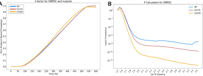
Figure 2.5: Path sampling 𝑣̇ and P factor 76
Figure 2.6: Reactive paths of decarboxylation for WT and mutants on PMF 79
Figure 2.7: Proton-transfer coordinate profiles for WT and mutants 81
Figure 2.S1: Visitation probability for WT and mutants 93
Chapter 3: Identifying the electronic determinants of reactivity in enzyme catalysis Figure 3.1: Hypothesized reaction mechanism of KARI 101
Figure 3.2: QM region and hypothesized transition-state inhibitors of KARI 102
Figure 3.3: Breaking bond and forming bond dynamics of KARI 103
Figure 3.4: Lewis representation of KARI reaction 111
Figure 3.5: Bond index of KARI reaction 112
Figure 3.6: Orbital representation of KARI reaction 113
Figure 3.7: Reactive and Non-reactive atomic orbital hybridization (C4) 118
Figure 3.8: Reactive and Non-reactive atomic orbital hybridization (C5) 119
Figure 3.9: Reactive and Non-reactive atomic orbital hybridization (C7) 120
Figure 3.10: Reactive and Non-reactive atomic orbital hybridization (O8) 121
Figure 3.11: Reactive and Non-reactive atomic orbital hybridization (O6) 122
Figure 3.12: Histograms of reactive and non-reactive O8-M17 distance 123
Figure 3.13: Histograms of reactive and non-reactive O6-M17 distance 124
Figure 3.14: Geometric classifier feature schematic …… ………. 126
Figure 3.15: Electronic classifier feature schematic 128
Figure 3.S1: Methyl orientation histogram for reactive and non-reactive 144
Figure 3.S2: C4-C5 σ-bond orbital energy stratified on torsion 145
Figure 3.S3: C5-C7 σ*-bond and 3C orbital energy stratified on torsion (reactives) 147
Figure 3.S4: Anti-correlation of methyl torsion with respect to C4 and C7 148
Figure 3.S5: OE1-C5 and OE1-H association ..……… ………. 149
Figure 3.S6: C4-C5 σ-bond orbital energy histogram and stratified on C5 torsion 150
Figure 3.S7: 2D histogram of C4-C5 bond index versus OE1-H distance and C5 torsion 151 Chapter 4: Dynamic drivers of catalytic strategy in OMPDC and mutants Figure 4.1: Active site of OMPDC and order parameters of reactive pathways 157
Figure 4.2: Time alignment coordinate of OMPDC 160
Figure 4.3: Example of pathways on WT and mutant PMFs 161
Figure 4.4: Schematic of (D75*/CG–OMP/O2’) and (D70/OD2–OMP/OX1) 169
Figure 4.5: Histogram distributions of (D75*/CG–OMP/O2’) and (D70/OD2–OMP/OX1) 171 Figure 4.6: Absolute N1 improper angle distributions 173
Figure 4.7: N1 improper angle populations stratified on two features 174
Figure 4.S1: Time alignment coordinate of mutant OMPDC 182
Figure 4.S2: Order parameters labeled by pathway 183
Figure 4.S3: Schematic of top 10 features of t = –20 fs classifier features 184
Figure 4.S4: Schematic of top 10 features of t = –10 fs classifier features 185
Figure 4.S5: Schematic of top 10 features of t = 0 fs classifier features 186
Figure 4.S6: Schematic of top 10 features of t = 10 fs classifier features 187
Figure 4.S7: Schematic of top 10 features of t = 20 fs classifier features 188
Figure 4.S8: Schematic of top 10 features of t = 30 fs classifier features 189
Figure 4.S9: Example structures of orotidyl ring 194
Figure 4.S10: Histograms of (D75*/CG–OMP/O2’) per pathway (all t)… 195
Figure 4.S11: Histograms of D75*/CG–OMP/O2’) per system (all t)… 196
Figure 4.S12: Histograms of (D70/OD2–OMP/OX1) per pathway (all t)… 197
Figure 4.S13: Histograms of (D70/OD2–OMP/OX1) per system (all t)… 198
Figure 4.S14: Distribution of closest D75 oxygen to 2’-hydroxyl proton per pathway 199
Figure 4.S15: C2-N1-C6-CX angle between reactive pathways 200
Figure 4.S16: C4-C5-C6-CX angle between reactive pathways 201
List of Tables
Chapter 2: Catalytic Strategies of OMPDC elucidated by path sampling methods
Table 1: OMPDC transition-state barrier heights of experimental and predicted rates 75
Table 2: TPS and experimental reaction rates 77
Chapter 3: Identifying the electronic determinants of reactivity in enzyme catalysis Table 1: Geometric to reactivity classifier performance and coefficients 125
Table 2: Electronic to reactivity classifier performance and coefficients 127
Table 3: Geometric to electronic classifier performance and coefficients (2-feature) 134
Table 4: Geometric to electronic classifier coefficients (10-feature) 134
Chapter 4: Dynamic drivers of catalytic strategy in OMPDC and mutants Table 1: Pathway classifier performance for six timepoints of OMPDC. …………. 165
Table 2: Top feature pairs for pathway classifiers 166
Table 3: Unique features across all timepoints 168
Table 4: Coefficients of (D75*/CG–OMP/O2’) and (D70/OD2–OMP/OX1) 170
Table S1: Coefficients of classifier at t = –20 fs…..…… …………. 190
Table S2: Coefficients of classifier at t = –10 fs…..…… …………. 190
Table S3: Coefficients of classifier at t = 0 fs…..…… … 191
Table S4: Coefficients of classifier at t = 10 fs…..…… …………. 191
Table S5: Coefficients of classifier at t = 20 fs…..…… …………. 192
Table S6: Coefficients of classifier at t = 30 fs…..…… …………. 192
Chapter 1: Introduction
Natasha Seelam1,2
1. Department of Chemical Engineering, Massachusetts Institute of Technology, Cambridge MA
2. Computer Science and Artificial Intelligence Laboratory (CSAIL), Massachusetts Institute of Technology, Cambridge MA
1.1 Overview
Of the diverse functions of proteins, enzymes perform the role of biological catalysts, facilitating reactions on molecules that are easily millions of times faster than their uncatalyzed version at physiological pH and temperature, and with high specificity [1, 32]. To date, enzymes are known to catalyze at least 5000 reactions, many critical to life as the product molecules of these reactions would be imperceptible at uncatalyzed rates [2, 3]. Harnessing the exquisite specificity of enzymes has long been a desirable goal for industrial purposes [30–33]. Problematically, it remains a major challenge to re–engineer proteins for custom function or repurpose them for novel substrates [34].
Several experimental and computational strategies exist toward the design of custom enzymes, including but not exclusive to directed evolution, computer–aided rational design, and a newly emerging branch employing machine learning. While such approaches have had remarkable successes, the complexity of design space and the highly optimized nature of enzymes for their native function thwart ubiquitous application of these approaches [26]. Often, engineered enzymes (or antibodies with catalytic function) are many orders of magnitude less efficient than their natural counterparts, and may still require rounds of directed evolution or random mutagenesis to improve their efficacy [29, 35–39], This suggests the need for further inquiry into the facets of enzyme chemistry to develop a holistic view on how to engineer an enzyme toward a desired goal (either de novo or repurposed), and to understand what structural facets of the enzyme govern successful catalysis. Three key directions toward that end include: (1) modeling the complex chemistries performed by enzymes in rich detail while preserving the underlying dynamics, (2) identifying the salient structural components most indicative of reactivity, and lastly (3) understanding the refined interplay between atomic positions within the active site and its influence on electronic structure
that encourage reactivity. The focus of this thesis is to delve into the catalytic nuances of enzyme reactivity, and to provide a computational framework in order to generate representative simulation data of enzyme–facilitated reactivity, analyze the catalytic strategies, and interpret the way enzymes achieve incredible chemistries.
1.2 Enzyme kinetics
1.2.1 Transition–state theory and origins
Inquiry into the origins of enzymes’ remarkable catalytic prowess has led to numerous theories as to how enzymes facilitate these great enhancements. The prevailing theory of enzyme catalysis has been traditionally couched in the language of transition–state theory (TST), arising from foundational work beginning in the 1940s [4–7]. Broadly, transition–state theory provides a framework in which to study chemical reactions: namely, in this framework, ‘activated– complexes’ (i.e. the transition state) exist in quasi–equilibrium with the reactant–state molecules, and they have the ability to convert to products [4–6, 40, 41]. While the quasi–equilibrium assumption differs from the classical interpretation of equilibrium, the same thermodynamic treatment can be used to express the formation of product [5, 42].
The definition of the transition–state structure relies on the concept of a potential energy surface (PES), in which the progress of a reaction is defined as a function of atomic positions and momenta. The 'reaction coordinate' is a hypothetical variable(s) often used to describe this reaction progress, and the transition state is often cast in the context of it. Identifying the transition–state structure then corresponds to identifying the saddle points on the PES of the reaction [40, 41]. Due to the nature of this high energy, unstable structure, the transition state is extraordinarily short– lived, with a proposed lifetime of barely 10–13 s, on the scale of a bond vibration [47].
Using the framework of TST, Pauling proposed that these biological catalysts exhibit tight– binding to transition–state structures, thereby reducing the activation energy and improving the rate of reactivity without altering the equilibria of the unbound reactant and product molecules [7, 12] (Figure 1.1).
1.2.2 Michaelis–Menten kinetics in enzyme–substrate catalysis
Figure 1.1 shows the description of enzyme kinetics in the Michaels–Menten formalization [86]. Free enzyme (E) and substrate (S) exist in equilibrium with the formation of the enzyme– substrate complex (ES), with this equilibrium association constant denoted as 𝐾$ =&&'
('. The ES
complex also has the ability to become product (P) with rate 𝑘*+, (measured in units of inverse
time for ‘unimolecular’ reactions, of which ES is considered to be). When considering catalysis, the prowess of the enzyme influences 𝑘*+,. The rate, 𝑘*+,, incorporates additional terms that quantify the equilibrium of the ES complex and the (activated) transition state (𝐸𝑆‡), as well as
the formation of 𝐸𝑃, or the enzyme–product bound state, that then separates into enzyme and product molecules (E + P).
Figure 1.1: Kinetics of enzyme–substrate chemistry, as described by Michaelis–Menten kinetics [86]. The free (unbound) enzyme is “E”, the unbound substrate is “S”, the unbound product is “P”, the bound enzyme–substrate complex is “ES”, and the activated (transition state) complex is “ES‡”. The left–hand portion denotes the equilibrium kinetics at which the enzyme and
substrate association leading to the enzyme substrate complex. The right–hand portion of the equation denotes the enzyme–substrate complex becoming unbound enzyme and product, for
E + S
ES
ES
‡
E+P
"
#"
$#"
%"
$%"
&Figure 1.2 illustrates these kinetics in the context of a free–energy diagram (i.e., potential energy surface). The corresponding schematic illustrates a hypothetical reaction coordinate that marks the progress of the reaction. The left–hand portion of the equation in Figure 1.1 is represented in the ‘binding’ stage of Figure 1.2, in which the ES complex is formed. Catalysis is the subsequent activation of the ES complex, marked by the transition state (TS, or 𝐸𝑆‡) with a
concomitant increase in energy, followed by the formation of the enzyme–product complex (EP). This transition–state complex is energetically unfavorable and it quickly dissociates to free enzyme and product, for favorable reaction attempts [7, 12, 86].
The region of the protein that surrounds the substrate molecule for binding and subsequent reactivity is characterized as the ‘active site’. The active site often has side–chain amino acids that are capable of exerting several forces (hydrogen bonds, van der Waals, and electrostatics) that provide an appropriate environment for catalysis, and that reduce the activation energy of the reaction. Comparisons of the uncatalyzed versus catalyzed rate of enzymatic enhancements have estimated that this reduction in activation energy (𝐸3) ranges from 11 to 38 kcal/mol from the uncatalyzed reaction [8–11].
Rigorous treatment of transition–state theory also suggests the inclusion of a transmission coefficient [4]. The transmission coefficient accounts for the fact that not every vibration may lead to successful barrier crossing [4]. While considerable work exists to characterize transmission coefficients in enzymes, it is not always practical to explicitly consider such effects [133–135].
1.2.3 Catalytic strategies employed by enzymes
Transition–state theory has played a pronounced role in the investigation of enzyme catalysis, and has led to the rational design of inhibitor drug molecules that mimic hypothesized transition states of their respective enzymes [47]. Figure 3 represents a schematic of a hypothetical reaction from the context of enzyme–substrate catalysis, contrasting both the uncatalyzed and catalyzed version of the reaction. Two prevalent catalytic strategies put forward in the context of transition–state theory are ground–state destabilization (GSD) and transition–state stabilization (TSS), both of which both aim to reduce the activation energy of the reaction [24, 55]. It should be noted that enzymes may also provide for an alternative reaction pathway compared to what may
E
a(enzyme)
E
a(without enzyme)
Figure 1.2 [Figure adapted from reference 141]: Schematic of the canonical interpretation of enzyme kinetics. The binding step incorporates free enzyme (E) and substrate (S) into the enzyme–substrate (𝐸𝑆) complex. This complex becomes activated into 𝐸𝑆‡, the transition–
state. The 𝐸𝑆‡ exists in quasi–equilibrium with 𝐸𝑆, but also has the ability to become 𝐸𝑃, or
the enzyme–product complex. Subsequently, the enzyme–product state rapidly dissociates to form free enzyme and product.
As the name suggests, GSD increases the free energy of the enzyme and/or substrate in the enzyme–substrate complex relative to the same molecules in the unbound substrate and enzyme, often through using some of the binding energy to induce a distortion. On the other hand, TSS decreases the free energy of the bound transition–state complex. Conventional mechanistic proposals that support GSD–based hypotheses are often centered around electronic strain, bond– distortion, desolvation–effects, and conformational restriction after binding the substrate [43]. Similarly, central tenets of TSS include favorable interactions promoted by electrostatic interactions, such as hydrogen bonding, solvent environment, and occasionally promoting efficient and rapid proton abstractions or additions [48].
While transition–state theory has provided an organized methodology toward studying reactivity in enzymes, increasingly, studies have shown that there are other contributions that influence enzyme reactivity [13–17]; thus, these necessitate extending upon the classic transition– state theory characterization of enzymes. Several hypotheses have been put forth to help fill out the complete picture of enzyme kinetics, including enzyme pre–organization, and the role of near– attack conformations (NACs) which are conformations of the ground state that lie on the transition path of the reaction [13–17].
The hypothesis of electrostatic preorganization describes the enzyme providing a (typically) polar environment that encourages catalysis [54, 55]. Traditionally, this region is defined to include generally the first and occasionally second coordination sphere residues (typically corresponding to the amino acids within the active site) [52, 53]. A core tenet of this hypothesis is that the enzyme positions residues to sample NACs more effectively; this rearrangement of the environment can be as effective in enabling catalysis as lowering the reaction barrier [53, 138–140].
Figure 1.3 [Figure used from reference 46] Hypothetical 1D reaction profile. Free enzyme is represented as “E”, unbound substrate as “S”, unbound product as “P”, enzyme–substrate complex as “ES”, and the bound enzyme–product complex as “EP”. Note, in all cases, these examples are simplified; enzymes can drastically change the reaction path compared to the solvated reaction. (A) The uncatalyzed reaction has some barrier indicated by the top-most dashed line. The activation energy would be the difference in energy from the TS, appearing at the topmost dashed black line, to the substrate “S”. (B) This hypothetical uniform binder enzyme is not considered a true catalyst, as it does not change the activation energy of the enzyme. The decrease in energy after binding substrate is the same as the decrease in energy in attaining the enzyme-assisted TS complex. Thus, the activation energy is unchanged. (C) This hypothetical enzyme stabilizes the transition state compared to Fig 3B, as the difference between TS and ES is smaller than in Fig 3A and 3B. (D) This enzyme additionally includes ground-state destabilizing interactions compared to Fig 3A–3C, as the ground state is higher in energy; the difference in energy from TS and ES is smaller than 3A–3C, also suggesting this is enzyme would be a more effective catalyst.
A parallel question emerging from these catalytic strategies is what role atomic motions and dynamics play in the context of enzyme catalysis [17, 50, 51]. Reactivity (bond breaking/formation) often occurs on time scales that span mere femtoseconds up to picoseconds – on the order of larger–scale atomic motions [29]. In contrast, binding events are often on the scale of microseconds to milliseconds, suggesting these events could be decoupled from the catalytic act [29, 49]. A compelling hypothesis put forth by Schwartz and Schramm is the idea that the enzyme active site “increases the probability of rare dynamic interactions that permit rapid barrier crossing” [29]. Stated otherwise, after the relatively slow events of substrate collision and binding (and any conformational changes that may accompany them), the active site of an enzyme may favor or even encourage rate–promoting vibrations that help the system cross the barrier and facilitate catalysis [17, 29, 50]. Consensus among enzymologists suggests that enzymes likely employ numerous strategies, not limited to just GSD, TSS, preorganization, or NACs, to attain their catalytic performance [17–24, 52–56].
1.3 Biophysical modeling and molecular simulation
1.3.1 Quantum–mechanical/molecular–mechanical methodsAs reactivity is a rapid event, careful experimental work has been able to supplement and support mechanistic proposals supporting these hypotheses [56–58]. However, many of these methods can be quite challenging to employ in experimental settings due to the short–lived nature of many of these states; hence computational approaches are an attractive strategy to investigate and quantify refined atomistic details about how enzymes facilitate their chemistries, and to inspire subsequent experiments that attempt to alter their functions. When considering theoretical studies, enzymes are large, high–dimensional, and complex molecules to model. With the advent of
increased processing power and specialized algorithms, biophysical modeling at the atomic level of detail is possible for many of these proteins [45]. Molecular mechanics employs the use of force fields to describe the physics of atomic interactions from a classical perspective; such approaches are versatile in describing dynamics and binding events [86–94]. However, force fields alone do not describe electronic structure phenomena, such as bond formation and destruction. Chemical reactions require quantum mechanical descriptions to characterize the transient changes in electronic structure. Quantum mechanical/molecular mechanical (QM/MM) methods were developed to efficiently address this discrepancy, in which a system is simultaneously modeled with both levels of theory, focusing the more expensive QM model only on the reactive portion [95–98]. For protein systems, a region within the active site where the reaction occurs (typically catalytic/conserved residues and the substrate) is often characterized at the quantum level of theory, while the remainder of the environment is quantified with an appropriate molecular– mechanics forcefield [45].
1.3.2 Enhanced sampling approaches to identify energetic landscapes
Most QM/MM simulations are run at ambient conditions; given a Boltzmann energy distribution centered around these conditions, sampling high–energy configurations, such as transition states, can be extraordinarily rare. To illustrate this point, consider orotidine 5´-monophosphate decarboxylase (OMPDC); its reaction barrier is nearly 17 kcal/mol [66]. The Boltzmann–associated likelihood of generating a configuration with this energy at 300 K would be 𝑒56789≅ 4 × 10⁵$? – a virtually impossible event!
To tackle this problem, clever physical methods have been developed that efficiently sample configurations in these high–energy regions. These methods encompass techniques such as umbrella sampling, blue–moon sampling, metadynamics, quantum–mechanical band methods, and empirical–valence bond theory [38, 59–62, 99–101]. A unifying element of these techniques is to apply either a biasing potential or higher temperatures to make rare configurations more accessible [59–62]. Methods like Weighted Histogram Analysis Method (WHAM) can then be used to estimate the unbiased potential at the desired temperature. Although these techniques provide accurate potential energy landscapes for reactive paths, they alter the dynamics of the simulation [63]. Moreover, the performance of these techniques in estimating barrier heights and reactivity is sensitive to the definition of the reaction coordinate(s) [63–65].
1.3.3 Transition path sampling methods for catalysis
Path algorithms, led by Transition Path Sampling (TPS), can explore rugged energy landscapes and reactivity without distorting dynamics. These methods harness Markov chain Monte Carlo (MCMC) techniques to sample ensembles of ‘transitions’, and they are formulated to be agnostic to reaction coordinates [63–65]. The only requirement for path sampling methods is the definition of an order parameter that appropriately identifies structures in the starting (reactant) basin and the ending (product) basin [63, 64] (Figure 4). Equipped with this parameter, one only needs an initial ‘seed’ path that connects between the starting and ending basins to generate ensembles of the transition of interest [63–65].
Generating ensembles with path sampling methodologies requires the use of several types of moves: this work specifically focuses on shifting and shooting moves, as indicated in Figure 4 [63–65]. Shifting moves preserve the majority of the trajectory, but alter a relatively few time steps
at the start or end of the trajectory [63–65]. By contrast, shooting moves can generate entirely de novo pathways. The Monte Carlo move chooses a time slice of a given trajectory, and creates a small perturbation to the velocity of all the atomic coordinates at this selected point. This perturbation is integrated forward and backward in time by the prescribed physics of the QM/MM simulation. A new candidate trajectory is accepted if the move resulted in a trajectory that successfully begins and ends in the appropriate starting and ending basins [63–65].
Prior studies have employed path–sampling to study several enzymatic systems due to the methods’ versatility and rich data generation process [67–76]. Additionally, a rigorous statistical mechanical formulation has been developed to analyze the trajectories computed by path sampling methodologies and compute rate constants from the generated ensembles [77, 78]. Historically, path–sampling simulation studies have often focused on the mechanistic details of enzymatic reactions as opposed to full rate computations [128–132].
A
B
C
Figure 1.4 [Figure used from reference 79] (A) Hypothetical potential energy surface (PES) and reactive paths drawn across the landscape. Path sampling strategies require the order parameter to provide a clear delineation between the starting basin “A”, and the product basin “B”. In the following diagram, the advantage of path sampling strategies is that the Markov chain Monte Carlo (MCMC) sampling procedures allow for new paths to be constructed that can navigate across rugged landscapes that conventional sampling may not traverse without an assisting biasing potential. The ensemble of paths that connect A with B can then be analyzed for insights. In the case of reactivity, the starting basin is the reactant, and the ending basin is the product. The paths that connect between these states represent the catalytic trajectories. (B) A schematic of a shooting move; a shooting move within the MCMC path sampling ensemble uses a slice chosen from the prior trajectory within the ensemble (guaranteed to connect between the starting and ending basins), and applies a momentum or velocity perturbation to all the atoms in the system. Molecular mechanics models can propagate the resulting forces due to this perturbation forward and backward in time to assess whether the new trial trajectory also connects between the desired basins. If it does, the trajectory is accepted into the ensemble and a new move is computed using this as the starting trajectory. If it is rejected, the original seed trajectory is added again to the ensemble. (C) A schematic representing a shifting move; the TPS shifting move temporally “shifts” a path so that the actual path connecting between the basins remains the same, the beginning and ending are changed by lengthening or shortening.
1.3.4 Quantum calculation and natural bonding orbitals (NBO)
The path sampling methodology preserves the underlying dynamics of crossing a reactive barrier. For this reason, the atomic positions of the enzyme reaction can be probed in richer detail to quantify the electronic transitions as the reaction proceeds. To analyze electronic structure, pure ab initio methods can be employed on a system of interest to construct a wavefunction (or proxy of one) that characterizes the electron density across the atoms. However, full quantum characterization of biological systems remains a difficult feat. The complexity and size of large biomolecules (for example, the fully solvated protein system in Chapter 2 with orotidine 5´-monophosphate and explicit water is nearly 54,000 atoms!) makes it difficult to overcome the computational requirements that often scale as order 3 or greater with respect to the system size (i.e. O(N3) where N is the number of electrons in the system) [38, 99–103]. However, analyses
may be focused on the critical region of reactivity (the substrate and the active site) to quantify the manner in which the enzyme facilitates reactivity [98, 104, 105]. While quantum techniques provide descriptions of electron density, further refinement of the orbital structure, as localized between atoms, can afford interpretable chemical insight. To that end, techniques such as Natural Bonding Orbital (NBO) theory allow the elucidation of mechanism through the lens of an organic chemist; namely, NBO theory quantifies bonding orbitals and lone–pair orbitals from a Lewis– like perspective [80–82].
NBOs aim to characterize the electron density between “centers”, typically one or two (occasionally three or more) atoms via an orthonormal set of “localized maximum occupancy orbitals” [80–82, 106–108]. This technique makes no pre–supposed hypotheses toward either the form, or location of the bonding orbitals. Instead, it searches across all possible ways of drawing bonds and lone pairs that describe the highest percentage of total electron density in leading
“Lewis–type” NBOs [106–108]. This description is considered the “Natural Lewis Structure” (NLS), which attributes the percentage of the computed wavefunction toward a “Lewis–like” interpretation, and the remaining orbitals are labeled as “non–Lewis” type NBOs that represent the residual effects not well–captured by Lewis theory (such as resonance or other types of delocalization).
The NBO approach works through a series of steps; first, the non–orthogonalized functions corresponding to the atomic wavefunctions calculated via some high level of theory (such as Density Functional Theory “DFT” or Hartree–Fock “HF”) are converted into individual, “atom– centered”, orthogonal natural atomic orbitals (NAOs) [81, 109]. These NAOs represent a localized, 1–center orbital that are ascribed to a given atom. The formulation of the NAOs allows for two physical attributes to be qualitatively described: (a) the spatial diffuseness of the orbital (i.e. the delocalized versus contracted nature) will depend on the molecular environment and (b) the valence NAOs properly incorporate nodal features from steric confinement. NAOs are strictly orthogonal, and are used in Natural Population Analysis (NPA) that appropriates the number of electrons associated to each atom in a more basis–insensitive way compared to the Mulliken Population Analysis [81, 109].
NAOs are then subsequently transformed into natural hybrid orbitals (NHOs), which represent atom–centered hybrids that are a linear combination of NAOs. The NHOs are eventually used in the two–centered natural bond orbital or NBO [110]. One advantage of NBOs is that they are uniquely associated to the wavefunction, with orbitals that are generally non–degenerate. Departures from classic Lewis–like descriptions also have meaningful interpretations, typically regarded as delocalization or resonance effects that a single Lewis structure cannot easily articulate. Common orbitals found in the NBO formulation include core shell, lone pair (“non–
bonded”), bonding, antibonding, and Rydberg orbitals. More recently, the formulation adopted a definition to encompass 3–center–2–electron (3C) descriptors [111, 112]. For biological systems, the NBO analysis has been performed on localized regions of enzymes in order to explore the chemical mechanism of catalysis [113], and to link key atomistic features and the way they influence the electronic environment of a reaction.
While quantum calculations provide refined interpretation of the electronic structure underlying a chemical reaction, understanding how a protein facilitates reactivity requires also capturing what geometric changes of the active site drive catalysis and are amenable to successfully crossing a reactive barrier. To highlight those geometric facets of the active site that give rise to reactivity, machine learning models can highlight key features that are linked to catalysis, which can then lead to structure and dynamics–based mechanistic hypotheses.
1.4 Machine learning methods in protein catalysis
Protein models are complex, diverse–atom, high–dimensional systems which are not necessarily intuitive, even to the keen enzymologist. Most classes of machine learning models are adept at parsing complex, multivariate relationships between key features within data, and can be applied to biology. Recently, several pioneering papers have employed various architectures of machine learning models to investigate relationships in structural biology in the fields of protein– protein interaction networks, optimized directed evolution, protein–ligand binding, protein structure and/or protein folding prediction [114–121].
Given the multitude of machine learning models, the choice of which algorithm depends on the problem posed. No single architecture or algorithm suffices for all tasks ubiquitously; thus, the formulation of the problem will drive the types of models deployed. The goal of understanding
the drivers of enzyme reactivity with machine learning requires two criteria be met: models should (i) identify whether or not a system crosses the barrier (reactive versus “non–reactive”), and (ii) select features most important toward predicting reactivity that are readily interpretable and/or easily accessible.
Given these criteria, neural networks are not necessarily appropriate for the enzyme reactivity problem. While they have excellent predictive performances [115–121], interpretability of the key features that influence predictive power in neural networks is still an open problem in the field [122–124]. Moreover, such models are often data hungry and require extensive training data in order to learn appropriate representations, given the number of parameters in the model [115–121]. Simpler machine learning models typically apply some type of linear transformation on the input features in order to predict a property or class of interest, and are often the first baseline predictors employed before more expressive models are used [117]. The work within chapters 3 and 4 employs the use of a simpler machine learning model, logistic regression, chosen for its ability to classify while directly underscoring the relevant features involved.
1.5 Thesis scope and organization
This thesis aims to synthesize the above progress in enzymology and computational techniques to investigate high–dimensional data in order to understand how enzymes facilitate their magnificent chemistries. Toward that end, the following three chapters describe key studies to explore these methods.
In Chapter 2, we studied the catalytic proficiency of orotidine 5´-monophosphate decarboxylase (OMPDC), an enzyme found in many biological systems that facilitates the decarboxylation of its substrate, orotidine 5´-monophosphate (OMP). This enzyme is known for
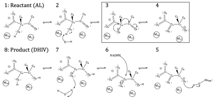
its incredible catalytic proficiency, enhancing reactivity by nearly 17 orders of magnitude from the solvated, uncatalyzed reaction [125]. Elegant experimental methods have dissected several key residues that facilitate reactivity in the enzyme, and our work focuses on 2 empirically verified mutants, S127A and V155D [66]. These variants, respectively, address the catalytic strategies of transition–state stabilization and ground–state destabilization. This investigation employed the use of potentials of mean force (PMFs) and path sampling strategies to provide both an energetic and a dynamic perspective on the decarboxylation of OMPDC. The PMF and path sampling trajectories showed reasonable agreement with the ranked, relative orders of the empirically identified rates, suggesting that both approaches were capable of reflecting systematic changes in catalysis from the local environment of the enzyme. The path sampling trajectories across WT, S127A, V155D revealed that there were at least two distinct reactive pathways to facilitate decarboxylation. WT and S127A frequently decarboxylated via a “simultaneous” mechanism, where the position of K72 was observed to be “coordinated” with the decarboxylation event. The V155D mutant displayed two types of mechanisms: one that was similar to WT and S127A, and another that explicitly broke the carbon–carbon bond before the proton transfers to the substrate and finishes the reaction (“stepwise”).
We applied quantum–chemical techniques to explore the electronic description of the drivers of reactivity in Chapter 3. This chapter investigated the catalysis performed by ketol–acid reductoisomerase (KARI), an enzyme that facilitates a methyl–transfer isomerization with the assistance of two magnesium ion cofactors and NADPH. This enzyme has been the target of several studies, as the reaction it catalyzes plays a critical role in the synthesis of biofuels [127, 137, 138]. Prior work in the group identified a subset of 30 geometric features, constructed from distances, angles, and torsions of the active site of KARI that predicted reactivity and influenced
the likelihood of crossing the barrier when sampled in the reactant basin [136]. Chapter 3 analyzed the successful, methyl transferring reactive simulations and the simulations that failed to cross the barrier from the work of Bonk et al. [136] and extended the chemical significance by revealing the mechanism of KARI via Natural Bonding Orbital (NBO) analyses to represent electronic structure in a Lewis–like representation. Our calculations revealed that methyl transfer occurred simultaneously with carbonyl formation and lone–pair formation with the adjacent oxygens (O6, O8) of the substrate, and that the reaction formed a three–center–two–electron bond, formed from the participating carbons (C4, C5, C7) of the substrate. In order to investigate the relationship between the geometric features of the active site and underlying electronic structure, we performed classification tasks to predict reactivity with both geometric and electronic features. Subsequent analyses identified a subset of 10 geometric features or a set of 6 electronic features, either of which were sufficient to predict reactive and non–reactive simulations with ROC AUC > 0.9, suggesting that while both types of features were predictive, fewer electronic features were required to predict reactivity with the same performance. We identified a possible catalytic strategy in which one of the geometric features involving a catalytically conserved glutamate, E319, and the distance to the transferring methyl (E319/OE1–C5) influenced the torsional orientation of the methyl prior to transferring such energy of the breaking bond orbital (C4–C5) increased, suggesting ground–state destabilization. This torsional orientation was also related to an electronic descriptor reporting on the electron density of the breaking bond (C4–C5 bond index), demonstrating that eclipsed orientations weakened the breaking bond prior to reacting. Lastly, we showed that pair–feature models of the 10 geometric features in predicting each of the 6 electronic features performed comparably to a full cumulative model, suggesting small subsets of geometric features were enough to predict the underlying electronic structure.
Chapter 4 revisited the OMPDC system and harnessed the framework of Chapter 3 in order to investigate the decarboxylation mechanism across the WT and mutant systems. From the results of chapter 2, energetic and dynamic characterization of the decarboxylation of OMPDC revealed two distinct pathways: a ‘simultaneous’ mechanism where the decarboxylation is coordinated with the positioning of catalytically important lysine, K72, and a ‘stepwise’ mechanism in which the decarboxylation is relatively independent of K72’s position. The reactive pathways of the WT, S127A, and V155D mutants were combined to create two aggregate ensembles for each pathway, and supervised machine learning models are trained to classify between the two mechanisms for several time points starting from the reactant basin to 30 fs after the systems were committed to crossing the reaction barrier. Pair–feature models were able to predict reactivity with (ROC) AUC > 0.8 across all time points tested in this work. Moreover, model–selected features underscored several mechanistic hypotheses, of which we showed that two features, the distance between residue D75 in proximity to the 2’–hydroxyl of the OMP substrate ribose and the distance between D70’s carboxylate oxygen and OMP’s carboxylate oxygen, influenced the ability to distort the planarity of the orotidyl prior to reacting. Taken together, this highlights the ability for machine learning models to recognize chemically meaningful features in enzyme catalysis, without explicit knowledge of the mechanism.
1.6 References
1. X. Zhang, K.N. Houk. Why Enzymes are Proficient Catalysts: Beyond the Pauling Paradigm. Acc. Chem. Res. 38, 5, 379–385, 2005.
2. Schomburg, A. Chang, S. Placzek, C. Söhngen, M. Rother, M. Lang, C. Munaretto, S. Ulas, M. Stelzer, A. Grote, M. Scheer, D. Schomburg. BRENDA in 2013: integrated reactions, kinetic data, enzyme function data, improved disease classification: new options and contents in BRENDA. Nucleic Acids Res. 41:D1 D764–D772, 2013.
3. J.M. Berg, J.L. Tymoczko, L. Stryer, L. Stryer. Biochemistry. New York: W.H. Freeman. 2002.
4. H.Eyring. The Activated Complex in Chemical Reactions. J. Chem. Phys. 3 (2): 107–115, 1935.
5. K.J Laidler, M.C. King. The development of Transition–State Theory. J. Phys. Chem. 87 (15): 2657–2664, 1983
6. M. G. Evans and M. Polanyi. Some applications of the transition state method to the
calculation of reaction velocities, especially in solution. Trans. Faraday Soc. 31, 875, 1935. 7. L. Pauling. Chemical achievement and hope for the future. Am. Scientist. 36, 51–58, 1948. 8. B.G. Miller, R. Wolfenden. Catalytic proficiency: the unusual case of OMP decarboxylase.
Annu. Rev. Biochem. 71: 847–885, 2002.
9.M.J. Snider, R. Wolfenden. The rate of spontaneous decarboxylation of amino acids. J. Am. Chem. Soc., 122, 11507–11508, 2000.
10. C. Lad, H. Williams, R. Wolfenden. The rate of hydrolysis of phosphomonoester dianions and the exceptional catalytic proficiencies of protein and inositol phosphatases. Proc. Natl. Acad. Sci. USA, 100, 5607–5610, 2003.
11. A.C. Reyes, A.P. Koudelka, T.L. Amyes, J.P. Richard. Enzyme Architecture: Optimization of Transition–State Stabilization from Cation–Phosphodianion Pair. J. Am. Chem. Soc. 137, 16, 5312–5315, 2015.
12. L., Pauling. Molecular Architecture and Biological Reactions. Chem. Eng. News. 24 (10): 1375–77, 1946.
13. M. Garcia–Viloca, J. Gao, M. Karplus, D.G. Truhlar. How enzymes work: analysis by modern rate theory and computer simulations. Science. 303:186–195, 2004.
14. S.J. Benkovic, S.A. Hammes–Schiffer. Perspective on Enzyme Catalysis. Science. 301(5637), 1196–11202, 2003.
15. K. Zinovjev, I. Tunon. Quantifying the limits of transition state theory in enzymatic catalysis. Proc. Natl. Acad. Sci. USA 114:12390–12395, 2017.
16. D.D. Boehr, R. Nussinov, P.E. Wright. The role of dynamic conformational ensembles in biomolecular recognition. Nat Chem Biol. 5:789–796, 2009.
17. S. Hur, T.C. Bruice. The near attack conformation approach to the study of chorismite to prephenate reaction. Proc. Natl. Acad. Sci. USA, 100(21) 12015–12020, 2003.
18. J.R. Knowles. To build an enzyme. Philos. Trans. R. Soc., B 332, 115−121, 1991 19. J.R. Knowles. Enzyme catalysis: Not different, just better. Nature 350, 121−124, 1991. 20. P. Carter, J.A. Wells. Functional interaction among catalytic residues in subtilisin BPN′.
Proteins 7, 335−342, 1990.
21. D.A. Kraut, K.S. Carroll, D. Herschlag, D. Challenges in enzyme mechanism and energetics. Annu. Rev. Biochem. 72, 517− 571, 2003.
22. G.G. Hammes, S.J. Benkovic, S. Hammes–Schiffer. Flexibility, diversity, and cooperativity: Pillars of enzyme catalysis. Biochemistry 50, 10422−10430, 2011.
23. A, R. Fersht. Structure and Mechanism in Protein Science, 2nd ed., W. H. Freeman and Co., New York, 1999.
24. W.P. Jencks. Catalysis in Chemistry and Enzymology, 2nd ed., Dover, New York, 1987. 25. J.P. Richard. Protein Flexibility and Stiffness Enable Efficient Enzyme Catalysis. J. Am.
Chem. Soc. 141, 3320–3331, 2019.
26. B. Kuhlman, D. Baker. Native protein sequences are close to optimal for their structures. Proc. Natl. Acad. Sci. USA, 97(19): 10383–103888, 2000.
27. M. Roca, A. Vardi–Kilshtain, A. Warshel. Toward accurate screening in computer aided enzyme design. Biochemistry 48:3046–305, 2009.
28. Warshel, P.K. Sarma, M. Kato, Y. Xiang, H. Liu, M.H.M. Olsson. Electrostatic basis for enzyme catalysis. Chem. Rev. 106:3210–3235, 2006.
29. S.D. Schwartz, V.L. Schramm. Enzymatic transition states and dynamic motion in barrier crossing. Nat Chem Biol. 5(8), 551–558, 2009.
30. H. Nam, N.E. Lewis, J. A. Lerman, D. Lee, R.L. Chang, D. Kim, B.O. Palsson. Network Context and Selection in the Evolution to Enzyme Specificity. Science, 337(6098): 1101– 1104, 2012.
31. S. Li, X. Yang, S. Yang, M. Zhu, X. Wang. Technology prospecting on enzymes: application, marketing and engineering. Comput Struct Biotechnol J. 2:1–11, 2012.
32. J.M. Choi, S.S. Han, H.S. Kim. Industrial applications of enzyme biocatalysis: current status and future aspect. Biotechnol Adv. 33:1443–1454, 2015.
33. C. Li, R. Zhang, J. Wang, L.M. Wilson, Y.Yan. Protein Engineering for improving and diversifying natural product biosynthesis. Trends in Biotechnol. 38(7):729–744, 2019.
34. S. Jemli, D. Ayadi–Zouari, H.B. Hlima, S.Bejar. Bioacatalysts: application and engineering for industrial purposes. Crit Rev Biotechnol. 36(2):246–58, 2016.
35. A.M. David, A.T. Plowright, E. Valeur. Directing Evolution: the next revolution in drug discovery?. Nat. Rev. Drug Discovery. 16(10):681–698, 2017
36. F,H. Arnold. The nature of chemical innovation: new enzymes by evolution. Q. Rev. Biophys. 48(4) 404–410, 2015.
37. T.C. Bruice, K. Kahn. Computational Enzymology. Curr Opin Chem Biol. 4(5) 540–544, 2000.
38. M.P. Frushicheva, M.J. Mills, P. Schopf, M.J. Singh, R.B. Prasad, A. Warshel. Computer aided enzyme design and catalytic concepts. Curr Opin Chem Biol. 21:56–62, 2014.
39. L. Regan, D. Cabellero, M.R. Hinrichsen, A. Virrueta, D.M. Williams, C.S. O'Hern. Protein Design: Past, Present, and Future. Biopolymers. 104(4): 334–350, 2015.
40. R. Marcelin. Contribution a l'étude de la cinétique physico–chimique. Annales de Physique. 9(3):120–231, 1915.
41. J. Bigeleisen. The effect of isotopic substitution on the rates of chemical reaction. J. Phys. Chem. 56(7):823–858, 1952.
42. J.L. Steinfeld, J.S. Francisco, W.L Hase. Chemical Kinetics and Dynamics (2nd ed.). Prentice–Hall. pp. 289–293, 1999.
43. V.E. Anderson. Ground State Destabilization. John Wiley and Sons. 2001. https://doi.org/10.1038/npg.els.0000625
44. J.G. Belasco, J.R. Knowles. Direct observation of substrate distortion by triosephosphate isomerase using Fourier transform infrared spectroscopy. Biochemistry 19: 472–477, 1980.
45. M.W. van der Kamp, A.J. Mulholland, Combined Quantum Mechanics/Molecular Mechanics (QM/MM) Methods in Computational Enzymology. Biochemistry, 52, 2708–2728, 2013 46. L.D. Andrews, T.D. Fenn, D. Herschlag. Ground–State Destabilization by Anionic
Nucleophiles Contributes to the Activity of Phosphoryl Transfer Enzymes. PLOS Biology, 11:7, 1–18, 2013.
47. V.L. Schramm, B.A. Horenstein, P.C. Kline. Transition State Analysis and Inhibitor Designs for Enzymatic Reactions. The Journal of Biological Chemistry, 269, 28, 18259–18262, 1994. 48. Schramm, VL. Enzymatic Transition States and Transition–State Analog Design. Annu. Rev.
Biochem. 693–720, 1998.
49. R. Wolfenden, M.J. Snider. The depth of chemical time and the power of enzymes as catalysts. Acc. Chem. Res, 34:938–945, 2001.
50. G. Bhabha, J. Lee, D.C. Ekiert, J. Gam, I.A. Wilson, H.J. Dyson, S.J. Benkovic, P.E. Wright. A Dynamic Knockout Reveals That Conformational Fluctuations Influence the Chemical Step of Enzyme Catalysis. Science. (2), 332, 6026, 234–238, 2011.
51. A.J. Adamcyzk, J. Cao, S.C.L. Kamerlin, A. Warshel, Catalysis by dihydrofolate reductase and other enzymes arises from electrostatic preorganization, not conformational motions. Proc. Natl. Acad. Sci. USA108, 34, 14115–14120, 2011.
52. Morgenstern, M. Jaszai, M.E. Eberhart, A.N Alexandrova. Quantified electrostatic preognization in enzymes using the geometry of electron charge density. Chem. Sci., 8, 5010–5018, 2017.
53. J. Fuller III, T.R. Wilson, M.E. Eberhart, A.N. Alexandrova. Charge Density in Enzyme Active Site as a Descriptor of Electrostatic Preorganization. J. Chem. Inf. Model. 59, 5, 2367–2373, 2019.
![Figure 1.2 [Figure adapted from reference 141]: Schematic of the canonical interpretation of enzyme kinetics](https://thumb-eu.123doks.com/thumbv2/123doknet/14433199.515558/22.918.117.797.110.406/figure-figure-adapted-reference-schematic-canonical-interpretation-kinetics.webp)
![Figure 1.3 [Figure used from reference 46] Hypothetical 1D reaction profile. Free enzyme is represented as “E”, unbound substrate as “S”, unbound product as “P”, enzyme–substrate complex as “ES”, and the bound enzyme–product](https://thumb-eu.123doks.com/thumbv2/123doknet/14433199.515558/24.918.95.812.122.468/figure-figure-reference-hypothetical-reaction-represented-substrate-substrate.webp)
![Figure 1.4 [Figure used from reference 79] (A) Hypothetical potential energy surface (PES) and reactive paths drawn across the landscape](https://thumb-eu.123doks.com/thumbv2/123doknet/14433199.515558/29.918.99.827.136.460/figure-figure-reference-hypothetical-potential-surface-reactive-landscape.webp)