HAL Id: hal-02956243
https://hal.archives-ouvertes.fr/hal-02956243
Submitted on 9 Oct 2020
HAL is a multi-disciplinary open access
archive for the deposit and dissemination of
sci-entific research documents, whether they are
pub-lished or not. The documents may come from
teaching and research institutions in France or
abroad, or from public or private research centers.
L’archive ouverte pluridisciplinaire HAL, est
destinée au dépôt et à la diffusion de documents
scientifiques de niveau recherche, publiés ou non,
émanant des établissements d’enseignement et de
recherche français ou étrangers, des laboratoires
publics ou privés.
oxygen and carbon in the cultured zooxanthellate coral,
<i>Acropora</i>: implications for coral-growth rates
A. Juillet-Leclerc, S. Reynaud
To cite this version:
A. Juillet-Leclerc, S. Reynaud. Light effects on the isotopic fractionation of skeletal oxygen and
carbon in the cultured zooxanthellate coral, <i>Acropora</i>: implications for coral-growth rates.
Biogeosciences, European Geosciences Union, 2010, 7 (3), pp.893-906. �10.5194/bg-7-893-2010�.
�hal-02956243�
Biogeosciences, 7, 893–906, 2010 www.biogeosciences.net/7/893/2010/
© Author(s) 2010. This work is distributed under the Creative Commons Attribution 3.0 License.
Biogeosciences
Light effects on the isotopic fractionation of skeletal oxygen and
carbon in the cultured zooxanthellate coral, Acropora: implications
for coral-growth rates
A. Juillet-Leclerc1and S. Reynaud2
1LSCE Domaine du CNRS, 91198 Gif sur Yvette, France
2CSM Avenue Saint-Martin, 98000 Monaco, Principality of Monaco
Received: 25 September 2009 – Published in Biogeosciences Discuss.: 3 November 2009 Revised: 18 February 2010 – Accepted: 26 February 2010 – Published: 8 March 2010
Abstract. Skeletal isotopic and metabolic measurements of
the branching coral Acropora cultured in constant conditions and subjected to two light intensities were revisited. We in-dividually compared the data recorded at low light (LL) and high light (HL) for 24 colonies, all derived from the same parent colony. Metabolic and isotopic responses to the dif-ferent light levels were highly variable. High light led to pro-ductivity enhancement, reduction of surface extension, dou-bling of aragonite deposited weight and increased δ18O lev-els in all nubbins; responses in respiration and δ13C were not clear. The partitioning of the colonies cultured at HL into two groups, one showing a δ13C enrichment and the other a δ13C decrease revealed common behaviors. Samples showing an increase in δ13C were associated with the co-variation of low surface extension and high productivity while samples show-ing a decrease in δ13C were associated with the co-variation of higher surface extension and limited productivity.
This experiment, which allowed for the separation of tem-perature and light effects on the coral, highlighted the signif-icant light influences on both skeletal δ18O and δ13C. The high scattering of inter-colony δ18O observed at one site could be due to the differing photosynthetic responses of symbiotic algal assemblages.
We compared our results with observations by Gladfelter on Acropora cervicornis (1982). Both set of results high-light the relationships between coral-growth rates, micro-structures and photosynthetic activity. It appears that exten-sion growth and skeleton thickening are two separate growth
Correspondence to: A. Juillet-Leclerc
modes, and thickening is light-enhanced while extension is light-suppressed. There are multiple consequences of these findings for paleoclimatic reconstructions involving corals.
1 Introduction
As early as 1972, Weber and Woodhead demonstrated that the variability in oxygen-isotope compositions (δ18O) of scleractinian coral skeletons, although showing very negative values compared with the isotopic equilibrium, was essen-tially due to sea-surface temperature (SST), (Epstein et al., 1953). Correlations of SST with aragonite δ18O also differed among coral genera (Weber and Woodhead, 1972). Anal-ysis of samples taken along the main growth axis of a coral head revealed that monthly δ18O signals were correlated with seasonal SST and seawater δ18O variations (Fairbanks and Dodge, 1979; McConnaughey, 1989a). Because it exhibited a strong seasonal signal such a sampling has been system-atically used for paleoclimatic reconstructions of SST from coral δ18O measurements (e.g., Cole et al., 1993; Quinn et al., 1993; Dunbar et al., 1994). However, many heads of
Porites lobata growing in close proximity at Clipperton Atoll
showed isotopic discrepancies of up to 0.4‰ (Linsley et al., 1999), equivalent to a 2◦C isotopic effect for the same
pe-riod using the δ18O/SST relationship estimated by Gagan et al. (1994). This discrepancy reached 1.28‰ (more than 6◦C) for Porites spp. from the Gulf of Aqaba (Felis et al., 2003). It was concluded that18O concentrations were also colony dependent, this effect being commonly called the “vital ef-fect” (Urey et al., 1951). Moreover, isotopic profiles may also change according to the axis sampled on a single coral
head (Maier et al., 2004). Such sources of variability could strongly compromise the validity of δ18O as an accurate en-vironmental proxy.
Compared to the skeletal δ18O signature, interpretation of the variability of the carbon isotopic ratio (δ13C) within coral skeletons has long been a matter of debate. δ13C variabil-ity has therefore scarcely been considered for climatic re-construction (Guzman and Tudhope, 1998). In contrast to δ18O, which was assumed to essentially depend on external factors, δ13C has been generally considered as affected by coral physiology either via respiration rate (McConnaughey et al., 1997), or via the photosynthetic activity of the sym-biotic zooxanthellae (Swart, 1983; McConnaughey, 1989a). Different observations led Goreau (1977) and Erez (1978) to propose two different models to explain carbon isotopic fractionation. The first author observed that δ13C increased with augmented light level. Indeed, algae and coral were thought to extract their inorganic carbon from the same reser-voir for both photosynthesis and calcification processes. As photosynthesis is the faster reaction, lighter carbon isotopes were used preferentially; thus, the reservoir enriched in13C caused a δ13C increase with light (Goreau, 1977). The sec-ond author, after the observing the opposite, i.e., that δ13C decreased with light, proposed that during intense photo-synthetic activity, it was possible that corals incorporated depleted metabolic carbon into their skeleton (Erez, 1978). None of these assumptions has been yet validated.
To explain the positive correlation between oxygen and carbon ratio, McConnaughey (1989a, b) assumed that the kinetic isotopic fractionation was strongly linked with cal-cification rate. After these publications, several theoretical models were put forth explaining how the observed coral-skeleton isotopic fractionations were derived from a combi-nation of kinetic and metabolic effects (Heikoop et al., 2000; McConnaughey, 2003; Omata et al., 2005).
Experiments conducted in the laboratory by Weil et al. (1981) showed a negative correlation between skeletal δ13C and light, i.e., the supply of autotrophic energy in the coral Montipora. Conversely, field experiments conducted by Swart et al. (1996) exhibited only weak correlations be-tween skeletal δ13C and the supply of autotrophic energy, measured as the P/R ratio (photosynthesis/respiration). Grot-toli and Wellington (1999) later found a negative correlation between skeletal δ13C and the heterotrophic energy supply in the zooplankton and a positive correlation with light, i.e., the autotrophic energy supply. In addition, δ13C variabil-ity seemed decoupled from coral growth (Grottoli, 2002). Among these studies, only the laboratory experiments of Weil et al. (1981) deciphered the relations between the light and temperature effects and could document the effect of a single factor on metabolic activity and thus provide clear re-sponses for the isotopic fractionations of oxygen and carbon. It has been generally assumed that the geochemical re-sponse derived from several colonies is more significant than data provided by a single colony. Thus, authors have
usu-ally considered averaged metabolic and chemical data from several colonies (Grottoli and Wellington, 1999; Reynaud-Vaganay et al., 1999, 2001; Grottoli, 2002; Suzuki et al., 2005). However, individual metabolic and isotopic responses can differ markedly. For instance, Acropora nubbins col-lected from a single parent colony and cultured in controlled SST conditions exhibited an inter-colony variability of 1‰ (Reynaud-Vaganay et al., 1999). This has been confirmed for cultured Porites sp. (Suzuki et al., 2005), which showed similar variability at various temperature settings. In culture experiments, as in the field, coral δ13C showed larger inter-colony variability than δ18O, often ≥2‰ (Reynaud-Vaganay et al., 1999; Suzuki et al., 2005).
The present work is based on data previously published by Reynaud-Vaganay et al. (2001), which examined the effect of light on the mean skeletal isotopic signatures (δ18O and δ13C) of several nubbins of Acropora sp. The effect of light was also measured on metabolic activities, such as photosyn-thesis, respiration, calcification rate and surface extension. In contrast with the preceding study, we now examined the in-dividual coral responses. The results of this experiment con-ducted on Acropora were then compared with observations made by Gladfelter (1982) and we considered the possible relationship between skeletal growth and the relative roles of two crystalline microstructures. Finally, for climatic pur-poses, we compared the effects of a change in light intensity on a branched colony versus the time response of samples collected along the main growth axis of Porites.
2 Materials and methods 2.1 Biological materials
The experiment was conducted in the laboratory using colonies of the branching zooxanthellate scleractinian coral,
Acropora sp. Tips from 24 branches were sampled from a
single parent colony. The specimens were glued onto glass slides (3×6×0.2 cm) using underwater epoxy (Devcon) as described by Reynaud-Vaganay et al. (1999), and randomly distributed in two aquaria (15 L). The tanks were supplied with heated Mediterranean seawater (24◦C) pumped from a depth of 50 m. The seawater renewal rate was approximately five times per day and the seawater was continuously mixed with a Rena pump (6 L min−1). Metal halide lamps (Philips
HPIT, 400 W) provided light of 260 or 130 µmol m−2s−1
on a 12:12 photoperiod. Seawater was continuously aerated with outside air. The culture temperature (25◦C) was con-trolled to within ±0.1◦C using a temperature controller (EW, PC 902/T).
All colonies were initially cultured for six weeks under a light intensity of 130 µmol m−2s−1 (referred to as Low Light, LL). Thereafter, colonies were cultured for six ad-ditional weeks under a light intensity of 260 µmol m−2s−1 (High Light, HL). At the completion of each period and to
A. Juillet-Leclerc and S. Reynaud: Acropora: implications for coral-growth rates 895 determine the isotopic composition, the newly deposited ring
skeleton which formed on the glass slide was collected with a scalpel (Reynaud-Vaganay et al., 1999), dried overnight at room temperature and stored in glass containers pending iso-topic analysis.
During the experiment all potential variables (other than temperature and light) such as the chemical properties of sea-water (pH, salinity. . . ) were strictly controlled and kept con-stant.
2.2 Measurements of environmental parameters
Irradiance was measured using a 4-π quantum sensor (Li-Cor, LI-193SA) once a week (Table 1). Temperature (pre-cision: ±0.05◦C) was logged at 10-min intervals using a Seamon temperature recorder.
Light intensities used in this experiment (130 and 260 µmoles photons m−2s−1) correspond to 5.6 and 11.2 mol m−2d−1, respectively. Davies (1991) estimated that during a typical sunny day on a tropical reef a coral receives about 14.4 mol m−2d−1 of sunlight at a depth of three meters in turbid water. On a cloudy day, the coral receives about 6.2 mol m−2d−1of sunlight. Thus, the light intensities in our experiments imitated a range from quite low to very strong natural illumination levels.
2.3 Photosynthesis and respiration
Photosynthesis and respiration were measured using the respirometry technique, which consists of measuring the changes in oxygen concentration during the incubation.
The experimental sequence was identical for each coral: each nubbin was taken from the culture aquarium, placed in a Perspex chamber (240 ml) containing filtered sea-water, for a 30-min pre-incubation in the light (130 or 260 µmol photons m−2s−1, depending on culture condition). The coral nubbin was then incubated for 1 h in the same chamber to measure the rate of photosynthesis. The chamber was then flushed and the coral pre-incubated for 30 min in the dark and then for 1 h in the dark to measure the respiration rate. During the incubation, the medium was continuously agitated using a magnetic stirrer and was changed after each incubation.
The respirometric chamber was kept at 25◦C in a thermo-static water bath. All incubations took place between 08:00 and 14:00 h. The colonies were subsequently returned to the culture aquarium. Oxygen concentration was monitored in the chamber and recorded every 1 min using a data-logger (LI-1000, Li-Cor Inc.). Dissolved O2 was measured using
a Ponselle polarographic electrode calibrated daily against air-saturated seawater (100%) and a saturated solution of sodium sulfite (zero oxygen). Rates of net photosynthesis and respiration were estimated using a linear regression of O2against time. Photosynthesis and respiration values were
then normalized to the skeletal surface area as estimated by the aluminum-foil technique (Marsh, 1970).
2.4 Growth rates 2.4.1 Calcification rate
Corals were weighed using the buoyant-weight technique (Jokiel et al., 1978; Davies, 1989) at the beginning and end of the experiment. The calcification rate was measured using the following formula:
G = n s
Pn
P0
−1
Where G is the calcification rate, n is the number of the cul-ture days, Pnis the dry weight after n days of culture and P0
is the initial dry weight. Such a value indicates the percent-age of weight increase relative to the initial weight per day.
2.4.2 Surface extension
Measurements are performed on the skeleton formed on the glass slide (Marsh, 1970). Thus, the corresponding size in-crease is the surface occupied by the newly-formed skeleton, receiving light perpendicularly.
2.4.3 Thickening
Thickening refers to the addition of new aragonite filling-in porosity or strengthening skeletal structure without notice-able horizontal surface extension.
2.5 Isotopic measurements
The isotopic values were calibrated against those determined by conventional methods using an Optima-VG mass spec-trometer. Results are given in the conventional notation, ex-pressed as per mil (δ ‰) against the V-PDB standard (Vienna Pee Dee Belemnite), where:
δ(sample) = [(Rsample−RStandard) −1] × 103
The external precision, estimated using an internal standard, was ±0.11 and 0.08‰ vs. V-PDB for carbon and oxygen, respectively. The reproducibility of carbon and oxygen iso-topic measurements, calculated from replicate coral samples, was 0.10 and 0.08‰ vs. V-PDB, respectively.
3 Results
All data are given in Table 1. This new interpretation of the data reported in Table 1 was based on a comparison of the behavior of each nubbin. Although nubbins were collected from a single parent colony, all measured parameters were highly variable. Measurements of some parameters were
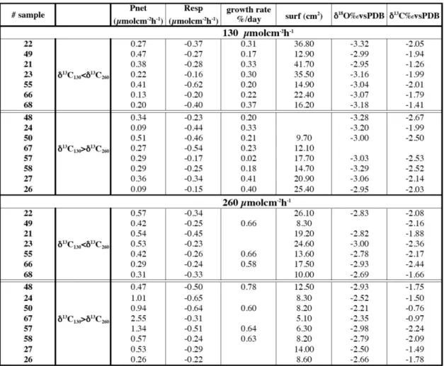
Table 1. Metabolic and isotopic measurements for each colony after 6 weeks of culture. All the nubbins have been submitted successively to
low light and high light intensity during the same duration. For each light value, nubbins showing higher δ13C at HL than at LL are separated from the others (see Fig. 1).
lacking due to difficulties related with experimental condi-tions (Table 1). Herein, we examined metabolic and isotopic data provided by a single colony successively submitted to the two light conditions (from LL to HL; Fig. 1).
3.1 The effect of light
We note that metabolic and chemical data are highly scat-tered (Fig. 1a). For instance, Pnet variability was lower
at LL (0.5 µmol cm−2h−1) than at HL (the amplitude was >2 µmol cm−2h−1), likewise δ18O varied by 0.5‰ at LL and 1‰ at HL. But respiration variability was high for the two light conditions, from −0.6 to −0.2 µmol cm−2h−1, as well as δ13C values showing for the two conditions around 1.5‰ of amplitude. The trend shown by all nubbins between LL and HL was identical for all the measured parameters, except for R and δ13C. The significance of a unanimous metabolic or chemical response is stronger than a trend revealed after averaging data.
Figure 1b clearly indicates that light increase led to inten-sified net photosynthesis and increased calcification (in terms of percentage of growth per day), but also led to a decrease of surface extension and δ18O enrichment. Respiration variabil-ity showed identical range during the two light conditions. The δ13C change was more confusing with half of the
nub-bins exhibiting increased values and the other half showing decreased values between low and high light.
3.2 Partitioning into two groups
We expected an unique response, thus in order to high-light our results, we divided the nubbins into two groups: those showing a δ13C increase from LL to HL (the expected response according to the global carbon-pool assumption, Goreau, 1977) (Fig. 1c, light color) and those showing a δ13C decrease (i.e., the expected response in case of uptake of13C depleted metabolic carbon, Erez, 1978) (Fig. 1c, dark color).
A. Juillet-Leclerc and S. Reynaud: Acropora: implications for coral-growth rates 897
Copernicus Publications
Bahnhofsallee 1e 37081 Göttingen Germany
Martin Rasmussen (Managing Director) Nadine Deisel (Head of Production/Promotion)
Contact [email protected] http://publications.copernicus.org Phone +49-551-900339-50 Fax +49-551-900339-70 Legal Body Copernicus Gesellschaft mbH Based in Göttingen Registered in HRB 131 298 County Court Göttingen Tax Office FA Göttingen USt-IdNr. DE216566440
Page 1/1
Fig. 1. Values of measured metabolic and geochemical parameters. (a) Values of each nubbin are plotted for high and low light. Light (dark)
color corresponds with colonies showing higher (lower) δ13C at HL than at LL. To underline the individual variability shown by one nubbin, HL and LL dots are linked. (b) At each light condition values of all nubbins have been averaged. Darker bars are associated with LL and lighter ones with HL (plotted error: 2σ ). We notice that in this case, R or δ13C were almost identical at LL and HL. (c) Averaged values for each light condition calculated after splitting nubbins into two groups: light (dark) color corresponds with colonies showing higher (lower) δ13C at HL than at LL. Darker bars are associated with LL and lighter ones with HL (plotted error: 2σ ).
At LL (Fig. 1c, darker bars), the two groups exhibited sim-ilar net photosynthesis, respiration, growth rate and δ18O. Values were more scattered for surface extension and δ13C. By examining the responses at HL (Fig. 1c, lighter bars), we realized that metabolic and isotopic values shown by these two groups presented common features. Colonies showing lower δ13C at HL than at LL (Fig. 1c, dark color, lighter bar) were associated with larger surface extension and the others characterized by higher δ13C than at HL (Fig. 1c, light color, lighter bar) exhibited the smallest surfaces.
Colonies displaying higher δ13C at HL (Fig. 1c, light color, lighter bar) showed higher photosynthetic activity and the most enriched δ18O and δ13C values. Surface exten-sions of the nubbins showing lower δ13C (Fig. 1c, dark color, lighter bar) were almost all greater than that measured on the nubbins showing higher δ13C but always lower than that measured at LL. For the two groups (Fig. 1a, clear and dark color, lighter bar), the weight of colonies at least doubled during the incubation, while surface extension was weaker by ca. 40% (Fig. 1c). The δ18O of all colonies increased, the enrichment being more pronounced for colonies showing an increase in δ13C (Fig. 1c, light color, lighter bar).
The averaged values calculated after partitioning were more significant than earlier values published for all the
colonies (Fig. 1b) (Reynaud-Vaganay et al., 2001). We noticed that all intermediate values between the highest and lowest δ13C-differences between LL and HL were recorded. This explains why the difference in the δ13C av-erage (Fig. 1b) was not significant (Reynaud-Vaganay et al., 2001).
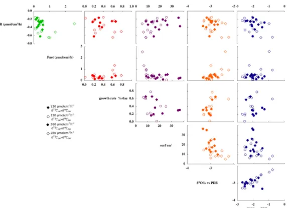
We plotted on Fig. 2 all the possible relationships between the measured parameters. There is not noticeable correla-tion. In opposite to what is usually thought, there is no clear correlation between δ13C and Pnet, neither between δ13C and
R.
3.3 Duality of metabolic and isotopic responses at HL
The partitioning of the colonies into two groups, according to the sign of the δ13C change between LL and HL, underlines various metabolic responses previously ignored.
At HL, the nubbins showing lower δ13C (Fig. 1c, dark color, lighter bar) corresponded to lower productivity asso-ciated with the highest surface extension (Fig. 3). Pnet and
surface extension (S) are roughly linearly correlated: S =0.01 · Pnet+0.25 with R2=0.46 for N = 8 (1)
Fig. 2. Comparison of all measured metabolic and isotopic parameters. Circles correspond to data obtained at LL, diamonds to HL. Clear
(dark) symbols are associated to colonies showing lower (higher) δ13C at HL than at LL. Linear correlation is clear between Pnetand R for
the highest values of Pnet, between surface and growth rate and between δ18O and δ13C.
At HL, colonies showing higher δ13C (Fig. 1c, light color, lighter bar) were those displaying higher productivity associ-ated with the lowest surface extension (Fig. 3). The parame-ters are also weakly correlated:
S = −0.17 · Pnet+2.48 with R2=0.48 for N = 7 (2)
The correlation coefficients were not significant; however they do not invalidate the suggestion of two different behav-iors related to light intensity. Under HL, corals may display one of the two behaviors linked with photosynthetic activity: either photosynthesis activity slightly increases and surface extension remains noticeable (although lower than at LL) or photosynthesis is clearly enhanced and surface extension strongly reduced.
Photosynthetic activity and respiration were correlated for colonies showing the highest photosynthesis (Pnet>0.6)
(Fig. 2). However, as there were only four data points, this relationship needs to be confirmed by additional exper-iments. There was no correlation between isotopic data and metabolic indicators (Fig. 2).
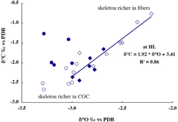
For δ13C versus δ18O (Fig. 4), there was no relation at LL (circles) but at HL (diamonds) the relationship was sig-nificant: δ13C=1.92·δ18O+3.41 with R2=0.86 for N =14 (3) (Fig. 4). We note that skeletal δ18O values were also more positive in those showing a more positive δ13C at HL than at LL (clear circles), whereas the other δ18O values were
Fig. 3. Photosynthetic activity versus surface covered by new-formed aragonite at HL. Darker symbols correspond to colonies showing higher δ13C at HL than at LL. Pnet values measured
on colonies showing lower δ13C than at LL, are limited to 1.6 µmol cm−2h−1and are associated with development on larger surface than colonies showing higher δ13C than at LL and respond-ing with higher photosynthetic activity, though all the nubbins re-spectively almost doubled their skeleton weight. Correlation coeffi-cients of the lines plotted on the figure are not significant, however, full and empty diamonds clearly show two distinct behaviors.
A. Juillet-Leclerc and S. Reynaud: Acropora: implications for coral-growth rates 899
Fig. 4. δ18O value of individual specimen plotted against δ13C. At HL (diamonds) δ18O and δ13C are linearly correlated, with a significant correlation coefficient. The highest δ18O and δ13C data correspond to the nubbins responding strongly to HL. These sam-ples could be rich in fibers, whereas the samsam-ples showing both low δ18O and δ13C could be richer in COC.
roughly within the same range as for LL (Figs. 2 and 4). In-deed, at HL few δ18O showed lower values than −3.0‰.
We highlighted the unexpected light effects on δ18O (Reynaud-Vaganay et al., 2001). The discrepancies in metabolic behavior at HL exhibited by δ13C variability were also not expected. We sought to highlight how modifications in the host metabolism may impact chemical properties of the coral skeleton.
4 Discussion
This experiment confirmed that calcification is light-enhanced (Goreau and Goreau, 1959; Chalker, 1981; Gat-tuso et al., 1999) because each nubbin showed an increased calcification with increased light. At HL, all nubbins at least doubled their initial weight in respect with calcification at LL, whereas surfaces covered by the newly-formed skeleton remained smaller. As such, we consider that surface exten-sion is light-suppressed.
4.1 Metabolic imprint on skeleton chemistry
The culture technique used here allowed us to separate tem-perature from light effects on the skeletal isotopic signature and metabolic parameters. Clode and Marshall (2004) have tested the role of light on the calcification rate of a zoox-anthellate (Galaxea) and azooxzoox-anthellate (Dendrophyllia) coral, using the45Ca technique. Galaxea and
Dendrophyl-lia presented similar Ca2+incorporation versus temperature over the range of 18–29◦C, and authors have concluded that the calcification process was affected by temperature but was probably not associated with photosynthesis. This previous
experiment, however, used different species displaying dif-ferent metabolic specificities and moreover, temperature and light effects were mixed. Thus, the data could not be com-pared and it could not demonstrate the solely photosynthetic influence. Other factors could explain the difference of Ca incorporation, e.g., temperature or differences in calcifica-tion rate among species. Conversely, our experiment avoided the temperature effect and examined the response of nubbins originating from one species, even a single parent colony, thus all were supposed to present identical metabolic and morphological characteristics.
As the experiment was conducted on nubbins originating from a single parent colony the isotopic scattering observed might be compared with inter and even intra-colony δ18O variability exhibited by Porites collected at Clipperton (Lins-ley et al., 1999) or in Indonesia (Maier et al., 2004). More-over, by considering isotopic effects of a single nubbin suc-cessively submitted to two light intensities we have an iden-tical approach than when we examine δ18O variation along close corallites representing mineral deposited during two successive months. The results obtained showed that the av-erage of several responses could mask the significance of the individual behavior (Weil et al., 1981; Grottoli and Welling-ton, 1999; Grottoli, 2002).
4.2 Light effects on growth
Increased light was systematically associated with an in-crease of skeletal weight and a dein-crease of surface exten-sion of newly-formed skeleton during identical duration. The coupled effects generated an increase in skeletal den-sity. Acropora usually does not show clear annual density bands; however, this experiment presents proof that light af-fects Acropora density. This could also be related to obser-vations made on Porites most often producing low-density bands during winter (Lough and Barnes, 2000), but it is im-portant to keep in mind that, in the field, light effects may be obscured by other factors such as temperature and/or repro-ductive cycles (Mendes, 2004).
This light effect observed can provide an explanation for data from the Caribbean Sea. When Montastrea annularis was submitted to unfavorable conditions (lower light and an-thropogenic influence), they appeared to “sacrifice skeletal density while maintaining or increasing skeletal extension, despite having a lower calcification rate” (Cruz-Pi˜non et al., 2003) (see also Carricart-Ganivet, 2004). Although in our experiment we measured surface extension and not linear extension, these field observations are roughly in agreement with what we observed at LL. Is the extension an expression of growth compensation when luminosity is insufficient, or is there a competition between the two growth rates? At LL, the extension rate was always higher than at HL (Fig. 2), and, at HL, when photosynthesis was intense, extension was re-duced (Fig. 3). Nubbins showing higher extension than the other colonies at LL seemed to roughly keep this specificity
at HL without being able to produce strong photosynthetic activity. However, among these colonies, there was a positive correlation between extension and net productivity (Fig. 3).
The partitioning of the colonies into two groups stresses that the two growth features, surface extension rate and weight of deposited aragonite per time unit (calcification rate), are distinct processes (Fig. 1c). It has been noted previ-ously that these two measurements of coral growth are not re-dundant and may provide complementary information (Scof-fin et al., 1992). However, we wanted to understand why nubbins originating from one parent colony presented two different behaviors.
4.3 The role of zooxanthellae
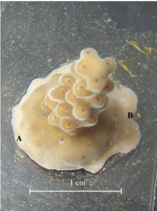
Earlier we noted that the δ13C responses exhibited all in-termediate values between the two extremes, likewise, the metabolic responses, especially photosynthesis and surface extension were highly scattered (Fig. 1). By examining Fig. 5 we observe that one side of the colony (A side) presented thickened skeleton (not expanded on the glass side) sur-rounding the axial corallite. This illustrates the growth mode called “thickening” in the first part of this article, which is observed on the inter-corallite spaces and at the base of corallite. The other side of the axial corallite (B side) is composed of newly-formed corallites (Juillet-Leclerc et al., 2009). By comparison with the description of Acropora
hy-acinthus given by Nothdurft and Well (2007), they could be
called radial corallites, which are growing vertically in the field. The surface extension measured during our experiment might be assimilated to linear extension. We suggest that the aragonite collected on the glass slide (Fig. 4) integrated vari-able amounts of skeleton fragments, with metabolic and also isotopic measurements integrating the relative responses; this could also explain the large scatter of our data.
Although we did not measure algal abundance, we could relate the different growth modes observed at HL to highly variable efficiency of photosynthesis due to different densi-ties of zooxanthellae. Figure 3 indeed suggests that skele-tons showing the maximal extension are poorer in algal den-sity, leading to a low Pnet. Weber et al. (1976) stressed this
specificity in Acropora cervicornis. They observed that the abundance of symbiotic zooxanthellae increased from the tip to the base of a branch. They associated this difference with rather confused isotopic behaviors for O and C. This obser-vation, which supports our interpretation, could be counter-intuitive. Indeed, the upper surface of the coral, where skele-togenesis seems to start, receiving more direct light incidence could be supposed to be richer in algae than the sides of colonies, which receive less intense light.
We assumed that the highest O and C isotopic values were provided by samples essentially composed of skeleton por-tions corresponding to the sides of corallites or to the skele-tal zone called inter-corallites, having the ability to strongly photosynthesize, while the samples characterized by low
Fig. 5. Acropora nubbin after few week growth at HL. New-formed
aragonite is deposited on the colony and the glass slide. On side A, the layer of new mineral is reduced and thick, whereas on side B it extends further the initially stuck nubbin and new corallites are visible. We cannot attribute such a discrepancy to different light incidences, on the glass slide, all the portions of the new-formed skeleton received identical light intensity. On the A side at the level of the section of the branched coral is essentially formed by fibers capable to thicken the skeleton under HL. On the B side, the section of the branch coral contain COC, of which growth is light-repressed. Such portion of nubbin could be much more developed under LL.
δ13C contained greater amounts of aragonite forming the ra-dial and central corallite tips according to observations made by Weber et al. (1976).
It seems that at HL, when photosynthesis was clearly ac-tive, respiration proportionally decreased (Fig. 2). This ob-servation could be due to a part of the respired CO2
be-ing used for photosynthesis, as in the carbon-translocation hypothesis of Muscatine and Porter (1977). However, we have no information to document a mechanism linking the metabolic activity of algae with coral metabolism leading to modifications in skeletogenesis.
4.4 Is there competition between photosynthetic activity and surface extension?
Figure 3, obtained with samples maintained at HL, highlights an inverse relationship between photosynthesis and surface extension. Indeed, the group of colonies showing reduced photosynthesis and δ13C depletion with the augmented light
A. Juillet-Leclerc and S. Reynaud: Acropora: implications for coral-growth rates 901 also presented an increase in surface extension, while the
group showing an active photosynthesis was characterized by the lowest extension. On one hand, this could illustrate com-petition between surface extension and photosynthetic activ-ity at HL, while, on the other hand, it could indicate that there are two biomineralization strategies: submitted to HL, some parts of the skeleton are dedicated to spatial growth and the others to strengthening skeletal structure. We note that the relative growth weight doubled in both cases.
4.5 Axial and lateral portions of a corallite
Gladfelter (1982, 1983, 1984) investigated the skeletal growth pattern of Acropora cervicornis. She described the wall of an axial corallite, surrounding the calyx, formed by vertical spines connected together tangentially and radially by a porous mineral (Gladfelter, 1982). She identified two different growth units: massive, randomly oriented crystals, called fusiform crystals, and numerous needle-like crystals projecting in many directions from the fusiform crystals. She noted that the needles, which were gathered into bundles oriented perpendicularly to the spine, showed a progressive filling-in of pore space from the tip of the corallite to the base. She deduced from these observations that skeletogen-esis could be the result of two processes: the deposition of fusiform crystals and the progressive thickening of the ini-tial framework by needle-like crystals. She attributed these two modes of deposition to a dichotomy in growth axes: the axial growth most often expressed as a linear extension re-sulting from the deposition of fusiform crystals, with lateral accretion by fibers ensuring the strengthening of the skeleton (Gladfelter, 1982, 1984). These investigations led her to con-clude that the first step of skeletogenesis was performed dur-ing the night, while the main parameter influencdur-ing the sec-ond was the duration of sunlight per day (Gladfelter, 1983).
The differentiation of coral-skeleton growth relative to its shape could be a specific feature of branched corals such as
Acropora. Skeletal structure has also been studied in Porites
(Barnes and Lough, 1993). Like Gladfelter (1983), these authors also proposed skeletal development in two steps. Lough and Barnes (2000) observed different rates of exten-sion and calcification between the top and sides of colonies in numerous massive Porites from the Great Barrier, which led them to attribute the observed discrepancy to light avail-ability. Like Gladfelter (1982), they noted the coral growth dichotomy.
SEM microstructural observations of several coral gen-era led Cuif and Dauphin (1998) to also suggest that coral-skeleton deposition operated into two successive steps. Also from SEM observations, Nothdurft and Webb (2007) un-derlined common features and discrepancies shown by sev-eral coral genera, the presence of two growth units and two growth modes appearing as common features to everyone. At another size scale, it has been demonstrated that calcifi-cation of Stylophora pistillata also operates by two
mecha-nisms: first extension and secondly progressive filling-in of the previously deposited structure (Raz-Bahat et al., 2006).
4.6 The link between light, skeletal microstructures and growth modes
Isotopic measurements conducted at the micrometer scale on cultured Acropora, similar to the colonies studied here, (Juillet-Leclerc et al., 2009) confirmed that the fusiform crystals stressed by Gladfelter (1982), abundant around the theca of Acropora, were identical with centers of calcifica-tion (COC) or early mineralizacalcifica-tion zones (EMZ) like those observed along the trabecula by other authors (Cuif and Dauphin, 1998, 2003; Raz-Bahat et al., 2006; Nothdurft and Webb, 2007).
Although earlier observations and the present study are based on two different size scales, our experiment indicated that coral growth followed one main axis and a perpendic-ular plan corresponding to two separate processes. Our re-sults also suggest that extension ensured by COC is light-suppressed whereas thickening, in term of filling-in or con-solidation by fiber bundles, is light-enhanced. Photosynthetic activity of the lateral portion of a corallite, assumed to be less exposed to light, was more intense than at the coral-lite tips because the sides would be likely richer in algae (Weber et al., 1976). The same paradox at the crystal scale was stressed in Stylophora pistillata observations by combin-ing Field Emission Scanncombin-ing Electron Microscopy (FESEM) and supplementary fluorescence techniques (Tambutt´e et al., 2007).
As shown in Fig. 5, there was a side (A side) where the newly deposited mineral corresponded essentially to accre-tion of fibers with some spinules containing few COC but free of calyces, forming a thick and dense aragonite layer around the axial polyp, while on the other side (B side) the new mineral was rich in new calyces with COC at their tip and also in the radial septae. Thus, the relative proportion be-tween COC and fibers depends on the location of the nubbin section, which explains why superficial extension of several nubbins may be as different as noted during this experiment. Juillet-Leclerc et al. (2009) demonstrated that, in a colony grown at constant conditions, COC δ18O is almost constant and centered on the lowest value, while fiber δ18O varies over a 5‰-amplitude range, from the equilibrium value to the lowest one, the latter isotopic behavior likely being linked with kinetics of deposition process. This could explain the high variability exhibited by nubbins from a single parent colony due to the highly variable relative crystal amounts, which is a crucial factor determining isotopic value. Addi-tionally, the skeleton developed on the glass slide received identical light over the whole surface (which is not the case on the side of a wild colony), thus, the effect on the different portions of the skeleton is amplified.
We emphasize that, in the case of Acropora, the space be-tween each calyx is more developed than in other genera such
as Porites (Nothdurft and Webb, 2007). This skeletal differ-ence may also likely serve to amplify the discrepancies be-tween several nubbins.
4.7 Relationship between growth and isotopic values
We demonstrated that HL enhanced fiber formation (Juillet-Leclerc et al., 2009). Therefore, knowing that δ18O in fibers is always higher than δ18O in COC, we suggest that the sys-tematic positive δ18O response to light increase (Fig. 1a) was due to fiber enrichment in all the colonies. This conclusion is supported by the fact that the positive isotopic response was more pronounced in colonies responding actively to light through photosynthesis (Fig. 1c). Additionally, we note that when photosynthesis was strongly active, δ18O signature was higher. Moreover, the oxygen reservoir was isotopically con-stant over the incubation period, as the atoms came from sea-water where they are abundant.
Meibom et al. (2006) showed that the COC δ13C of
Colpophyllia sp. were lower than that of the fibers. This
could explain why at HL, colonies strongly sensitive to pho-tosynthesis (likely richer in zooxanthellae) exhibited higher values than the others. At LL, the δ13C of colonies richer in COC would then be lower than those containing mainly fibers. Curiously, we observed the opposite (Fig. 4). In addi-tion, these nubbins showed lower δ13C at HL (Fig. 4). In the case of carbon, the origin of the atoms might vary between the two conditions. Indeed, Rollion-Bard et al. (2003a) demonstrated that δ18O and δ13C measured in a single sam-ple at the micrometer scale were not always correlated. We previously noted that the results considered in the present study were formed by the integration of numerous isotopi-cally heterogeneous microstructures. At the macro scale, due to natural integration, δ18O and δ13C may thus be correlated. Therefore, to explain δ13C variations, we must assume that, in addition to the fractionation due to kinetic processes, the carbon reservoir has to differ. Biological investigations us-ing double labelus-ing (14C and45Ca) (Erez, 1978; Furla et al., 2000) demonstrated that in the case of active photosynthesis, the amount of carbon present in the symbiotic system is not sufficient to sustain physiological activities, and this lack of carbon is compensated by an increase of seawater-uptake by the coral tissue. Considering that Dissolved Inorganic Car-bon DIC seawater δ13C is much higher than metabolic δ13C, skeletal δ13C should be enriched, supporting the macroscale observations.
In addition, deposition processes of each crystal type dif-fer as well as their isotopic signatures (Juillet-Leclerc et al., 2009). Therefore, we also can explain isotopic discrepan-cies existing between different nubbins (Fig. 1). Some of our samples contained mainly fibers, thus showing at HL an increase of δ18O and δ13C associated with the lowest exten-sion rate (Fig. 1c). For the other nubbins richer in COC, HL enhanced only the growth of their fibrous portion, enriching the δ18O signature but decreasing the extension rate. The
pri-mary skeletal carbon origins then changed between LL and HL, and the global effect on the δ13C values of “bulk” sam-ples was variable.
4.8 δ18O and δ13C correlation
The high δ18O and δ13C correlations of seasonal samples and samples collected horizontally around Pavona clavus heads and simultaneously deposited led McConnaughey (1989a and b) to assume kinetic isotopic fractionation in the coral skeleton. The distribution of δ18O and δ13C in Porites arag-onite sampled along the faster growing axis appeared to be caused by the relative variability of temperature and light. The two interpretations were consistent: the upper surface of the coral head received more sunshine, thus grew faster and showed depleted δ18O while the lateral surfaces, receiv-ing less light, exhibited a slower growth rate and higher δ18O (McConnaughey, 1989a). Therefore, it has been concluded that when δ18O and δ13C were correlated this could indicate the skeleton deposition following a kinetic process; negative oxygen fractionation has been associated with high growth rate and high light, δ18O being affected by temperature and δ13C by photosynthesis. Our experiment highlights that it is not so simple.
δ18O and δ13C are both affected by light (Fig. 1). How-ever, light effects are not equivalent on all skeletal mi-crostructures and thus the isotopic analyses are more or less impacted by the integration of micro-isotopic signatures. This leads to non-homogeneous isotopic distribution, with strong variations according to the morphology. Linear exten-sion and calcification are distinct kinetic processes, respond-ing differently to light. Thus, relationships between kinetics and isotopic fractionation and between kinetics and light are more complex: in Acropora sampled at the macro-scale, the kinetic imprint on isotopic fractionation due to fiber growth is sensitive because fibers are the most numerous skeletal components. Only oxygen is purely fractionated through a kinetic process; δ13C is determined by the combination of reservoir change and kinetic fractionation according to the metabolic activity. We note that oxygen and carbon isotope correlation was essentially significant under HL because it was only in these conditions that photosynthesis activity and thus thickening, responsible for the O and C correlation, was really active (Fig. 4).
4.9 Consequences for paleoclimatic investigations
The present experiment performed on cultured Acropora highlights the behavior of a single coral clone, potentially inhabited by different concentrations of zooxanthellae and maintained under two light conditions. Each aragonite mea-surement includes an axial corallite plus some radial coral-lites. Our conclusions make sense only by comparing the same initial corallite subject to different conditions. Al-though morphology and micro-structure distribution differ
A. Juillet-Leclerc and S. Reynaud: Acropora: implications for coral-growth rates 903 between Acropora and Porites, the conclusions inferred from
our study may be extended to the time series usually devel-oped for Porites paleoclimatic studies.
The common sampling method used for paleoclimatic studies systematically follows the major growth axis of the coral. By examining samples collected on a profile over time, we record the chemical response to environmental sea-sonal changes of corallites derived from identical clones and probably inhabited by similar symbiont assemblages. Addi-tionally, this means that the sample profile corresponds to an area where COC constitute the largest portion of microstruc-tures and where photosynthetic activity influence is reduced, in contrast to the initial assumptions commonly formulated. This confirms conclusions earlier proposed for Acropora and probably for other genera (de Villiers et al., 1995; Cardinal et al., 2001) that extension is not linear during the year. We add that extension decreases with more light. Thus, we should take into account such a feature to correctly record and con-vert environmental changes in a given chronology. This em-phasizes the importance of the sampling mode along coral-lites always being oriented along a similar direction during the study period and showing an apparent continuity by X-ray imaging.
In contrast with the commonly held belief, linear exten-sion of coral does not respond positively to light. We cannot apply systematic corrections to δ18O as proposed by Maier et al. (2004). However, we must assume that metabolic ac-tivity, essentially photosynthetic acac-tivity, is roughly repro-ducible each year, even when the algal assemblage is slightly modified over time. Two adjacent areas horizontally sampled on a single coral head may host different algal assemblages and algal concentrations and this could explain the great vari-ability in isotope signals as reported by Linsley et al. (1999), Felis et al. (2003) and Maier et al. (2004).
At the millimeter scale, it would be difficult to ob-serve simple relationships between isotopic signatures and metabolic activity due to the presence in the bulk sample of variable relative amounts of crystals characterized by differ-ent isotopic fractionations.
As light and temperature often vary in parallel but have opposite effects, the amplitude of δ18O fluctuations only due to temperature will always be smoothed by the effect of light through photosynthetic activity. This could explain the observed high variability of time series and calibrations (Wellington et al., 1996; Linsley et al., 1999; Maier et al., 2004). It could also explain the correlation of δ18O versus SST showing lower absolute slope values than −0.20‰/◦C
(Juillet-Leclerc and Schmidt, 2001; Suzuki et al., 2005). Thickening of the corallite wall could also smooth δ18O by the addition of aragonite fibers. Due to a specific morpholog-ical organization, especially a reduced inter-corallite surface, this effect would likely remain limited for Porites compared with Acropora; sampling along the axis of maximum growth rate would also reduce this effect.
We note that linear extension and δ18O are affected by light in the same way; the direct temperature effect and that caused through photosynthesis are opposite. It would be in-formative to compare δ18O and linear-extension fluctuations over a year, but extension variability is not easy to measure over a year. The light influence on δ13C is more complex and depends on the corallite portion sampled.
Our findings help explaining the contrasting observations made on different colonies (Goreau, 1977; Erez, 1978). However, on a Porites head, as sampling is centered along the main growth axis where COC are abundant, a clear δ13C decrease is almost always recorded with light enhancement during the summer (Swart et al., 1996).
5 Summary
1. Rates of photosynthesis are not constant or equal on the surface of a coral skeleton. Zooxanthellae could also be more abundant on the sides than on the apex of corallites. Light enhancement leads to decreased ex-tension rate and increased aragonite weight deposited. Thus, HL causes a skeleton-density increase. Although the absence of algae on the apical part of the skeleton is counter-intuitive following classical geochemical hy-pothesis, several lines of evidence suggest this conclu-sion.
2. Both δ18O and δ13C are impacted by light. In the field, light-induced increase in δ18O is masked by the temper-ature effect. δ13C may increase in the lateral corallite portion while simultaneously decreasing at the apex. 3. We did not observe a direct correlation between
metabolism and isotopic data. This could be attributed to the systemic mixture of microstructures, for which the relative ratios varied in the measured samples. 4. Significant correlations between δ18O and δ13C were
likely due to the kinetic deposition process of fibers (Juillet-Leclerc et al., 2009) and are not directly related to classical growth-rate measurements, density and lin-ear extension. The strong relationships between oxygen and carbon isotopic fractionation, growth rate and light are much more complex than previously thought. 5. Light influence on O and C isotopic ratios is part of a
vital effect, essentially inducing high colony variabil-ity and also horizontal variabilvariabil-ity on the surfaces of colonies in some species (McConnaughey, 1989). This could be due to the variable abundance of zooxanthel-lae, variable light incidence and/or the sampled morpho-logical parts of corallites.
6. Variability of isotopic ratios is significant over the time only when measurements are performed along individ-ual successive specimens, in this case corallites.
These results provide evidence underlining the importance of the coral-algae symbiosis in coral-reef formation, espe-cially concerning the strong and complex relationships be-tween calcification and photosynthetic activity (Gattuso et al., 1999). Estimation of the relative importance of temper-ature and light effect on skeletal isotopic signtemper-atures will be investigated further in the future.
Vital effects are particularly complex in corals due to the impact of the zooxanthellate metabolism on the chemical fea-tures of the coral skeleton and the collective growth of a colony of multiple organisms. However, as we highlighted here, each specimen shows its own signature. Such a prop-erty is not a coral specificity, indeed inter- and intra-specimen variability of Mytilus edulis and Pecten maximus has still been stressed by Freitas et al. (2008).
Acknowledgements. The authors thank J. Lough and C.
Ferrier-Pag`es for fruitful contributions, which significantly improved the quality of the manuscript. This work benefited greatly from the constructive comments of E. Gladfelter and an anonymous reviewer and encouraging J. Bijma (associate Editor) comments. Additional thanks to Cecilia Garrec for English-language editing.
Edited by: J. Bijma
The publication of this article is financed by CNRS-INSU.
References
Barnes, D. J. and Lough, J. M.: On the nature and causes of density banding in massive coral skeletons, J. Exp. Mar .Biol. Ecol., 167, 91–108, 1993.
Cardinal, D., Hamelin, B., Bard, E., and P¨atzold, J.: Sr/Ca, U/Ca and δ18O records in recent massive corals from Bermuda: rela-tionships with sea surface temperature, Chem. Geol., 176, 213– 233, 2001.
Carricart-Ganivet, J. P.: Sea surface temperature and the growth of the West Atlantic reef-building coral Montastraea annularis, J. Exp. Mar .Biol. Ecol., 302, 249–260, 2004.
Chalker, B. E.: Simulating light-saturation curves for photosynthe-sis and calcification by reef-building coral, Mar. Biol., 63, 135– 141, 1981.
Marshall, A. T. and Clode, P.: Calcification rate and the effect of temperatre in a zooxanthellate and an azooxanthellate sclerac-tinian ref coral, Coral Reefs, 23, 218–224, 2004.
Cole, J. E., Fairbanks, R. G., and Shen, G. T.: Recent variability in the Southern Oscillation: Isotopic results from a Tarawa Atoll coral, Science, 260, 1790–1793, 1993.
Cruz-Pi˜non, G., Carricart-Ganivet, J. P., and Espinoza-Avalos, J.: Monthly skeletal extension rates of the hermatypic corals
Mon-tastraea annularis and MonMon-tastraea faveolata: biological and
environmental controls, Mar. Biol., 143, 491–500, 2003. Cuif, J.-P. and Dauphin, Y.: Microstructural and physico-chemical
characterisation of centres of calcification in septa of some Scle-ractinian corals, Pal. Zeit, 72, 257–270, 1998.
Cuif, J.-P., Dauphin, Y., Doucet, J., Salome, M., and Susini, J.: XANES mapping of organic sulphate in three Scleractinian coral skeletons, Geochim. Cosmochim. Acta, 67, 75–83, 2003. Davies, P. S.: Short-term growth measurements of corals using an
accurate buoyant weighing technique, Mar. Biol., 101, 389–395, 1989.
de Villiers, S., Nelson, B. K., and Chivas, A. R.: Biological controls on coral Sr/Ca and δ18O reconstructions of sea surface tempera-ture, Science, 269, 1247–1249, 1995.
Fairbanks, R. G. and Dodge, R. E.: Annual periodicity of the 18O/16O and 13C/12C ratios in the coral Montastrea annularis, Geochim. Cosmochim. Acta, 43, 1009–1020, 1979.
Dunbar, R. B., Wellington, G. M., Colgan, M. W., and Glynn, P. W.: Eastern Pacific sea surface temperature since 1600 A.D.: the δ18O record of climate variability in Galapagos corals, Paleo-ceanography, 9, 291–315, 1994.
Epstein, S., Buchsbaum, R., Lowenstam, H., and Urey, H. C.: Revised carbonate-water isotopic temperature scale, Bull. Geol. Soc. Am., 62, 417–425, 1953.
Erez, J.: Vital effect on stable-isotope composition seen in foraminifera and coral skeletons, Nature, 273, 199–202, 1978. Felis, T., P¨atzold, J., and Loya, Y.: Mean oxygen-isotope signatures
in Porites sp. corals: inter-colony variability and correction for extension-rate effects, Coral Reefs, 22, 328–336, 2003. Freitas, P. S., Clarke, L. J., Kennedy, H. A., and Richardson, C. A.:
Inter- and intra-specimen variability masks reliable temperature control on shell Mg/Ca ratios in laboratory- and field-cultured Mytilus edulis and Pecten maximus (bivalvia), Biogeosciences, 5, 1245–1258, 2008,
http://www.biogeosciences.net/5/1245/2008/.
Furla, P., Galgani, I., Durand, I., and Allemand, D.: Sources and mechanisms of inorganic carbon transport for coral calcification and photosynthesis, J. Exp. Biol., 203, 3445–3457, 2000. Gagan, M. K., Chivas, A. R., and Isdale, P. J.: High resolution
isotopic records from corals using ocean temperature and mass-spawing chronometers, Earth Planet. Sci. Lett., 121, 549–558, 1994.
Gattuso, J.-P., Allemand, D., and Frankignoulle, M.: Photosynthe-sis and calcification at cellular, organismal and community levels in coral reefs: a review on interactions and control by carbonate chemistry, Amer. Zool., 39, 160–183, 1999.
Gladfelter, E. H.: Skeletal development in Acropora cervicornis: I. Patterns of calcium carbonate accretion in the axial corallite, Coral Reefs, 1, 45–51, 1982.
Gladfelter, E. H.: Skeletal development in Acropora cervicornis: II. Diel patterns of calcium carbonate accretion, Coral Reefs, 2, 91–100, 1983.
Gladfelter, E. H.: Skeletal development in Acropora cervicornis: III. A comparison of monthly rates of linear extension and cal-cium carbonate accretion measured over a year, Coral Reefs, 3, 51–57, 1984.
Gladfelter, E. H.: Skeletal development in Acropora palmata (Lamarck 1816): a scanning electron microscope (SEM) com-parison demonstrating similar mechanisms of skeletal extension
A. Juillet-Leclerc and S. Reynaud: Acropora: implications for coral-growth rates 905
in axial versus encrusting growth, Coral Reefs, 26, 883–892, 2007.
Goreau, T. J.: Carbon metabolism in calcifying and photosynthetic organisms: theoritical models based on stable isotope data, Third international coral reef symposium, 395–401, 1977.
Goreau, T. J. and Goreau, N. I.: The physiology of skeleton forma-tion in corals. II. Calcium deposiforma-tion by hermatypic corals under various conditions on the reef, Biol. Bull. (Woods Hole), 118, 419–429, 1959.
Grottoli, A. G. and Wellington, G. M.: Effect of light and zooplank-ton on skeletal δ13C values in the eastern Pacific corals Pavona
clavus and Pavona gigantean, Coral Reefs, 18, 29–41, 1999.
Grottoli, A. G.: Effect of light anf brine shrimp on skeletal δ13C in Hawaiian coral Porites compressa: a tank experiment, Geochim.. Cosmochim. Acta, 66, 1955–1967, 2002.
Guzman, H. M. and Thudope, A. W.: Seasonal variation in skeletal extension rate and stable isotopic (13C/12C and18O/16O) com-position in response to several environmental variables in the Caribbean reef coral Siderastrea sidereal, Mar. Ecol. Prog. Ser., 166, 109–118, 1998.
Heikoop, J. M., Dunn, J. J., Risk, M. J., McConnaughey, T. A., and Sandman, I. M.: Separation of kinetic and metabolic effect in carbon-13 records preserved in reef coral skeletons, Geochim. Cosmochim. Acta, 6, 975–987, 2000.
Highsmith, R. C.: Coral growth rates and environmental control of density banding, J. Exp. Mar. Biol. Ecol., 37, 105–125, 1979. Jokiel, P. L,. Maragos, J. E., and Franzisket, L.: Coral growth:
buoy-ant weight technique, in: Coral Reef: Research Methods, edited by: Stoddart, D. R. and Johannes, R. E., Unesco, Paris, 379–396, 1978.
Juillet-Leclerc, A. and Schmidt G.: A calibration of the oxygen iso-tope paleothermometer of coral aragonite from Porites, Geophys. Res. Lett., 28, 4135–4138, 2001.
Juillet-Leclerc, A., Reynaud, S., Rollion-Bard, C., Cuif , J.-P., Dauphin, Y., Blamart, D., Ferrier-Pag`es, C., and Allemand, D.: Oxygen isotopic signature of the skeletal microstructures in cultured corals: identification of vital effects, Geochim. Cos-mochim. Acta, 73, 5320–5332, 2009.
Linsley, B. K., Messier, R. G., and Dunbar, R. B.: Assessing between-colony oxygen isotope variability in the coral Porites
lobata at Clipperton Atoll, Coral Reefs, 18, 13–27, 1999.
Lough, J. M. and Barnes, D. J.: Environmental control on growth of the massive coral Porites, Mar. Ecol. Prog. Ser., 245, 225–243, 2000.
Maier, C., Felis, T., P¨atzold, J., and Bak, R. P. M.: Effect of skeletal growth and lack of species effects in the skeletal oxygen isotope climate signal within the coral genus Porites, Mar. Geol., 207, 193–208, 2004.
Marsh, J. A. J.: Primary productivity of reef building calcareous red algae, Ecol., 51, 255–263, 1970.
McConnaughey, T. A.: C-13 and O-18 isotopic desequilibrium in biological carbonates: I. Patterns, Geochim. Cosmochim. Acta, 53, 151–162, 1989a.
McConnaughey, T. A.: C-13 and O-18 isotopic disequilibria in bi-ological carbonates: II. In vitro simulation of kinetic isotope ef-fects, Geochim. Cosmochim. Acta, 53, 163–171, 1989b. McConnaughey, T. A., Burdett, J., Whelan, J. F., and Paull, C. K.:
Carbon isotopes in biological carbonates: respiration and photo-synthesis, Geochim. Cosmochim. Acta, 61, 611–622, 1997.
Meibom, A., Yurimoto, H., Cuif, J.-P., Domart-Coulon, I., Houlbr`eque, F., Constantz B., Dauphin, B., Tambutt´e, E., Tambutt´e, S., Allemand, D., Wooden, J., and Dunbar, R.: Vital effect in coral skeletal composition display strict three-dimensional control, Geophys. Res. Lett., 33, L11608, doi:10.1029/2006GL025968, 2006.
Mendes, J.: Timing of skeletal band formation in Monstastrea
an-nularis: relationship to environmental and endogeneous factors,
Bull. Mar. Sci., 75, 423–437, 2004.
Muscatine, L. and Porter, J. W.: Reef corals: mutualistic symbiosis adapted to nutrient-poor environments, BioScience, 27, 454–459 1977.
Nothdurft, L. D. and Webb, G. E.: Microstructure of common reef-building coral genera Acropora, Pocillopora, Goniastrea, and
Porites: constraints on spatial resolution in geochemical
sam-pling, Facies, 53, 1–26, 2007.
Omata, T., Suzuki, A., Kawahat, H., and Okamoto, M.: Annual fluctuation in the stable carbon isotope ratio of coral skeletons: the relativeintensities of kinetic and metabolic isotope effect, Geochim. Cosmochim. Acta, 69, 3007–3016, 2005.
Quinn, T. M,. Taylor, F. W., and Crowley, T. J.: A 173 year stable isotope record from a tropical south Pacific coral, Quaternary Sci. Rev., 12, 407–418, 1993.
Raz-Bahat, M., Erez, J., and Rinkevich, B.: In vivo light-microscopic documentation for primary calcification processes in the hermatypic coral Stylophora pistillata, Cell. Tissue Res., 325, 361–368, doi:10.1007/s00441-006-0182-8, 2006.
Reynaud-Vaganay, S., Gattuso, J.-P., Cuif, J.-P., Jaubert, J., and Juillet-Leclerc, A.: A novel culture technique for scleractinian corals: application to investigate changes in skeletal δ18O as a function of temperature, Mar. Ecol. Prog. Ser., 180, 121–130, 1999.
Reynaud-Vaganay, S., Juillet-Leclerc, A., Gattuso, J.-P., and Jaubert, J.: Effect of light on skeletal δ13C and δ18O and inter-action with photosynthesis, respiration and calcification in two zooxanthellate scleractinian corals, Paleogeogr. Paleocl., 175, 393–404, 2001.
Rollion-Bard, C., Chaussidon, M., and France-Lanord, C.: pH con-trol on oxygen isotopic composition of symbiotic corals, Earth Planet. Sci. Lett., 215, 265–273, 2003.
Scoffin, T. P., Tudhope, A. W., and Brown, B. E.: Fluorescent and skeletal density banding in Porites lutea from Papua New Guinea and Indonesia, Coral Reefs, 7, 169–178, 1989.
Suzuki, A., Hibino, K., Iwase, A., and Kawahata, H.: Inter-colony variability of skeletal oxygen and carbon isotope signa-tures of cultured Porites corals: temperature-controlled experi-ments, Geochim. Cosmochim. Acta, 69, 4453–4462, 2005. Swart, P. K.: Carbon and oxygen isotope fractionation in
sclerac-tinian corals: a review, Earth-Sci. Rev., 19, 51–80, 1983. Swart, P. K., Leder, J. J., Szmant, A., and Dodge, R. E.: The
ori-gins of variations in the isotopic record of scleractinian corals II. Carbon, Geochim. Cosmochim. Acta, 60, 2871–2886, 1996. Tambutt´e, ´E., Allemand, D., Zoccola, D., Meibom, A., Lotto, S.,
Caminiti, N., and Tambutt´e, S.: Observations of the tissue-skeleton interface in the Scleractinian coral Stylophora pistillata, Coral Reefs, 26, 517–529, 2007.
Urey, H. C., Lowenstam, H. A., Epstein, S., and McKinney, C. R.: Measurements of paleotemperatures and temperatures of the Up-per Cretaceous of England, Denmark, and the southern United
States, Bull. Geol. Soc. Am., 62, 399–416, 1951.
Weber, J. N. and Woodhead, P. M. J.: Temperature dependence of Oxygen-18 concentration in reef coral carbonates, J. Geophys. Res., 7, 463–473, 1972.
Weber, J. N., Deines, P., Weber, P. H., and Baker, P. A.: Depth related changes in the 13C/12C ratio in skeletal carbonate de-posited by the Caribbean reef-frame building coral Monstatrea
annularis: futher implications of a model for stable isotope
frac-tionation by Scleractinian corals, Geochim. Cosmochim. Acta, 40, 31–39, 1976.
Weil, S. M., Buddemeier, R. W., Smith, S. V., and Kroopnick, P. M.: The stable isotopic composition of coral skeletons: control by environmental variables, Geochim. Cosmochim. Acta, 45, 1147– 1153, 1981.
Wellington, G. M., Dunbar, R. B., and Merlen, G.: Calibration of stable oxygen isotope signatures in Galapagos corals, Pale-oceanography, 11, 467–480, 1996.