Acute Stroke: Present and Future
of Catheter-Based Interventions
Jan Gralla
1, Caspar Brekenfeld
1, Marcel Arnold
2, Gerhard Schroth
1 AbstractAcute ischemic stroke is a major cause of morbidity and mortality in industrialized nations. The sequel of stroke ranges from mild to severe disability and even death. Since the impairment may be permanent, the costs to society from work incapacity and the need for long-term care can be high. Additionally, the burden of suffering associated with the disease may have devas-tating effects on individuals and families.
Following the occlusion of a cerebral vessel, the full extent of the infarction may not become clinically and radiologically apparent until days after. By then, the surrounding watershed zone, or penumbra, either survives or succumbs to necrosis over time. The natural history of this “tissue at risk” is determined by two
fac-tors: the collateralization from other vascular territo-ries and the possible occurrence of spontaneous re-canalization of the occluded vessel. The current treat-ment options for acute ischemic stroke are aiming at an early and sustained restoration of flow to the pen-umbra. The effect of the treatment is therefore time-dependent and the neurologic outcome is a func-tion of the time span between onset of symptoms and recanalization therapy as well as of the recanalization rate associated with a specific treatment.
This report summarizes the results of the major trials on catheter-based interventions, either using thrombolytic drugs or novel mechanical approaches being developed to treat patients with acute ischemic stroke.
Akuter zerebrovaskulärer Insult: Gegenwart und Zukunft katheterbasierter Eingriffe
ZusammenfassungDer akute ischämische zerebrovaskuläre Insult ist in den westlichen Industrieländern eine der häufigsten Ursachen von Morbidität und Mortalität. Die Folgen des Insults reichen für den Betroffenen von milden neurologischen Ausfällen über schwerste Pflegebe-dürftigkeit bis zum Tod mit erheblichen Kosten für das Gesundheitssystem.
Nach einem akuten Verschluss eines hirnversor-genden Gefäßes werden Teile des Parenchyms zunächst über Kollateralen vor der Nekrose bewahrt. Im Laufe von Tagen kann sich dieses Gewebe, die Penumbra, regene-rieren, oder es wird untergehen. Dies ist sowohl von der
Kollateralversorgung des Gewebes als auch von einer Reperfusion, z.B. durch spontane Thrombolyse, abhän-gig. Das Ziel der derzeitigen Therapieoptionen ist eine Wiederherstellung der Perfusion der Penumbra durch Rekanalisierung des Gefäßes. Der Therapieerfolg hängt dabei von der Rekanalisierungsrate und der Ischämie-zeit der Penumbra bis zur Reperfusion ab.
Dieser Artikel fasst die Ergebnisse der größeren Studien zu katheterbasierten Techniken zusammen und vergleicht die medikamentöse Thrombolyse mit neueren mechanischen Ansätzen zur Thrombektomie in der Behandlung des akuten ischämischen zerebro-vaskulären Insults. Schlüsselwörter: Akuter zerebrovaskulärer Insult · Interventionelle Therapie · Thrombekto-mie · Intraarterielle Thrombolyse · Katheter-basierte Eingriffe Key Words: Acute ischemic stroke · Interven-tional treatment · Mechanical throm-bectomy · Intraarte-rial thrombolysis · Catheter-based in-terventions 1 Department of
Inter-ventional and Diagnos-tic Neuroradiology, In-selspital, University of Bern, Switzerland. 2 Department of Neurol-ogy, Inselspital, University of Bern, Switzerland. Herz 2008;33:507–17 DOI 10.1007/ s00059-008-3153-x
Introduction
Acute ischemic cerebrovascular stroke is character-ized by neurologic deterioration caused by the sud-den occlusion of a brain-perfusing artery either by an embolic occlusion with thrombotic material or, less often, by a locally formed thrombus. The subsequent undersupply with oxygen (ischemia) causes a pro-gressive damage of the brain tissue distally to the oc-clusion site. Acute ischemic stroke is a common dis-ease in industrialized nations, characterized by a high prevalence of cardiovascular risk factors and a pro-gressive aging of the population. The natural course of stroke ranges from mild neurologic deficits to life-long disability and dependency or death.
To protect patients from the high rate of morbid-ity and mortalmorbid-ity associated with stroke, various sys-temic and local treatment options have been advo-cated.
Despite different approaches in the treatment strategy, all studies have underlined the crucial deter-minants for the patients’ neurologic outcome after acute ischemic stroke [1–5]:
(a) the size of the ischemic brain area perfused by the occluded vessel;
(b) the time span between onset of symptoms and re-vascularization;
(c) the recanalization rate associated with a specific treatment;
(d) the occurrence of symptomatic intracranial hem-orrhage (sICH).
Hence, fast and sufficient reperfusion in combina-tion with a low rate of sICH is the key to successful stroke treatment (Figures 1 and 2).
The intravenous administration of thrombo-lytic drugs (intravenous thrombolysis [IVT]) has been evaluated in various randomized studies ap-plying recombinant tissue-type plasminogen acti-vator (rtPA) to acute ischemic stroke patients [3, 6, 7]. The NINDS study [3, 6] was able to demon-strate the efficacy of systemic IVT therapy when initiated within 3 h from onset of stroke symptoms and if an intracranial hemorrhage was previously ruled out. The IVT has been permitted in the USA
since 1996. All thrombolytic therapies elevate the risk of bleeding. In the trials, in 6.4% (NINDS) to 8.8% (ECASS) of patients with IVT an sICH oc-curred.
Because of the narrow time window for treat-ment and multiple exclusion criteria, only 9–22% of ischemic stroke patients admitted to stroke centers may be treated with IVT.
In addition, a reanalysis of the NINDS study [8] revealed the limited effect of IVT in patients with severe stroke (National Institute of Health Stroke Scale [NIHSS] > 16). The NIHSS increases with the size of the vessel occluded. An NIHSS > 12 suggests an occlusion of a proximal, large ves-sel and therefore a high thrombus burden [9].
Figures 1a to 1f. Endovascular recanalization of an acute
right-sided M1 occlusion in a 59-year-old patient. The initial ICA angiogram in anteroposterior (a) and lateral (b) projec-tion demonstrates the complete M1 occlusion of the right MCA (➝). The extent of the occlusion can be angiographi-cally demonstrated (c) by passing the thrombus in the MCA and injecting contrast (*). During retrieval of the microcath-eter, contrast injection illustrates the proximal and distal end of the thrombus (d, **). After treatment, the target ves-sel (M1) is completely recanalized (TIMI 3, e), but thrombotic material is dislodged into the proximal M2 segments (f), hin-dering reperfusion of the complete MCA territory (➩). This distal dislocation is not assessed by the TIMI classification but relevant for the reperfusion success of the therapy. Some studies therefore additionally apply the Mori classification to document reperfusion.
Abbildungen 1a bis 1f. Endovaskuläre Rekanalisation eines
akuten M1-Verschlusses links bei einem 59-jährigen Pati-enten. Das initiale Angiogramm in frontaler (a) und lateraler (b) Projektion bestätigt den kompletten Verschluss (➝). Die Ausdehnung des Verschlusses kann nach Passage des Thrombus mit einem Mikrokatheter dargestellt werden (c). Dabei wird Kontrastmittel distal (*) und proximal (d, **) des Thrombus appliziert. Nach Therapie ist das Hauptgefäß (M1) komplett rekanalisiert (e), aber thrombotisches Material ist in einzelne M2-Segmente disloziert (f) und verhindert die Reperfusion in Teilen des abhängigen Stromgebiets (➩). Die Verschleppung von Thrombus nach distal wird von der TIMI-Klassifikation nicht berücksichtigt, ist aber für die zere-brale Reperfusion relevant. Aus diesem Grund verwenden einige Studie zusätzlich die Mori-Klassifikation.
a b
c d
These data indicate a large room for further im-provement in the treatment strategy and patient selection.
Endovascular Approaches
The local intraarterial techniques expand the time window for treatment and may increase the recanali-zation rate compared to the systemic IVT. The local approach is promising to reduce or eliminate the ex-posure to thrombolytic drugs and therefore to reduce the risk of accompanying hemorrhage. Various endo-vascular treatment options including a range of phar-macological and mechanical approaches have been advocated.
Intraarterial Thrombolysis
In intraarterial thrombolysis (IAT), a microcathe-ter is placed proximal to or directly into the throm-bus. Technically, a long 7-French (F) sheath is placed into the femoral artery and a 6- or 7-F guid-ing catheter is advanced into the internal carotid artery (ICA) or vertebral artery of the affected side. A microcatheter is then navigated to the oc-clusion site over a microwire.
Theoretical advantages of this route of appli-cation include:
(a) the angiographic evaluation reveals the precise occlusion site, the extent of collaterals and as-sesses the grade of recanalization during treat-ment;
Figures 2a to 2f. Diffusion-weighted imaging (DWI)
illus-trates an infarction in the basal ganglia (a) on the left in a 62-year-old male patient with acute onset of right-sided hemiparesis and dysphasia. Perfusion imaging (b) illustrates a reduced perfusion of the MCA territory with a large mis-match between MR perfusion (b) and DWI (a). The angiogram confirms the complete (TIMI 0) left M1 occlusion (c). Despite the complete recanalization (TIMI 3) after 1,000,000 U of urokinase and mechanical disruption of the thrombus (d), the patient developed an sICH 10 h later (e, f).
Abbildungen 2a bis 2f. Die Diffusionswichtung (DWI) zeigt
einen linksseitigen Basalganglieninfarkt (a) bei einem 62-jährigen Patienten mit akut aufgetretener rechtsseitiger Hemiparese und Dysphasie. Die Perfusionsbildgebung (b) beweist die Minderversorgung des Arteria-cerebri-media-Stromgebiets mit ausgedehnter Diskrepanz zwischen MR-Perfusion (b) und DWI (a). Das Angiogramm bestätigt den kompletten M1-Verschluss links (c). Trotz kompletter Reka-nalisierung des Gefäßes nach lokaler Gabe von 1 000 000 Einheiten Urokinase und mechanischer Manipulation des Thrombus (d) entwickelte der Patient 10 h später ein sym-ptomatisches intrazerebrales Hämatom (e, f).
a b
c d
(b) a higher effective dosage of thrombolytic agent is delivered directly to the thrombus reducing the systemic side effects;
(c) the approach facilitates the combination with the mechanical recanalization techniques. In open clinical series IAT has achieved a higher early recanalization rate compared to IVT [10, 11].
Down sides of IAT include:
(a) it is a time-consuming procedure which delays the initiation of treatment compared to IVT; (b) it involves manipulation of cervical and cranial
vessels with the risk of periinterventional com-plications;
(c) it demands highly specialized centers;
(d) it requires high human and financial resources; (e) the direct endovascular access to the distal in-tracranial vasculature, e.g., distal M2 and M3 segments of the middle cerebral artery (MCA), is limited.
PROACT. The Prolyse in Acute Cerebral Throm-boembolism I (PROACT I) study [5] was the first small clinical trial to evaluate the safety and effi-ciency of intraarterial administration in the treat-ment of acute ischemic stroke. The study included 40 patients with an angiographically proven M1 or M2 occlusion and a mean of 5.5 h from symptom onset. 14 patients (mean NIHSS: 19) were random-ized into the control placebo group, 26 patients (mean NIHSS: 17) were treated with a local admin-istration of prourokinase (6 mg) over 120 min into the proximal thrombus surface. Any mechanical manipulation of the clot was not allowed. The re-canalization was graded according to the Throm-bolysis in Myocardial Infarction (TIMI) classifica-tion and revealed a partial or complete recanaliza-tion (TIMI 2 and 3) in 57.7% of the patients treated with prourokinase compared to 14.3% in the con-trol group. An sICH within 24 h occurred in 15.5% of the treated patients and 7.1% of the placebo pa-tients. The number of patients was too small to demonstrate a statistically significant benefit in terms of clinical outcomes or mortality at 90 days. Nevertheless, an absolute increase in favorable clinical neurologic outcome over the placebo group was found in 10–12%. The mortality rate was de-creased from 42.9% in the placebo group to 26.9% in the prourokinase group. The results of PRO-ACT I suggested an enhanced recanalization with prourokinase and a positive trend toward better neurologic outcome and survival rate.
The following large-scale, multicenter, random-ized trial PROACT II [4] included 180 patients with angiographically confirmed M1 or M2 occlu-sion within the first 6 h after onset of symptoms.
121 patients (NIHSS: 17) received low-dose venous heparin (2,000 U bolus, 500 U/h) and intra-arterial prourokinase (9 mg) proximally to the thrombus; again, any mechanical disruption of the thrombus was not allowed. 59 patients of the con-trol group received only low-dose intravenous hep-arin.
The intraarterially treated group showed a sig-nificantly (p < 0.001) higher rate of partial or com-plete recanalization in 66% compared to 18% in the heparin-only group. Excellent neurologic out-come was achieved in 40% of the treated patients compared to 25% in the control group (p = 0.04). The rate of sICH within 24 h was elevated to 10% in the prourokinase group compared to 2% in the control group (p = 0.06), but no significant differ-ence in the 90-day mortality was found (25% in the treated and 27% in the control group).
The study was able to demonstrate the benefi-cial effect of IAT on the recanalization and clinical outcome of patients with M1 and M2 occlusions. Single-center experiences. However, nonrandom-ized single-center studies have reported on a high number of patients treated with IAT for acute isch-emic stroke. The largest study of Brekenfeld et al. [12] reported a retrospective analysis of 297 stroke patients treated with intraarterial urokinase within a 6-h time window for occlusions in the anterior and posterior circulation. Contrary to the PRO-ACT study protocol, the administration of throm-bolytics was not limited to the proximal surface of the occlusion site but allowed application within the thrombus. In addition, mechanical manipula-tion with the microwire and/or microcatheter was performed. The evaluated cohort had, compared to PROACT II, a longer median time span from symptom onset to treatment (318 vs. 255 min) and a slightly lower mean NIHSS (15 vs. 17). In this study, the mean dose of urokinase administered lo-cally was 1,000,000 U. The study demonstrated a higher recanalization rate (TIMI 2 and 3) of 70% (vs. 66% in PROACT II) and a lower rate of sICH of only 4.8% (vs. 10% in PROACT II). A failed reperfusion was identified as independent predic-tor of sICH. In patients with sICH a favorable clin-ical outcome (modified Rankin Score [mRS]: 0–2) was less likely compared to the group of patients without sICH (7% vs. 58%).
The study illustrated a favorable outcome of stroke patients treated with IAT despite the de-layed time to treatment in that group. The recana-lization rate was higher compared to other studies on IAT probably due to the mechanical manipula-tion of the thrombus performed by highly experi-enced operators.
Comparison IVT versus IAT
In many centers, accessible occlusions in the anteri-or circulation are treated with IAT, either in patients who did not reperfuse after IVT or even as a first line of treatment. However, there is no randomized trial comparing patients’ outcome after IVT and IAT.
A recent publication compared the outcome and morbidity of patients treated with IVT and IAT in two different stroke units [11]. The patients were selected by the presence of a hyperdense MCA sign on computed tomography, indicating an M1 occlu-sion. 55 patients were treated with IAT using uroki-nase, 59 patients underwent IVT. Although the time to treatment was significantly (p = 0.0001) longer in the IAT group (mean 244 min) than in the IVT group (mean 156 min), the study revealed a more frequent favorable outcome for the patients treated with urokinase (53%) compared to the patients treated with IVT (23%; p = 0.001). In addition, the mortality rate was reduced (4.7%) in the IAT group compared to the IVT group (23%; p = 0.001).
Although these studies suggest a benefit for IAT over IVT especially for large vessel occlusions in the anterior circulation, there is a need for more data on both approaches. Other possibilities for im-proving ischemic stroke therapy are the expansion of the time window using magnetic resonance imag-ing selection criteria for treatment (e.g., presence of a large penumbra) and the use of combined IVT and IAT, the so-called bridging concept [13].
Ischemic Stroke in the Posterior
Circulation
The data on IVT and IAT presented focuses on the most common site of intracranial vessel occlusion, the carotid distribution and the M1 and M2 seg-ments of the MCA in particular. Posterior circula-tion stroke, however, differs in several aspects. The evolution of clinical symptoms is often gradual. The precise assessment of the onset of symptoms and of the time window for treatment is difficult. Compared to ischemic stroke in the ICA territory, stroke in the posterior circulation is more often based on atherosclerotic disease (e.g., ruptured plaque) of the vasculature and locally formed thrombus. The risk of reocclusion after recanaliza-tion is therefore higher [14–17]. The natural history shows a poor outcome with a high mortality rate of 70–80% [14, 18]. Studies have suggested to extend the time window for treatment beyond the 6 h up to 24 h [16, 19, 20]. However, no randomized trial of IAT has been carried out to support this practice. In clinical practice, there is no consensus on the strategy and time window for treatment. However,
recanalization is crucial to improve the dismal out-come of patients with vertebrobasilar stroke. Therefore, the use of more aggressive treatment options such as extending the time window for the administration of thrombolytic drugs or mechani-cal approaches seem justified.
Mechanical Recanalization Techniques
Although thrombolysis is effective in some patients, the limited recanalization rate and the increase of complications give room for further improvement in the strategy of stroke treatment. Recent develop-ments focused on the possibilities of a mechanical recanalization in order to reduce revascularization time and to increase revascularization rate. Further-more, the waiving of thrombolytic drugs is considered to reduce the occurrence of sICH and might prolong the time window for treatment, especially in the ICA territory.Mechanical Thrombectomy
Intraarterial mechanical thrombectomy, in general, is a well-described procedure for peripheral vessels and first experiences are dated back to Fogarty in 1967 [21]. In spite of the wide use in peripheral vessels the application of this method to treat cerebral vessel oc-clusion is relatively new. Although the mechanical principles may be similar, the different composition of brain vessels as well as the possible fatal conse-quences in case of thrombus dislocation or device failure and the lack of fast surgical alternatives de-mand to adapt the principles of thrombectomy for the cerebral circulation.
Since 1999, descriptions of intracranial mechani-cal thrombectomy are found in the literature [22]. Initial attempts consisted on the use of proximal de-vices using a microcatheter for thrombus aspiration in combination with IAT [23, 24]. Since 2004, a few studies have reported on mechanical thrombectomy as a stand-alone therapy in acute stroke. This devel-opment was mainly driven by the fast and innovative device improvement on a parallel field of cerebro-vascular intervention, the intracranial aneurysm therapy. Since the ISAT trial [25, 26] stated a clear benefit in favor of endovascular treatment for cranial aneurysms, the spectrum of devices for intra-cranial application has been broadened dramatically. Furthermore, the experiences of cardiology in acute myocardial infarction treatment have demonstrated the superiority of mechanical strategies compared with IVT.
As a consequence, also in the field of mechanical thrombectomy the use of more complex devices has been advocated recently. All mechanical
thrombec-tomy devices are delivered by endovascular access proximal to the occlusion site.
The systems can be divided into two major groups according to where they apply force on the thrombus: (a) Proximal devices apply force to the proximal base of the thrombus. This group includes various aspi-ration catheters.
(b) Distal devices approach the thrombus proximally but then are advanced by guide wire and micro-catheter across the thrombus to be unsheathed distally to it, where force is applied to the distal base of the thrombus. This group includes snare-like, basket-like or coil-like devices [27– 29].
Both approaches have been evaluated in an in vivo animal study [30]. The evaluation has demonstrated that proximal devices were faster in application with a low complication rate. The distal devices were more successful at removing thrombotic material, but their method of application and attendant thrombus com-paction increases the risk of thromboembolic events and vasospasms [31].
The clinical success of the different approaches has been illustrated in multiple reports and small
clin-ical studies. So far, however, no randomized clinclin-ical data is available. This limits the comparison on the clinical effectiveness between different devices and to other therapies such as IVT and IAT.
Distal devices. Recent developments in mechanical stroke interventions mainly focus on the distal ap-proach. Compared to IAT and the use of proximal devices and IAT, the procedure is technically more complex. Clinical observations have shown that thrombectomy with distal devices bears the risk of dislodging thrombotic material from the occlusion site into a previously unaffected vascular territory, e.g., from the MCA to the anterior cerebral artery. This embolic event then might worsen the patient’s symptoms. Therefore, distal devices are regularly used in combination with proximal balloon occlusion in the ICA in addition to aspiration from the guiding catheter to reduce this risk. Although the recommen-dations differ with each device, in general, the proce-dure is carried out as follows:
An 8- to 9-F sheath and balloon catheter of simi-lar size are used. After placement of the balloon cath-eter in the ICA, a microcathcath-eter in combination with a microwire is navigated to the occlusion site. This catheter then has to pass the thrombus. In vivo stud-ies [32, 33] as well as clinical observations suggest that the microcatheter passes between the thrombus and the vessel wall, rather than penetrating the thrombus. For some devices a contrast injection is recommend-ed distally to the thrombus to estimate the length of occlusion and to illustrate the anatomy of the distal vessel. The device is then introduced into the micro-catheter and unsheathed behind the thrombus. The balloon at the tip of the guiding catheter is inflated. During slow retraction of the device and mobilization of the thrombus, aspiration is applied at the guiding catheter. The device and thrombus are retrieved into the guiding catheter and the balloon is deflated. In clinical practice, the total procedure often has to be repeated to recanalize the vessel. Furthermore, the application of the balloon catheter might be limited in case of high-grade ICA stenosis.
Although the approach is similar for all distal de-vices, the design and performance of the available device might differ significantly. Various different devices have been advocated; the following compila-tion is an overview of the most frequently applied dedicated thrombectomy devices.
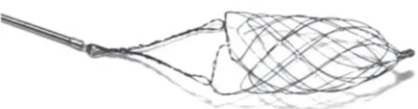
Catch device. The design of the Catch device
(Balt, Montmorency, France) nicely illustrates the idea of the distal approach. The basket-like self-ex-pandable device (Figure 3) is designed to mobilize and retrieve clot from cerebral vessels. It is available in different diameters (4 mm and 9 mm). To our knowledge, it was the first device obtaining the CE
Figure 3. The
bas-ket-like Catch device is a self-expandable mesh that can easily be delivered distally to the thrombus. The distal devices shift the site of force effect to the dis-tal surface of the thrombus.
Abbildung 3. Das
Catch-Device besteht aus einem selbstex-pandierenden Nitinol-geflecht. Es wird hinter dem Thrombus freige-setzt und übertragt die Kraft bei Zug auf den distalen Anteil des Thrombus.
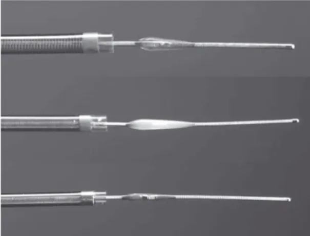
Figures 4a and 4b. The
Merci Retrieval System is available in various diameters and shapes. The corkscrew shape of the X-type (a) is slowly pulled back to encase the thrombus. The add-ed microfilaments of the L-type (b) are de-signed to reinforce this effect.
Abbildungen 4a und 4b. Das
schraubenar-tige Merci-Retrieval-System (Typ X, a) ist in verschiedenen Durch-messern erhältlich und wird langsam von dis-tal in den Thrombus gezogen, um diesen einzuhüllen. Das neue-re Model (Typ L, b) ver-fügt zusätzlich über Mikrofilamente, um diesen Effekt zu ver-stärken.
a
mark. The device has been successfully applied for thrombectomy in vitro, in vivo in animal studies and in clinical reports [32]. A registry for stroke patients treated with this device has been set up and clinical data on the recanalization rate and patients’ outcome after thrombectomy will be available in the near fu-ture.
Merci device. The Merci Retrieval System
(Con-centric Medical, Inc., Mountain View, CA, USA) is a shaped wire constructed of nitinol. The flexible cork-screw-like tip can easily be delivered through a mi-crocatheter into the vessel distally to the occlusion site. When deployed, it returns to its coiled shape to ensnare the thrombus. The thrombus is bypassed and the retriever deployed from inside the catheter distal to the thrombus. The corkscrew-like tip is pulled back while being slowly rotated to ensnare the clot as a corkscrew would ensnare a cork. The retriever is then retracted into the catheter under proximal flow ar-rest. Different versions of the device are available. The initial X-type (X6) consists of a bare-metal coil, the successor L-type applies additional filaments to increase the encasement of the clot (Figure 4). The devices are available in various diameters from 1.5 to 3 mm depending on the treated vessel.
The Merci Retrieval System (X-type) is some-how the protagonist of intracranial device develop-ment. In 2004, the Food and Drug Administration (FDA) of the USA approved its use for clot removal from intracranial vessels in patients with ischemic stroke. The device was subsequently also CE-marked. This first approval has undoubtedly accelerated the whole commercial development of mechanical thrombectomy devices in the recent years.
FDA approval was based on a review of data obtained in the multicenter Mechanical Embolus Removal in Cerebral Ischemia (MERCI) trial that involved 141 patients (mean age: 60 years, mean NIHSS: 21) ineligible for standard thrombolytic ther-apy [27]. The time window between onset of clinical symptoms and endovascular treatment was extended to 8 h, compared to the 6-h window usually aplied for IAT.
This trial reported a recanalization rate (TIMI 2 and 3) using the X-type MERCI retriever of 46%. An sICH was found in 7.8% of the patients mainly follow-ing ICA and MCA occlusions (90%). In the study, the number of attempts to retrieve the clot was limited to six; a mean of 2.9 attempts was performed for recanali-zation. The mean procedural time was 2.1 h. With re-spect to device-related complications the study report-ed vessel perforations (4.2%), subarachnoid hemor-rhages (2.1%) and embolization of thrombotic material (2.1%) [27, 28, 34].
The recently published Multi MERCI trial [35] was a prospective, multicenter, single-arm registry
that included 131 patients (mean age: 68 years, mean NIHSS: 19) treated with different Merci Retrieval Systems (X5, X6, and the novel L5). Again, the time window between onset of clinical symptoms and en-dovascular treatment was extended to 8 h and the majority of patients were treated for an ICA or MCA occlusion (92%). Patients with persistent large vessel occlusion after IVT (with rtPA) were also included in the study and adjunctive IAT using rtPA was al-lowed.
In 55% of the interventions, the mechanical thrombectomy led to recanalization (TIMI 2 and 3). After adjunctive IAT 68% of the target vessels were recanalized. Clinically significant device-related com-plications occurred in 5.5% and the rate of sICH was 9.8%. At 90 days 36% of the patients had a favorable outcome (mRS: 0–2) and the mortality rate was 34%.
Phenox device. An interesting new approach is
the Phenox clot retriever (pCR, Phenox GmbH,
Bo-Figures 5a to 5c. The Phenox pCR device (a) applies force to the distal surface of the
thrombus only by flexible polyamide microfilaments mounted on a core wire. This flexible device is applicable even in small side branches. The newly developed CRC device (b) additionally carries a self-expandable nitinol mesh on the proximal seg-ment. When applied to the vessel, as simulated in a glass tube (c), the stent-like mesh extends and increases the force on the thrombus.
Abbildungen 5a bis 5c. Das Phenox-pCR-Device (a) verfügt lediglich über
Mikrofi-lamente, um Kraft auf den distalen Anteil des Thrombus auszuüben. Dieser flexi-ble Aufbau erlaubt die Anwendung auch zur Behandlung von Verschlüssen in kleineren Seitenästen. Bei dem neueren CRC-Device (b) wurde zusätzlich proximal ein selbstexpandierendes Nitinolgeflecht aufgebracht. Im Gefäß freigesetzt, hier simuliert in einem Glasröhrchen (c), verstärkt dieses die Struktur des Device und somit die Kraftübertragung auf den Thrombus.
a
b
chum, Germany, CE-marked). The pCR device re-duces the stiff mechanical components and applies force to the distal surface of the thrombus by devel-oping a dense palisade of perpendicular-oriented polyamide microfilaments mounted on a core wire
(Figure 5a). This flexible design allows the use of two devices simultaneously (e.g., in bifurcations or larger vessels) and the microfilaments might be able to re-duce distal emboli due to a filter effect [36]. The de-vice is introduced into the target vessel through a 0.021" or 0.027" microcatheter, deployed distally to the thrombus, and slowly pulled back under continu-ous aspiration via the guiding catheter. Different di-ameters and lengths are available (diameter: 2 mm proximally, 2–5 mm distally, length: 10–20 mm). The great flexibility of devices may allow to treat more distal branches of the intracranial vasculature such as M2 and M3 branches of the MCA.
The newly developed CRC version of the device (Figures 5b and 5c) applies additional force to the thrombus by an attached proximal self-expandable nitinol cage.
Few clinical reports on the use of the device in vitro and in clinical application are available [36, 37]. A clinical trial on 55 stroke patients has just been completed and the data will be published soon. Proximal devices. From the procedural point of view, proximal devices are comparable to IAT. Access is usually gained with a 7- to 8-F sheath. After place-ment of the guiding catheter, the device is navigated to the proximal surface of the clot. Force is applied to the thrombus either by aspiration and pulling of the catheter or in combination with mechanical disrup-tion of the clot. This approach omits repetitive pass-ing of the occlusion site.
Aspiration catheters. Simple aspiration devices
are microcatheters, flexible enough to pass the tortu-osity of cranial vessels (e.g., carotid siphon) and ex-hibit a braiding that prevents collapse during aspira-tion with, e.g., a 60-ml syringe. The diameter of these devices range between 4.5 and 5.5 F and they are equipped with a blunt tip. Only a few aspiration cath-eters dedicated to intracranial occlusions are avail-able and none of them has been systematically evalu-ated in a clinical trial.
Vasco 35 Aspi microcatheter. Few clinical data is
available on the Vasco 35 Aspi microcatheter (Balt, Montmorency, France). An in vivo animal study [32] has demonstrated the feasibility of clot retrieval using this device in the setting of acute vessel occlusion. Furthermore, the mechanically simple approach, avoiding the passage of the occlusion site, seems to be less traumatic to the vessel wall. The Vasco 35 Aspi is a 5-F braided catheter available with a preshaped curved tip. It allows easy navigation through the ca-rotid siphon and has been applied for thrombus aspi-ration in the MCA and ICA (Figure 6). Furthermore, it can be used in a triaxial system to advance larger guiding catheters into the intracranial ICA for fur-ther aspiration and/or stenting [38].
Figures 6a to 6f. Complete distal M1 occlusion (➝) on the left side in a 71-year-old
female patient (a, b). Although the carotid siphon exhibits a moderate elongation, a Vasco 35 aspiration catheter (*) is advanced into the proximal M1 segment (c, d). With aspiration (60-ml syringe), the thrombus is retrieved from the vessel result-ing in a complete recanalization of the M1 segment (➩, TIMI 3, e, f).
Abbildungen 6a bis 6f. Kompletter linksseitiger M1-Verschluss (➝) bei einer
71-jährigen Patientin (a, b). Der Karotissiphon erscheint etwas elongiert, dennoch kann der Vasco-35-Aspirationskatheter (*) problemlos in das proximale M1-Seg-ment eingebracht werden. Unter Aspiration mit einer 60-ml-Spritze wird der Thrombus aus dem Gefäß entfernt und dieses somit komplett rekanalisiert (➩, TIMI 3, e, f).
a b
c d
Fragmentation and aspiration. The disadvantage
of the aspiration catheters is the fragmentation of the thrombus while applying force to the proximal sur-face of the clot. The fragment then obstructs the cath-eter and hinders further aspiration and demands re-trieval and repositioning of the catheter.
An further development of the aspiration tech-nique is the controlled fragmentation and aspiration of the debris. Various more or less mechanically com-plex devices have been tested experimentally in stroke models.
Penumbra device. The most remarkable and
promising device of this group is the Penumbra Sys-tem (Penumbra, Alameda, CA, USA). As the second device available, it has received both FDA and CE approval for use in the revascularization of patients with acute ischemic stroke.
The Penumbra System consists of a microcathe-ter attached to continuous aspiration via a dedicated pumping system. A microwire/separator with an ol-ive-shape tip is used to fragmentize the thrombus from proximally to distally (Figure 7). Both, micro-catheter and separator are available in various sizes and diameters to adjust the device to different ana-tomic settings.
A prospective, single-arm, independently moni-tored trial was performed to assess the efficiency and safety of the system [39]. A total of 23 patients (mean age: 60 years, mean NIHSS: 21) were enrolled within 8 h after onset of symptoms. Six patients included in the study were refractory to IVT. Unlike other stud-ies on thrombectomy, a large number of patients had occlusions in the posterior circulation (43%). In nine patients additional IAT was performed using rtPA. In all patients (100%) the target vessel was recana-lized (TIMI 2 or 3). The study did not exhibit any pro-cedural complications. The high mortality rate (45%) is likely related to the patient population enrolled. An sICH occurred in 15% of the patients.
The reported recanalization rate underlines the potential of this approach.
Laser recanalization and Angiojet. Laser
recana-lization and intracranial application of the Angiojet device (Possis Medical, Inc., Minneapolis, MN, USA) have been investigated experimentally. Despite the potential advantages of the proximal approach with fragmentation and aspiration, the laser and Angiojet devices are not currently approved for the intracra-nial circulation due to the rigidity of these mechani-cally complex devices.
Study groups have tested laser recanalization, in which light absorption photo energy within the cath-eter tip is changed into acoustic energy that emulsi-fies the thrombus. Berlis et al. [40] tested the efficacy and safety of this method in 34 acute ischemic stroke patients. 18 patients were treated with laser
recanali-zation alone; in these 18 patients the recanalirecanali-zation rate was 61.1%. In 16 patients treatment was rated as incomplete because of vessel tortuosity, perforation, technical failure, or stenosis. Overall, the hemorrhage rate was 5.9%, the mortality 38.2%. Laser recanaliza-tion had the decisive advantage of allowing fast re-canalization within minutes (mean application time: 9.65 min).
The Angiojet is a catheter designed to perform hemolytic thrombectomy based on the Venturi effect. In addition to the catheter, the system has a high-pres-sure water pump to pump a saline solution in a 0.5-mm metal tube to the catheter tip. The catheter lumen is located in a 4-F catheter, whose main lumen is used to house the microwire and for aspiration. At the cathe-ter tip wacathe-ter can be sprayed in small jets backward into the catheter, thus creating negative pressure that sucks in the endovascular thrombotic material and conveys it in a backflow out of the catheter. The poor flexibility of the first Angiojet catheter limited its use to segments of the large vessels supplying the brain. Nevertheless, this method has been successfully used to treat some patients with ICA occlusions or basilar artery thromboses [41]. A safety study of a smaller version of the Angiojet, designed to enable catheter-ization of the MCA, was terminated early due to ves-sel dissections. Modifications of both the catheter and
Figure 7. The Penumbra System consists of a microcatheter
for continuous aspiration and a separator, an olive-shaped corpus mounted on a flexible microwire. The manipulation of the thrombus via the proximal surface fragmentizes the thrombus. The continuous aspiration clears the debris from the vessel. The system is available in different sizes for inter-ventions in cranial vessels of various diameters.
Abbildung 7. Das Penumbra-System besteht aus einem
Mi-krokatheter zur Applikation der kontinuierlichen Aspiration und einem Separator. Durch Vor- und Zurückziehen des Se-parators wird der Thrombus fragmentiert und bei konstan-ter Aspiration aus dem Gefäß entfernt. Das System ist in Größen für verschiedene Gefäße verfügbar.
the study protocol are currently undergoing evalua-tion in a phase I trial, the Thrombectomy in Middle Cerebral Artery Embolism (TIME) study.
Other Mechanical Approaches
PTA and stent. The most recent approach adopts an angiologic/cardiologic procedure for the treatment of vessel occlusions known as percutaneous balloon an-gioplasty (PTA), sometimes in combination with stent placement. By omitting the repetitive retrieval attempts needed for the mechanical thrombectomy, this approach may reduce severe complications and thromboembolic events.
PTA achieves fast recanalization of occluded vessels, while placement of the mesh stent stabilizes the result by pinning the thrombus to the vessel wall. This combined technique has a high recanalization rate in peripheral vessel and coronary occlusions and is considered a standard procedure in these cardio-vascular territories. Therefore, it has great potential for achieving fast intracranial recanalization, since the pathophysiological background may be similar. Unlike peripheral or coronary vessels, however, in-tracranial occlusions usually represent purely throm-boembolic events without underlying stenosis. Moreover, the risks associated with intracranial in-terventions are greater, since the patient can suffer permanent neurologic damage from vessel dissection or embolism. One of the main differences with the coronary circulation is the extremely thin wall of in-tracranial vessels, which may favor vessel perfora-tion. In addition, even a small residual thrombus bur-den may jeopardize a sufficient recanalization or cause a permanent occlusion of perforating vessels. Therefore, to date combined PTA and stent place-ment have not been routinely used to treat patients with acute ischemic stroke.
An in vivo animal study evaluated the recanali-zation and complication rate of PTA and stenting in the setting of acute cerebrovascular occlusion [32]. The recanalization rate of PTA was not found to be sufficient for a stand-alone therapy in the treatment of acute ischemic stroke. Compared to stenting, the recanalization rate was significantly lower immedi-ately after application as well as during the time course of the study (3-h observation interval). Fur-thermore, PTA revealed a significantly lower and de-creasing recanalization rate over time. By contrast, stent application in combination with postdilatation is a fast and safe option to restore permanent recana-lization in the occluded vessel.
An ongoing study reports for the first time the use of intracranial stent placement in 19 patients with acute ischemic stroke in whom thrombolysis or me-chanical recanalization attempts had been
unsuccess-ful [42]. The small study employs three different bal-loon-mounted stent systems. The reported recanali-zation rate was 79%, clearly superior to any other approach. The intracranial hemorrhage rate is 5% and mortality 32%.
Avoiding typical complications of thrombolysis and mechanical thrombectomy, this approach might have a high clinical potential.
Conclusion
Patients’ outcome after acute ischemic stroke de-pends on the territory affected, the time span between onset of symptoms and revascularization, the recana-lization rate of any specific revascularization strategy, and the occurrence of intracranial hemorrhage or major procedure-related complications. At present, in most of the centers, IVT or IAT are the standard treatment, sometimes combined in a bridging con-cept.
Although being established, thrombolytic thera-py is not effective in all patients, carries a risk of intra-cranial hemorrhage, and has a limited time window for treatment. Current research therefore focuses on various mechanical revascularization techniques to achieve fast and sustained reperfusion. New-genera-tion thrombolytic drugs are believed to reduce the incidence of symptomatic intracranial hemorrhage and prolong the time window for treatment. Initial clinical results on mechanical recanalization are en-couraging, but more clinical data on the various dif-ferent devices and approaches are necessary.
The future in the treatment of acute ischemic stroke is likely to be a combination of different me-chanical and thrombolytic techniques, probably in a staged escalation concept.
Disclosure: The authors declare that they have no financial
or personal relations to other parties whose interests could have affected the content of this article in any way, either positively or negatively.
References
1. Alexandrov AV, Burgin WS, Demchuk AM, et al. Speed of in-tracranial clot lysis with intravenous tissue plasminogen activator therapy: sonographic classification and short-term improvement. Circulation 2001;103:2897–902. 2. Sobel BE. Intracranial bleeding, fibrinolysis, and
anticoagu-lation. Causal connections and clinical implications. Circu-lation 1994;90:2147–52.
3. Tissue plasminogen activator for acute ischemic stroke. The National Institute of Neurological Disorders and Stroke rt-PA Stroke Study Group. N Engl J Med 1995;333:1581–7. 4. Furlan A, Higashida R, Wechsler L, et al. Intra-arterial
pro-urokinase for acute ischemic stroke. The PROACT II study: a randomized controlled trial. Prolyse in Acute Cerebral Thromboembolism. JAMA 1999;282:2003–11.
5. Del Zoppo GJ, Higashida RT, Furlan AJ, et al. PROACT: a phase II randomized trial of recombinant pro-urokinase by direct arterial delivery in acute middle cerebral artery stroke. PROACT Investigators. Prolyse in Acute Cerebral Thrombo-embolism. Stroke 1998;29:4–11.
6. Grotta JC, Welch KM, Fagan SC, et al. Clinical deterioration following improvement in the NINDS rt-PA stroke trial. Stroke 2001;32:661–8.
7. Hacke W, Donnan G, Fieschi C, et al. Association of outcome with early stroke treatment: pooled analysis of ATLANTIS, ECASS, and NINDS rt-PA stroke trials. Lancet 2004;363: 768–74.
8. Ingall TJ, O’Fallon WM, Asplund K, et al. Findings from the re-analysis of the NINDS tissue plasminogen activator for acute ischemic stroke treatment trial. Stroke 2004;35:2418–24. 9. Fischer U, Arnold M, Nedeltchev K, et al. NIHSS Score and
arteriographic findings in acute ischemic stroke. Stroke 2005;36:2121–5.
10. Alexandrov AV, Demchuk AM, Burgin WS, et al. Ultra-sound-enhanced thrombolysis for acute ischemic stroke: phase I. Findings of the CLOTBUST trial. J Neuroimaging 2004;14:113–7.
11. Mattle HP, Arnold M, Georgiadis D, et al. Comparison of in-traarterial and intravenous thrombolysis for ischemic stroke with hyperdense middle cerebral artery sign. Stroke 2008;39:379–83.
12. Brekenfeld C, Remonda L, Nedeltchev K, et al. Symptomatic intracranial haemorrhage after intra-arterial thrombolysis in acute ischaemic stroke: assessment of 294 patients treated with urokinase. J Neurol Neurosurg Psychiatry 2007;78:280–5.
13. IMS-Study Investigators. Combined intravenous and in-tra-arterial recanalization for acute ischemic stroke: the Interventional Management of Stroke Study. Stroke 2004; 35:904–11.
14. Zeumer H, Freitag HJ, Grzyska U, et al. Local intraarterial fibri-nolysis in acute vertebrobasilar occlusion. Technical develop-ments and recent results. Neuroradiology 1989;31:336–40. 15. Hacke W, Zeumer H, Ferbert A, et al. Intra-arterial
thrombo-lytic therapy improves outcome in patients with acute ver-tebrobasilar occlusive disease. Stroke 1988;19:1216–22. 16. Becker KJ, Monsein LH, Ulatowski J, et al. Intraarterial
thrombolysis in vertebrobasilar occlusion. AJNR Am J Neu-roradiol 1996;17:255–62.
17. Jahan R. Hyperacute therapy of acute ischemic stroke: in-traarterial thrombolysis and mechanical revascularization strategies. Tech Vasc Interv Radiol 2005;8:87–91.
18. Archer CR, Horenstein S. Basilar artery occlusion: clinical and radiological correlation. Stroke 1977;8:383–90. 19. Zeumer H, Hacke W, Ringelstein EB. Local intraarterial
thrombolysis in vertebrobasilar thromboembolic disease. AJNR Am J Neuroradiol 1983;4:401–4.
20. Zeumer H, Freitag HJ, Zanella F, et al. Local intra-arterial fi-brinolytic therapy in patients with stroke: urokinase versus recombinant tissue plasminogen activator (r-TPA). Neuro-radiology 1993;35:159–62.
21. Fogarty TJ. Catheter technic for arterial embolectomy. J Car-diovasc Surg (Torino) 1967;8:22–8.
22. Kuether TA, Nesbit GM, Barnwell SL. Other endovascular treatment strategies for acute ischemic stroke. Neuroim-aging Clin N Am 1999;9:509–25.
23. Chapot R, Houdart E, Rogopoulos A, et al. Thromboaspira-tion in the basilar artery: report of two cases. AJNR Am J Neuroradiol 2002;23:282–4.
24. Nedeltchev K, Remonda L, Do DD, et al. Acute stenting and thromboaspiration in basilar artery occlusions due to
em-bolism from the dominating vertebral artery. Neuroradiol-ogy 2004;46:686–91.
25. Molyneux A, Kerr R, Stratton I, et al. International Subarach-noid Aneurysm Trial (ISAT) of neurosurgical clipping versus endovascular coiling in 2143 patients with ruptured intra-cranial aneurysms: a randomised trial. Lancet 2002;360: 1267–74.
26. Molyneux AJ, Kerr RS, Yu LM, et al. International Subarach-noid Aneurysm Trial (ISAT) of neurosurgical clipping versus endovascular coiling in 2143 patients with ruptured intra-cranial aneurysms: a randomised comparison of effects on survival, dependency, seizures, rebleeding, subgroups, and aneurysm occlusion. Lancet 2005;366:809–17.
27. Smith WS, Sung G, Starkman S, et al. Safety and efficacy of mechanical embolectomy in acute ischemic stroke: results of the MERCI trial. Stroke 2005;36:1432–8.
28. Gobin YP, Starkman S, Duckwiler GR, et al. MERCI 1: a phase 1 study of Mechanical Embolus Removal in Cerebral Isch-emia. Stroke 2004;35:2848–54.
29. Wikholm G. Transarterial embolectomy in acute stroke. AJNR Am J Neuroradiol 2003;24:892–4.
30. Gralla J, Schroth G, Remonda L, et al. A dedicated animal model for mechanical thrombectomy in acute stroke. AJNR Am J Neuroradiol 2006;27:1357–61.
31. Gralla J, Burkhardt M, Schroth G, et al. Occlusion length is a crucial determinant of efficiency and complication rate in thrombectomy for acute ischemic stroke. AJNR Am J Neu-roradiol 2008;29:247–52.
32. Gralla J, Schroth G, Remonda L, et al. Mechanical thrombec-tomy for acute ischemic stroke: thrombus-device interac-tion, efficiency, and complications in vivo. Stroke 2006;37: 3019–24.
33. Brekenfeld C, Schroth G, El Koussy M, et al. Mechanical thromboembolectomy for acute ischemic stroke: compari-son of the Catch thrombectomy device and the Merci Re-triever in vivo. Stroke 2008;39:1213–9.
34. Smith WS. Safety of mechanical thrombectomy and intra-venous tissue plasminogen activator in acute ischemic stroke. Results of the Multi Mechanical Embolus Removal in Cerebral Ischemia (MERCI) trial, part I. AJNR Am J Neuro-radiol 2006;27:1177–82.
35. Smith WS, Sung G, Saver J, et al. Mechanical thrombectomy for acute ischemic stroke: final results of the Multi MERCI trial. Stroke 2008;39:1205–12.
36. Liebig T, Reinartz J, Hannes R, et al. Comparative in vitro study of five mechanical embolectomy systems: effective-ness of clot removal and risk of distal embolization. Neuro-radiology 2008;50:43–52.
37. Henkes H, Reinartz J, Lowens S, et al. A device for fast me-chanical clot retrieval from intracranial arteries (Phenox clot retriever). Neurocrit Care 2006;5:134–40.
38. Nedeltchev K, Brekenfeld C, Remonda L, et al. Internal ca-rotid artery stent implantation in 25 patients with acute stroke: preliminary results. Radiology 2005;237:1029–37. 39. Bose A, Henkes H, Alfke K, et al. The Penumbra System: a
mechanical device for the treatment of acute stroke due to thromboembolism. AJNR Am J Neuroradiol 2008;29: 1409–13.
40. Berlis A, Lutsep H, Barnwell S, et al. Mechanical thromboly-sis in acute ischemic stroke with endovascular photoacous-tic recanalization. Stroke 2004;35:1112–6.
41. Mayer TE, Hamann GF, Schulte-Altedorneburg G, et al. Treatment of vertebrobasilar occlusion by using a coronary waterjet thrombectomy device: a pilot study. AJNR Am J Neuroradiol 2005;26:1389–94.
42. Levy EI, Ecker RD, Horowitz MB, et al. Stent-assisted intra-cranial recanalization for acute stroke: early results. Neuro-surgery 2006;58:458–63. Address for Correspondence Jan Gralla, MD Department of Diagnostic and Interventional Neuroradiology Inselspital University of Bern Freiburgstraße 20 3010 Bern Switzerland Phone (+41/31) 632-2655, Fax -4872 e-mail: jan.gralla@ insel.ch