HAL Id: hal-02158159
https://hal.archives-ouvertes.fr/hal-02158159
Submitted on 17 Jun 2019
HAL is a multi-disciplinary open access
archive for the deposit and dissemination of
sci-entific research documents, whether they are
pub-lished or not. The documents may come from
teaching and research institutions in France or
abroad, or from public or private research centers.
L’archive ouverte pluridisciplinaire HAL, est
destinée au dépôt et à la diffusion de documents
scientifiques de niveau recherche, publiés ou non,
émanant des établissements d’enseignement et de
recherche français ou étrangers, des laboratoires
publics ou privés.
alleviates peripheral neuropathic pain in mice
Cyril Rivat, Chamroeun Sar, Ilana Mechaly, J.P. Leyris, Lucie Diouloufet,
Corinne Sonrier, Yann Philipson, Olivier Lucas, Sylvie Mallié, Antoine
Jouvenel, et al.
To cite this version:
Cyril Rivat, Chamroeun Sar, Ilana Mechaly, J.P. Leyris, Lucie Diouloufet, et al.. Inhibition of neuronal
FLT3 receptor tyrosine kinase alleviates peripheral neuropathic pain in mice. Nature Communications,
Nature Publishing Group, 2018, 9 (1), pp.1042. �10.1038/s41467-018-03496-2�. �hal-02158159�
Inhibition of neuronal FLT3 receptor tyrosine kinase
alleviates peripheral neuropathic pain in mice
Cyril Rivat
1,2
, Chamroeun Sar
1,2
, Ilana Mechaly
1,2
, Jean-Philippe Leyris
1,3
, Lucie Diouloufet
1
, Corinne Sonrier
1,3
,
Yann Philipson
4
, Olivier Lucas
1
, Sylvie Mallié
1,2
, Antoine Jouvenel
1,2
, Adrien Tassou
1,2
, Henri Haton
1,2
,
Stéphanie Venteo
1
, Jean-Philippe Pin
5
, Eric Trinquet
6
, Fabienne Charrier-Savournin
6
, Alexandre Mezghrani
1
,
Willy Joly
1
, Julie Mion
1
, Martine Schmitt
4
, Alexandre Pattyn
1
, Frédéric Marmigère
1
, Pierre Sokoloff
3
,
Patrick Carroll
1
, Didier Rognan
4
& Jean Valmier
1,2
Peripheral neuropathic pain (PNP) is a debilitating and intractable chronic disease, for which
sensitization of somatosensory neurons present in dorsal root ganglia that project to the
dorsal spinal cord is a key physiopathological process. Here, we show that hematopoietic
cells present at the nerve injury site express the cytokine FL, the ligand of fms-like tyrosine
kinase 3 receptor (FLT3). FLT3 activation by intra-sciatic nerve injection of FL is suf
ficient to
produce pain hypersensitivity, activate PNP-associated gene expression and generate
short-term and long-short-term sensitization of sensory neurons. Nerve injury-induced PNP symptoms
and associated-molecular changes were strongly altered in
Flt3-deficient mice or reversed
after neuronal FLT3 downregulation in wild-type mice. A
first-in-class FLT3 negative allosteric
modulator, discovered by structure-based in silico screening, strongly reduced nerve
injury-induced sensory hypersensitivity, but had no effect on nociception in non-injured animals.
Collectively, our data suggest a new and specific therapeutic approach for PNP.
DOI: 10.1038/s41467-018-03496-2
OPEN
1Institute for Neurosciences of Montpellier, INSERM, Institut National de la Santé et de la Recherche Médicale, UMR1051, Hôpital Saint-Eloi, Montpellier
34000, France.2Université de Montpellier, Montpellier 34000, France.3Biodol Therapeutics, Cap Alpha, Clapiers 34830, France.4Laboratoire d’Innovation Thérapeutique, UMR7200, CNRS-Université de Strasbourg, Illkirch 67400, France.5Institut de Génomique Fonctionnelle, CNRS, INSERM, Univ. Montpellier,
34094 Montpellier, France.6Cisbio Bioassays, Parc Marcel Boiteux, BP8417530200 Codolet, France. These authors contributed equally: Cyril Rivat,
Chamroeun Sar, Ilana Mechaly, Jean-Philippe Leyris. Correspondence and requests for materials should be addressed to D.R. (email:rognan@unistra.fr) or to J.V. (email:jean.valmier@umontpellier.fr)
123456789
P
eripheral neuropathic pain (PNP), for which specific and
effective therapies are lacking, is a broad public health
problem particularly due to its high prevalence (estimated
to be 6.9–10% in the general population
1), debilitating effects, and
its high social cost
2,3. Currently, PNP treatments consist
essen-tially in repurposed depressant drugs (e.g., tricyclic
anti-depressants and serotonin-noradrenaline uptake inhibitors) and
anti-epileptic drugs of the class of
α2δ−1 voltage-gated calcium
channels blockers, both having poor efficacy and producing
several side effects
4. There is a crucial need for specific PNP
medications, which requires the identification of new specific
targets implicated in the initiation and maintenance of PNP.
PNP arises from aberrant functioning of somatosensory
neu-rons present in dorsal root ganglia (DRG) that project to the
dorsal spinal cord (DSC)
5,6. Environmental stimuli are converted
into voltage changes in somatosensory neurons by ionic
trans-ducer channels that respond to specific thermal, mechanical, and
chemical stimuli and activate sodium channels that generate and
propagate action potentials to the DSC. Nerve injury rapidly
induces peripheral sensitization due to reduced thresholds of both
transducers and voltage-activated channels, increasing
respon-siveness to stimuli, and axonal hyperexcitability
7,8. Within hours,
multiple adaptive modifications occur in the DRG, including gene
expression changes, post-translational protein alterations, and
modifications of protein trafficking. For example, after nerve
damage, TRPV1, a member of the TRP transducer family
9, with a
well-established role in inflammatory pain, is upregulated in
different models of PNP both at DRG peripheral and central
synapses. This upregulation is correlated with the development
and the maintenance of thermal hypersensitivity
10. In addition,
decreasing TRPV1 levels or inhibiting its activity reduces part of
the neuropathic hypersensitivity
11,12. Peripheral sensitization
leads to central sensitization in the DSC that is the cornerstone of
PNP chronification. Currently, how peripheral sensitization
develops and persists is incompletely understood.
Neuro-immune interactions are key regulators of local
per-ipheral sensitization
13,14. They are mediated by immune cells,
which invade the lesion site after nerve blood barrier
permeabi-lization, secrete sensitizers (cytokines, chemokines, growth
fac-tors) that contribute to the development of the nerve
injury-induced hypersensitivity and the maintenance of PNP
13,14.
Cytokines and their receptors have been identified as important
actors in these interactions
15. Among them, with the notable
exception of the cytokine FL
16,17and its cognate receptor FLT3
18,
all the members of the class III receptor tyrosine kinase (RTK)
family, which comprises stem cell factor (SCF) receptor (c-Kit),
colony-stimulating factor type-I (CSF1) receptor (CSF1R), and
platelet-derived growth factor (PDGF) receptors (PDGFR) have
been shown to be involved in normal nociception and/or
pain
19–22. For example, nerve injury induces de novo neuronal
expression of CSF1, which is transported to the DSC where it
targets CSF1R expressed by microglia cells
19. Microglia activation
is considered as a major factor in central PNP chronification
13,14.
However, the mechanism leading to CSF1 induction is
unidentified.
FLT3 is expressed in most hematopoietic organs, such as
spleen, thymus, peripheral blood and bone marrow, and its
gain-of-function mutation promotes hematopoietic cells proliferation
that is targeted for therapy of hematologic cancers
23. However,
Flt3
KOmice have a normal number of mature hematopoietic cells
in all hematopoietic organs, even though some discrete
popula-tions of cell progenitors, but not more mature cells, are reduced
24.
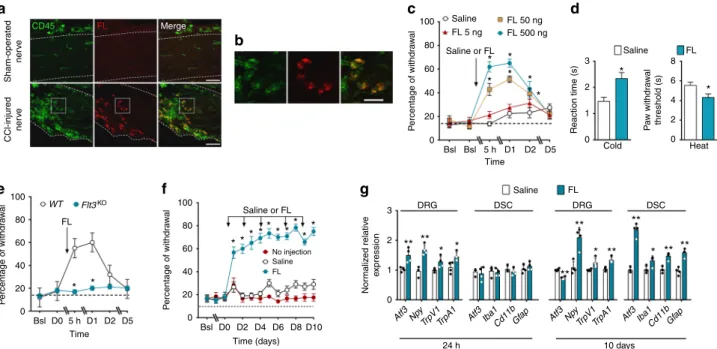
Sham-oper ated ner v e P e rcentage of withdr a w al Nor maliz ed relativ e e xpression P e rcentage of withdr a w al P e rcentage of withdr a w al Reaction time (s) P a w withdr a w al threshold (s) 100 Saline FL 5 ng FL 50 ng FL 500 ng Saline or FL Saline or FL Saline Saline DRG ** ** ** ** ** ** ** * * ** * * * * * * * * * * * * * * DRG DSC DSC * * * * * * * * 80 60 40 20 2 2 1 0 0 4 6 8 3 0 100 80 60 40 20 0 100 3 2 1 0 24 h 10 days Atf3 Npy TrpA1
TrpV1 Atf3 Iba1Cd11bGfap Atf3 Np y TrpA1 TrpV1 Atf3 Iba1Cd11bGfap 80 No injection Saline FL 60 40 20 0 Bsl FL WT Flt3KO Bsl 5 h D1 D2 D5 Bsl Bsl D0 D0 D2 D4 D6 D8 D10 5 h D1 D2 D5 Time Time (days) Cold Heat Time CCl-injured ner v e FL FL
a
b
c
d
e
f
g
CD45 FL MergeFig. 1 Infiltration of CCI-injured nerve by CD45/FL-positive cells and effects of intrathecal FL injections on pain sensitivity and PNP-related biomarkers. a CD45- and FL-positive cells at the site of nerve injury in the sciatic nerve, delineated by dotted lines, 10 days post-CCI. Scale bars= 50 µm. b Enlargement of the dotted square areas ina. Bars= 20 µm. c Mechanical pain sensitivity, measured as the percentage of withdrawal to application of von Frey filament, 5 h (5 h) to 5 days (D5) after a single intrathecal saline or different doses of FL injection (concentrations in ng/10µl). Bsl basal score before injection. d Cold and heat thermal sensitivity 24 h after intrathecal saline or FL injection (500 ng).e Mechanical sensitivity after a single intrathecal injection of FL (500 ng/10µl) in wild-type (WT) or Flt3-deficient (Flt3KO) mice.f Mechanical pain sensitivity after repeated intrathecal injections of saline or FL at the dose of
(500 ng/10µl), measured 1–10 days after FL injections performed on days 1, 2, 4, 7, and 9. Control animals received no injections. g Normalized gene expressions of PNP biomarkers in the DRG and DSC measured by q-PCR 24 h after a single saline or FL injection (500 ng/10µl) and 10 days after repeated injections as inf. Results are means ± s.e.m. of data from 8 animals c–f or 4 animals (g). Two-way ANOVA and Dunnett’s test (c, e, f). Unpaired Student’s t-test (d, g).*P < 0.05; **P < 0.01 vs. saline or WT or no injection
Flt3 transcripts are expressed in various regions of the nervous
system, in particular in human and mice DRGs
25–28. FLT3
modulates the in vitro survival of embryonic mice DRG
neu-rons
28but its role in the adult nervous system is not known. Here
we demonstrate that neuronal FLT3 in DRG is a critical actor for
PNP initiation and maintenance in different mouse models. In
addition, we show that PNP can be alleviated by specific FLT3
inhibition using a new chemical entity, thus identifying a novel
therapeutic strategy for PNP treatment.
Results
FL induces PNP-related behavioral and molecular changes. We
used the chronic constriction injury (CCI) model in mice as a
model of persistent PNP, consisting in three chronic ligatures tied
loosely around the sciatic nerve
29. On longitudinal sections of
sciatic nerve 10 days after nerve injury, intense FL
immunor-eactivity was present in the nerve at the site of injury in CCI mice,
whereas it was absent in normal, sham-operated nerve. All of the
FL-positive cells also expressed CD45, a marker of the immune
hematopoietic lineage
30(Fig.
1
a, b; Supplementary Fig.
1
a).
Further analysis showed that 60% of the FL-positive cell
popu-lation express CD11b, a marker of myeloid cells, but not CD68, a
marker of macrophages (Supplementary Fig.
1
b, c). No
co-localization was found with CD3, a marker of T lymphocytes
(Supplementary Fig.
1
d). Therefore, FL-expressing cells that
penetrate the nerve at the lesion site can be identified as
mono-cytes, neutrophils, and/or natural killer cells but not macrophages
or T lymphocytes.
To assess whether FL could participate in the generation of
PNP symptoms, uninjured mice were injected intrathecally with
recombinant FL and their sensitivity to mechanical stimulation of
the hind-paw was measured. A single FL injection induced a
dose-dependent increase in the percentage of paw withdrawal to a
calibrated von Frey
filament (Fig.
1
c), i.e., mechanical
hypersen-sitivity, a hallmark of PNP
31, which was present 5 h post-injection
and persisted for at least 2 days. In a separate experiment, the
onset of FL-induced mechanical pain hypersensitivity was
determined to be 120 min: the percentage of paw withdrawal
after an intrathecal injection of 500 ng of FL were (means ± s.e.m.,
n
= 8) 31.3 ± 4.5, 18.8 ± 2.6 and 51.3 ± 3.2% at 30, 90 and 120 min
post-FL injection, respectively, whereas those after saline injection
were 30.0 ± 3.7, 21.3 ± 3.4, and 21.3 ± 3.9% (P < 0.0001 for
comparison of FL vs. saline at 120 min by two-way ANOVA,
not significant at other time points).
FL-injected mice also displayed thermal hypersensitivity in
response to cold and hot stimuli (Fig.
1
d). Mice with a
homozygous deletion of Flt3 (Flt3
KOmice) failed to develop
mechanical hypersensitivity following FL injection (Fig.
1
e),
whereas they displayed normal proportions of sensory neurons
expressing typical molecular markers, motor behaviors, and
pain responses to nociceptive stimuli (Supplementary Fig.
2
a, b).
This indicates that FL-induced hypersensitivity resulted from
activation of its cognate FLT3 receptor. Repeated injections
of FL every 2 days during 10 days maintained mechanical
hypersensitivity that persisted as long as the treatment continued
(Fig.
1
f).
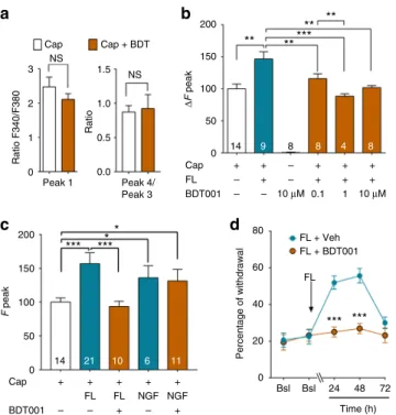
1.5 1.0 300 *** *** *** *** *** *** 200 100 0 Cap FL Cap FL 5.6 0.056 0.56 5.6 nM WT Flt3KO + + + + + + + + – – + + + + + + – – 17 17 8 8 8 14 7 14 11 0.5 Ratio F340/F380 Δ F peak 300 250 TRPV1 TRPA1 TRPM8 *** ** ** 200 150 0.0 8 Amplitude P3/P2 Duration of responses (s) 7 0.5 1.0 1.5 2.0 100 80 60 40 NS NS 20 0 0 30 min 3 h 24 h Time *** * * 100 50 0 Cap Cap + FL Menthol + FL Cina + FL Cina Menthol 200 100 0 Δ F peak Δ F peak 0 0 200 400 600 Time (s) 14 15 19 21 5 7 CapCap Cap Cap
Cap Cap Cap+FL 3 X Cap 4 X Cap 500 pA 1 min Cap
Cap Cap Cap+FL
Cap+FL Cap +FL Peak 3 Peak 2 Peak 1 Peak 4
a
b
c
d
e
f
g
Fig. 2 FL potentiates TRP function in vitro and in vivo. a Traces of [Ca2+]iresponses to repeated bath applications of capsaicin (Cap, 2.5µM), a selective TRPV1 activator, or capsaicin combined with FL (0.56 nM) on [Ca2+]ilevels measured by real-time Ca2+cell imaging in cultured DRG neurons. Ratio F340/F380 is the ratio offluorescence signals measured at 340 and 380 nm excitation wavelengths. b Effects of capsaicin (Cap, 2.5 µM) or FL at the indicated concentrations, either alone or in combinations on [Ca2+]ilevels, measured at the fourth application of capsaicin as ina. Results are expressed as response amplitudes normalized to capsaicin alone (ΔF peak). c Effects of FL on capsaicin-induced increased [Ca2+]ilevels, measured as ina, in DRG neurons from wild-type (WT) orFlt3KOmice.d Potentiation by FL (0.56 nM) of increase in [Ca2+]ilevels induced by capsaicin (Cap, 2.5µM),
cinnamaldehyde, Cina (50µM), and menthol (1 µM). e Traces of voltage-clamp whole-cell recording of capsaicin (Cap, 1 µM)-induced TRPV1 currents and their potentiation by FL (10 nM).f Quantification of FL effects measured as in e. g In vivo potentiation by FL of capsaicin-induced pain-related behaviors. Animals received either 4 repeated identical injections of capsaicin (15 ng) or a combination of Cap and FL (2 ng) followed by 3 repeated capsaicin injections in the paw, at the indicated times. Results are means ± s.e.m. of data from the number of cells indicated in columns (b–d, f) or data from 8 animals (g). Unpaired Studentt-test. NS non-significant; *P < 0.05; **P < 0.01; ***P < 0.001
We next examined changes in nerve injury-associated genes in
DRG and DSC. Twenty-four hours after a single FL injection,
expression levels in DRG of the stress-induced gene transcript
Atf3
32and several important neuronal pain-related gene
transcripts, e.g., neuropeptide Y (NpY)
33and transient receptor
potentials TrpV1
34and TrpA1
35were increased (Fig.
1
g).
Expression levels of PNP-associated gene transcripts in the DSC
at this time point (24 h) showed no change compared to
saline-injected animals (Fig.
1
g). In contrast, repeated injections of FL
over 10 days caused striking changes in the DSC expression of
genes associated with the process of central pain sensitization
36,
notably the activated microglia markers Iba1 and Cd11b
37and
the activated astrocyte marker Gfap
38(Fig.
1
g). Thus, FLT3
activation causes, in the short-term, upregulation of PNP-related
genes in the DRG, and in the long-term, molecular changes in the
DSC typical of those occurring during chronification of PNP.
In cultured DRG neurons, application of capsaicin, a specific
TRPV1 agonist, increased intracellular Ca
2+levels ([Ca
2+]
i) and
repeated capsaicin applications attenuated TRPV1 responses,
which reflects receptor desensitization (Fig.
2
a)
39. FL alone had
no effect on basal [Ca
2+]
i(Fig.
2
b), but markedly potentiated, in a
concentration-dependent manner, the TRPV1 response to
repeated capsaicin applications (Fig.
2
b). Although the capsaicin
response was normal in cultures established from Flt3
KOmice,
the potentiating effect of FL was completely abolished (Fig.
2
c).
Similarly to its effect on TRPV1 function, FL potentiated both the
[Ca
2+]
iresponse to cinnamaldehyde and menthol, specific
activators of TRPA1 and of TRPM8 channels, respectively
(Fig.
2
d). Voltage-clamp whole-cell recording confirmed that
capsaicin-induced TRPV1 currents were effectively potentiated by
FLT3 activation (Fig.
2
e, f). Furthermore, hind-paw FL injection
also potentiated capsaicin-induced spontaneous pain-related
behaviors (Fig.
2
g).
Thus, acute and chronic in vivo FLT3 activation by FL
recapitulates in mice some of the molecular and functional
changes in sensory neurons and behavioral alterations that are
normally induced by peripheral nerve injury. Altogether, these
data strongly suggest that FL directly acts on primary sensory
neurons. Indeed, injection of FL directly into the sciatic nerve, but
not systemic FL augmentation by intravenous injection
(Supple-mentary Fig.
3
a, b), caused mechanical hypersensitivity, showing
that systemic peripheral activation of FLT3 is not involved in pain
behavior. This indicates that FLT3 triggering PNP-like symptoms
is present in the nerve and/or DRG. In agreement with this
observation, Western blot detected FLT3 in DRG tissue from WT,
but not Flt3
KOmice, (Supplementary Fig.
3
c) and Flt3 mRNA was
visualized in DRG neurons by in situ hybridization
(Supplemen-tary Fig.
3
d).
Neuronal FLT3 controls PNP development and maintenance.
Considering the similarities in the effects of FLT3 activation and
those induced by peripheral nerve injury, we then asked whether
downregulation of FLT3 functioning could influence the
mole-cular, cellular, and behavioral responses to nerve injury. In the
CCI model, injury of the sciatic nerve produced mechanical
hypersensitivity that lasted for more than 2 months in WT mice,
whereas Flt3
KOmice failed to develop pain-related behavior over
the same period (Fig.
3
a). Note that the repetition of painful
mechanical stimulus applications is unlikely to produce
sensiti-zation or tolerance since the maximal change in paw withdrawal
threshold was achieved after 2 weeks and was maintained over
time until the end of the experiment. The heat hypersensitivity
and mechanical allodynia observed in WT mice after CCI were
also absent in Flt3
KOmice (Fig.
3
b, c). The conditioned place
preference paradigm has been used to reveal the presence of
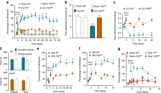
non-100 Sham WT WT SNI WT SNI Flt3KO SNI WT SNI Flt3KO Saline WT Oxal. WT Saline Flt3KO Oxal. Flt3KO Sham
or CCI Nerveinjury
Nerve injury Nerve injury Saline or oxal. CCI WT CCI Flt3KO Flt3KO Sham Flt3KO Sham WT CCI WT
CCI WT CCI Flt3KO CCI Flt3
KO Sham Flt3KO 80 60 15 10 2.5 2.0 1.0 1.5 0.5 0.0 0 5 10 15 20 Time (days) 0 5 10 15 Time (days) 0 –4 0 4 8 12 16 20 24 5 10 15 Time (days) Time (days) 5 0 * * * * * * * * * * * * * * * * * * * * * * *** * * * * * * * * * * * 40 Percentage of withdrawal
Paw withdrawal threshold (s)
Paw withdrawal threshold (g)
Percentage of withdrawal Percentage of withdrawal Percentage of withdrawal 20 0 100 80 60 40 20 0 100 80 60 40 20 0 100 80 60 40 20 0 0 200 400 600 0 200 400 600 Pretest Test Time (s) Time (s) 0 8 16 24 32 40 48 56 64 72 Time (days) Clonidine-paired Vehicle-paired 80
a
b
c
d
e
f
g
Fig. 3 FLT3 is critical for the development of pain hypersensitivity. a–g Flt3-knockout (Flt3KO), compared to wild-type (WT) animals, showed marked reductions of hypersensitivity to mechanical nociceptive stimuli as measured by the von Frey test (a), thermal stimuli as measured via the Hargreaves test 14 days post-injury (b) and punctate tactile stimuli (c), and of conditioned place preference induced by clonidine-evoked analgesia 14 days post-injury (d).Flt3KOmice also showed decrease of mechanical pain hypersensitivity in the spared nerve injury (SNI) model (e), in the sciatic nerve ligature (SNL) model (f) and in the oxaliplatin model of generalized peripheral neuropathic pain (g). All the values are means ± s.e.m. (n = 8 except in d, n = 11). Two-way ANOVA and Dunnett’s test (a–c, f); Student’s t-test (d). Two-way ANOVA and Bonferroni’s test (e, g), *P < 0.05; ***P < 0.001 vs. Sham WT, Sham CCI, CCIFlt3KO, or vehicle paired
evoked ongoing pain in nonverbal animals
40. Administration of
the non-rewarding and rapidly-acting analgesic drug clonidine,
referred to as clonidine-induced analgesia, produced a place
preference in wild-type CCI animals, which was totally abolished
in Flt3
KOCCI mice (Fig.
3
d), suggesting a loss of non-evoked
ongoing pain in Flt3
KOmice. Furthermore, the results on nerve
injury-induced mechanical hypersensitivity could be extended to
different PNP models, the spared nerve injury (SNI)
41and spinal
nerve ligation (SNL)
42models, and also to the oxaliplatin model
of chemotherapy-induced generalized PNP (Fig.
3
e–g).
Never-theless, Flt3
KOmice have normal responses to
chemically-induced nociception in the formalin test and in the complete
Freund’s adjuvant (CFA) chronic inflammatory pain model
(Supplementary Fig.
4
a, b).
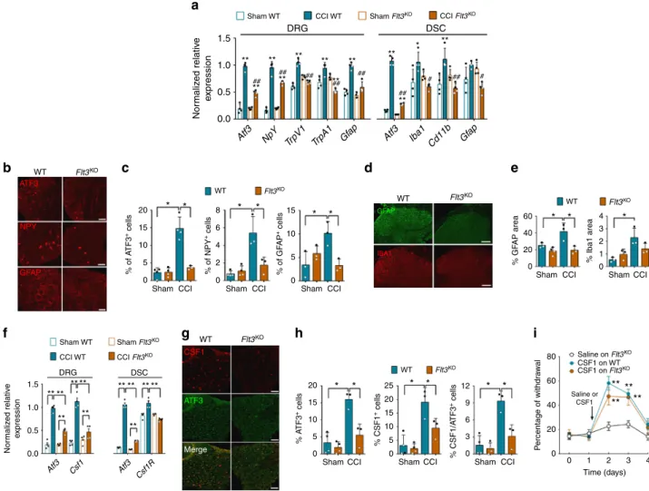
We next assessed whether the deletion of Flt3 could modulate
the expression of pain-related factors induced by nerve injury
involved in peripheral and central sensitization responsible for
PNP chronification
14. The injury-induced increases in transcripts
of Atf3, NpY, TrpV1, and Gfap in DRG and Iba1, Cd11b in DSC
were abrogated or diminished after Flt3 deletion, at 3 days
post-CCI (Fig.
4
a). These variations were confirmed by
immunochem-istry analysis, showing that, in DRG, Flt3 deletion inhibited the
injury-induced increases in ATF3, NPY, and GFAP, seen in WT
mice at 14 days post-CCI (Fig.
4
b, c). In DSC, Flt3 deletion also
diminished nerve injury-induced changes of astrocyte (GFAP)
and a similar trend was seen for microglial (IBA1) activation
markers (Fig.
4
d, e). Flt3 deletion in CCI mice attenuated the
upregulation of Csf1 ligand transcript in DRG and Csf1R in DSC
(Fig.
4
f). These results were confirmed by the quantification of
reduced numbers of DRG CSF1-positive cells using
immunohis-tochemistry (Fig.
4
g, h). Finally, intrathecal CSF1 injection
induced mechanical hypersensitivity in both WT and Flt3
KOmice
(Fig.
4
i), suggesting that FLT3 acts upstream of CSF1/CSF1R, a
major signaling pathway in neuropathic pain chronification
19.
Downregulation of
Flt3 expression reverses established PNP.
To determine whether PNP-like symptoms, once established,
could be reversed by Flt3 downregulation, we injected
Sham WT DRG 1.5 Nor maliz ed relativ e e xpression Nor maliz ed relativ e e xpression 1.0 ** ** ** ** ** ** ** ** ** ** * ## ## ## ## ## 0.5 0.0 WT ATF3 NPY GFAP IBA1 CSF1 ATF3 Merge GFAP 20 * * * * * * * * * 15 10 5 % of A TF3 + cells % of NPY + cells % of GF AP + cells 0 20 * * * * * * 20 15 10 5 % A TF3 + cells % CSF1 + cells 0 15 25 10 12 9 80 Saline on Flt3KO CSF1 on WT Saline or CSF1 ** ** ** ** CSF1 on Flt3KO 60 P e rcentage of withdr a w al 40 20 0 0 1 2 3 4 Time (days) 6 3 0 % CSF1/A TF3 + cells 5 0 0 8 15 10 20 40 60 0 % GF AP area 5 0 6 4 4 3 2 1 0 % Iba1 area 2 Sham Sham WT Sham Flt3KO CCI Flt3KO CCI WT DRG 1.5 1.0 0.5 0.0 Atf3 Csf1 Atf3 Csf1R ** ** ** ** ** ** ** ** ** ** ** DSC
CCI Sham CCI Sham CCI
Sham CCI Sham CCI Sham CCI
Sham CCI Sham CCI
Flt3KO WT Flt3KO WT Flt3KO WT Flt3KO WT Flt3KO WT Flt3KO Atf3 NpY TrpV1 TrpA1 Gf ap Atf3 Iba1 Cd11b Gf ap DSC
CCI WT Sham Flt3KO CCI Flt3KO
a
b
c
d
e
f
g
h
i
## # ## ** #Fig. 4 FLT3 is critical for upregulation of markers associated with peripheral nerve injury. a Changes in PNP-related biomarkers mRNA expression, measured by q-PCR 3 days after sham or CCI surgery in wild-type (WT) orFlt3-knockout (Flt3KO) mice. Values are means ± s.e.m of data from 4 animals. One-way ANOVA and Bonferroni’s test, *P < 0.05; **P < 0.01 vs. respective Sham,#P < 0.05;##P < 0.01 vs. CCI WT. b ATF3, NPY, and GFAP
immunoreactivity in DRG from WT andFlt3KOCCI mice, 14 days post-CCI. Scale bars= 100 µm except for GFAP: 50 µm. c Quantification of experiments as inb. d GFAP and IBA1 immunoreactivity in DSC from WT andFlt3KOCCI mice. Scale bars= 100 µm. e Quantification of experiments as in d. f Normalized gene expressions ofCsf1 and Csf1R measured by q-PCR in DRG and DSC from WT and Flt3KOsham or 3 days post-CCI mice. Means ± s.e.m. of
data from 4 animals. One-way ANOVA and Bonferroni’s test, *P < 0.05; **P < 0.01 vs. respective Sham,#P < 0.05;##P < 0.01 vs. CCI WT. g CSF1 and ATF3 immunoreactivity in DRG from WT andFlt3KOCCI mice at 3 days post-injury. Bars= 100 µm. h Quantification of experiments as in g. i CSF1-induced mechanical pain hypersensitivity in WT andFlt3KOmice. Means ± s.e.m. of data from 8 animals.c, e, f, h, i NS non-significant; unpaired Mann–Whitney
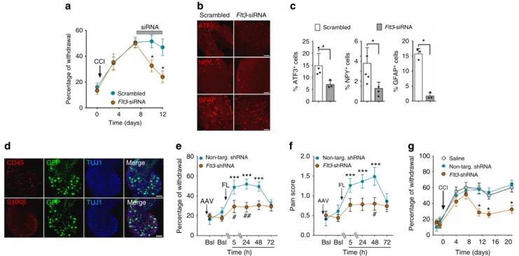
intrathecally Flt3-directed siRNA in CCI mice via mini-pumps
during a 6-day period starting 8 days after the nerve injury.
Flt3-siRNA (but not scrambled Flt3-siRNA) effectively down-regulated Flt3
expression in vitro and FLT3 function ex vivo (Supplementary
Fig.
5
a–c), as well as CCI-induced mechanical hypersensitivity
(Fig.
5
a), without affecting normal mechanical nociception
(Supplementary Fig.
5
d). These changes were accompanied by
reductions of PNP-related mRNAs (Supplementary Fig.
5
e) and
protein levels, as shown by reductions in numbers of cells
expressing ATF3, NPY, and GFAP in the DRG (Fig.
5
b, c). To
determine whether neuronal FLT3 in DRG is necessary and
sufficient to regulate PNP symptoms, we constructed an AAV9
virus vector co-expressing an Flt3 shRNA (Flt3-sh) and the green
fluorescence protein (GFP). When injected intrathecally,
virus-derived GFP expression in DRG was restricted to neurons
(Fig.
5
d) and in DSC appeared only in
fiber-like processes, most
likely originating from sensory neuron projections. GFP
expres-sion was not present in DSC cell bodies (Supplementary
6
a, b).
Following reduction of FLT3 protein levels in the DRG after
treatment with the Flt3-directed shRNA-expressing AAV9 virus
(Supplementary Fig.
6
c, d), intrathecal FL injections failed to
produce evoked mechanical pain hypersensitivity (Fig.
5
e), as well
as increase pain score, which takes into account pain-related
behaviors (Fig.
5
f), thus showing that the effect of FL is exerted
directly via neuronal FLT3. Furthermore, Flt3-directed shRNA
largely reduced percentage of withdrawal in CCI mice, whereas an
AAV9 virus expressing a non-targeting shRNA had no effect
(Fig.
5
g). Thus, inhibition of Flt3 expression reduced mechanical
pain hypersensitivity produced by either FL injection or nerve
injury. Altogether, these results show that PNP symptoms
induced by peripheral nerve injury are mediated by FLT3
expressed in DRG neurons and that PNP, once established, can be
reversed by acute downregulation of Flt3 gene expression.
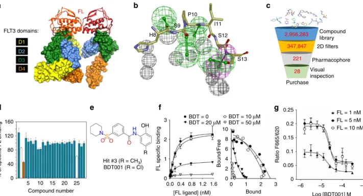
BDT001, an FLT3 inhibitor, reverses PNP symptoms. The
X-ray structure of human FL-FLT3 complex
23,43(Fig.
6
a) was used
to design a pharmacophore recapitulating all molecular
interac-tions between the FLT3 D3 domain and the N-terminal part
(H8-S13) of the FL ligand. Owing to the surprisingly compact
FL-binding epitope (FL-FLT3 interface area of 749 Å
2), the FL–FLT3
interaction pharmacophore (Fig.
6
b) is simple enough to be
fitted
by small molecular weight compounds. In silico screening of ca. 3
million commercially available compounds afforded 221 unique
compounds fulfilling the pharmacophore, out of which 28
che-mically representative hits (Fig.
6
c; Supplementary Table
1
) were
selected for purchase.
One compound (compound Hit #3, Fig.
6
d, e) effectively
prevented extracellular FL binding to FLT3, as measured by
time-resolved
fluorescence resonance energy transfer (trFRET, see
Supplementary Fig.
7
a, b for binding assay validation). Among
the 21 commercial and close structural analogs of compound 3
(Supplementary Table
2
), seven compounds were more potent
than parent compound 3 in the competition binding assay
(Supplementary Fig.
8
a). Notably compounds 66 and 75 inhibited
FL binding to FLT3 with IC
50values of 11 and 17 µM,
respectively (Supplementary Fig.
8
a). Binding affinities of the 21
analogs permitted to establish preliminary structure–activity
relationships (SAR) on the chemical series (Supplementary
Fig.
8
b). After in-house synthesis (Supplementary Fig.
9
a),
siRNA ATF3 NPY GFAP 60 40 Percentage of withdrawal
Percentage of withdrawal Percentage of withdrawal
CCI Scrambled Flt3-siRNA Scrambled Flt3-siRNA Scrambled 25 6 4 2 0 * * * 20 % ATF3 + cells % NPY + cells % GFAP + cells 15 10 5 0 20 15 10 5 0 Flt3-siRNA * * 20 0 0 4 8 12 80 60 2.0 1.5 100 80 60 40 20 0 0 4 8 12 16 20 Time (days) 1.0 Pain score 0.5 0.0 40 20 0 Bsl Bsl 5 24 48 72 Time (h) Bsl Bsl 5 24 48 72 Time (h) Non-targ. shRNA *** *** *** * * * *** *** *** # ## # FL FL AAV AAV CCI Flt3-shRNA Non-targ. shRNA
Flt3-shRNA Non-targ. shRNA
Flt3-shRNA Saline Time (days) CD45 S100β GFP GFP TUJ1 TUJ1 Merge Merge
a
b
c
d
e
f
g
Fig. 5 Curative effects ofFlt3 downregulation on PNP-related genes, and nerve injury-induced and FL-induced pain hypersensitivity. a Mechanical pain hypersensitivity after intrathecal infusion for 4 days of scrambled- orFlt3-siRNA in CCI mice. Means ± s.e.m. of data from 8 animals. Two-way ANOVA and Dunnett’s test, *P < 0.05 vs. Scrambled siRNA animals. b ATF3, NPY, and GFAP immunoreactivity in lumbar DRG after intrathecal infusion for 4 days of scrambled- orFlt3-siRNA in CCI mice. Bars = 100 µm except for GFAP: 50 µm. c Quantification of experiments as in b. Means ± s.e.m. of data from 4 animals. Mann–Whitney t-test, *P < 0.05. d Localization of the AAV9 virus expressing anti-Flt3 shRNA and GFP in neurons (TUJ1-positive cells), but not in CD45- or S100β-positive cells in DRG of WT mice. Scale bars = 100 µm. e, f Effects of Flt3-shRNA on FL-induced mechanical pain hypersensitivity (e), and on pain-related behaviors (f). FL (500 ng/10µl) was injected intrathecally 17 days after the virus. Means ± s.e.m. of data from 8 animals. One-way ANOVA and Dunnett’s test, *P < 0.05; **P < 0.01; ***P < 0.001 vs. Bsl; two-way ANOVA with repeated measures and Dunnett’s test,#P < 0.05 vs. non-targeting shRNA.g Effects ofFlt3-shRNA on mechanical pain hypersensitivity in CCI mice. The virus was injected intrathecally 48 h before CCI. Means ± s.e.m. of data from 8 animals. Two-way ANOVA with repeated measures and Dunnett’s test, *P < 0.05 vs. non-targeting shRNA
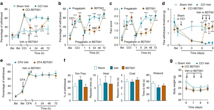
compound 66 (hereafter referred to as BDT001, Fig.
6
e) was
confirmed as a true FLT3 inhibitor, which inhibited FL binding in
a non-competitive manner and disrupted positive cooperativity of
FL binding (Fig.
6
f). BDT001 inhibited FL-induced FLT3
phosphorylation in leukemia-derived RS4–11 cells, also measured
by trFRET (see Supplementary Fig.
7
a, b for phosphorylation
assay validation) with an IC
50of 18–24 µM that was almost
unchanged with increasing FL concentrations (Fig.
6
g). These
results suggest that BDT001 is an FLT3 negative allosteric
modulator.
In primary cultures of adult DRG neurons, BDT001 affected
neither capsaicin-induced TRPV1 activation (Fig.
7
a) nor basal
[Ca
2+]
i(Fig.
7
b), but reversed in a dose-dependent manner the
potentiation by FL, with a maximal effect at 1 µM, and a partial
effect at 0.1 µM (Fig.
7
b). Thus, the effective BDT001
concentra-tion required for funcconcentra-tional inhibiconcentra-tion of FLT3 in neurons was
significantly lower than that required for inhibiting FL binding
and FL-induced FLT3 auto-phosphorylation in RS4–11 cells.
Among the rare examples of extracellular RTK inhibitors, two
similar situations have already been encountered (Supplementary
Note
1
). The effect of BDT001 was FLT3-dependent, as the
compound at 10 µM had no effect when TRPV1 potentiation was
induced by NGF (Fig.
7
c).
After systemic administration of BDT001 (5 mg/kg i.p.) in
mice, FL-induced mechanical hypersensitivity was completely
abrogated (Fig.
7
d). In the CCI model, a single injection of
BDT001 (5 mg/kg i.p.) reversed mechanical hypersensitivity
for 2 days (Fig.
8
a). As compared to pregabalin, a standard of
care for PNP, BDT001 produced longer effects when considering
either paw withdrawal thresholds (Fig.
8
b) or pain-related
behaviors (Fig.
8
c). Repeated injections of BDT001 every day
during 3 days fully reversed mechanical allodynia as long as the
treatment continued (Fig.
8
d). In agreement with the data on
Flt3
KOmice, BDT001 did not change CFA-induced inflammatory
mechanical
pain
hypersensitivity
(Fig.
8
e).
Neither
sensory–motor functions in naive mice (Fig.
8
f) nor body weight
in nerve-injured mice (Fig.
8
g) were affected by single and
repeated BDT001 (5 mg/kg i.p.) injections during 4 days,
respectively. In a preliminary pharmacokinetics study, the
BDT001 plasma level reached 0.19 ± 0.06 µM (N
= 3), 30 min
after administration at a dose of 5 mg/kg i.p.
Tested at a concentration of 10 µM, BDT001 shows a high
selectivity for the FLT3 kinase as neither binding to 25 diverse
RTKs (Supplementary Table
3
) nor inhibitory functional
effects on 49 intracellular kinases (Supplementary Table
4
)
could be demonstrated, with the exception of Interleukin-1
receptor-associated kinase 4 (IRAK4), for which a moderate
binding (64% inhibition of staurosporine binding at 10 µM)
was detected. Apart from a poor metabolic stability
(Supplemen-tary Fig.
9
b), BDT001 exhibits promising properties suggesting
that the clinical development of close analogs is indeed
feasible.
FLT3 domains: H8 S9 P10 I11 S12 S13 Compound library 2D filters Pharmacophore Visual inspection Purchase 160 120 % of specific FL binding 80 40 0 5 10 15 20 25 Compound number Hit #3 (R = CH3) OH 3 3 –6 –5 –4 –3 Log [BDT001] M 10 8 6 4 2 0 0 0 BDT = 0 BDT = 20 μM BDT = 10 μM 0.25 FL = 1 nM FL = 5 nM FL = 10 nM 0.2 0.15 0.05 0.1 BDT = 50 μM 2 2FL specific binding Bound/Free
Ratio F665/620 1 1 0 0.0 0.4 0.8 1.2 1.6 [FL ligand] (nM) Bound R H N N S O O O BDT001 (R = CI) 28 221 347,847 2,956,283 FL D1 D2 D3 D4
a
b
c
d
e
f
g
Fig. 6 Identification of BDT001 as an extracellular FLT3 inhibitor. a X-ray structure of human FL–FLT3 complex. The FL dimer is represented by ribbons (monomer 1,firebrick; monomer 2, redorange) and the FLT3 extracellular D1-D4 domains by solid surfaces (D1, yellow; D2, blue; D3, green; D4, orange). The region delineated in black represents the targeted FL/FLT3 interface.b FL–FLT3 interaction pharmacophore, comprising four hydrogen-bond acceptors (green), one hydrogen-bond donor (magenta) and 12 exclusion spheres (gray) placed at FLT3 key side chain atoms.c Virtual screeningflow-chart. Three million commercially available compounds werefirst filtered according to simple molecular counts and 2D properties and converted into 3D structures (Supplementary Methods). Remaining 347,847 compounds werefitted to the pharmacophore and the best 221 hits selected. Overall, 28 representative chemically diverse hits werefinally selected after clustering and visual inspection. d Hit experimental validation by inhibition of Red-FL binding to Lumi4-Tb-SNAP-FLT3 in HEK293 cells. Compounds were tested at 100µM. Compounds #2 and #25 altered the fluorescence emission of the donor at 620 nm. Results are means ± s.e.m. of quadruplicate determinations in a single experiment.e Chemical structure of hit #3 and BDT001. f Saturation curves of FL binding in the presence of BDT001 (BDT) at the indicated concentrations (left) and Scatchard plots showing non-competitive inhibition and positive cooperativity of FL binding, the latter was lost in the presence of BDT001 at 20 and 50µM (right). g BDT001 inhibited FL-induced FLT3 auto-phosphorylation in RS4–11 cells with IC50values of 18, 24, and 22µM at FL concentrations of 1, 5, and 10 nM, respectively. In f, g results are means ± s.e.m. of quadruplicate determinations in a single experiment, which was repeated once with similar results
Discussion
Using different approaches aimed at selectively activating or
inhibiting neuronal FLT3 signaling, we disclose a novel role for
neuronal FLT3 as a master hub protein mediating neuro-immune
interactions known to be crucial in PNP physiopathology
13.
Indeed, disruption of the blood–nerve barrier produced by nerve
injury seems to be responsible for the presence of CD45 positive
FL-expressing hematopoietic cells at the site of nerve lesion. We
demonstrate that sensory neuron FLT3 is necessary and sufficient
for the development and maintenance of the PNP state.
Activa-tion of FLT3 by exogenous FL in mice mimics many aspects of
nerve injury-induced PNP, i.e., mechanical and thermal
hyper-sensitivity, pain-related behaviors, rapid upregulation and
potentiation of TRP family transducers in the DRG (likely
involving phosphorylation
7) that may lead to neuron
hyper-excitability, and delayed transcriptional modifications in the DSC
that have been reported to be associated with central pain
sen-sitization. Disruption of FLT3 functioning by various means, e.g.,
gene deletion, small interfering RNAs, an AAV9 virus vector
expressing Flt3 shRNA into neurons or a newly designed
inhi-bitor, reverse changes associated with PNP, without affecting
normal sensory–motor system functioning. Our results also show
that FLT3 signaling modulates the expression of the cytokine
CSF1, known to activate microglial cells in the DSC
19, a key
process in central sensitization thought to underlie PNP
main-tenance. Hence, FLT3 in sensory neurons appears as a key
upstream trigger and controller of PNP.
FLT3 activation by FL indeed requires peripheral nerve lesion,
rupture of the blood–nerve barrier and presence of FL-expressing
cells. Thus, our data demonstrate that FLT3 is involved in nerve
injury-induced hyperalgesia and pain sensitization, but not in
normal nociception nor in inflammatory pain, which does not
involve nerve lesion. We have therefore discovered a specific
therapeutic target for PNP.
Many high-affinity inhibitors targeting the intracellular
ATP-binding site of the FLT3 kinase domain have been developed and
are currently under clinical evaluation for the treatment of
FLT3-mutated acute myeloid leukemia
44,45. However, even the most
potent and selective FLT3 inhibitor to date (quizartinib) still inhibits
several other receptor tyrosine kinases (e.g., c-KIT, RET, PDGFRB,
CSF1R) at single digit nanomolar concentrations
46. Severe side
effects associated with the therapeutic use of existing FLT3
inhibi-tors are acceptable in oncology patients, but not in PNP patients in
the perspective of a long-lasting treatment. Using a structure-based
virtual screening approach, we targeted the unique extracellular
FL–FLT3 interface. This strategy of extracellular RTK inhibition
47by low-molecular weight compounds was shown to be successful in
at least two examples in which potent TrkB
48and FGFR
49extra-cellular negative allosteric modulators were discovered. We have
identified a low-molecular weight compound (BDT001) as an FLT3
negative allosteric modulator that specifically inhibits FL binding
and blocks FL-induced FLT3 receptor phosphorylation with
mod-erate potency (ca. 10–20 µM) in artificial in vitro assays. However,
BDT001 exhibits a much higher potency for inhibiting FL-induced
effects in DRG neurons, e.g., TRPV1 potentiation compared to
inhibition of binding and phosphorylation in RS4–11 cells. The
concentrations of BDT001 needed to inhibit TRPV1 potentiation
(0.1 µM) are similar to those reached in plasma after systemic
administration at a dose that also produces robust anti-hyperalgesic
and anti-allodynic effects in an PNP model in mice. At this low
concentration, BDT001 does not affect any kinase binding (23
receptor tyrosine kinases tested) nor functional activity (49 kinases
tested) and therefore seems to display the needed pharmacological
selectivity. Indeed, BDT001 completely inhibits FL-induced
mechanical hypersensitivity, an effect shown here to be
depen-dent on FLT3 activation. This may suggest that BDT001 is a potent
agent against PNP, due to the particular environment of neuronal
FLT3 not present in the myeloid cell line RS4–11, or to the high
sensitivity of PNP mechanisms to FLT3 inhibition. Thus, although
the precise molecular mode of interaction of BDT001 with FLT3
remains to be elucidated, this compound represents a prototypical
selective FLT3 inhibitor. Remarkably, BDT001 has anti-hyperalgesic
effects without altering sensory–motor functions, such as
nocicep-tive sensitivity or motor balance and coordination, which supports
the selectivity of the compound. Furthermore, BDT001 does not
affect inflammatory pain hypersensitivity, which suggests that
BDT001 or an analog may be a specific treatment for PNP.
Moreover, the effects of BDT001 last 48 h after a single
adminis-tration. Such a long-lasting effect, in spite of rapid decrease in
plasma drug levels, may be due to the compound remaining in a
somatosensory compartment or locked in an FLT3 inactive
con-formation after its elimination from the systemic circulation.
Another more likely hypothesis is that this long-lasting effect, may
be due to prolonged inhibition of FLT3 phosphorylation, as was
shown in the case of in vivo administration of the small-molecule
FLT3 inhibitor sunitinib in a tumor xenograft model
50.
By specifically targeting the upstream trigger and controller of
PNP, the novel FLT3-based therapy may be more efficacious than
Cap NS NS 3 1.5 1.0 0.5 0.0 Ratio F340/F380 Ratio 2 1 0 Peak 1 Peak 4/ Peak 3 Cap + BDT 200 ***** ** ** ** 150 100 Δ F peak 50 0 200 80 FL + Veh FL + BDT001 *** *** FL 60 40 Percentage of withdrawal 20 0 Bsl Bsl 24 48 72 Time (h) * * *** *** 150 100 F peak 50 0 Cap BDT001 – – – + FL FL NGF NGF 14 21 10 6 11 + + + + + + 14 9 8 8 4 8 Cap FL BDT001 – – – – – + + + + + + + + + 10 μM 10 μM 0.1 1
a
b
c
d
Fig. 7 BDT001 inhibited FL-induced TRPV1 potentiation in DRG neurons in vitro and FL- induced pain symptoms in vivo.a In the absence of FL, BDT001 (10µM), did not inhibit capsaicin (Cap)-induced increases in [Ca2+]ilevels, in a protocol identical to that described in Fig.2a. Results are means ± s.e.m. of data from 9 neurons. Unpaired Student’s t-test. b BDT001 inhibited potentiation by FL (0.056 nM) of capsaicin-induced increases in [Ca2+]ilevels in a concentration-dependent manner, but had no effect alone on [Ca2+]ilevels.c BDT001 (10µM), inhibited potentiation by FL (0.056 nM), but not NGF (10 nM) of capsaicin-induced increases in [Ca2+]ilevels. Inb, c, results are means ± s.e.m. of data obtained from the number of neurons indicated in columns and are expressed as response amplitudes normalized to capsaicin (0.5µM) alone (ΔF peak). Unpaired Student’s t-test, *P < 0.05; **P < 0.01; ***P < 0.001. d BDT001
(5 mg/kg i.p.) inhibited mechanical pain hypersensitivity induced by FL (50 ng/10µl, injected intrathecally). Bsl basal scores before FL injection. Results are means ± s.e.m. of data from 8 animals per group. One-way ANOVA with repeated measures and Dunnett’s test, *P < 0.05; **P < 0.01; ***P < 0.001 vs. Bsl
current therapies targeting either non-specific PNP mechanisms
(e.g., anti-epileptics or antidepressants), or mechanisms operating
on narrow aspects of PNP, such as excitability of sensory neurons
(e.g., voltage-dependent sodium channel blockers) or
inflamma-tion (e.g., interleukins or chemokines inhibitors).
In conclusion, our data support peripheral sensory neuron
FLT3 as an innovative and specific target for PNP management.
BDT001, a rationally designed FLT3 negative allosteric
mod-ulator, is consequently a novel prototypical therapeutic tool to
alleviate PNP symptoms.
Methods
Animals. Experiments were performed in C57BL/6 naive mice (Janvier, France) or mice carrying a homozygous deletion of Flt3 (Flt3KOmice)24and their littermates
(WT) weighing 25–30 g. All the procedures were approved by the French Ministry of Research (authorization #1006). Animals were maintained in a climate-controlled room on a 12 h light/dark cycle and allowed access to food and water ad libitum. Male and female mice werefirst considered separately in behavioral procedures. Both sexes showed mechanical hypersensitivity of same intensity after intrathecal FL injection and nerve injury and were similarly affected by Flt3 deletion (ANOVA followed by Bonferroni’s test, n = 8 for both sexes and genotypes for each experi-ment). Thereafter, experiments were performed only on male mice.
Chronic pain models. Four different models of peripheral neuropathic pain and one model of chronic inflammatory pain were used. All surgical procedures were performed under deep isoflurane anesthesia.
The CCI model was performed as described previously29and adapted for mice51. Briefly, skin was incised and the sciatic nerve was exposed unilaterally at
the mild-high level by dissecting through the biceps femoris. Three ligations (catgut 6.0) were loosely tied around the sciatic nerve with about 1 mm spacing to reduce
bloodflow. The skin was then closed with staples. In sham-operated animals the sciatic nerve was exposed without ligation.
The SNI procedure was performed as already described41. Briefly, the three terminal branches of the sciatic nerve were exposed after incision of the lateral surface of the thigh through the biceps femoris muscle. Then, the common peroneal and the tibial nerves were tightly ligated with 6.0 silk thread and sectioned distal to the ligation. The sural nerve was left intact.
The SNL procedure was performed as described previously42. Briefly, the L6
transverse process was removed to expose the L4 and L5 spinal nerves. The L5 spinal nerve was then isolated and tightly ligated with 6.0 silk thread. For sham operations, the L5 spinal nerve was exposed but not ligated.
The induction of oxaliplatin-induced peripheral neuropathy was performed according to Descoeur et al.52Oxaliplatin (3 mg/kg) was dissolved in saline and intraperitoneally injected in the animals. Sham animals were injected with an equivalent volume of saline.
The model of complete Freund adjuvant (CFA)-induced pain53has been used for assessing chronic inflammatory pain. Briefly, under isoflurane anesthesia, an intraplantar injection (20 µl) of a solution of 1 mg of mycobacterium tuberculosis (Sigma-Aldrich) per ml was performed in the left hind-paw of wild-type or Flt3KO
animals.
Behavioral testing. Before testing, mice were acclimatized for 60 min in the temperature and light-controlled testing room within a plastic cylinder or on wire mesh. Experimenters were blinded to the genotype or the drug administered.
Three different types of tests were performed to evaluate mechanical sensitivity. A 0.6 g-von Freyfilament was used to test hind-paw mechanical
hypersensitivity. Sharp withdrawal of the stimulated hind-paw was considered as a positive response. The procedure was applied 10 times and the percentage of positive responses was calculated. According to Ducourneau et al.54, we quantified
pain-related behaviors in response to mechanical stimulation by recording a pain score as followed. 0; no withdrawal, 1; movements of the toes without withdrawal, 2; slow withdrawal, 3; sharp withdrawal, 4; withdrawal with nociceptive behaviors such asflinching, shaking/or licking. Tactile withdrawal threshold was also determined in response to probing of the hind-paw with eight calibrated von Frey
80 60 40 Percentage of withdrawal 20 0 80 2.5 3 Sham Veh Sham or CCI CCI BDT001 CCI Veh Sham Veh Body weight (g) Time to fall (s) Reaction time (s) Reaction time (s) % of withdrawal 30 Rotarod Cold 80 Naive Heat Von Frey CFA Veh 100 80 60 40 20 0 Bsl Bsl CFA 3 24 48 72 Time (h) CFA Percentage of withdrawal CFA BDT001 Veh or BDT001 Veh BDT001 4 10 40 30 20 10 0 5 0 3 2 1 0 60 40 20 0 28 26 24 22 20 6 7 8 9 Time (days) CCI BDT001 CCI Veh Veh or BDT001 Veh or BDT001 2 1 0 2.0 1.5 1.0 0.5 Pregabalin or BDT001 0.0 Pregabalin BDT001 Pregabalin BDT001 60 40 Percentage of withdrawal
Paw withdrawal threshold (g)
Pain score 20 0 Bsl Bsl CCI Veh or BDT001 Pregabalin or BDT001 CCI BDT001 CCI
Sham Veh CCI Veh
* *** * ****** *** *** *** *** *** *** *** *** *** ****** *** *** *** ** ** ** ** ** ** * ## ** ** *** *** *** * #### * ** *** *** *** *** ** 4 24 72 Time (h) 48 Bsl CCI 1 5 24 72 Time (h) 48 Bsl Bsl 0 3 Time (days) 6 9 CCI 1 5 24 72 Time (h) 48
a
b
c
d
e
f
g
Fig. 8 BDT001 inhibited CCI-induced pain symptoms in vivo, without altering body weight in CCI-mice or sensory–motor behaviors in non-injured mice (a) BDT001 (5 mg/kg i.p.) inhibited mechanical hypersensitivity in CCI-mice. After two basal scorings (Bsl), CCI or Sham was performed and mechanical hypersensitivity measured 10 days later (CCI). Eleven days after sham or CCI, vehicle (Veh) or BDT001 (5 mg/kg i.p.) was injected and mechanical hypersensitivity measured 4, 24, 48, or 72 h after injection.b, c Comparison of BDT001 (5 mg/kg i.p.) and pregabalin (10 mg/kg) on mechanical hypersensitivity (b) and pain-related behaviors (c) in CCI-mice. a–c One-way ANOVA with repeated measures and Dunnett’s test, *P < 0.05; **P < 0.01; ***P < 0.001 vs. Bsl; two-way ANOVA with repeated measures and Bonferroni’s test#P < 0.05;##P < 0.01;###P < 0.001 vs. FL-Veh or CCI-Veh or
pregabalin.d BDT001 (5 mg/kg i.p.) inhibited punctuate tactile pain in CCI-mice. Treatment with BDT001 or vehicle (Veh) was administered immediately after scoring 6, 7, and 8 days after CCI. Two-way ANOVA with repeated measures and Bonferroni’s test. *P < 0.05; **P < 0.01; ***P < 0.001 vs. CCI Veh. e BDT001 (5 mg/kg i.p.) did not affect mechanical hypersensitivity to chronic inflammatory pain induced by paw injection of complete Freund’s adjuvant (CFA). Two-way ANOVA with repeated measures (treatment effect,p = 0.8265). f In non-injured mice, BDT001 (5 mg/kg i.p.) did not significantly change mechanical sensitivity and heat and cold sensitivity, as measured by the von Frey, Hargreaves and acetone tests, respectively, nor performance in the rotarod test. One-way ANOVA (p = 0.1764, p = 0.3478, p = 0.7772, and p = 0.2908, respectively). g BDT001 did not significantly change body weight in mice utilized ind, One-way ANOVA with repeated measures (p = 0.9285). a–g Results are means ± s.e.m. of data from 8 animals per group
filaments (Stoeling, Wood Dale, IL, USA) in logarithmically spaced increments ranging from 0.41 to 15 g (4–150 mN). Filaments were applied perpendicularly to the plantar surface of the paw. The 50% paw withdrawal threshold was determined in grams by the Dixon nonparametric test55. The protocol was repeated until three
changes in behavior occurred. In the dynamic von Frey procedure, animals were placed on a metal mesh surface in an enclosed area. A stainless steelfilament (0.5 mm diameter) was automatically applied to the hind-paw. Thefilament exerted an increasing force to the plantar surface until paw withdrawal. The latency until withdrawal, in seconds, and the force, in grams, at which the paw was withdrawn were recorded.
For assessing cold sensitivity, acetone (60 µl) was appliedfirst on the left hind-paw for all animals then on the right hind-hind-paw. The time spent licking or biting the paw was recorded with a stopwatch and reported as the cumulative time of licking/ biting for the two hindpaws. A cut-off time of 45 s was used in each trial.
For assessing heat sensitivity, a radiant heat source (plantar test Apparatus, IITC Life Science, Woodland Hills, USA) was focused onto the plantar surface of the paw. The paw withdrawal latency was recorded and nociceptive behaviors were scored as followed: 1: no reaction, 2; paw withdrawal, 3; sharp withdrawal with shaking or licking of the paw. Each paw was tested 3 times with 10 min-intervals between each trial. A maximal cut-off of 20 s was used to prevent tissue damage. The data are expressed as paw withdrawal latency or pain score calculated by dividing the paw withdrawal latency by the score of nociceptive behavior measured for each animal.
Intraplantar capsaicin (10 µl of a solution at 5 µM in 1%-DMSO) was performed in unanaesthetized mice. Immediately after the injection, each animal was observed for 20 min and spontaneous pain-related behaviors were evaluated through the time spent in shaking and licking the injected hind-paw. Data are represented as the duration of the responses. After afirst injection of capsaicin associated with intrathecal saline or FL (500 ng/10 µl), capsaicin injection was repeated three times, 30 min, 3 h, and 24 h after thefirst injection.
The model of formalin-induced pain56,57was used to assess acute inflammatory
pain as followed. In wild-type or Flt3KOunanaesthetized mouse, an intraplantar
injection (10 µl) of 2.5% formalin was performed in the left hind-paw. Spontaneous nociceptive behaviors were evaluated for 45 min by measuring the time spent in shaking and licking the injected hind-paw. Data are expressed as duration of responses every 5 min.
In the Rotarod test, the speed was set at 10 rpm for 60 s and subsequently accelerated to 80 rpm over 5 min. The time taken for mice to fall after the beginning of the acceleration was recorded.
Conditioned place preference (CPP) was performed as follows. Tonic-aversive state in neuropathic pain can be unmasked by the administration of non-rewarding and rapidly-acting analgesic drugs such as clonidine. All experiments were conducted by using the single trial CPP protocol as described previously for rodents40,58. CPP apparatus (Bioseb, Vitrolles, France) consists of two equally sized
chambers (20 × 18 × 25 cm) interconnected by a rectangular corridor (20 × 7 × 25 cm). The chambers are differentiated by the wall pattern (dotes versus stripes) and color (different shades of gray versus black). Fourteen days after CCI surgery, animals went through a 3-day pre-conditioning period with full access to all chambers for 20 min. On day 3, a pre-conditioning bias test was performed to determine whether a preexisting chamber bias existed. In this test, mice were placed into the middle chamber and allowed to explore openfield with access to all chambers for 15 min. No animal spending more than 80% or <20% of the total time in an end chamber was found and all animals tested were retained for this CPP experiment. On conditioning day (day 4), micefirst received intrathecal saline (10 µl) paired with a randomly chosen chamber in the morning. Four hours later, the same animals received intrathecal clonidine (10 µg/10 µl.) paired with the other chamber in the afternoon. Conditioning sessions lasted 15 min, each mice did not have access to the other chamber. On the test day (d5), 20 h after the afternoon pairing, mice were placed in the middle chamber of the CPP box with all doors open so animals could have free access to all chambers. The time spent in each chamber was recorded for 20 min for analysis of chamber preference.
Drug delivery. BDT001 (5 mg/kg, i.p.) or pregabalin (10 mg/kg, i.p., R&D Systems Europe, France) was administered intraperitoneally. For continuous siRNA intra-thecal delivery, a polyurethane catheter (Alzet #0007743) with the following spe-cifications: 2.5 cm 32 G (0.23 mm OD; 0.09 mm ID) connected to 1 cm (0.76 mm OD; 0.38 mm ID), connected to a 2.5 cm (1.02 mm OD; 0.61 mm ID) ALZET connection with Teflon-coated stylet wire was inserted into the subarachnoid space between S1 and L6 level and anchored with histoacryl tissue adhesive to L6 ver-tebrae and attached to muscles surrounded the spine with 4.0 silk suture. After a 7 day-recovery period, the minipump was connected to the catheter and implanted into the dorsal subcutaneous space. Behavioral experiments were performed 10 days after minipump (Model 1002, Alzet Osmotic pump, Charles River) implantation. For FL or virus acute intrathecal injection, a 30 G needle attached to a microsyringe was inserted between L4 and L5 vertebrae in lightly restrained, unanaesthetized mice. The reflexive tail flick was used to confirm the punction. A total volume of 10 µl was injected. After intrathecal injection, 2 mice were excluded from the data analysis because they presented motor dysfunction and/or paralysis. In Fig.2g, FL injection was performed in the paw. For intra-nerve injection (Supplementary Fig.3a), under isoflurane anesthesia, the left sciatic nerve was exposed as described above for CCI surgery. A Hamilton syringe (10 µl) was connected to an electronic pump by polyethylene tubing. To avoid damaging the
nerve we used a 33 G needle to disrupt the epineurium of the sciatic nerve and a polyurethane tubing (Alzet #0007743) was inserted to slowly deliver the solutions into the sciatic nerve. One µl of a solution of FL (50 µg/ml) or saline was injected in mice at a rate of 30 µl/h. After the injection, the skin was closed with staples. After intrathecal injection, no mouse presented motor dysfunction and all the animals were retained for the data analysis. In Supplementary Fig.3b, FL (5 mg/100 µl) was injected intravenously.
FLT3 transcript knockdown experiments with siRNAs. Scrambled control small interfering (siRNA) based on the Flt3 sequence and a pool of 4 specific siRNAs against Flt3 (Flt3-siRNA) were used (On-target Plus SMART pools from Dharmacon, Perbio Science, Brebières, France; Dharmacon Catalog L-002000-00-0005 targeting FLT3). Anti-Flt3 siRNA was validated in vitro in HEK293M cells transfected with a SNAP-tagged-Flt3 cDNA and the relevant siRNA or a control siRNA. HEK293M cells were maintained in DMEM Glutamax (Invitrogen) supplemented with antibiotics (penicillin 50 U/ml, streptomycin 50 µg/ml) and 10% heat-inactivated Fetal Calf Serum. Transfection mixes were prepared using 0.5 µg of mouse SNAP-FLT3 plas-mid, 9.5 pmol of mouse Flt3-siRNA, 5 µl of Lipofectamine 2000 (Invitrogen) and 1000 µl Opti-MEM per well. The SNAPfluorescent signal was detected after incu-bation with Tb-labeled benzylguanine (100 nM), using an advancedfluorescence microplate reader (CLARIOstar, BMG Labtech) equipped with a HTRF optic module allowing a donor excitation at 337 nm and a signal collection both at 620 nm. A frequency of 300flashes/well was selected for the lamp excitation. For in vivo injections, 5 µg of specific or non-specific siRNAs were complexed with 1.8 µl of 200 µM linear low-molecular weight polyethylenimine ExGen 500 (Euromedex, Souffel-weyersheim, France). Flt3-siRNA (12.53 ng/ml) or a scrambled siRNA was admi-nistered via osmotic minipump infusion (0.25 µl/h) for 4 days.
Real-time PCR. Mice lumbar (L4–L6) dorsal root ganglia and dorsal spinal cord were dissected at different stages post-surgery and stored at−80 °C until RNA was extracted using the RNAqueous-4PCR Kit (Ambion). One µg of total RNA was reverse-transcribed with 100 U of Superscript II reverse transcriptase (Invitrogen), 5 µM random hexamers (Promega), 0.5 mM of each dNTPs (Promega), 10 mM of dithiothreitol, and 20 U of recombinant RNase inhibitor (Promega) 1 h at 37 °C. Real-time PCR was carried out as described previously59using SYBR Green I dye
detection on the Light Cycler system (Roche Molecular Biochemicals). PCR reac-tions were carried out in 96-well plates in a 10 µl volume containing 3 µl of cDNA product (final dilution 1/30), 0.5 µM of forward and reverse primers, and 2 µl of QuantiTect SYBR Green PCR Master Mix (Roche Diagnosis). Amplified products were sequenced at least once (Beckman Coulter Genomics, UK). The relative amounts of specifically amplified cDNAs calculated on at least three independent experimental replicates using the delta-CT method59–61were normalized with
RNA polymerase II polypeptide J (Polr2j) and DEAD box polypeptide 48 (Ddx48) as stable control genes.
Sequences of the primer pairs used are as follows:
Polr2j: F-ACCACACTCTGGGGAACATC, R-CTCGCTGATGAGGTCT GTGA; NM_011293
Ddx48: F-GGAGTTAGCGGTGCAGATTC, R-AGCATCTTGATAGCCC GTGT; NM_138669
Atf3: F-ACAACAGACCCCTGGAGATG, R-CCTTCAGCTCAGCATTCACA; NM_007498
TrpV1: F-GGATCCCTCGGAAGAAGAAG, R-GCAGGACAAGTGGGACA GAT; NM_001001445
TrpA1: F-GCGGAGACTTGGACATGATT, R-TCTGTGAAGCAGGGTCTC CT; NM_177781
NpY: F-TGGACTGACCCTCGCTCTAT, R-TGTCTCAGGGCTGGATCTCT; NM_023456
Iba1 alias Aif1: F-GGATCAACAAGCAATTCCTCGA, R- AGCCACTGGA CACCTCTCTA; NM_019467
Gfap: F-GCCACCAGTAACATGCAAGA, R- GCTCTAGGGACTCGTTC GTG; NM_010277
Cd11b alias Itgam: F-ACATGTGAGCCCCATAAAGC, R-AATGACCC CTGCTCTGTCTG; NM_001082960
Csf1: F-GCCTGTGTCCGAACTTTCCA, R-GATCCCTCATGCTGCTCCAC; NM_001113529
Csf1R: F-TCTTGTGTGGCCAGCAATGA, R-GCTTGCGCTGGTCTTC AAAG; NM_001037859
Adult sensory neuron culture. For both whole-cell patch-clamp recordings and calcium imaging, neuron cultures were established from lumbar (L4–L6) dorsal root ganglia59. Briefly, ganglia were successively treated by two incubations with
col-lagenase A (1 mg/ml, Roche Diagnostic, France) for 45 min each (37 °C) and trypsin-EDTA (0.25%, Sigma, St Quentin Fallavier, France) for 30 min. They were mechanically dissociated through the tip of afire-polished Pasteur pipette in neu-robasal (Life Technologies, Cergy-Pontoise, France) culture medium supplemented with 10% fetal bovine serum and DNase (50 U/ml, Sigma). Isolated cells were collected by centrifugation and suspended in neurobasal culture medium supple-mented with 2% B27 (Life Technologies), 2 mM glutamine, penicillin/streptomycin (20 U/ml, 0.2 mg/ml) plated at a density of 2500 neurons per coverslip and were
![Fig. 2 FL potentiates TRP function in vitro and in vivo. a Traces of [Ca 2+ ] i responses to repeated bath applications of capsaicin (Cap, 2.5 µ M), a selective TRPV1 activator, or capsaicin combined with FL (0.56 nM) on [Ca 2+ ] i levels measured by real-](https://thumb-eu.123doks.com/thumbv2/123doknet/14649580.551107/4.892.121.766.74.466/potentiates-function-responses-applications-capsaicin-selective-activator-capsaicin.webp)