HAL Id: hal-01873329
https://hal.sorbonne-universite.fr/hal-01873329
Submitted on 13 Sep 2018
HAL is a multi-disciplinary open access
archive for the deposit and dissemination of
sci-entific research documents, whether they are
pub-lished or not. The documents may come from
teaching and research institutions in France or
abroad, or from public or private research centers.
L’archive ouverte pluridisciplinaire HAL, est
destinée au dépôt et à la diffusion de documents
scientifiques de niveau recherche, publiés ou non,
émanant des établissements d’enseignement et de
recherche français ou étrangers, des laboratoires
publics ou privés.
Mutation of frizzled8a delays neural retinal cell
differentiation and results in microphthalmia in zebrafish
Xiao-Ning Cheng, Ming Shao, De-Li Shi
To cite this version:
Xiao-Ning Cheng, Ming Shao, De-Li Shi. Mutation of frizzled8a delays neural retinal cell
differen-tiation and results in microphthalmia in zebrafish. International Journal of Developmental Biology,
University of the Basque Country Press, 2018, 62 (4-5), pp.285 - 291. �10.1387/ijdb.170167ds�.
�hal-01873329�
Mutation of frizzled8a delays neural retinal cell differentiation
and results in microphthalmia in zebrafish
XIAO-NING CHENG
1, MING SHAO
1and DE-LI SHI*
,21School of Life Sciences, Shandong University, Jinan, China and
2Sorbonne Universités, UPMC Univ Paris 06, CNRS UMR7622, IBPS-Developmental Biology Laboratory, France
ABSTRACT Eye formation in vertebrates involves highly coordinated processes, and the differen-tiation of various eye tissues is regulated by conserved transcription factors and signalling path-ways. Mutations in key genes of the regulatory hierarchy lead to congenital disorders and ocular diseases. The Wnt signalling pathway plays a key role in different aspects of eye development, and several Wnt receptors of the Frizzled family are required for eye specification and differentiation. However, their precise function in these processes remains elusive. Here we show that mutation of the frizzled8a gene in zebrafish leads to microphthalmia. The differentiation of retinal layers is delayed, and retinal progenitor cells in microphthalmic embryos fail to normally exit the cell cycle to enter into the post-mitotic state. They exhibit delayed differentiation associated with enhanced apoptosis, which results in abnormal lamination of retinal layers, reduction in the number of reti-nal cells, and small eye phenotype. These findings suggest that Frizzled8a plays a specific role in regulating cell cycle progression during the differentiation of retinal progenitor cells.
KEY WORDS: frizzled, retina, microphthalmia, cell cycle, zebrafish
Introduction
The vertebrate eye is a complex organ composed of three principle tissues: cornea, lens and retina. Wnt signalling regulates different aspects eye development (Graw, 2010). There are many lines of evidence indicating that the Wnt/b-catenin pathway controls proliferation of retinal progenitor cells (RPCs) in various species (Kubo and Nakagawa, 2008; Denayer et al., 2008; Agathocleous et
al., 2009; Borday et al., 2012), and is required for the maintenance
of RPCs in a proliferative state (Meyers et al., 2012). Several Wnt receptors of the Frizzled (Fz) family are shown to be implicated in these processes. In Xenopus, both fz3 and fz5 are expressed in the early optic vesicle, knockdown of fz3 prevents the early induction of the eye field, while knockdown of fz5 impairs cell proliferation in the developing retina (Shi et al., 1998; Sumanas and Ekker, 2001; Rasmussen et al., 2001; Van Raay et al., 2005). There is also evidence indicating that different Fz proteins may play both redundant and distinct function during eye development. In zebrafish, functional analyses suggest that fz8a interacts with
wnt8b to antagonize eye specification, whereas fz5 interacts with wnt11 to promote eye development (Kim et al., 2002; Cavodeassi et al., 2005). Nevertheless, whether they regulate the differentiation
process has not been studied.
*Address correspondence to: De-Li Shi. UPMC Univ Paris 06, CNRS UMR7622, IBPS-Developmental Biology Laboratory, 9 quai Saint-Bernard, 75005 Paris, France.
Tel: 33-1-44272772. Fax: 33-1-44273445. E-mail: de-li.shi@upmc.fr - https://orcid.org/0000-0002-6104-9137
Supplementary Material (four figures) for this paper is available at: http://dx.doi.org/10.1387/ijdb.170167ds
Abbreviations used in this paper: CMZ, ciliary marginal zone; Fz, Frizzled; Fz8a,
Frizzled8a; GCL, ganglion cell layer; hpf, hours post-fertilisation; INL, inner nuclear layer; MZ, maternal-zygotic; ONL, outer nuclear layer; RPCs, retinal progenitor cells; RPE, retinal pigment epithelium; TALENs, transcription activator-like effector nucleases.
In this study, we examined the implication of fz8a gene in retinal differentiation in zebrafish. Two fz8 genes (fz8a and fz8b) are present in the zebrafish genome, but only fz8a is expressed in the developing retina. We find that mutation of fz8a leads to microphthalmia. The affected embryos display smaller eye size with impaired retinal differentiation. Noticeably, RPCs remain in a proliferative state, but they fail to exit the cell cycle and to undergo differentiation. As a consequence, retinal differentiation is delayed and the lamination of retinal layers is disorganized. These results suggest that Fz8a may be involved in regulating the cell cycle exit in RPCs during retinal differentiation.
Results
Loss-of-function of fz8a in zebrafish reduces eye size
We used transcription activator-like effector nucleases (TALENs) genome-editing approach to inactivate fz8a gene and generated a
B
C
A
D
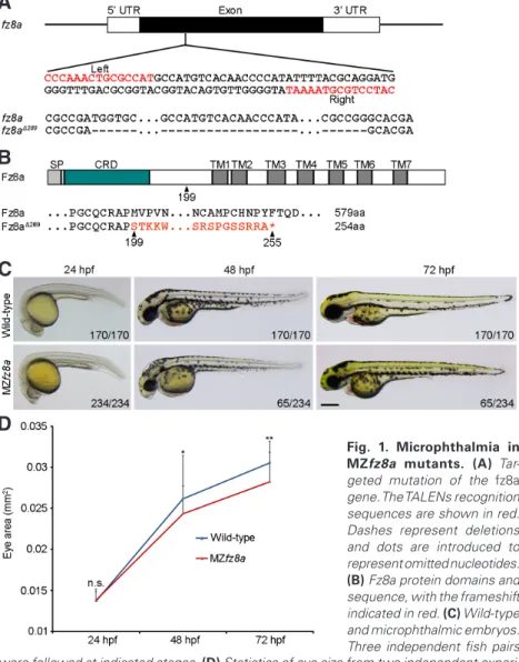
mutation with 289 nucleotides deletion in its unique exon (Fig. 1A). This leads to a frameshift after amino acid 198 and a premature stop codon after amino acid 254 (Fig. 1B). Genotyping indicated that both the gene and the corresponding transcript were truncated around the target sites (Supplementary Fig. S1).
Maternal-zygotic (MZ) fz8a (MZfz8a) mutants developed normally until the end of somite seg-mentation period (26 somites). At 24 hpf (hours post-fertilization), no morphological difference could be observed between wild-type and MZfz8a embryos. However, at 48 hpf, there was a clear reduction of the eye size in some MZfz8a mutants, and at 72 hpf, this became more evident (Fig. 1C,D). Analysis from three independent fish pairs indicated that MZfz8a mutants exhibited smaller eye phenotype with 25% to 30% penetrance (Fig. 1C, Supplementary Fig. S2), and those embryos developed microphthalmia.
A detailed examination of microphthalmic em-bryos revealed eye development defects prior to the reduction of eye size. At 24 hpf, compared with wild-type embryos or normal siblings, they failed to close the choroid fissure (Fig. 2 A,A’,A”). At 36 hpf, the choroid fissure in microphthalmic embryos became closed, but the thickness of the retina was obviously reduced and there was an accumulation of blood around the lens (Fig. 2 B,B’,B”), which resembles persistent fetal vasculature (PFV). Dur-ing subsequent stages, it was evident that retina pigmentation and the eye size became reduced in these embryos (Fig. 2 C-E,C’-E’,C”-E”). At 96 hpf, microphthalmic embryos also displayed lens development defects (Fig. 2 F,F’,F”). Since mutation of MZfz8a did not affect the specification of the eye field (Supplementary Fig. S3), these optic defects imply that fz8a is required for eye differentiation.
Fig. 1. Microphthalmia in MZfz8a mutants. (A) Tar-geted mutation of the fz8a gene. The TALENs recognition sequences are shown in red. Dashes represent deletions and dots are introduced to represent omitted nucleotides.
(B) Fz8a protein domains and sequence, with the frameshift indicated in red. (C) Wild-type and microphthalmic embryos. Three independent fish pairs
Fig. 2. Defective eye development in mi-crophthalmic embryos. (A-F) Eye development in wild-type embryos.
(A’-F’) MZfz8a siblings show normal eye develop-ment. (A”-F”) Defective eye development in mi-crophthalmic embryos. The retina is outlined at 24 hpf to show the persistence of the cho-roid fissure (arrow). Ar-rowheads indicate blood accumulation around the lens. A reduction of the retina and eye size is evident at 36 hpf, lens differentiation defect can be observed at 96 hpf. Scale bar, 100 mm. were followed at indicated stages. (D) Statistics of eye size from two independent
Microphthalmic embryos display delayed retinal lamination and reduced retinal cell numbers
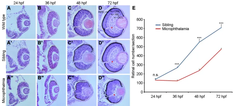
To examine how the eye size is reduced in microphthalmic embryos, we first performed histological analysis of eye structure. At 24 hpf, compared with wild-type embryos and normal siblings, no discernible alteration was observed in the microphthalmic retina (Fig. 3 A,A’,A”). However, at 36 hpf, neural retina became obviously thinner and lens size was reduced, whereas the retinal
pigment epithelium (RPE) seemed to be normally laminated (Fig. 3 B,B’,B”). At 48 hpf, when the ganglion cell layer (GCL) became laminated in wild-type embryos and normal siblings, there was a clear disorganization of the neural retina in microphthalmic mutants, which was formed by a pseudostratified epithelium. The differentiation of lens fibers was also delayed, as revealed by the presence of nuclei (Fig. 3 C,C’,C”). At 72 hpf, the outer nuclear layer (ONL), inner cell layer (INL), and GCL in the neural retina were
Fig. 3. Delayed lamination and reduced retinal cell number in microphthalmic mutants. (A-D) Retina structure and lamination in wild-type em-bryos. (A’-D’) MZfz8a siblings show aspects of retina and lens differentiation which are similar to those of wild-type emem-bryos. (A”-D”) Microphthalmic embryos display disorganized retinal layers. The retina is thinner at 36 hpf and pseudostratified at 48 hpf. GCL lamination can be distinguished at 72 hpf. Arrowheads indicate the optic nerve. (E) Statistics of retinal cells show the delay in cell number increase from 24 hpf to 36 hpf. Bars represent the mean ± s.d. from three sections (***, P<0.001; n.s., not significant). Scale bar: 50 mm.
Fig. 4. Neuronal differentiation is delayed in the microphthalmic retina. (A-E) The expression of
ath5 in normal siblings picks at 36 hpf and becomes
restricted to the retina ganglion progenitor cells from 48 hpf onward. (A’-E’) The expression of ath5 in microphthalmic retina ganglion progenitor cells is shifted to 72 hpf. (F-H’) Analysis of the expression of brn3a in the retina ganglion progenitor cells in normal sibling and microphthalmic eyes (outlined) shows a similar shift. Scale bar, 100 mm.
well differentiated and organized in wild-type and sibling eyes (Fig. 3 D,D’). However, the ONL and INL were not distinguishable, only some GCL cells began to be laminated and the optic nerve began to be differentiated in microphthalmic embryos (Fig. 3D”), indicating an impaired retinal lamination.
This apparent delay in retinal differentia-tion may be due to a reduced proliferadifferentia-tion of neural retina cells. We thus compared the cell number in the neural retina between microphthalmic and normal sibling eyes. By examining comparable histological sections, we found that, in normal siblings, there was
a steady increase in retinal cell number from 24 hpf to 72 hpf, while in microphthalmic embryos, there was no such increase between 24 hpf and 36 hpf. During subsequent stages, although there was a similar kinetics of increase in retinal cell number between normal siblings and microphthalmic embryos, the total number of microphthalmic retinal cells remained significantly low (Fig. 3E). Although the three major neural retina cell layers eventually laminated at 96 hpf to 120 hpf, they were significantly thicker compared to those in wild-type embryos or normal siblings (Supplementary Fig. S4). This indicates that mutation of fz8a delays retinal cell differentiation.
Both ath5 and brn3a are markers of neuronal production in the retina. We thus analyzed their expression to see how neuronal differentiation is affected. In normal siblings, ath5 expression was low at 27 hpf but peaked at 36 hpf (Fig. 4 A,B). It then decreased progressively to become restricted in the retinal ganglion progeni-tor cells (Fig. 4 C-E). In microphthalmic embryos, however, this temporal expression pattern exhibited a shift by at least 12 hours (Fig. 4 A’-E’). Interestingly, brn3a expression showed a similar decrease as ath5 in normal siblings from 48 hpf to 72 hpf (Fig. 4 F-H), while it began to be expressed at 60 hpf and increased at 72 hpf in microphthalmic embryos (Fig. 4 F’-H’). These
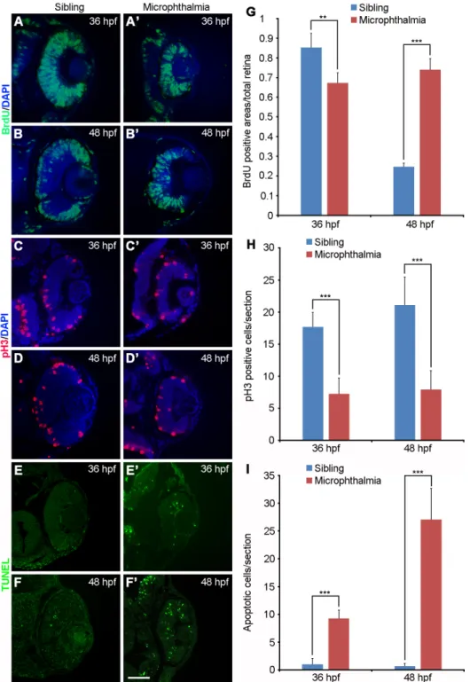
analy-Fig. 5. Sustained proliferation and abnormal apoptosis of microphthalmic retinal cells.
(A-B’) Cell proliferation in normal siblings at 48 hpf is restricted to the RPCs of the CMZ, while the entire microphthalmic retinal cells are prolifera-tive. (C-D’) Reduced pH3-positive dividing cells in microphthalmic embryos at 36 hpf and 48 hpf.
(E-F’) Increased apoptotic microphthalmic cells at 36 hpf and 48 hpf. (G-I) Statistics of BrdU-positive area, pH3-BrdU-positive cells, and apoptotic cells. All data were obtained from three sections at a comparable position between normal siblings and microphthalmic embryos (**, P<0.01; ***, P<0.001; n.s., not significant). Scale bar: 50 mm.
ses clearly show that the differentiation of neural retina cells is delayed, but not abolished, in microphthalmic embryos.
Abnormal retinal cell proliferation and apoptosis in microphthalmic embryos
We next employed different markers to characterize molecularly the delayed retinal cell differentiation in microphthal-mic embryos. Neural retina cells were incubated in BrdU for 30 minutes and then cultured for 2 hours. Whereas cell prolif-eration (BrdU incorporation) decreased in sibling eyes between 36 hpf and 48 hpf as retinal progenitors exited cell cycle to differentiate (Fig. 5 A,B,G), we observed a maintenance in the number of proliferative retinal progenitor in microphthalmic eyes (Fig. 5 A’,B’,G), indicating a sustained retinal cell proliferation. However, since the total number of neural retina cells in microphthalmic embryos was low (see Fig. 3), the sustained proliferation may not be accompanied by an active cell division. Indeed, phosphorylated histone H3 (pH3) staining showed that, both at 36 hpf and 48 hpf, there were significantly less dividing cells, essentially localized at the apical side of the microphthalmic retina (Fig. 5 C-D’,H). This indicates a reduced division rate of neural retina cells. However, the possibility of an increased apoptosis of RPCs cannot be excluded. Analysis by TUNEL assay indicated that
very few fluorescent apoptotic spots were present in normal siblings (Fig. 5 E,F,I), by contrast, significantly increased apop-totic cells were observed in the microphthalmic embryos (Fig. 5 E’,F’,I). These observations suggest that reduced division rate and increased apoptosis in neural retina cells may account for the microphthamia phenotype.
(Fig. 6 I’,J’), but was detected at high level in the proximal neural retina at 48 hpf and 60 hpf (Fig. 6 K’,L’). Thus, compared with wild-type RPCs, there was an expanded p57Kip2 expression in the microphthalmic RPCs at 48 hpf and 60 hpf, indicating that they start to exit the cell cycle only at later stages. Together, these results suggest that microphthalmic RPCs are delayed to exit
Fig. 6. Delayed cell cycle exit in microphthalmic embryos. (A-D’) The expression of myca in wild-type embryos is restricted to RPCs of the CMZ from 36 hpf onward, while it persists in the entire microphthalmic retina at 36 hpf and 48 hpf. (E-H) The expression of cyclinD1 in wild-type embryos is restricted to the RPCs of the CMZ at 48 hpf and 60 hpf. (E’-H’) In microphthalmic embryos, cyclinD1 remains to be expressed in the entire retina at all stages examined. (I-L) The expression of p57Kip2 in the wild-type retina increases at 36 hpf, and is progressively restricted in the RPCs of the CMZ from 48 hpf to 60 hpf. (I’-L’) In microphthalmic embryos, the expression of p57Kip2 increases at 48 hpf and persists at 60 hpf. Scale bar: 50 mm.
Cell cycle exit is delayed in microphthal-mic embryos
The sustained proliferation state of RPCs in microphthalmic embryos is not consistent with the reduced division of these cells. One possibility that accounts for this discrepancy may be a delay of cell cycle exit. The expres-sion of myca is correlated with retinal cell proliferation. We found that myca expression in the wild-type retina began to be predominantly localized to the ciliary marginal zone (CMZ) at 24 hpf, and became entirely restricted in the CMZ from 36 hpf onward (Fig. 6 A-D). By contrast, it was detected nearly in the entire microphthalmic retina at 24 hpf and 36 hpf (Fig. 6 A’,B’). The expression level of myca in the proximal region of microphthalmic retina began to decrease at 48 hpf, and restricted
myca expression could be observed in the
CMZ at 60 hpf (Fig. 6 C’,D’). Thus, both the pattern and level of myca expression in mi-crophthalmic embryos show a delay of neural retina cell differentiation by about 24 hours, likely caused by a sustained proliferation of these cells.
Both cyclinD1 and the cyclin-dependent kinase inhibitor p57Kip2 are important regula-tors of RPC proliferation. The expression of
cyclinD1 becomes rapidly down-regulated in
emerging post-mitotic cells, while conversely, the expression of p57Kip2is up-regulated in a subset of RPCs as they exit the cell cycle (Das et al., 2009; Dyer and Cepko, 2001; Shkumatava and Neumann, 2005). At 24 hpf and 36 hpf, similar intensity and distribution of
cyclinD1 transcripts were detected in wild-type
and microphthalmic embryos (Fig. 6 E,F,E’,F’). Interestingly, from 48 hpf to 60 hpf, low level of cyclinD1 expression was detected in the CMZ of wild-type embryos (Fig. 6 G,H). By contrast, high level of cyclinD1 expression was maintained in the entire microphthalmic retina (Fig. 6 G’,H’). Complementary to cyclinD1,
p57Kip2 was expressed at low level in the
wild-type retina at 24 hpf (Fig. 6I). After an increase in the proximal neural retina at 36 hpf (Fig. 6J), indicating that these cells exit the cell cycle, its expression decreased and became restricted in the RPCs of the CMZ at 48 hpf and 60 hpf (Fig. 6 K,L). By contrast,
p57Kip2 expression was not detected in the
the cell cycle. As a result, neural differentiation is defective due to increased apoptosis and reduced amount of retinal cells.
Discussion
We show that knockout of fz8a results in microphthalmia in zebraf-ish. RPCs in these mutants have prolonged period of proliferation without increase in cell number. These cells exhibit a delayed exit from the cell cycle, which likely prevents them to enter into the post-mitotic state for neuronal differentiation.
In MZfz8a mutants, about 30% of the embryos progressively develop microphthalmia. This low penetrance could not due to the compensation by fz8b, which is not expressed in the developing eye. Rather, at least fz3 and fz5 are expressed early in the eye region (Shi et al., 1998; Cavodeassi et al., 2005). They may functionally interact with fz8a to regulate eye development. Single knockout of
fz5 in mice results in mild coloboma and microphthalmia with about
50% penetrance (Liu and Nathans, 2008), while single knockout of
fz8 causes similar but weaker phenotypes (Liu et al., 2012). However,
compound fz5 and fz8 mutant exhibits severe retinal coloboma and microphthalmia (Liu et al., 2012). Thus, the ocular defects caused by fz8 loss-of-function are closely similar between zebrafish and mice, indicating a conserved function of fz8 in eye development. There should be also a functional redundancy between Fz proteins that have similar ligand-binding and signalling specificity.
The Wnt pathway during eye development shows a high degree of complexity, depending largely on the cellular context. Our pres-ent work suggests that fz8a is implicated in RPC proliferation and differentiation through regulation of cell cycle exit. Indeed, Wnt signalling regulates cell proliferation and cell cycle progression by activating target genes c-myc and cyclinD1. Consistently, we find an altered expression of cyclinD1 in the microphthalmic RPCs. In particular, cyclinD1 expression is initially down-regulated in the RPCs, and is recovered with a significant delay. This indicates that RPCs remains in a mitotic state for a longer period, and as a consequence, their differentiation becomes delayed. Thus, we conclude that fz8a functions in the step of neuronal production and differentiation.
The microphthalmic retina remains pseudostratified, with a significant delay of retinal lamination. This may be caused by a sustained proliferation of neural retina cells and/or by a delayed cell cycle exit. As a result, these cells are maintained in an undifferenti-ated state for a prolonged period. However, the process that shifts neuronal differentiation to later stages of eye development may be more complex. Indeed, the increased proliferation of retinal cells and delayed cell cycle exit are also accompanied by an enhanced apoptosis. Furthermore, although pH3 staining is essentially local-ized to the apical surface of the microphthalmic retina, it clearly shows a reduced number of dividing cells. Thus, the decrease of cell division associated with an increase of apoptotic cells may ac-count for the reduction of retinal cells and for the microphthalmia phenotype. Nevertheless, it remains to be determined whether those non-dividing cells eventually enter into apoptosis.
The phenotype of microphthalmic MZfz8a embryos also re-sembles PFV. Since this occurs earlier than the appearance of abnormal retinal cell proliferation and apoptosis, it suggests that Fz8a may regulate other aspects of eye development. Thus, further analysis of its function may help to understand the cause of human ocular disease.
Materials and Methods
Zebrafish embryos
Zebrafish embryos were cultured at 28.5°C. Phenylthiourea (Sigma-Aldrich) at 50 mM was added to E3 solution to prevent pigment devel-opment.
Targeted mutation
TALEN recognition sequences are located in the 5′ region of the unique exon of fz8a locus (see Fig. 1). The targeting efficiency was determined as described (Cheng et al., 2017). Genotyping PCR primers are as follow:
F1, 5′-ATGAGGTGCGATCTACTGC-3′ F2, 5′-ACCAACTACCCCAGCAAAG-3′ Fw, 5′-CGCTCTACAACCGCGTTAAG-3′ R3, 5′-GAAGTAGATAAGCAGGAACAC-3′ R4, 5′-TGCTCCACGTCATACTCCC-3′ and R5, 5′-GTTGCCAACGTAGCAGATC-3′.
In situ hybridization and RT-PCR
In situ hybridization was performed as described (Shao et al., 2017).
Antisense probes were labelled using digoxigenin-11-UTP. Wild-type and mutant fz8a transcripts were analyzed by semi-quantitative PCR using primers F2 and R4, with b-actin (5′-CACAGTGCTGTCTGGAGGTAC-3′, 5′-GAGGGCAAAGTGGTAAACG-3′) as a loading control.
Histology and immunofluorescence
Embryos were fixed in 4% paraformaldehyde for 2 hours at room tem-perature, and were embedded in paraffine. Sections of 5 mm were made and stained with hematoxylin and eosin (HE). For immunofluorescence, sections were incubated with G3G4 monoclonal anti-BrdU antibody (Developmental Studies Hybridoma Bank, 1:300), or anti-pH3 antibody (Abcam, 1:1000), followed by Alexa Fluor488 rabbit anti-mouse lgG or Fluo594 goat anti-rabbit lgG (Interchim, 1:1000). They were analyzed under a fluorescence microscope (Leica DM2000).
BrdU incorporation, cell proliferation and apoptosis assays
Embryos were dechorionated and incubated in Ringer’s solution containing 10 mM BrdU (Roche Diagnostics) and 15% dimethylsulfoxide for 30 minutes at 6°C. They were washed 3 times with Ringer’s solu-tion (10 minutes each) and recovered at 28.5°C for at least 2 hours, and were then fixed with 4% paraformaldehyde overnight at 4°C for paraffine sectioning. Detection of mitotic cells was performed through anti-pH3 antibody staining. Labelling for apoptosis was performed using the TUNEL kit (Roche Diagnostics).
For each of the staining described above, at least 3 retinal sections at a comparable position in wild-type and mutant embryos from differ-ent experimdiffer-ents were selected. BrdU-positive area relative the differ-entire neural retina surface, pH3-positive cells, and TUNEL-labelled fluorescent apoptotic bodies were analyzed.
Retinal cell number counting
At each stage, 3 retinal sections from sibling and mutant embryos at similar position of the eye were identified by the presence of optic nerve.
Determination of eye size
Synchronously spawned wild-type and mutant embryos (50 at each stage) were fixed and individually imaged at the eye region, along with a known reference area. The eye size was analyzed by ImageJ (NIH Image) and the real size was calculated by comparison with the reference area.
Determination of neural retina layer width
The total width of the retina and the width of each retinal layer were measured in sectioned embryos at 120 hpf from three independent spawning using a total of 20 sections with optic nerve.
Acknowledgements
We thank members of our laboratory for technical assistance and ze-brafish care. This research was supported by the NSFC grant (31671509).
References
AGATHOCLEOUS, M., IORDANOVA, I., WILLARDSEN, M.I., XUE, X.Y., VETTER, M.L., HARRIS, W.A. and MOORE, K.B. (2009). A directional Wnt/beta-catenin-Sox2-proneural pathway regulates the transition from proliferation to differentiation in the Xenopus retina. Development 136: 3289-3299.
BORDAY, C., CABOCHETTE, P., PARAIN, K., MAZURIER, N., JANSSENS, S., TRAN, H.T., SEKKALI, B., BRONCHAIN, O., VLEMINCKX, K., LOCKER, M. and PERRON, M. (2012). Antagonistic cross-regulation between Wnt and Hedgehog signalling pathways controls post-embryonic retinal proliferation. Development 139: 3499-3509.
CHENG, X.N., SHAO, M., LI, J.T., WANG, Y.F., QI, J., XU, Z.G. and SHI, D.L. (2017). Leucine repeat adaptor protein 1 interacts with Dishevelled to regulate gastrulation cell movements in zebrafish. Nat Commun 8: 1353.
CAVODEASSI, F., CARREIRA-BARBOSA, F., YOUNG, R.M., CONCHA, M.L., AL-LENDE, M.L., HOUART, C., TADA, M. and WILSON, S.W. (2005). Early stages of zebrafish eye formation require the coordinated activity of Wnt11, Fz5, and the Wnt/beta-catenin pathway. Neuron 47: 43-56.
DAS, G., CHOI, Y., SICINSKI, P. and LEVINE, E.M. (2009). Cyclin D1 fine-tunes the neurogenic output of embryonic retinal progenitor cells. Neural Dev 4: 15. DENAYER, T., LOCKER, M., BORDAY, C., DEROO, T., JANSSENS, S., HECHT, A.,
VAN ROY, F., PERRON, M. and VLEMINCKX, K. (2008). Canonical Wnt signaling controls proliferation of retinal stem/progenitor cells in postembryonic Xenopus eyes. Stem Cells 26: 2063-2074.
DYER, M.A. and CEPKO, C.L. (2001). p27Kip1 and p57Kip2 regulate proliferation in distinct retinal progenitor cell populations. J Neurosci 21: 4259-4271.
GRAW, J. (2010). Eye development. Curr Top Dev Biol 90: 343-386.
KIM, S.H., SHIN, J., PARK, H.C., YEO, S.Y., HONG, S.K., HAN, S., RHEE, M., KIM, C.H., CHITNIS, A.B. and HUH, T.L. (2002). Specification of an anterior neuroecto-derm patterning by Frizzled8a-mediated Wnt8b signalling during late gastrulation in zebrafish. Development 129: 4443-4455.
KUBO, F. and NAKAGAWA, S. (2008). Wnt signaling in retinal stem cells and regen-eration. Dev Growth Differ 50: 245-251.
LIU, C. and NATHANS, J. (2008). An essential role for frizzled 5 in mammalian ocular development. Development 135: 3567-3576.
LIU, C., BAKERI, H., LI, T. and SWAROOP, A. (2012). Regulation of retinal progeni-tor expansion by Frizzled recepprogeni-tors: implications for microphthalmia and retinal coloboma. Hum Mol Genet 21: 1848-1860.
MEYERS, J.R., HU, L., MOSES, A., KABOLI, K., PAPANDREA, A. and RAYMOND, P.A. (2012). β-catenin/Wnt signaling controls progenitor fate in the developing and regenerating zebrafish retina. Neural Dev 7: 30.
RASMUSSEN, J.T., DEARDORFF, M.A., TAN, C., RAO, M.S., KLEIN, P.S. and VETTER, M.L. (2001). Regulation of eye development by frizzled signaling in
Xenopus. Proc Natl Acad Sci USA 98: 3861-386.
SHAO, M., WANG, M., LIU, Y.Y., GE, Y.W., ZHANG, Y.J. and SHI, D.L. (2017). Veg-etally localised Vrtn functions as a novel repressor to modulate bmp2b transcrip-tion during dorsoventral patterning in zebrafish. Development 144: 3361-3374. SHI, D.L., GOISSET, C. and BOUCAUT, J.C. (1998). Expression of Xfz3, a Xenopus
frizzled family member, is restricted to the early nervous system. Mech Dev 70: 35-47.
SHKUMATAVA, A. and NEUMANN, C.J. (2005). Shh directs cell-cycle exit by activat-ing p57Kip2 in the zebrafish retina. EMBO Rep 6: 563-569.
SUMANAS, S. and EKKER, S.C. (2001). Xenopus frizzled-5: a frizzled family mem-ber expressed exclusively in the neural retina of the developing eye. Mech Dev 103: 133-136.
VAN RAAY, T.J., MOORE, K.B., IORDANOVA, I., STEELE, M., JAMRICH, M., HARRIS, W.A. and VETTER, M.L. (2005). Frizzled 5 signaling governs the neural potential of progenitors in the developing Xenopus retina. Neuron 46: 23-36.