Journal of Pathology
J Pathol 2015
Published online in Wiley Online Library (wileyonlinelibrary.com)DOI: 10.1002/path.4644
INVITED REVIEW
Regeneration versus scarring in vertebrate appendages and heart
Anna Ja´zwi´nska* and Pauline Sallin
Department of Biology, University of Fribourg, Switzerland
*Correspondence to: A Ja´zwi´nska, Department of Biology, University of Fribourg, Chemin du Musée 10, 1700 Fribourg, Switzerland. E-mail: anna.jazwinska@unifr.ch
Abstract
Injuries to complex human organs, such as the limbs and the heart, result in pathological conditions, for which we often lack adequate treatments. While modern regenerative approaches are based on the transplantation of stem cell-derived cells, natural regeneration in lower vertebrates, such as zebrafish and newts, relies predominantly on the intrinsic plasticity of mature tissues. This property involves local activation of the remaining material at the site of injury to promote cell division, cell migration and complete reproduction of the missing structure. It remains an unresolved question why adult mammals are not equally competent to reactivate morphogenetic programmes. Although organ regeneration depends strongly on the proliferative properties of cells in the injured tissue, it is apparent that various organismic factors, such as innervation, vascularization, hormones, metabolism and the immune system, can affect this process. Here, we focus on a correlation between the regenerative capacity and cellular specialization in the context of functional demands, as illustrated by appendages and heart in diverse vertebrates. Elucidation of the differences between homologous regenerative and non-regenerative tissues from various animal models is essential for understanding the applicability of lessons learned from the study of regenerative biology to clinical strategies for the treatment of injured human organs.
© 2015 The Authors. The Journal of Pathology published by John Wiley & Sons Ltd on behalf of Pathological Society of Great Britain and Ireland.
Keywords: fin; limb; zebrafish; urodele; epimorphosis; cytodifferentiation; cellular specialization; blastema; wound epidermis; fibroblast; cardiomyocyte
Received 9 July 2015; Revised 15 September 2015; Accepted 18 September 2015
No conflicts of interest were declared.
Introduction
Restoration of tissue integrity following injury is a fun-damental property of multicellular organisms. Animals respond to traumatic organ loss either by reparative seal-ing of the wound or by regeneration. Non-regenerative healing often involves synthesis of a scar to repair the interrupted continuity of the organ without reproduction of the missing tissue. In contrast, regeneration recre-ates the architecture and function of the damaged body part, either without scarring or with transient fibrosis. This process depends on cellular plasticity, accessibility to developmental programmes and functional integra-tion of newly formed tissues with the pre-existing organ remnants. Despite extensive research in various model animals, it remains poorly understood what specific fac-tors predispose for non-regenerative repair versus com-plete reconstruction of complex vertebrate organs, such as limbs and heart [1–7].
In humans, injury followed by inadequate regener-ation and scarring is a common pathological mecha-nism. This is the case for an irreversible limb loss and for a heart infarct. By contrast, certain non-mammalian adult vertebrates, such as zebrafish, newts and axolotls,
can reconstitute fully functional appendages and a car-diac ventricle [6,8–16]. These animals provide unique in
vivo model systems to understand how damaged mature
organs are naturally capable of a rapid switch between resting and re-growing phases. A great deal of atten-tion has been dedicated to the molecular mechanisms of regeneration, as discussed in many recent reviews [10–12,17–27]. Here we focus on the cellular and anatomical features of appendage and heart regeneration in anamniotic vertebrates (fish and amphibians) and how this information is relevant for the goals of regenerative medicine.
Regeneration versus scarring from the functional
and adaptive perspective
The absence of an appendage, such as a limb or a tail, impedes efficient locomotion, resulting in a decreased survival rate. Myocardial infarction disrupts the per-formance of the blood-pumping organ, leading to a life-threatening condition. Given that the organism sur-vived the initial trauma, complete organ regeneration intuitively provides an advantage to regain fitness after
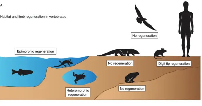
injury. Ironically, lower vertebrates possess an innate ability to restore their damaged appendages and the heart, while mammals lack this capability. This uneven phylogenic distribution of regeneration could be due to various proximate constraints of anatomical, physiolog-ical and molecular nature, such as the high complexity of the mammalian organs, a cytokine profile of inflam-matory cells and inaccessible morphogenetic informa-tion, which arose as adaptive traits or as side-effects of other evolutionary changes [2,23,28–30]. Among many concepts, the classic hypothesis proposes that the regenerative capability declines whenever the lost part of organ becomes absolutely indispensable for survival:
To qualify for replacement, a structure … must be important enough to be missed when it is gone, but not so vital that an animal cannot survive its loss long enough to grow a replacement [31]. Accordingly, the loss of limb regeneration in the evolution of vertebrates may be associated with the move onto land, when limbs become indispensable for locomotion, much more than when an animal is suspended in water (Figure 1A). The limbs of terrestrial animals have an essential function to carry the body mass while remaining in direct contact with rough ground, which might biophysically preclude regenera-tive outgrowth formation from the stump [25]. Among urodeles, the efficiency and fidelity of regeneration become drastically lower in land-phase animals as compared to water-phase forms [32,33]. The adult aquatic urodeles, eg newts and paedomorphic axolotls, can complete regeneration within 30–60 days after amputation, whereas the terrestrial forms, eg certain salamanders and postmetamorphic axolotls, require 200–400 days [32–34]. In addition, a proper terrarium with wet and soft substrates is a prerequisite for limb regeneration in the land-phase urodeles, as adverse environmental conditions impair this process [32,35]. It is possible that during the evolution of terrestrial amniotic vertebrates, beginning with reptiles, the limb regenerative capability has been replaced with scarring as a more suitable strategy for repairing stumps while walking on rough land. Nevertheless, some degree of appendage restoration can evolve locally, exemplified by the gain of digit tip regeneration in mice and humans [36–38].
Although the heart is obviously a life-essential organ, a partial loss of cardiac muscle can be differentially tol-erated among vertebrates. Among fish, medakas were reported to react with high lethality and health impair-ment to ventricular surgery in comparison to zebrafish [39]. Zebrafish can generally survive genetic ablation of 60% of cardiomyocytes or damage to 20% of the ven-tricle [40–46], which would have fatal consequences in adult mammals [47]. Thus, a portion of the myocardium can be of low/intermediate importance for the global function of the heart in certain vertebrates. In the line of the above-cited hypothesis, proposed by Goss [31,48],
such animals might have a better predisposition for car-diac regeneration than mammals, especially in a milieu of low metabolism and with a trabecular myocardium typical of poikilotherms. In poikilothermic vertebrates, a significant portion of cardiomyocyte oxygen supply derives from the blood flow in cavities of the ventricular chamber. This is in contrast to the compact myocardium of homeothermic mammals, which depends strongly on coronary circulation [49–51] (Figure 1B). Indeed, the thickness of the mammalian compact myocardium pre-vents gas diffusion from luminal blood to subepicardial cardiomyocytes, a mechanism that could possibly atten-uate the damage during left ventricular ischaemia. The trabecular architecture, such as in zebrafish and newt heart, may account for a permissive environment for a regenerative response at the site of injury [11,52]. Mammals, which are homeothermic animals, require a higher performance of the blood-pumping organ. Remarkably, the ventricular blood pressure in adult zebrafish reaches the value of approximately 2.5 mmHg [53], as compared to an average of 120 mmHg in adult humans and mice. It is possible that the biomechan-ical properties of the ventricular wall and the mode of oxygen supply might limit the choice of the tis-sue type to restore the interrupted myocardium, while the heart has to beat continuously during the healing process.
Studies of anamniotes revealed that the basic regen-erative principles are similar in the appendages and the heart [3,6,9]. Nevertheless, specific cellular programmes are poorly comparable between the appendages and the heart, as these body parts are built with different ’blocks’. Therefore, we discuss the current knowledge on regeneration of these two distinct organs in separate sections.
Appendage: the blastema and the
pro-regenerative environment
Complete restoration of the amputated vertebrate appendage, called epimorphic regeneration, involves the creation of two key structures, a blastema and a wound epithelium [21,54]. Importantly, neither is fundamentally different from the stump tissues. The wound epidermis derives from the migrating epithe-lial cells at the site of injury, whereas the blastema arises by the conversion of local non-dividing cells into lineage-restricted proliferating cells, which give rise to the outgrowth [55–60]. To date, there is no evidence for the involvement of remote cell sources. To initiate epimorphic regeneration, the participating cells change gene expression and reactivate developmental signalling pathways to guide cell dedifferentiation, proliferation and migration, as recently reviewed [10,12,17,18]. The wound epidermis serves not only for healing of the interrupted organ, but also forms a specialized layer that acts as a signalling centre by producing cytokines, such as Wnts, Fgfs and Shh
Regeneration versus scarring in vertebrate appendages and heart 3
Figure 1. Potential impact of external and internal milieux on the regenerative feasibility of vertebrate appendages and heart. (A) The transition onto land coincides with the reduction of limb-regenerative capability. Fish and aquatic urodeles possess the ability to completely and efficiently reproduce amputated appendages. Postmetamorphic terrestrial frogs and toads completely lack this capability, while aquatic Xenopus froglets are capable of heteromorphic regeneration of a cartilaginous ’spike’ from the amputation plane. Amniotic vertebrates, ie reptiles, birds and mammals, which adapted to life and reproduction on the land, display no limb regeneration. A certain capacity of appendage restoration can be regained, as exemplified by digit tip regeneration in mice and humans under certain circumstances. (B) The rise in cardiac workload in endothermic vertebrates correlates with compaction of the myocardial wall and elaboration of the coronary vasculature; schematic representation of heart anatomy with a transverse section of the ventricle in fish (left) and mammals (right). In the poikilothermic vertebrates, ie fish, amphibians and reptiles, the ventricle is a trabecular, sponge-like chamber. This architecture allows for oxygenation of ventricular cardiomyocytes directly from blood flowing through many miniscule cavities of the trabecular wall. The outer compact myocardial layer is typically thin, with fine coronary vasculature. Among animals with the trabecular heart, zebrafish, newts and axolotls display heart regeneration. A radical change of cardiac anatomy is observed in homeothermic mammals and birds, in which the ventricles are composed predominantly of a compact myocardium and a discrete central lumen. Oxygenation of cardiomyocytes is dependent on coronary circulation. This architecture has been associated with no regenerative capabilities
[17,18]. The reciprocal communication between the wound epidermis, nerves and the underlying tissues is a prerequisite for appendage regeneration [17,18,61–63]. The interruption of epithelial–mesenchymal interaction by covering the amputation plane with a mature skin
graft prevents regeneration [64,65]. A contribution of the wound epidermis and the mesenchymal blastema has been observed during digit tip regeneration in mice and humans, suggesting parallel regenerative strate-gies between diverse vertebrates [36,66]. In humans,
it is assumed that dermal scarring and the inability to form the blastema are responsible for the lack of limb regeneration. Thus, the mechanisms regulating wound healing and the plasticity of mature cells are of central interest in the field of regenerative biology and medicine.
Blastema formation is associated with cellular de-differentiation of specialized appendage tissues, such as bone-forming osteoblasts in zebrafish [56–60,67,68] and skeletal muscles in urodeles [69–72]. However, dedifferentiation is not the only mechanism for the generation of new cells for both of these specialized tissues. In the fin, genetic ablation of all osteoblasts, using a nitroreductase system, does not prevent bone regeneration, suggesting a possible secondary source of osteoblasts under certain restrictive conditions [59]. Although myofibre dedifferentiation occurs during urodele limb restoration, Cre–loxP genetic fate map-ping in the axolotl revealed that new muscle mainly originates from endogenous Pax7-positive satellite cells [69]. These examples indicate a conditional plasticity and a species-specific variability of possible sources of cells for appendage regeneration.
By contrast to bone and muscle tissues, dermal fibrob-lasts do not appear to dedifferentiate markedly dur-ing regeneration, as they are originally at a relatively low cytospecialization level. Instead, the connective tis-sue undergoes disorganization and remodelling of the extracellular matrix (ECM) to facilitate fibroblast pro-liferation and migration. In urodeles, the population of mesenchymal cells is highly over-represented in the blastema in comparison to other cell types [73]. More-over, mesenchymal cells are the pioneers promoting the protrusion of the regenerative outgrowth. This sug-gests that dermal fibroblasts play a leading role in appendage regeneration. Thus, it is important to iden-tify the structural differences of dermal tissue between blastema-forming versus scar-forming model organ-isms.
As the skin acts as a protective barrier, the surface of the body in terrestrial vertebrates underwent specializa-tion from mucogenesis to cornificaspecializa-tion, while the dermis shifted toward a more robust collagen fibre-rich matrix (Figure 2A, F). In most mammals, cutaneous injuries heal by scarring. In contrast, the surface of wet anatom-ical sites in humans, such as the oral mucosa, is capable of scar-free wound closure, which is associated with a higher proliferative capacity of local keratinocytes and fibroblasts, beneficial growth factor production and appropriate matrix deposition, as compared to skin [74]. Based on these observations, it can be proposed that cellular specialization between homologous structures may explain the regenerative differences, even in the same organism.
The zebrafish fin is a non-muscularized dermal fold that encompasses multiple bony rays spaced by soft interrays [10]. The stratified epidermis contains only a few cell layers, while the dermis is subdivided into two compartments: (a) dermal bone, produced by osteoblasts; and (b) a network of fibroblasts with
little collagenous matrix (Figure 2B, G). The dermal bone is located directly underneath the basal layer of the epidermis, suggesting a contact between both tissues. Remarkably, a structural connection between the bone and the epidermis has been reported in the regenerative part of the mammalian limb, viz. in the distal tip of the last phalangeal element (P3) in mice [36]. The significance of the link between the bone and epidermis during appendage regeneration in vertebrates remains to be determined. In aquatic urodeles, the dermis contains relatively few collagen fibrils, espe-cially in its subepidermal layer [75,76]. Mammals with exceptionally elastic skin and little fibrillar collagen, such as the African spiny mouse, can also regenerate large skin holes perfectly [77]. These examples suggest a correlation between a low content of collagen fibrils and regenerative success. Thus, a dense accumulation of fibrous matrix in the human dermis could cause one of the barriers for intercellular communication and tissue plasticity, which are essential factors for organ regeneration.
The fin and urodele limb initiate regeneration remark-ably quickly, as the re-epithelialization and local disorganization of mesenchyme occurs within the first 12 h post-amputation [75,76,78]. During this time, tenascin C and matrix metalloproteinases become up-regulated to stimulate tissue remodelling [79,80]. In zebrafish, a regenerative outgrowth becomes clearly vis-ible at 3 days post-amputation (dpa) (Figure 2C, H). The apical zone is considered to be the upstream signalling organizer of the regenerate, which includes a columnar wound epithelium and mesenchymal progenitor cells [81] (Figure 2E, J). The proximal part of the blastema contains highly proliferative mesenchymal cells and regenerating osteoblasts that initiate the deposition of new bone matrix in a proximal–distal direction of the outgrowth [56] (Figure 2D, I). The progression of regen-eration depends on a balance between prolifregen-eration and redifferentiation, which is regulated by a combination of multifunctional signalling pathways, such as FGF, Wnt, TGFβ, Activin, BMP, IGF, Notch, Shh, retinoic acid (RA) and Hippo [9,17,68,82–84], and epigenetic mechanisms [85]. Depending on the temperature, fin regeneration is completed approximately 3 weeks after injury [10,86]. It is still poorly understood how the amputation event triggers the programme that generates the specialized wound epithelium and the blastema from the stump tissues. At least, the histological com-parison of the dermis between intact and regenerating fins suggests an absence of extracellular obstacles, such as excessive collagen fibres, which could potentially impede the execution of the intrinsic regenerative programme.
What is the particular function of fibroblasts during blastema formation? Grafting experiments in urodeles demonstrated an inductive role of dermal fibroblasts during reconstruction of the appropriate structure along the body axis [24,87]. A juxtaposition of cells from the proximal and distal blastemas elicits a segrega-tion of cells according to their original source. In
Regeneration versus scarring in vertebrate appendages and heart 5
Figure 2. Histological comparison reveals excessive collagenous matrix in human dermis as compared to mesenchyme of the fin. Postnatal human skin (section of a cheek) and zebrafish fin (longitudinal section of a ray) were incubated with the same histological staining solutions. (A–E) Haematoxylin (purple-brownish, nuclear marker) and eosin (orange, protein marker) (H&E) staining. (A) The multilayered epidermis of human skin comprises a stratum corneum with anuclear keratinocytes; the dermis is rich in ECM that is produced by loosely distributed fibroblasts. (B) The stratified epidermis of the fin is composed of only a few cell layers and lacks a stratum corneum; it contains a basal cell layer (BC) and specialized epithelial cells, such as mucous cells (MuCs) and alarm-substance cells (ASCs); the dermis of the fin ray comprises segmented bone produced by osteoblasts and vascularized mesenchymal tissue; Mel, melanocytes. (C–E) A fin regenerate at 3 days post-amputation (dpa); the regenerate is located to the right of the amputation plane (dashed line); wound epidermis (WEp) surrounds the outgrowth; the basal wound epithelium (BWEp) contains cuboidal cells at the level of the proximal blastema (Prox.Bl) or columnar cells at the level of the distal blastema (Dist.Bl). (D) Higher magnification of regenerating bone with osteoblasts (Ob). (E) The cells of the distal blastema are tightly packed under the wound epithelium, with little intercellular space. (F–J) Aniline blue, acid Fuchsin and Orange G (AFOG) staining reveals collagen or mucous in blue, cornified epidermis in red, mineralized bone matrix in magenta and cytoplasm in orange; histological procedures were performed in parallel on human skin and fin sections. (F) Dermis of human skin contains a high amount of ECM with collagen fibrils. (G) The dermal bone of the fin is mineralized (magenta); a thin layer of collagen (blue) covers the bone surface, the bone canal and joints; dermal fibroblasts of the ray do not deposit extensive collagen fibres, consistent with the mesenchymal character of this tissue. (H–J) A section adjacent to the specimen shown in (C–E): collagen is detected in the basement membrane (BMem) and around the osteoblasts (Ob). Scale bars = 100 μm
some instances, the positional discontinuity between cells stimulates regrowth of the missing intermediate structures, resulting in intercalary regeneration of an ectopic appendage that bridges the gap between the positional disparities [88,89]. Notably, transcriptome analyses provide evidence that positional memory, which was established during embryogenesis, is also present in mature mammalian fibroblasts [90,91]. It will be essential to elucidate how the coordinates are commu-nicated between cells to guide pattern formation during regeneration. Although cell transplantation has been practised in regenerative medicine, the key unsolved problem is the creation of such regeneration-competent progenitor cells that can recognize the position in the stump, integrate with the host organ and/or act as ’pattern-forming’ cells. The fin and amphibian limb regeneration models can provide some hints on how to establish a regeneration-competent environment and ’plasticizing’ agents to enhance survival, intercellular communication and integration of the transplanted cells into the stump.
Heart: cardiomyocyte plasticity in vertebrates
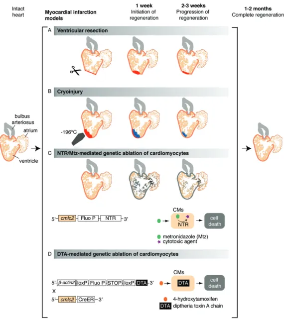
The zebrafish heart can fully regenerate within 30–60 days, either after removal of up to 20% of the ventricle [92], after cardiomyocyte-specific genetic ablation that causes the loss of up to 60% of the myocardium [40–43] or after cryoinjury-induced car-diac infarction of 20–25% of the ventricle [44–46] (Figure 3). In the newt, complete myocardial restoration has been observed within 2–7 months after ventricular apex amputation or mechanical disruption of> 50% of the ventricle [8,52,93–96]. Cardiac regeneration has been reported in axolotls 3 months after ventricular resection [15,16]. These findings have raised several fundamental questions about the origin of the new myocardium and signalling pathways involved in this process and, importantly, whether these findings are transposable to humans.
Similarly to mammals, there is no evidence of regeneration-competent cardiac progenitor cells in
Figure 3. Cardiac injury models in zebrafish. Schematic representation of different experimental procedures to induce cardiac damage in the zebrafish; illustration shows a longitudinal section of the heart. (A) The ventricular resection method was developed in 2002 [92]; in this model, the apex of the ventricle (about 20% of the heart volume) is amputated with scissors. (B) The cryoinjury procedure was established in 2011 [44,46,182,183] and is based on rapid freezing– thawing of the cardiac tissue by the application for 20–25 s of a stainless steel cryoprobe precooled in liquid nitrogen; as a result, massive cell death occurs in the frozen area (about 20% of the heart volume). (C) The nitroreductase (NTR)/metronidazole (Mtz) genetic ablation system enables depletion of specific cardiac cell types in an inducible manner [40,42,43,149]; in this method, first applied to zebrafish in 2007 [43,184], a bacterial nitroreductase fused to a fluorescent protein (Fluo P) is expressed under the control of a tissue-specific promoter, such as the cardiac myosin light chain 2 (cmlc2), which is specific for cardiomyocytes (CMs) [43,185]. This enzyme catalyses the conversion of the prodrug metronidazole into a cytotoxic agent, thereby inducing cell death in the NTR-expressing cells. (D) The Cre/loxP transgenic system, developed by Wang and colleagues in 2011, is based on the inducible expression of diphtheria toxin A chain (DTA) in cardiomyocytes (CMs) [41]; in brief, this procedure is based on the use of a double transgenic system, including Tg(𝛽actin2:loxP-Fluo P-STOP-loxP-DTA), which contains an inducible cytotoxic DTA gene preceded by a loxP-flanked reporter gene and stop codon, and the Tg(cmlc2:CreER) [98], which allows expression of the 4-hydroxytamoxifen (4-HT)-inducible Cre recombinase in cardiomyocytes. Treatment of the intercrossed fish cmlc2:CreER;𝛽actin2:loxP-Fluo P-STOP-loxP-DTA results in a massive myocardial cell depletion (up to 60% of the ventricle)
the post-embryonic stages in lower vertebrates. In zebrafish, genetic fate-mapping experiments with
Cre –loxP transgenic lines have demonstrated that the
regenerated myocardium is derived from pre-existing adult cardiomyocytes [97,98]. Consistently, differ-ent proliferative assays based on the detection of G1–S-phase or mitotic markers have revealed an accu-mulation of proliferating cardiomyocytes in injured myocardium [44,45,92,97–99]. In newts, experiments
demonstrating the origin of the new cardiac muscle are still missing. However, evidence of the cell-cycling activity in endogenous cardiomyocytes after injury strongly suggests a regenerative mechanism similar to that in zebrafish [8,96].
The main question is why adult cardiomyocytes can multiply to restore a damaged myocardium in fish and urodeles, but not in mammals. Obstacles for cell-cycle entry can exist at the molecular, cellular and
Regeneration versus scarring in vertebrate appendages and heart 7
organismic levels. During development, cardiomy-ocytes are functional as contractile cells but remain at a low differentiated state that is compatible with cell division and morphogenesis [26,100]. After the com-pletion of development, cardiomyocytes differentiate and adopt a mature cytoarchitecture, which can exhibit different properties in distinct species. This develop-mental transition has been associated with a reduction of proliferative potential in higher vertebrates, but not in zebrafish and newts. In mammals, the proliferative capacity becomes dramatically limited at birth, when the heart switches from a hyperplastic to a hypertrophic mode of growth [101]. Accordingly, studies in mice and rats have demonstrated that the regenerative capacity is restricted to 1 week of life [102–104]. In contrast to rodents, sheep have already undergone hypertrophic transition 10 days before birth [105]. Thus, myocardial maturation follows a distinct temporal frame in altricial and precocial non-primate mammals, suggesting a possible correlation between cardiomyocyte differenti-ation and the increased heart workload associated with locomotion. In humans, the enlargement of cardiac cells during early childhood is accompanied by remarkable cardiomyocyte proliferation [106], which contrasts with the rapid perinatal switch from proliferation to binucleation/hypertrophy in other mammalian species.
Adult mammalian cardiomyocytes become enlarged and more complex compared to their perinatal form (Figure 4A, B). The tightly packed myofibrils are separated by invaginations of the plasma membrane that form transverse T-tubules [107–109]. Cardiac cytospecialization is less advanced in fish and urode-les, whereby cardiomyocytes form a rod shape but remain relatively thin (ca. 5 μm) and lack T-tubules [110–112] (Figure 4C). In vitro and in vivo experiments have demonstrated that such adult cardiomyocytes can undergo cell division (Figure 4C) [96,111]. Despite these morphological differences, action potential char-acteristics of cardiomyocytes in zebrafish closely resemble those in mammals [110,113]. While the majority of adult cardiomyocytes in fish are mononu-cleated/diploid, the rodent heart contains predominantly binucleated/diploid cardiomyocytes, whereas the majority of human cardiac cells remain mononucle-ated/polyploid (Figure 4D) [11,27,104,114–116]. Thus, the degree of cardiomyocyte cytospecialization varies across species, probably in correlation with the specific organismic demands. In primates, it is possible that a transition from tetra- to bipedal locomotion with vertical blood pumping might have imposed polyploidy as an adaptation for the increase in myocyte size [117].
In mammals, it has been suggested that the enlarged size, complex membranous architecture and dense intracellular sarcomere network might impede cell division [118]. This assumption has been refined by studies of cultured cardiomyocytes, showing that the constraints for cell division are represented by the loss of centrosome integrity and defective contractile ring formation [119,120]. Nevertheless, measurements of nucleotide analogue incorporation and analysis of
Figure 4. Comparison between mammalian and zebrafish diomyocytes in vitro. (A) Image of an isolated adult rat car-diomyocyte stained with DAPI (false-coloured pink) and antibody against α-Actinin (green, Z-disc). (B) Image of an isolated neonatal rat cardiomyocyte stained with DAPI (pink) and antibody against Myomesin (green, M-band). (A and B) Courtesy of E. Ehler. (C) Image of adult zebrafish cardiomyocytes after 7 days in culture and 3 days of BrdU treatment; the cells were stained with DAPI and antibodies against BrdU (green) and Myosin (red); the presence of BrdU-positive nuclei (green arrows) indicates proliferative activ-ity of the cultured cardiomyocytes; non-proliferating cardiomoy-cytes are indicated by white arrows. (D) Schematic representation of different levels of cardiomyocyte specialization in the adult heart across species: differentiated myocytes of zebrafish and newts dis-play a less complex cytoarchitecture than their mammalian coun-terparts; the specialization level is adapted to organismic demands
14C levels have provided evidence of DNA synthesis
activity and low cardiomyocyte turnover in the adult mammalian heart [121–124]. Moreover, several labo-ratories have demonstrated a proliferative response of adult cardiac cells after various experimental manipu-lations, such as transfection with specific micro-RNAs (miRNAs) [125–127], modulation of cyclin expression [128,129] or administration of specific extracellular factors [130–134]. In general, these findings suggest an inherent potential of differentiated mammalian car-diomyocytes to access the proliferative machinery under appropriate circumstances. Boosting of this potential represents an interesting perspective for regenerative medicine.
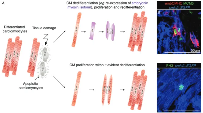
Although adult cardiomyocytes in newts and zebrafish are capable of proliferation, cardiac injury has been
Figure 5. Zebrafish heart regeneration involves proliferation of mature and dedifferentiated cardiomyocytes. (A) Model showing two possible modes of cardiomyocyte proliferation during cardiac regeneration in zebrafish. At the vicinity of the injured zone, cell-cycling activity is observed in dedifferentiated cardiomyocytes, which re-express embryonic markers; this response predominantly occurs during the initial stages (1–2 weeks) of regeneration [99]. Cardiomyocytes can also enter mitosis without a visible modification of their structural differentiation status. This process occurs in an organ-wide pattern during regeneration but also in intact hearts during ontogenetic growth. (B, C) Confocal images of heart sections from transgenic fish cmlc2:EGFP (blue) at 7 days post-cryoinjury (dpci). (B) A population of cardiomyocytes (blue) at the injury border expresses a proliferation marker, Minichromosome maintenance complex component 5 (MCM5; green) and reactivates the expression of an embryo-specific isoform of Cardiac myosin heavy chain (embCMHC; red), which indicates cellular dedifferentiation. (C) The immunostaining for phospho-(Ser10)-Histone H3 (PH3; green) reveals the presence of mitotic cardiomyocytes at the injury-remote position; in this case, cell cycle re-entry occurs without a re-expression of embCMHC [99,186]
shown to trigger several features of cellular dedif-ferentiation in these model organisms. Specifically, transmission electron microscopy imaging in the zebrafish has revealed that proliferating cardiomyocytes show disassembled sarcomeric structures in the vicinity of the injury [97]. Moreover, certain developmental genes, such as the cardiac transcription factors gata4 and hand2, or embryo-specific myosin heavy chain, are up-regulated in the regenerating myocardium in zebrafish [97–99,135–138]. Ventricular injury in newts has been associated with the down-regulation of cardiac muscle genes [8,93–96]. In these species, dediffer-entiation has been suggested to facilitate mitotic and morphogenetic activity during reconstitution of the lost part of the myocardium [96,97,99,136]. Never-theless, a clear interdependency between these two processes has not yet been established. Dedifferen-tiation of adult mammalian cardiomyocytes has also been observed during cardiac remodelling under diverse circumstances, including heart ischaemia [139–142]. In addition to sarcomere disorganization, this con-version is also characterized by a metabolic switch to carbohydrates to preserve cardiac function under pathophysiological stress. However, such modulation of structural and metabolic activities is not asso-ciated with enhanced cardiomyocyte proliferation [139,140,143–146]. Further comparative analysis between different model organisms would shed light on
the mechanisms governing plasticity of cardiomyocytes in response to heart injury.
In summary, several in vitro and in vivo studies have shown that mammalian, zebrafish and newt car-diomyocyte cell division occurs either with or without any evident sign of dedifferentiation [96,99,111]. In zebrafish, the local generation of less-differentiated cardiomyocytes in the proliferative zone of the injury border is accompanied by stimulation of mitotic divisions in the remote part of the ventricle, where dedifferentiation was not detected (Figure 5). The organ-wide proliferative response resembles a compen-satory mode of regeneration, such as in mammalian liver [147]. Further studies are needed to determine the mech-anisms and significance of enhanced cardiomyocyte proliferation in the injury-remote part of the ventricle. Moreover, the level of independence between dediffer-entiation and proliferation has to be thoroughly assessed under various conditions, such as cardiac regeneration, normal ontogenetic growth or homeostatic turnover.
Heart: cellular interactions in cardiac
regeneration
Cardiac regeneration in lower vertebrates involves essential molecular interplays between cardiomyocytes
Regeneration versus scarring in vertebrate appendages and heart 9
and other cardiac components, such as epicardium, endocardium and nerves [148–150]. Among these interactions, FGF, IGF and retinoic acid (RA) signalling represent key communicative elements between epi/ endocardial cells and cardiomyocytes [138,151–153]. Shortly after injury, the epi/endocardium start to re-express embryonic markers, including the tran-scription factors tbx18, wt1 or the RA-synthetizing enzyme retinaldehyde dehydrogenase 2 (raldh2), and release RA, which represents an important mitogen for cardiomyocytes during development and regeneration [138,152,154]. In addition, epicardial cells have been shown to enhance the migration and integration of cardiomyocytes in the injured area [155,156] and to promote neovascularization in this zone through the production of epicardial-derived cells (EPDCs), which migrate in the regenerating myocardium and form perivascular cells [138,157,158]. Thus, epicardial cells play pleiotropic roles during cardiac regeneration in zebrafish. Interestingly, a developmental reactivation of the adult epicardium has also been observed after myocardial infarction in adult mammals [148,159,160]. Therefore, the potential of epicardial cells might be fur-ther considered in the treatment of myocardial infarction in humans.
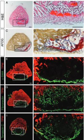
In opposition to the concept of ’scarless regenera-tion’ in vertebrate appendages, accumulation of fibrotic tissue in the post-infarcted heart is thought to play a beneficial structural role by providing robustness to the force-generating contractile organ [161,162]. In mammals, dense layers of collagen are deposited to replace a damaged part of the ventricular wall, which is challenged to withstand the high internal blood pres-sure. In comparison to humans and rodents, fish and amphibian ventricles are confronted by a lesser work-load [53,163,164], which may permit heart regener-ation without the involvement of compact scarring. Instead, a transient collagenous trabecular network is sufficient and necessary to support the function of the injured ventricle and to provide a guidance scaffold for invading cardiomyocytes in zebrafish (Figure 6A–D) and newts [52]. Our laboratory has demonstrated that the suppression of collagen deposition by inhibition of TGFβ signalling coincided with regenerative fail-ure and geometrical deformation of infarct shape [165]. Thus, collagen-deficient infarcts can lead to detrimen-tal remodelling in both mammals and fish. A balance between transient fibrosis and progressive replenishing of cardiomyocytes has to be achieved to synchronize the reparative and regenerative processes according to phys-iological conditions.
Transdifferentiation of fibroblasts into myofi-broblasts, which express α-Smooth muscle actin (α-SMA), is associated with fibrosis in mammalian heart [166–170]. In injured hearts of zebrafish, α-SMA-positive myofibroblasts accumulate along a peripheral rim of the post-infarct and only a few of these cells penetrate the central portion of the damaged zone [165] (Figure 6E, F). This distribution suggests a role of the contractile fibroblasts in providing mechanical stability
Figure 6. Transient connective tissue in the post-infarcted in zebrafish; transverse sections of the zebrafish ventricle at 14 days post-cryoinjury (dpci); the post-infarcted area is encircled with a dashed line. (A, B) Haematoxylin (dark purple, nuclei) and eosin (red, proteins) (H&E) staining; the infarct contains acellular rem-nants of fibrin that were deposited shortly after cryoinjury (pink) and granulation tissue. (C, D) Aniline blue, acid Fuchsin and Orange G (AFOG) staining reveals collagen in blue, fibrin in red and cyto-plasm in orange; the infarct tissue is supported by fibrin (red) and loose network of collagen fibres that serves as a provisional struc-tural matrix and a scaffold for migrating cardiomyocytes (orange) [44]. (E–J) Confocal images of heart sections immunostained with antibody against Tropomyosin (TPM; red) to highlight the intact myocardium and a marker of connective tissue. (E, F) α-Smooth muscle actin (α-SMA)-positive myofibroblasts (green) accumulate along the outer border of the infarct. (G, H) Fibronectin (green) forms a reticular pattern in the infarct zone. (I, J) Tenascin C (green) is predominantly detected at the interface of connective tissue and regenerating myocardium. Scale bars = 100 μm
to the outer border of the damaged ventricular wall in zebrafish. Other fibroblast populations infiltrate the mid-dle portion of the post-infarct, where they produce the ECM that is known not only to have structural functions but also to regulate many cellular processes, including cell migration, proliferation and differentiation, via, for instance, integrin signalling [171–174]. Cell–substrate communication has been attributed to various adhesive and matricellular proteins, including fibronectin and
tenascin C, respectively [166,175–177]. During heart regeneration in zebrafish, the entire post-infarct area dis-plays abundant expression of fibronectin, while tenascin C remains predominantly enriched at the interface between the regenerating myocardium and connective tissue [155,165] (Figure 6G–J). Similarly, fibronectin, tenascin-C and hyaluronic acid form a pro-regenerative matrix in the newt heart [178]. During regeneration, this connective matrix progressively resolves by mech-anisms that still remain poorly understood. Defining the intrinsic properties of fibroblasts and the external factors that determine the composition and dynamics of the ECM in the myocardial post-infarcts would be highly relevant for regenerative medicine.
Outlook
The appendage and heart cells of some anamniotic vertebrates possess an intrinsic ability to respond to injury by proliferation and reactivation of morpho-genetic programmes to replace the missing structures. This remarkable regenerative capability correlates with the relatively low cytospecialization of the adult organs in teleost fish and urodele amphibians in comparison to mammals. Histological analyses indicate that the adult differentiated cells of the non-mammalian and mam-malian species cannot be directly placed on the same scale. Although mature appendage and cardiac cells in fish and urodeles maintain an intrinsic proliferative potential, cellular dedifferentiation is often observed at the site of injury of the regenerating organs. This sug-gests a general significance of the separation of tissues from one another and a function of tissue isolation for regaining of a certain degree of cellular plasticity. In this context, dedifferentiation does not need to imply a com-plete reversion to an embryonic state, but a temporary reduction of the cellular complexity, which may facil-itate proliferation and morphogenesis. Mimicking this strategy in injured mammalian organs would demand a molecular toolkit to impose a transient and partial reversion of the local tissue complexity without repro-gramming into the pluripotent state. The recent advances of cell fate conversion suggest that the manipulation of cellular specialization seems to be an achievable goal [179–181]. The challenging part of this task concerns the understanding of organismic factors that impact tissue regeneration at molecular and biomechanical levels. The combination of findings across different animals will help to recognize the permissive and restrictive factors to establish a regeneration-competent environment. These advances will move the field of regenerative biology closer to clinical implementation.
Acknowledgements
We thank L Mène-Saffrané, A Buchala, T Carney, D König and C Pfefferli for critical reading of the review,
V Duruz for preparing a figure, E Ehler for images of mammalian cardiomyocytes and M Celio for pro-viding histological material. This work was supported by the Schweizerische Stiftung für die Erforschung der Muskelkrankheiten and the Swiss National Sci-ence Foundation (Grant Nos 310030_159995 and CRSII3_147675).
Author contributions
All authors contributed equally to this review and revised the final manuscript.
References
1. Galliot B, Ghila L. Cell plasticity in homeostasis and regeneration.
Mol Reprod Dev 2010; 77: 837–855.
2. Bely AE, Nyberg KG. Evolution of animal regeneration: re-emergence of a field. Trends Ecol Evol 2010; 25: 161–170. 3. Poss KD. Advances in understanding tissue regenerative capacity
and mechanisms in animals. Nat Rev Genet 2010; 11: 710–722. 4. Tanaka EM, Reddien PW. The cellular basis for animal
regenera-tion. Dev Cell 2011; 21: 172–185.
5. Kumar A, Brockes JP. Nerve dependence in tissue, organ, and appendage regeneration. Trends Neurosci 2012; 35: 691–699. 6. Godwin JW, Rosenthal N. Scar-free wound healing and
regenera-tion in amphibians: immunological influences on regenerative suc-cess. Differentiation 2014; 87: 66–75.
7. Sanchez Alvarado A, Tsonis PA. Bridging the regeneration gap: genetic insights from diverse animal models. Nat Rev Genet 2006; 7:873–884.
8. Borchardt T, Braun T. Cardiovascular regeneration in non-mammalian model systems: what are the differences between newts and man? Thrombosis Haemostasis 2007; 98: 311–318. 9. Gemberling M, Bailey TJ, Hyde DR, et al. The zebrafish as a model
for complex tissue regeneration. Trends Genet 2013; 29: 611–620. 10. Pfefferli C, Ja´zwi´nska A. The art of fin regeneration in zebrafish.
Regeneration 2015; 2: 72–83.
11. Kikuchi K, Poss KD. Cardiac regenerative capacity and mecha-nisms. Annu Rev Cell Dev Biol 2012; 28: 719–741.
12. Brockes JP, Gates PB. Mechanisms underlying vertebrate limb regeneration: lessons from the salamander. Biochem Soc Trans 2014; 42: 625–630.
13. McCusker C, Bryant SV, Gardiner DM. The axolotl limb blastema: cellular and molecular mechanisms driving blastema formation and limb regeneration in tetrapods. Regeneration 2015; 2: 54–71. 14. Ausoni S, Sartore S. From fish to amphibians to mammals: in search
of novel strategies to optimize cardiac regeneration. J Cell Biol 2009; 184: 357–364.
15. Vargas-Gonzalez A, Prado-Zayago E, Leon-Olea M, et al. [Myocar-dial regeneration in Ambystoma mexicanum after surgical injury].
Arch Cardiol Mex 2005; 75(suppl 3): S3 21–29.
16. Cano-Martinez A, Vargas-Gonzalez A, Guarner-Lans V, et al. Functional and structural regeneration in the axolotl heart (Ambystoma mexicanum) after partial ventricular amputation. Arch
Cardiol Mex 2010; 80: 79–86.
17. Wehner D, Weidinger G. Signaling networks organizing regenera-tive growth of the zebrafish fin. Trends Genet 2015; 31: 336–343. 18. Simon A, Tanaka EM. Limb regeneration. Wiley Interdiscip Rev Dev
Biol 2013; 2: 291–300.
19. Kikuchi K. Advances in understanding the mechanism of zebrafish heart regeneration. Stem Cell Res 2014; 13: 542–555.
Regeneration versus scarring in vertebrate appendages and heart 11
20. Lien CL, Harrison MR, Tuan TL, et al. Heart repair and regener-ation: recent insights from zebrafish studies. Wound Repair Regen 2012; 20: 638–646.
21. Stocum DL, Cameron JA. Looking proximally and distally: 100 years of limb regeneration and beyond. Dev Dyn 2011; 240: 943–968.
22. Tornini VA, Poss KD. Keeping at arm’s length during regeneration.
Dev Cell 2014; 29: 139–145.
23. Seifert AW, Monaghan JR, Smith MD, et al. The influence of fun-damental traits on mechanisms controlling appendage regeneration.
Biol Rev Camb Philos Soc 2012; 87: 330–345.
24. McCusker CD, Gardiner DM. Understanding positional cues in salamander limb regeneration: implications for optimizing cell-based regenerative therapies. Dis Model Mech 2014; 7: 593–599.
25. Monaghan JR, Maden M. Cellular plasticity during vertebrate appendage regeneration. In New Perspectives in Regeneration, Heber-Katz E, Stocum DL (eds). Springer: Berlin, Heidelberg, 2013; 53–75.
26. Xin M, Olson EN, Bassel-Duby R. Mending broken hearts: cardiac development as a basis for adult heart regeneration and repair. Nat
Rev Mol Cell Biol 2013; 14: 529–541.
27. Laflamme MA, Murry CE. Heart regeneration. Nature 2011; 473: 326–335.
28. Mescher AL, Neff AW. Regenerative capacity and the developing immune system. Adv Biochem Eng Biotechnol 2005; 93: 39–66. 29. Godwin JW, Brockes JP. Regeneration, tissue injury and the
immune response. J Anat 2006; 209: 423–432.
30. Brockes JP, Kumar A, Velloso CP. Regeneration as an evolutionary variable. J Anat 2001; 199: 3–11.
31. Goss RJ. Heads and tails. In Principles of Regeneration. Academic Press: New York, 1969; 191.
32. Young HE, Bailey CF, Dalley BK. Environmental conditions pre-requisite for complete limb regeneration in the postmetamorphic adult land-phase salamander, Ambystoma. Anat Rec 1983; 206: 289–294.
33. Monaghan JR, Stier AC, Michonneau F, et al. Experimentally induced metamorphosis in axolotls reduces regenerative rate and fidelity. Regeneration 2014; 1: 2–14.
34. Young HE. Existence of reserve quiescent stem cells in adults, from amphibians to humans. Curr Top Microbiol Immunol 2004; 280: 71–109.
35. Scadding SR. Phylogenic distribution of limb regeneration capacity in adult Amphibia. J Exp Zool 1977; 202: 57–67.
36. Simkin J, Sammarco MC, Dawson LA, et al. The mammalian blastema: regeneration at our fingertips. Regeneration 2015; 2: 93–105.
37. Takeo M, Chou WC, Sun Q, et al. Wnt activation in nail epithe-lium couples nail growth to digit regeneration. Nature 2013; 499: 228–232.
38. Shieh S-J, Cheng T-C. Regeneration and repair of human digits and limbs: fact and fiction. Regeneration 2015; DOI: 10.1002/reg2.41. 39. Ito K, Morioka M, Kimura S, et al. Differential reparative
phe-notypes between zebrafish and medaka after cardiac injury. Dev
Dynam 2014; 243: 1106–1115.
40. Zhang R, Han P, Yang H, et al. In vivo cardiac reprogramming contributes to zebrafish heart regeneration. Nature 2013; 498: 497–501.
41. Wang J, Panakova D, Kikuchi K, et al. The regenerative capacity of zebrafish reverses cardiac failure caused by genetic cardiomyocyte depletion. Development 2011; 138: 3421–3430.
42. Curado S, Stainier DY, Anderson RM. Nitroreductase-mediated cell/tissue ablation in zebrafish: a spatially and temporally con-trolled ablation method with applications in developmental and regeneration studies. Nat Protoc 2008; 3: 948–954.
43. Curado S, Anderson RM, Jungblut B, et al. Conditional targeted cell ablation in zebrafish: a new tool for regeneration studies. Dev Dyn 2007; 236: 1025–1035.
44. Chablais F, Veit J, Rainer G, et al. The zebrafish heart regener-ates after cryoinjury-induced myocardial infarction. BMC Dev Biol 2011; 11: 21.
45. Gonzalez-Rosa JM, Martin V, Peralta M, et al. Extensive scar formation and regression during heart regeneration after cryoinjury in zebrafish. Development 2011; 138: 1663–1674.
46. Schnabel K, Wu CC, Kurth T, et al. Regeneration of cryoin-jury induced necrotic heart lesions in zebrafish is associated with epicardial activation and cardiomyocyte proliferation. Plos One 2011; 6: e18503.
47. Mendis S, Puska P, Norrving B. Global Atlas on
Cardiovascu-lar Disease Prevention and Control. World Health Organization:
Geneva, 2011.
48. Goss RJ. Anatomy of antler regeneration. In Principles of
Regener-ation. Academic Press: New York, 1969; 234–235.
49. Burggren WW, Christoffels VM, Crossley DA, 2nd, et al. Com-parative cardiovascular physiology: future trends, opportunities and challenges. Acta Physiol (Oxf) 2014; 210: 257–276.
50. Farrell AP, Farrell ND, Jourdan H, et al. A perspective on the evolution of the coronary circulation in fishes and the transition to terrestrial life. In Ontogeny and Phylogeny of the Vertebrate Heart, Sedmera D, Wang T (eds). Springer: New York, 2012; 75–102. 51. Reese DE, Mikawa T, Bader DM. Development of the coronary
vessel system. Circ Res 2002; 91: 761–768.
52. Piatkowski T, Muhlfeld C, Borchardt T, et al. Reconstitution of the myocardium in regenerating newt hearts is preceded by transient deposition of extracellular matrix components. Stem Cells Dev 2013; 22: 1921–1931.
53. Hu N, Yost HJ, Clark EB. Cardiac morphology and blood pressure in the adult zebrafish. Anat Rec 2001; 264: 1–12.
54. Goss RJ. The natural history (and mystery) of regeneration. In
A History of Regeneration Research: Milestones in the Evolution of a Science, Dinsmore CE (ed.). Cambridge University Press:
Cambridge, 1991; 7–23.
55. Kragl M, Knapp D, Nacu E, et al. Cells keep a memory of their tissue origin during axolotl limb regeneration. Nature 2009; 460: 60–65.
56. Knopf F, Hammond C, Chekuru A, et al. Bone regenerates via dedifferentiation of osteoblasts in the zebrafish fin. Dev Cell 2011; 20:713–724.
57. Tu S, Johnson SL. Fate restriction in the growing and regenerating zebrafish fin. Dev Cell 2011; 20: 725–732.
58. Sousa S, Afonso N, Bensimon-Brito A, et al. Differentiated skeletal cells contribute to blastema formation during zebrafish fin regener-ation. Development 2011; 138: 3897–3905.
59. Singh SP, Holdway JE, Poss KD. Regeneration of amputated zebrafish fin rays from de novo osteoblasts. Dev Cell 2012; 22: 879–886.
60. Stewart S, Stankunas K. Limited dedifferentiation provides replace-ment tissue during zebrafish fin regeneration. Dev Biol 2012; 365: 339–349.
61. Campbell LJ, Crews CM. Wound epidermis formation and function in urodele amphibian limb regeneration. Cell Mol Life Sci 2008; 65: 73–79.
62. Geraudie J, Singer M. Nerve dependent macromolecular synthesis in the epidermis and blastema of the adult newt regenerate. J Exp
Zool 1978; 203: 455–460.
63. Simoes MG, Bensimon-Brito A, Fonseca M, et al. Denervation impairs regeneration of amputated zebrafish fins. BMC Dev Biol 2014; 14: 780.
64. Tassava RA, Garling DJ. Regenerative responses in larval axolotl limbs with skin-grafts over the amputation surface. J Exp Zool 1979; 208:97–110.
65. Mescher AL. Effects on adult newt limb regeneration of partial and complete skin flaps over the amputation surface. J Exp Zool 1976; 195:117–128.
66. Carlson BM. Some principles of regeneration in mammalian sys-tems. Anat Rec B New Anat 2005; 287: 4–13.
67. Stewart S, Gomez AW, Armstrong BE, et al. Sequential and oppos-ing activities of Wnt and BMP coordinate zebrafish bone regenera-tion. Cell Rep 2014; 6: 482–498.
68. Thorimbert V, Konig D, Marro J, et al. Bone morphogenetic protein signaling promotes morphogenesis of blood vessels, wound epider-mis, and actinotrichia during fin regeneration in zebrafish. FASEB J 2015; 29: 4299–4312.
69. Sandoval-Guzman T, Wang H, Khattak S, et al. Fundamental dif-ferences in dedifferentiation and stem cell recruitment during skele-tal muscle regeneration in two salamander species. Cell Stem Cell 2014; 14: 174–187.
70. Kumar A, Velloso CP, Imokawa Y, et al. The regenerative plasticity of isolated urodele myofibers and its dependence on MSX1. PLoS
Biol 2004; 2: E218.
71. Echeverri K, Clarke JDW, Tanaka EM. In vivo imaging indicates muscle fiber dedifferentiation is a major contributor to the regener-ating tail blastema. Dev Biol 2001; 236: 151–164.
72. Wang H, Loof S, Borg P, et al. Turning terminally differentiated skeletal muscle cells into regenerative progenitors. Nat Commun 2015; 6: 7916.
73. Muneoka K, Fox WF, Bryant SV. Cellular contribution from dermis and cartilage to the regenerating limb blastema in axolotls. Dev Biol 1986; 116: 256–260.
74. Glim JE, van Egmond M, Niessen FB, et al. Detrimental dermal wound healing: what can we learn from the oral mucosa? Wound
Repair Regen 2013; 21: 648–660.
75. Levesque M, Villiard E, Roy S. Skin wound healing in axolotls: a scarless process. J Exp Zool B Mol Dev Evol 2010; 314: 684–697.
76. Seifert AW, Maden M. New insights into vertebrate skin regenera-tion. Int Rev Cell Mol Biol 2014; 310: 129–169.
77. Seifert AW, Kiama SG, Seifert MG, et al. Skin shedding and tissue regeneration in African spiny mice (Acomys). Nature 2012; 489: 561–565.
78. Richardson R, Slanchev K, Kraus C, et al. Adult zebrafish as a model system for cutaneous wound-healing research. J Invest
Dermatol 2013; 133: 1655–1665.
79. Jazwinska A, Badakov R, Keating MT. Activin-βA signaling is required for zebrafish fin regeneration. Curr Biol 2007; 17: 1390–1395.
80. Seifert AW, Monaghan JR, Voss SR, et al. Skin regeneration in adult axolotls: a blueprint for scar-free healing in vertebrates. PLoS One 2012; 7: e32875.
81. Wehner D, Cizelsky W, Vasudevaro MD, et al. Wnt/β-catenin sig-naling defines organizing centers that orchestrate growth and differ-entiation of the regenerating zebrafish caudal fin. Cell Rep 2014; 6: 467–481.
82. Blum N, Begemann G. Osteoblast de- and redifferentiation is con-trolled by a dynamic response to retinoic acid during zebrafish fin regeneration. Development 2015; 142: 2894–2903..
83. Blum N, Begemann G. Retinoic acid signaling spatially restricts osteoblasts and controls ray–interray organization during zebrafish fin regeneration. Development 2015; 142: 2888–2893.
84. Mateus R, Lourenco R, Fang Y, et al. Yap control of tissue growth relies on cell density and F-actin in zebrafish fin regeneration.
Development 2015; 142: 2752–2763.
85. Pfefferli C, Muller F, Jazwinska A, et al. Specific NuRD compo-nents are required for fin regeneration in zebrafish. BMC Biol 2014; 12:30.
86. Akimenko MA, Mari-Beffa M, Becerra J, et al. Old questions, new tools, and some answers to the mystery of fin regeneration. Dev Dyn 2003; 226: 190–201.
87. Nacu E, Glausch M, Le HQ, et al. Connective tissue cells, but not muscle cells, are involved in establishing the proximo-distal outcome of limb regeneration in the axolotl. Development 2013; 140:513–518.
88. Bryant SV, French V, Bryant PJ. Distal regeneration and symmetry.
Science 1981; 212: 993–1002.
89. Endo T, Bryant SV, Gardiner DM. A stepwise model system for limb regeneration. Dev Biol 2004; 270: 135–145.
90. Chang HY, Chi JT, Dudoit S, et al. Diversity, topographic differen-tiation, and positional memory in human fibroblasts. Proc Natl Acad
Sci USA 2002; 99: 12877–12882.
91. Rinn JL, Bondre C, Gladstone HB, et al. Anatomic demarcation by positional variation in fibroblast gene expression programs. PLoS
Genet 2006; 2: e119.
92. Poss KD, Wilson LG, Keating MT. Heart regeneration in zebrafish.
Science 2002; 298: 2188–2190.
93. Witman N, Murtuza B, Davis B, et al. Recapitulation of devel-opmental cardiogenesis governs the morphological and functional regeneration of adult newt hearts following injury. Dev Biol 2011; 354:67–76.
94. Bader D, Oberpriller JO. Repair and reorganization of minced cardiac muscle in the adult newt (Notophthalmus viridescens).
J Morphol 1978; 155: 349–357.
95. Bader D, Oberpriller J. Autoradiographic and electron microscopic studies of minced cardiac muscle regeneration in the adult newt,
Notophthalmus viridescens. J Exp Zool 1979; 208: 177–193.
96. Laube F, Heister M, Scholz C, et al. Re-programming of newt cardiomyocytes is induced by tissue regeneration. J Cell Sci 2006; 119:4719–4729.
97. Jopling C, Sleep E, Raya M, et al. Zebrafish heart regeneration occurs by cardiomyocyte dedifferentiation and proliferation. Nature 2010; 464: 606–609.
98. Kikuchi K, Holdway JE, Werdich AA, et al. Primary contribution to zebrafish heart regeneration by gata4+cardiomyocytes. Nature 2010; 464: 601–605.
99. Sallin P, de Preux Charles AS, Duruz V, et al. A dual epimorphic and compensatory mode of heart regeneration in zebrafish. Dev Biol 2015; 399: 27–40.
100. Rana MS, Christoffels VM, Moorman AF. A molecular and genetic outline of cardiac morphogenesis. Acta Physiol (Oxf) 2013; 207: 588–615.
101. Leu M, Ehler E, Perriard JC. Characterisation of postnatal growth of the murine heart. Anat Embryol (Berl) 2001; 204: 217–224. 102. Porrello ER, Mahmoud AI, Simpson E, et al. Regulation of neonatal
and adult mammalian heart regeneration by the miR-15 family. Proc
Natl Acad Sci USA 2013; 110: 187–192.
103. Porrello ER, Mahmoud AI, Simpson E, et al. Transient regener-ative potential of the neonatal mouse heart. Science 2011; 331: 1078–1080.
104. Li F, Wang X, Capasso JM, et al. Rapid transition of cardiac myocytes from hyperplasia to hypertrophy during postnatal devel-opment. J Mol Cell Cardiol 1996; 28: 1737–1746.
105. Jonker SS, Louey S, Giraud GD, et al. Timing of cardiomyocyte growth, maturation, and attrition in perinatal sheep. FASEB J 2015; 29:4346–4357.
106. Mollova M, Bersell K, Walsh S, et al. Cardiomyocyte proliferation contributes to heart growth in young humans. Proc Natl Acad Sci
Regeneration versus scarring in vertebrate appendages and heart 13
107. Brette F, Orchard C. Resurgence of cardiac t-tubule research.
Phys-iology (Bethesda) 2007; 22: 167–173.
108. Ibrahim M, Gorelik J, Yacoub MH, et al. The structure and function of cardiac t-tubules in health and disease. Proc Biol Sci 2011; 278: 2714–2723.
109. Hirschy A, Schatzmann F, Ehler E, et al. Establishment of cardiac cytoarchitecture in the developing mouse heart. Dev Biol 2006; 289: 430–441.
110. Brette F, Luxan G, Cros C, et al. Characterization of isolated ventricular myocytes from adult zebrafish (Danio rerio). Biochem
Biophys Res Commun 2008; 374: 143–146.
111. Bettencourt-Dias M, Mittnacht S, Brockes JP. Heterogeneous pro-liferative potential in regenerative adult newt cardiomyocytes. J Cell
Sci 2003; 116: 4001–4009.
112. Shiels HA, White E. Temporal and spatial properties of cellular Ca2+flux in trout ventricular myocytes. Am J Physiol Regul Integr
Comp Physiol 2005; 288: R1756–1766.
113. Zhang PC, Llach A, Sheng XY, et al. Calcium handling in zebrafish ventricular myocytes. Am J Physiol Regul Integr Comp Physiol 2011; 300: R56–66.
114. Olivetti G, Cigola E, Maestri R, et al. Aging, cardiac hypertro-phy and ischemic cardiomyopathy do not affect the proportion of mononucleated and multinucleated myocytes in the human heart.
J Mol Cell Cardiol 1996; 28: 1463–1477.
115. Adler CP, Costabel U. Cell number in human heart in atrophy, hypertrophy, and under the influence of cytostatics. Recent Adv Stud
Cardiac Struct Metab 1975; 6: 343–355.
116. Zebrowski DC, Engel FB. The cardiomyocyte cell cycle in hyper-trophy, tissue homeostasis, and regeneration. Rev Physiol Biochem
Pharmacol 2013; 165: 67–96.
117. Borisov AB. Regeneration of skeletal and cardiac muscle in mam-mals: do nonprimate models resemble human pathology? Wound
Repair Regen 1999; 7: 26–35.
118. Pluess M, Ehler E. Cardiac cytoarchitecture in health and disease. In
Cardiac Cytoarchitecture: How to Maintain a Working Heart, Ehler
E (ed.). Springer: Berlin, Heidelberg, 2015; 1–15.
119. Zebrowski DC, Vergarajauregui S, Wu CC, et al. Developmental alterations in centrosome integrity contribute to the post-mitotic state of mammalian cardiomyocytes. Elife 2015; 4: e05563. 120. Engel FB, Schebesta M, Keating MT. Anillin localization defect
in cardiomyocyte binucleation. J Mol Cell Cardiol 2006; 41: 601–612.
121. Soonpaa MH, Field LJ. Survey of studies examining mammalian cardiomyocyte DNA synthesis. Circ Res 1998; 83: 15–26. 122. Soonpaa MH, Rubart M, Field LJ. Challenges measuring
cardiomy-ocyte renewal. Biochim Biophys Acta 2013; 1833: 799–803. 123. Senyo SE, Steinhauser ML, Pizzimenti CL, et al. Mammalian
heart renewal by pre-existing cardiomyocytes. Nature 2013; 493: 433–436.
124. Bergmann O, Bhardwaj RD, Bernard S, et al. Evidence for car-diomyocyte renewal in humans. Science 2009; 324: 98–102. 125. Eulalio A, Mano M, Dal Ferro M, et al. Functional screening
identifies miRNAs inducing cardiac regeneration. Nature 2012; 492:376–381.
126. Pandey R, Ahmed RP. MicroRNAs Inducing proliferation of quies-cent adult cardiomyocytes. Cardiovasc Regen Med 2015; 2: e519. 127. Yin VP, Lepilina A, Smith A, et al. Regulation of zebrafish heart
regeneration by miR-133. Dev Biol 2012; 365: 319–327. 128. Pasumarthi KB, Nakajima H, Nakajima HO, et al. Targeted
expres-sion of cyclin D2 results in cardiomyocyte DNA synthesis and infarct regression in transgenic mice. Circ Res 2005; 96: 110–118. 129. Woo YJ, Panlilio CM, Cheng RK, et al. Therapeutic delivery of cyclin A2 induces myocardial regeneration and enhances car-diac function in ischemic heart failure. Circulation 2006; 114: I206–213.
130. Bersell K, Arab S, Haring B, et al. Neuregulin1/ErbB4 signaling induces cardiomyocyte proliferation and repair of heart injury. Cell 2009; 138: 257–270.
131. Gemberling M, Karra R, Dickson AL, et al. Nrg1 is an injury-induced cardiomyocyte mitogen for the endogenous heart regeneration program in zebrafish. Elife 2015; 4: e05871. 132. Kuhn B, Del Monte F, Hajjar RJ, et al. Periostin induces
prolifera-tion of differentiated cardiomyocytes and promotes cardiac repair.
Nat Med 2007; 13: 962–969.
133. Engel FB, Schebesta M, Duong MT, et al. p38 MAP kinase inhi-bition enables proliferation of adult mammalian cardiomyocytes.
Genes Dev 2005; 19: 1175–1187.
134. Engel FB, Hsieh PC, Lee RT, et al. FGF1/p38 MAP kinase inhibitor therapy induces cardiomyocyte mitosis, reduces scarring, and res-cues function after myocardial infarction. Proc Natl Acad Sci USA 2006; 103: 15546–15551.
135. Sleep E, Boue S, Jopling C, et al. Transcriptomics approach to investigate zebrafish heart regeneration. J Cardiovasc Med
(Hager-stown) 2010; 11: 369–380.
136. Jopling C, Boue S, Izpisua Belmonte JC. Dedifferentiation, transd-ifferentiation and reprogramming: three routes to regeneration. Nat
Rev Mol Cell Biol 2011; 12: 79–89.
137. Schindler YL, Garske KM, Wang J, et al. Hand2 elevates cardiomy-ocyte production during zebrafish heart development and regenera-tion. Development 2014; 141: 3112–3122.
138. Lepilina A, Coon AN, Kikuchi K, et al. A dynamic epicardial injury response supports progenitor cell activity during zebrafish heart regeneration. Cell 2006; 127: 607–619.
139. Szibor M, Poling J, Warnecke H, et al. Remodeling and dediffer-entiation of adult cardiomyocytes during disease and regeneration.
Cell Mol Life Sci 2014; 71: 1907–1916.
140. Kubin T, Poling J, Kostin S, et al. Oncostatin M is a major mediator of cardiomyocyte dedifferentiation and remodeling. Cell Stem Cell 2011; 9: 420–432.
141. Dispersyn GD, Mesotten L, Meuris B, et al. Dissociation of car-diomyocyte apoptosis and dedifferentiation in infarct border zones.
Eur Heart J 2002; 23: 849–857.
142. Ausma J, Thone F, Dispersyn GD, et al. Dedifferentiated cardiomy-ocytes from chronic hibernating myocardium are ischemia-tolerant.
Mol Cell Biochem 1998; 186: 159–168.
143. Taegtmeyer H, Sen S, Vela D. Return to the fetal gene program: a suggested metabolic link to gene expression in the heart. Ann N Y
Acad Sci 2010; 1188: 191–198.
144. Dirkx E, da Costa Martins PA, De Windt LJ. Regulation of fetal gene expression in heart failure. Biochim Biophys Acta 2013; 1832: 2414–2424.
145. Rajabi M, Kassiotis C, Razeghi P, et al. Return to the fetal gene program protects the stressed heart: a strong hypothesis. Heart Fail
Rev 2007; 12: 331–343.
146. Hein S, Arnon E, Kostin S, et al. Progression from compensated hypertrophy to failure in the pressure-overloaded human heart: structural deterioration and compensatory mechanisms. Circulation 2003; 107: 984–991.
147. Fausto N, Campbell JS, Riehle KJ. Liver regeneration. Hepatology 2006; 43: S45–53.
148. Masters M, Riley PR. The epicardium signals the way towards heart regeneration. Stem Cell Res 2014; 13: 683–692.
149. Wang J, Cao J, Dickson AL, et al. Epicardial regeneration is guided by cardiac outflow tract and Hedgehog signalling. Nature 2015; 522:226–230.
150. Mahmoud AI, O’Meara CC, Gemberling M, et al. Nerves regulate cardiomyocyte proliferation and heart regeneration. Dev Cell 2015; 34:387–399.