ORIGINAL PAPER
Comparison of faecal techniques including FLOTAC
for copromicroscopic detection of first stage larvae
of Angiostrongylus vasorum
Manuela Schnyder&Maria P. Maurelli&
Maria E. Morgoglione&Lucia Kohler&Peter Deplazes&
Paul Torgerson&Giuseppe Cringoli&Laura Rinaldi
Received: 23 November 2010 / Accepted: 6 December 2010 / Published online: 22 December 2010 # Springer-Verlag 2010
Abstract Angiostrongylus vasorum is a metastrongylid nematode that resides in the pulmonary arteries and the right heart chambers. In dogs, infection results in respira-tory, bleeding and neurological disorders and further clinical signs. In the present study, FLOTAC was evaluated for the detection of first-stage larvae (L1) of A. vasorum in canine faecal samples. This technique is based on the counting of parasitic stages (eggs, larvae, oocysts and cysts) in chambers after spinning of faecal samples onto a surface. In a first step, nine flotation solutions were evaluated using faeces of two experimentally infected dogs. Zinc sulphate (specific gravity (s.g.) 1.2) and zinc sulphate plus potassium iodomercurate (s.g. 1.45) gave good results. However, with the latter technique, the larvae were slightly deformed. Subsequently, FLOTAC, using zinc sulphate, was com-pared through a randomisation technique with McMaster,
flotation in tube and Baermann–Wetzel technique. The
mean larvae per gramme (LPG) obtained by the FLOTAC for both dogs was significantly higher (P<0.05) than those obtained by the other three techniques (the means of the other techniques all lie below the 95% CI of the mean LPG of the FLOTAC technique). In addition, the FLOTAC results were consistent across replicates with only Poisson (or random) variation between individual replicates. The other techniques appear to be less consistent with evidence of extra-Poisson variation in at least one of the two dogs across the replicates within each technique. The FLOTAC technique may contribute to an improvement of the ability to diagnose canine lungworm infections and represent a valuable alternative for larval counting of A. vasorum in faecal samples, especially following transport or storage where there may be limited larvae viability, and larval migration techniques cannot be used.
Introduction
Angiostrongylus vasorum is a metastrongylid nematode that resides as adult stage in the pulmonary arteries and the right heart in dogs and other carnivores. Snails and slugs are the obligatory intermediate hosts (Eckert and Lämmler 1972; Guilhon and Bressou1960). The distribution of A. vasorum includes Europe, South and North America and Africa (Morgan et al. 2010). Respiratory, bleeding and neurolog-ical disorders, as well as non-specific signs, such as anorexia and exercise intolerance, are frequent (Chapman et al.2004; Mason1987; Patteson et al.1993; Staebler et al.
2005). Sudden death is also reported (Chapman et al.2004; Denk et al. 2009; Garosi et al. 2005; Wessmann et al.
2006). In particular, chronic angiostrongylosis with gradual onset and unpredictable progression is a diagnostic
chal-M. Schnyder
:
L. Kohler:
P. DeplazesInstitute of Parasitology, Vetsuisse Faculty, University of Zurich, Zurich, Switzerland
M. P. Maurelli
:
M. E. Morgoglione:
G. Cringoli:
L. Rinaldi Dipartimento di Patologia e Sanità Animale,Università degli Studi di Napoli Federico II, Naples, Italy
P. Torgerson
Section of Veterinary Epidemiology, Vetsuisse Faculty, University of Zurich,
Zurich, Switzerland M. Schnyder (*)
Institute of Parasitology, University of Zurich, Winterthurerstrasse 266a,
CH-8057, Zurich, Switzerland
lenge (Staebler et al.2005; Traversa and Guglielmini2008). At present, diagnosis relies on clinical manifestations, diagnostic imaging, bronchial washings and on the detec-tion of the first-stage larvae (L1) in faecal samples. Faecal smears can be used for diagnosis in general practise (Humm and Adamantos 2010), but larval migration techniques, such as the Baermann–Wetzel technique (Eckert et al.
2008) are recommended. These do not require sophisticated technologies and are highly specific if larval morphology is
considered (Eckert et al. 2008; McGarry and Morgan
2009). Limitations of the Baermann–Wetzel technique for the
detection of L1 in naturally infected dogs are the prepatent period and irregular larval shedding after onset of patency, possibly due to low worm burdens. Fatal clinical cases with negative Baermann migration results are reported (Denk et al.
2009; Patteson et al.1993). In experimentally inoculated dogs, larval excretion has been described as irregular, and animals may present with periods of negative or very low faecal larval output (Oliveira-Junior et al. 2006). The sensitivity of Baermann–Wetzel analysis may also be reduced by larval deformation or death, due to delayed analysis of faecal samples due to postal transport to the laboratory. Recently, biomolecular (Al-Sabi et al.2010; Jefferies et al.2009;) and serological (Schnyder et al. 2010b; Verzberger-Epshtein et al. 2008) techniques have also been evaluated for the diagnosis of canine angiostrongylosis.
The FLOTAC techniques are described for the copromi-croscopic diagnosis of parasites in humans and animals (Cringoli 2006; Cringoli et al. 2010). In particular, the detection of Aelurostrongylus abstrusus in cats (Gaglio et al. 2008), Crenosoma vulpis in dogs (Rinaldi et al. 2007) and of lungworms in sheep (Rinaldi et al.2010) have been evaluated. For these three larval stages of lungworms, the FLOTAC techniques were shown to detect significantly greater numbers of larvae per gramme of faeces (LPG) than produced by other more widely used diagnostic tools, indicating a potential for higher coproscopic sensitivity. The aim of this work was to compare the FLOTAC technique with the Baermann–Wetzel and other two standard copromicroscopic techniques, McMaster and simple flotation in tube, in faecal samples of dogs experimentally inoculated with A. vasorum.
Materials and methods
Animals, experimental inoculation with third stage larvae (L3) of A. vasorum and sample collection
Faecal samples were collected during an experimental study from purpose-bred beagle dogs. The study was carried out at the experimental units of the Vetsuisse Faculty at the University of Zurich (Switzerland) and approval by the
Cantonal Veterinary Office of Zurich was obtained prior to study start (permission number 13/2008). The dogs aged 11 months were inoculated with 200 L3 of A. vasorum from experimentally infected Biomphalaria glabrata snails through intragastric administration, as previously described (Schnyder et al.2009). Dogs were examined weekly clinically and daily coproscopically starting from 40 days post inoculation (dpi) by the Baermann–Wetzel technique (Eckert et al.2008). A composite fresh faecal sample (about 300 g) from each of the two dogs was collected on 99–100 dpi for a first evaluation of different flotation solutions (FS) and on 166–167 dpi for a comparison of four different copromicroscopic techniques. The composite samples were accurately homogenised with an electric mixer (Kenwood) to break clusters in faeces.
Evaluation of flotation solutions for FLOTAC
Forty grammes of faecal sample were weighted and 360 ml of tap water (dilution ratio, 1:10) were added. The suspension was then homogenised using a household mixer, filtered through a wire mesh (aperture, 250 μm), and the debris discarded. The filtered suspension was divided into 54 aliquots of 6 ml to have six replicates for each of the nine FS tested. Each aliquot was placed in 15 ml tubes and the tubes were centrifuged for 3 min at 1,500 rpm (170 g). The supernatant was poured off and discarded. The tube was then filled with the FS to the previous 6-ml level and slowly agitated. The resulting suspension was used to load one of the two chambers of the FLOTAC-400 (volume of each chamber, 5 ml). After centrifugation of the FLOTAC apparatus at 1,000 rpm (120 g) for 5 min, the top of the flotation chambers were translated and the larvae were counted. Thus, a single flotation chamber of the FLOTAC-400 was utilised for each replicate (analytic sensitivity, two LPG).
The following nine aqueous FS (FS1–FS9), (Cringoli et al. 2010) were utilised: (FS1) sucrose and formaldehyde, specific gravity (s.g.) 1.200; (FS2) sodium chloride, s.g. 1.200; (FS3) zinc sulphate, s.g. 1.200; (FS4) sodium nitrate, s.g. 1.200; (FS5) sucrose and mercury II iodide and potassium iodide, s.g. 1.250; (FS6) magnesium sulphate, s.g. 1.280; (FS7) zinc sulphate, s.g. 1.350; (FS8) mercury II iodide and potassium iodide, s.g. 1.440; (FS9) zinc sulphate and mercury II iodide and potassium iodide, s.g. 1.450. All solutions were prepared at room temperature on the same day, and the densities were measured with a hydrometer at 20°C.
Comparison between FLOTAC, McMaster, simple flotation in tube and Baermann techniques
Four different copromicroscopic techniques were compared for their efficiency in terms of sensitivity, mean LPG and
variation between samples with two individual samples from the dogs collected 166 and 167 dpi. The following four techniques were compared:
1. FLOTAC technique ( Cringoli et al.2010),
2. McMaster technique (MAFF1986),
3. Simple flotation technique (MAFF1986), and 4. Baermann–Wetzel technique (Eckert et al.2008).
For all techniques, L1 of A. vasorum were detected and counted, using a light microscope (×10 or ×40 magnifica-tion) for all replicates. In particular, ten replicates of the Baermann technique were first performed and analysed, as described in Eckert et al. (2008). Thirty grammes of the remaining faecal samples were suspended in tap water (dilution ratio, 1:10). The suspension was then poured through a wire mesh screen, having an aperture of 250μm and was divided into 30 aliquots of 6 ml in order to have ten replicates of each of the three flotation-based methods (techniques 1, 2, and 3). All tubes were centrifuged for 3 min at 1,500 rpm (170 g), and the supernatant was poured off and discarded (MAFF 1986), leaving a pellet in the tube. Each tube was then randomly assigned to a technique. FS3 (s.g. 1.200) was used for techniques 1, 2, and 3. This solution was chosen based on the results of the evaluation of flotation solutions for FLOTAC described above.
For the FLOTAC-400 the tubes were filled with the FS to the previous 6-ml level and slowly agitated. The resulting suspension was used to load the FLOTAC-400 as described for the calibration. Similarly, two chambers of the McMaster slide (Weber Scientific International, England; volume, 1.0 ml) were loaded, left for 10 min and larvae present in the whole slide were counted (analytic sensitivity, ten LPG).
For the simple flotation technique, tubes were filled with the FS until a convex meniscus was formed, then covered with a 18×18-mm coverslip and left for 15 min. The coverslip was then transferred on a slide and entirely examined (analytic sensitivity, one LPG).
Statistical analyses
All statistical analyses were undertaken with the statistical software R 2.4.0 (R Development Core Team,2006) using a randomisation technique which was similar to that described by Ruegg et al. (2008). Initially, the arithmetic mean LPG was calculated for the nine different FS utilised for the FLOTAC calibration. As the FLOTAC technique as described has a sensitivity of two LPG (i.e. each observed larvae in the chamber represents two larvae per gramme), the raw counts observed in the chamber were used for analysis (with subsequent multiplication factor of 2). Assuming the sample is well mixed and the techniques give consistent results, the raw larval counts of the six
repeat samples should follow a Poisson distribution with variations between replicates resulting from random varia-tion. The replicates were first tested by a randomisation test that the samples were Poisson distributed. Briefly, 10,000 samples, each containing six replicates, were generated by a Poisson random number generator with the observed mean of the raw count of the replicates. The variance of each of these 10,000 replicates was calculated, and the lower and upper 95% confidence intervals (CI) of the variances were computed. If the observed variance of the raw count is within these 95% CI, then, the observed data can be assumed to be Poisson distributed. If the variance is outside these limits, then the data is under- or over-dispersed. Likewise, the differences between different solutions can be calculated by comparing the observed mean of the raw count to the relevant percentile of the means of the randomly generated replicates from the comparison group. Data of comparison between the four techniques were double-entered and cross-checked. The mean and 95% CI of the detected LPG were calculated as described above, but in this case, using 10,000 random Poisson samples, each generating ten replicates. For each technique, the detection limit was used as a multiplier when undertaking the analysis. Thus, for the McMaster technique, the detection limit was ten LPG. The raw count (effectively larvae per 0.10 g) was first manipulated as described to calculate the percentile confidence limits of count per 0.10 g. The raw count was also tested for consistency with a Poisson distribution. When there was evidence of significant departure from the Poisson distribution, a suitable alternative distribution was used to calculate confidence limits of the observed mean (for example, using the negative binomial distribution when the data was over-dispersed). Once the percentile distributions of the mean of the raw count data had been calculated, these were multiplied by the detection limit to give the percentile limits of the counts per gramme. In the case of the McMaster test, this was a factor of 10. This was repeated for all techniques using the relevant detection limit in each case. If the mean of the counts of one technique were outside the 95% CI of the mean of the count of a comparison technique, they were assumed to be signifi-cantly different. The exact p value was calculated from the relevant term of the generated percentiles. The coefficient of dispersion for the raw (untransformed) count data was calculated as the variance of the count/mean of the count.
Results
The results of the comparison between the nine FS for the detection of L1 of A. vasorum in a collective faecal sample
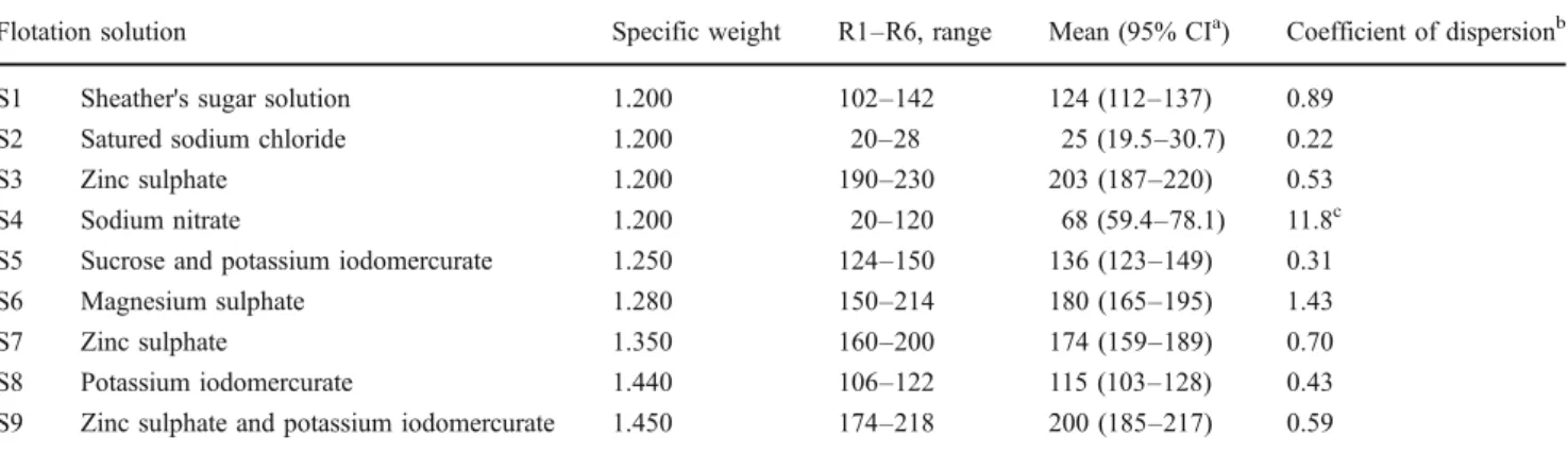
were present in all replicates analysed by FLOTAC. However, the best results, in terms of mean LPG and consistency, were obtained using FS3 (203 L1), i.e. zinc sulphate (s.g. 1.2). Also with FS9, zinc sulphate plus potassium iodomercurate (s.g. 1.45), good results were obtained, but the larvae resulted slightly deformed.
The comparative results between four different tech-niques for the detection of L1 of A. vasorum in individual faecal samples are shown in Table 2, according to the copromicroscopic techniques and the multiplication fac-tors used for each technique. The mean LPG obtained by the FLOTAC for both dogs was significantly higher (P< 0.05) than those obtained by the other three techniques (the means of the other techniques all lie below the 95% CI of the mean LPG of the FLOTAC technique). In addition, the FLOTAC results were consistent across replicates with only Poisson (or random) variation be-tween individual replicates. The other techniques appear to be less consistent with evidence of extra-Poisson variation in at least one of the two dogs across the replicates within each technique.
Discussion
Globally, A. vasorum was described to be present in enzootic foci; however, an increasing number of reports indicate a possibly increasing distribution, particularly in Europe and North America. Recent studies predicted wide and global areas with potential for parasite establishment based on eco-climatic data (Morgan et al.2009). Particularly in known endemic regions, prophylactic anthelmintic treat-ment (Conboy 2004; Koch and Willesen2009; Willesen et al. 2007) or routine screening of dogs for A. vasorum infection (Verzberger-Epshtein et al. 2008) are recommen-ded. A. vasorum affected dogs can be infected for months to years prior to developing clinical signs. Experimental infections have demonstrated that clinical signs, and even echocardiographic changes, may be subtle, even if patho-logical changes of the lungs were considerable (Kranjc et al.
2010; Schnyder et al.2010a)
A valid and affordable diagnostic method intended for early detection of A. vasorum-infected animals would improve not only prognosis of anthelmintic-treated animals,
Table 1 Comparison of nine flotation solutions using the FLOTAC technique (six replicates R1–R6) for the detection of first-stage larvae in a collective sample of faeces from dogs experimentally inoculated with third-stage larvae of Angiostrongylus vasorum
Flotation solution Specific weight R1–R6, range Mean (95% CIa) Coefficient of dispersionb S1 Sheather's sugar solution 1.200 102–142 124 (112–137) 0.89
S2 Satured sodium chloride 1.200 20–28 25 (19.5–30.7) 0.22 S3 Zinc sulphate 1.200 190–230 203 (187–220) 0.53 S4 Sodium nitrate 1.200 20–120 68 (59.4–78.1) 11.8c S5 Sucrose and potassium iodomercurate 1.250 124–150 136 (123–149) 0.31 S6 Magnesium sulphate 1.280 150–214 180 (165–195) 1.43 S7 Zinc sulphate 1.350 160–200 174 (159–189) 0.70 S8 Potassium iodomercurate 1.440 106–122 115 (103–128) 0.43 S9 Zinc sulphate and potassium iodomercurate 1.450 174–218 200 (185–217) 0.59
a CI confidence interval
b Variance/mean of raw data before multiplication by detection limit c Significant extra-Poisson variation
Technique R1–R10, range Mean LPG (95% CIs) Coefficient of dispersiona Dog 1 FLOTAC 286–336 310.8 (296–327) 0.45 McMaster 190–380 258.0 (219–304) 2.07b Flotation in tube 5–27 13.2 (9.7–18.1) 3.78b Baermann-Wetzel 141.3–183.7 154.7 (147–162) 11.03b Dog 2 FLOTAC 94–124 100.6 (91.6–109) 1.32 McMaster 40–120 81.0 (65–100) 0.89 Flotation in tube 1–7 4.1 (3.0–5.5) 1.59 Baermann-Wetzel 30.4–40.8 35.4 (33.3–37.6) 3.69b Table 2 Comparison of four
different copromicroscopic techniques (ten replicates each, R1–R10) for the determination of first-stage larvae per gramme of faeces (LPG) from two dogs (dog 1 and 2) experimentally inoculated with 200 third-stage larvae of Angiostrongylus vasorum
a Variance/mean of raw data before
multiplication by detection limit
b
Significant extra-Poisson varia-tion in raw count data
but may also be a useful tool for the reduction of parasite importation by, for example, restricting movement of dogs, unless tested or treated for A. vasorum. Additional advantages given by a quantitative analysis of parasitic stages are possible correlations with the effective worm burden or a follow-up of the efficacy of anthelmintic treatments, as previously shown (Schnyder et al.2010a).
An optimal coproscopy technique for determining the numbers of parasite larvae in faeces should be specific and sensitive, accurate and consistent. Sensitivity of copro-scopic methods was suggested to be increased by multiple faecal examinations, as performed for recent prevalence studies (Barutzki and Schaper 2009; Taubert et al. 2008). However, due to low compliance of animal owners for multiple faecal collections and the impossibility to always guarantee proper storage of faecal samples, alternative methods are needed. Recently, the FLOTAC technique was evaluated for the detection of lungworm larvae in faecal samples of sheep. In particular, the examination of fresh samples was compared with samples stored in
formalin 5% or 10% and at−20°C. The FLOTAC technique
showed to give higher larval counts even with frozen samples if compared with the McMaster technique and simple flotation (Rinaldi et al.2010), and may, therefore, represent a valid technique, overcoming the necessity of having viable larvae as in the case for larval migration techniques such as the Baermann–Wetzel method.
Canine faecal samples may contain different kinds of larvae, such as Crenosoma vulpis, Filariodes hirthi, Filaroides osleri or several kinds of free-living nematodes, in particular if faeces were collected from the soil. Their differentiation is performed microscopically based on larval
morphology (Eckert et al. 2008; McGarry and Morgan
2009). However expertise is needed, particularly in case of low numbers of larvae, the presence of damaged larvae, or co-infections of more than one species. Therefore, a sensitive coproscopic technique that allows the recovery of undamaged, intact larvae would be of great advantage.
Zinc sulphate (s.g. 1.200) or zinc sulphate plus potassi-um iodomercurate (s.g. 1.450) gave the best recovery rate of L1 of A. vasorum using the FLOTAC technique; however, zinc sulphate appeared to be less likely to deform the larvae morphologically, and hence, is the better technique. Similarly, FLOTAC had the best results with zinc sulphate solution for the recovery of C. vulpis larvae (Rinaldi et al. 2007). Nevertheless, it may be useful to further evaluate sensitivity and specificity of FLOTAC with faecal material from clinical cases.
In this simple comparison, the FLOTAC technique appears to be the most sensitive of the methods compared. In addition, the technique was the most consistent. In repeated counts from the same faecal sample, there was only a random (or Poisson) variation. This indicates that the
technique is consistent in detecting the larvae. With the other techniques, there was some evidence of extra-Poisson variation between replicates. The cause of this variation can only be speculated. The high coefficient of dispersion indicates that there was aggregation or clumping of the larvae within the sample, thus, resulting in some replicates having a substantially higher or lower larval count than the expected mean. If this occurs on a routine basis, it could lead to significant under- or overestimation of the LPG's. Such errors are much less likely where there is only random variation between replicates.
Morgan et al. (2005), using the McMaster technique, reported that there was only Poisson variation between replicates for helminth eggs. In our study, there was evidence of extra Poisson variation between replicates for counts of A. vasorum larvae. The reasons for the differences are not obvious, although larvae, unlike eggs, may remain motile during the processing, and thus, may contribute to the variation observed in our study. Morgan et al. (2005) also recommended the use of four replicates for faecal egg counts (FEC) in composite samples to avoid errors resulting from the random distribution of eggs in the faeces. Such errors could result in an inaccurate estimate of the FEC. Evidence for potential inaccuracies, even when the count may be consistent, can be seen in our data. Examination of the tables indicates that some techniques or solutions may have wide confidence limits for the estimates of LPG, yet still be consistent with random error between samples, whilst narrow confidence intervals do not preclude extra-Poisson variance. This is because of the multiplication factors in converting the raw count into LPG. For example, the McMaster technique had a CI of 65–100 LPG. However the raw count data varied between a minimum of 4 and 12 larvae per 0.1 g (mean 8.1), which is consistent with a Poisson distribution, and hence, random variation between replicates. In contrast, the Baermann techniques on the same dog had a CI of 33.3–37.6 LPG, but were over dispersed as the raw count data varied between 304 and 408 larvae per 10 g. Thus, the multiplication by a factor of 10 of the raw data with the McMaster technique magnifies the variance between replicates, whilst the division by a factor of 10 for the Baermann reduces the variance between replicates. Therefore, even with an accurate and consistent technique, Poisson errors may still result in substantial variability between replicates when the raw counts are low. A further alternative for improvement of A. vasorum diagnostics may be represented by the employment of novel molecular tools such as PCR (Traversa and Guglielmini
2008). A SYBR green real-time PCR has been described
for detection of circulating DNA in EDTA-blood and canine faeces and was compared with Baermann analysis. The lowest number of positive samples was detected by faecal PCR, while blood PCR detected a greater number of
A. vasorum-positive samples compared with the Baermann method; however, some discordance between blood and faecal PCR were observed (Jefferies et al.2009). Recently, a sieve-PCR method was established for faecal samples and was recommended as a non-invasive tool for surveillance of the reservoir for A. vasorum, i.e. foxes, and for confirmative diagnosis in dogs (Al-Sabi et al.
2010). This method is of special value if field investigations are performed with old faecal samples, in which larval motility is probably reduced. Furthermore, the serological detection of circulating A. vasorum antigen (Schnyder et al.
2010b; Verzberger-Epshtein et al. 2008) or of antibodies directed against adult A. vasorum (Cury et al. 1996) has shown good performances.
In conclusion, the findings of the comparison study showed that the FLOTAC technique, together with the just-mentioned methods, may contribute to an improvement of the ability to diagnose canine lungworm infections. The FLOTAC technique may, in particular, be a valuable alternative for faecal samples in which larval viability is not warranted.
References
Al-Sabi MN, Deplazes P, Webster P, Willesen JL, Davidson RK, Kapel CM (2010) PCR detection of Angiostrongylus vasorum in faecal samples of dogs and foxes. Parasitol Res 107:135– 140
Barutzki D, Schaper R (2009) Natural infections of Angiostrongylus vasorum and Crenosoma vulpis in dogs in Germany (2007– 2009). Parasitol Res 105(Suppl 1):39–48
Chapman PS, Boag AK, Guitian J, Boswood A (2004) Angiostrongy-lus vasorum infection in 23 dogs (1999–2002). J Small Anim Pract 45:435–440
Conboy G (2004) Natural infections of Crenosoma vulpis and Angiostrongylus vasorum in dogs in Atlantic Canada and their treatment with milbemycin oxime. Vet Rec 155:16–18
Cringoli G (2006) FLOTAC, a novel apparatus for a multivalent faecal egg count technique. Parassitologia 48:381–384
Cringoli G, Rinaldi L, Maurelli MP, Utzinger J (2010) FLOTAC: new multivalent techniques for qualitative and quantitative copromi-croscopic diagnosis of parasites in animals and humans. Nat Protoc 5(3):503–515
Cury MC, Lima WS, Vitor RWA (1996) Enzyme-Linked Immuno-sorbent Assay (ELISA) for the diagnosis of Angiostrongylus vasorum (Baillet, 1866) infection in dogs. Rev Med Vet 147:525–530
Denk D, Matiasek K, Just FT, Hermanns W, Baiker K, Herbach N, Steinberg T, Fischer A (2009) Disseminated angiostrongylosis with fatal cerebral haemorrhages in two dogs in Germany: a clinical case study. Vet Parasitol 160:100–108
Eckert J, Lämmler G (1972) Angiostrongylose bei Mensch und Tier. Z Parasitenk 39:303–322
Eckert J, Friedhoff KT, Zahner H, Deplazes P (2008) Lehrbuch der Parasitologie für die Tiermedizin. Enke, Stuttgart
Gaglio G, Cringoli G, Rinaldi L, Brianti E, Giannetto S (2008) Use of the FLOTAC technique for the diagnosis of Aelurostrongylus abstrusus in the cat. Parasitol Res 103:1055–1057
Garosi LS, Platt SR, McConnell JF, Wrayt JD, Smith KC (2005) Intracranial haemorrhage associated with Angiostrongylus vaso-rum infection in three dogs. J Small Anim Pract 46:93–99 Guilhon J, Bressou C (1960) Rôle des Limacidés dans le cycle
évolutif d'Angiostrongylus vasorum (Baillet, 1866). C R Acad Sc 251:2252–2253
Humm K, Adamantos S (2010) Is evaluation of a faecal smear a useful technique in the diagnosis of canine pulmonary angiostrongy-losis? J Small Anim Pract 51:200–203
Jefferies R, Morgan ER, Shaw SE (2009) A SYBR green real-time PCR assay for the detection of the nematode Angiostrongylus vasorum in definitive and intermediate hosts. Vet Parasitol 166:112–118
Koch J, Willesen JL (2009) Canine pulmonary angiostrongylosis: an update. Vet J 179:348–359
Kranjc A, Schnyder M, Dennler M, Fahrion A, Makara M, Ossent P, Morgan J, Deplazes P, Glaus TM (2010) Pulmonary artery thrombosis in experimental Angiostrongylus vasorum infection does not result in pulmonary hypertension and echocardiographic right ventricular changes. J Vet Intern Med 24:855–862 MAFF (1986) Manual of Veterinary Parasitological Laboratory
Techniques 3rd Edition. HMSO, London, p 166
Mason KV (1987) Canine neural angiostrongylosis: the clinical and therapeutic features of 55 natural cases. Austr Vet J 64:201–203 McGarry JW, Morgan ER (2009) Identification of first-stage larvae of
metastrongyles from dogs. Vet Rec 165:258–261
Morgan ER, Cavill L, Curry GE, Wood RM, Mitchell ESE (2005) Effects of aggregation and sample size on composite faecal egg counts in sheep. Vet Parasitol 131:79–87
Morgan ER, Jefferies R, Krajewski M, Ward P, Shaw SE (2009) Canine pulmonary angiostrongylosis: the influence of climate on parasite distribution. Parasitol Int 58:406–410
Morgan ER, Jefferies R, van Otterdijk L, McEniry RB, Allen F, Bakewell M, Shaw SE (2010) Angiostrongylus vasorum infection in dogs: presentation and risk factors. Vet Parasitol 173:255–261 Oliveira-Junior SD, Barcante JM, Barcante TA, Dias SR, Lima WS (2006) Larval output of infected and re-infected dogs with Angiostrongylus vasorum (Baillet, 1866) Kamensky, 1905. Vet Parasitol 141:101–106
Patteson MW, Gibbs C, Wotton PR, Day MJ (1993) Angiostrongylus vasorum infection in seven dogs. Vet Rec 133:565–570 R Development Core Team (2006) R: A Language and Environment
for Statistical Computing. Vienna, Austria: R Foundation for Statistical Computing.
Rinaldi L, Calabria G, Carbone S, Carrella A, Cringoli G (2007) Crenosoma vulpis in dog: first case report in Italy and use of the FLOTAC technique for copromicroscopic diagnosis. Parasitol Res 101:1681–1684
Rinaldi L, Maurelli MP, Musella V, Santaniello A, Coles GC, Cringoli G (2010) FLOTAC: an improved method for diagnosis of lungworm infections in sheep. Vet Parasitol 169:395–398 Ruegg SR, Heinzmann D, Barbour AD, Torgerson PR (2008) Estimating
the transmission dynamics of Theileria equi and Babesia caballi in horses using Monte Carlo techniques. Parasitology 135:555–565 Schnyder M, Fahrion A, Ossent P, Kohler L, Webster P, Heine J,
Deplazes P (2009) Larvicidal effect of imidacloprid/moxidectin spot-on solution in dogs experimentally inoculated with Angios-trongylus vasorum. Vet Parasitol 166:326–332
Schnyder M, Fahrion A, Riond B, Ossent P, Webster P, Kranjc A, Glaus T, Deplazes P (2010a) Clinical, laboratory and patholog-ical findings in dogs experimentally infected with Angiostrongy-lus vasorum. Parasitol Res 107:1471–1480
Schnyder M, Tanner I, Webster P, Barutzki D, Deplazes P (2010b) An ELISA for sensitive and specific detection of circulating antigen of Angiostrongylus vasorum in serum samples of naturally and experimentally infected dogs, submitted
Staebler S, Ochs H, Steffen F, Naegeli F, Borel N, Sieber-Ruckstuhl N, Deplazes P (2005) Autochthonous infections with Angiostrongylus vasorum in dogs in Switzerland and Germany (in German). Schweiz Arch Tierheilkd 147:121–127
Taubert A, Pantchev N, Vrhovec MG, Bauer C, Hermosilla C (2008) Lungworm infections (Angiostrongylus vasorum, Crenosoma vulpis, Aelurostrongylus abstrusus) in dogs and cats in Germany and Denmark in 2003–2007. Vet Parasitol 159:175–180 Traversa D, Guglielmini C (2008) Feline aelurostrongylosis and
canine angiostrongylosis: a challenging diagnosis for two emerging verminous pneumonia infections. Vet Parasitol 157:163– 174
Verzberger-Epshtein I, Markham RJ, Sheppard JA, Stryhn H, Whitney H, Conboy GA (2008) Serologic detection of Angiostrongylus vasorum infection in dogs. Vet Parasitol 151:53–60
Wessmann A, Lu D, Lamb CR, Smyth B, Mantis P, Chandler K, Boag A, Cherubini GB, Cappello R (2006) Brain and spinal cord haemorrhages associated with Angiostrongylus vasorum infection in four dogs. Vet Rec 158:858–863
Willesen JL, Kristensen AT, Jensen AL, Heine J, Koch J (2007) Efficacy and safety of imidacloprid/moxidectin spot-on solution and fenbendazole in the treatment of dogs naturally infected with Angiostrongylus vasorum (Baillet, 1866). Vet Parasitol 147:258– 264