Characterization of Tuba, a Novel Ena/VASP Ligand, and Function of Ena/VASP Proteins in Mouse Development
by
Adam Vincent Kwiatkowski
B.S. Biology
Cornell University, 1996
Submitted to the Department of Biology in Partial Fulfillment of the Requirement for the Degree of
Doctor of Philosophy in Biology
at the
Massachusetts
Institute of Technology
February 2005
02004 Massachusetts Institute of Technology. All Rights Reserved.
Signature of Author:
Department of Biology
__
December 15, 2004
Certified by: 7.1
Frank Gertler
Associate
Professor of Biology
Thesis Supervisor
Accepted by:
Stephen Bell
Professor of Biology
Chair, Committee for Graduate Students ACiHJVEWv *f· - Z-; - - -As MASSACHUSETTS INSTITUTE OF TECHNOLOGY FEB 3 2005
LIBRARIES
__ _Characterization of Tuba, a Novel Ena/VASP Ligand, and Function of Ena/VASP Proteins in Mouse Development
by
Adam Vincent Kwiatkowski
Submitted to the Department of Biology on December 15, 2004
in Partial Fulfillment of the Requirements for the Degree of Doctor
of Philosophy in Biology
Abstract
Regulated actin assembly drives cells movement, adhesion and shape
change. The EnaNASP family of proteins controls actin filament elongation and
are important regulators of axon guidance and cell motility. In vertebrates, the family consists of Mena (Mammalian Enabled), VASP (Vasodilator Stimulated
Phosphoprotein), and EVL (Ena-VASP-Like). This thesis work focused on
understanding the vertebrate EnaNASP protein family by discovering pathways that
regulate EnaNASP function and by defining the role of EnaNASP proteins in vertebrate development. Characterization of the EVL locus revealed a new EVL
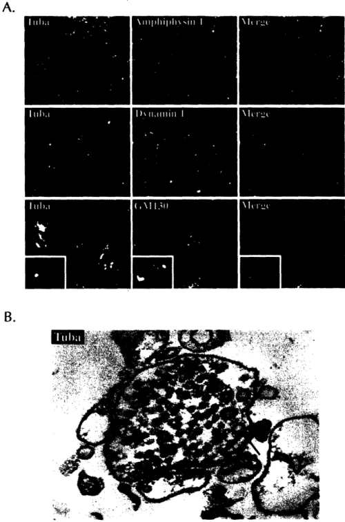
isoform. A protein interaction screen for new EnaNASP ligands produced Tuba, a
novel scaffold protein that associates
with EnaNASP proteins
in vivo.
Tuba is a
unique guanine nucleotide exchange factor (GEF) for Cdc42 that binds dynamin
and a number of actin regulatory proteins in addition to Ena/VASP proteins. A
knockout of EVL was made to determine the requirement for EVL in mouse
development. Genetic analysis of EnaNASP function in the mouse revealed a requirement for Ena/VASP proteins in neuronal layering, spinal and cranial nerve
formation, and cardiovascular development.
Thesis Supervisor: Frank Gertler
Table of Contents Title Page 1 Abstract 2 Table of Contents 3 Acknowledgements 4 Chapter 1. Introduction 5
Chapter 2. Molecular Characterization of EnaNASP-Like (EVL) 37 Chapter 3. Tuba is a Novel Rho-Family Guanine Nucleotide Exchange 56
Factor (GEF) That Binds EnaNASP Proteins and Links
Dynamin to Regulation of the Actin Cytoskeleton
Chapter 4. The Role of Ena/VASP Proteins in Mouse Development 102
Acknowledgements
There are many people to thank. I have had the amazing good fortune of meeting and interacting with many wonderful and interesting people during my years as a graduate student, all of whom impacted my life here at MIT in some manner. I will never forget my time here, and although trying at times, it has been a memorable journey.
First of all, thank you so much Frank. For all of your help, guidance, inspiration and motivation through the years. You have had an immeasurable impact on my development as scientist, and for that, I am forever grateful.
In no particular order, many thank yous to...
Dan - for your enthusiasm and hard work on the two hybrid screen. My first and only UROP, you taught me a lot, and I hope you learned a thing or two from me.
Jon - without your help, there would be EVL knockout mice. You came along just in the nick of time and helped me keep this project going when I had just about given up. Your hard work and determination made it happen.
Doug - what can I say, I owe you big. You have been invaluable in moving the knockout project forward. I have enjoyed working with you over the last year, and eagerly await the day when we can reap the benefits of our hard work. And stop setting up plugs.
Joe - for everything over the years. You have been a great friend and labmate, and were always there when I needed you most. You too have had an immeasurable impact on my development as a scientist.
Bronson - your help with the analysis of the knockout mice has been great.
Angelique, Kara, and Gretchen - an extra big thank you for all the help with genotyping and other mouse issues!
Jill, Caroline, Jen and the rest of the wonderful DCM staff - thank you for all the great work you do keeping our mice happy and healthy.
Jim and Matthias- for all the help and guidance over the years. Members of my committee - for all the helpful advice over the years.
Labmates - you have made my time in the Gertler lab interesting and unforgettable.
And last but certainly not least, my family - for the unconditional love and support over the years. I couldn't have done it without you.
Chapter
1
Chapter 1
Regulated cell movement is required for normal development and
physiology. Cell movement is driven largely by dynamic changes in the underlying
actin cytoskeleton that affect cell morphology and adhesion. A growing list of
proteins are known to regulate actin assembly, stability, and disassembly, many of
which are thought to play an important role in regulating cell motility. Included in
this list are the EnaNASP proteins, a highly conserved protein family that controls
actin filament elongation and regulates cell movement. The thesis work presented is a molecular, cell biological, and genetic analysis of EnaNASP proteins. It begins
in Chapter Two with the molecular characterization of one of three vertebrate
EnaNASP proteins, Ena-VASP-Like (EVL). Chapter Three describes the discovery and characterization of a novel EnaNASP ligand, Tuba, that links EnaNASP
proteins to Rho-family GTPases. Finally, Chapter 4 details a genetic analysis of
EnaNASP proteins that reveals new requirements for EnaNASP function in the
developing mouse.
Actin dynamics in cell motility
Cell motility can be broken down into 4 steps: protrusion, adhesion, retraction, and de-adhesion. The first step, protrusion, is characterized by the
Chapter 1
lamellipodium contains a branched network of actin filaments that provides the
mechanical force for membrane extension. Filamentous actin (F-actin) is made up
of individual globular actin (G-actin) monomers. Actin filaments are polar, having
a barbed or plus end and pointed or minus end. Actin monomers incorporate more
readily onto the barbed end of the actin filament. Within the extending
lamellipodium, actin filaments are oriented with fast-growing barbed ends facing
towards the membrane (Pollard and Borisy, 2003).
A dendritic nucleation model has been proposed to account for the
formation of the actin filament network within lamellipodia (Pollard and Borisy,
2003; Svitkina and Borisy, 1999). In this model, a defined set of proteins work in
concert at the front or leading edge of the extending lamellipodium to regulate
actin dynamics. Though cells possess
a large number of proteins that bind actin or
regulate actin assembly, basic actin assembly can be achieved with a small set of core proteins (Pantaloni et al., 2001). They are actin, profilin, the Arp2/3 complex,
an activator of the Arp2/3 complex, heterodimeric capping protein (CP), and
cofilin. The role of these proteins in a dendritic nucleation model of lamellipodial
protrusion is discussed in detail below.
Cells contain a large pool of monomeric actin, most of which is bound to
sequestering proteins, such as profilin. Profilin is a small protein found in all cell types that binds to actin monomer and inhibits spontaneous nucleation (Carlsson et al., 1977). Importantly, profilin catalyzes the exchange of ADP for ATP in the bound monomer (Goldschmidt-Clermont et al., 1992). This ATP-bound actin
Chapter 1
monomer is primed for incorporation onto the growing barbed end of an actin
filament. Thus, it is thought that profilin functions to maintain a pool of
unpolymerized actin ready for addition to newly created filaments during
protrusion.
Protrusion can be initiated when extracellular migratory cues bind to cell
surface receptors. Receptor activation initiates signaling pathways that stimulate PIP2 and Rho-family GTPases (Pollard and Borisy, 2003). The importance of both PIP2 and Rho GTPases in the regulation of the actin cytoskeleton is well
documented, and Rho family GTPases are discussed later in this chapter. PIP2 and activated Rho GTPases function to stimulate members of the
WASP/WAVE superfamily of proteins. This protein family activates the actin
nucleating Arp2/3 complex. The Arp2/3 complex is an assembly of seven proteins, including the two actin-related proteins Arp2 and Arp3 (Machesky et al., 1994). PIP2 and Cdc42, a Rho family GTPase, bind to N-WASP and release it from an auto-inhibited state (Prehoda et al., 2000). The C-terminus of N-WASP contains a binding site for actin monomer and an acidic region that activates the Arp2/3 complex and induces actin nucleation (Machesky and Insall, 1998).
The complex of N-WASP, Arp2/3 and monomeric actin binds to the sides of
existing actin filaments and promotes the formation of new actin filaments through
the nucleating activity of the Arp2/3 complex. New filament growth at the barbed
end is supported through the pool of profilin-actin complexes. In this model,
filament growth by actin monomer addition provides the mechanical force for
Chapter 1
membrane protrusion (Pollard and Borisy, 2003). Filament growth is quickly
terminated by the binding of CP to the barbed end, functioning to maintain
filament length and number, an important principle in this model (Cooper and Schafer, 2000).
Newly incorporated actin subunits hydrolyze their bound ATP and slowly
release the gamma-phosphate. Release of the gamma-phosphate induces filament disassembly and promotes the severing activity of ADF/cofilin at or near the pointed end. Freshly released ADP-actin monomers are bound by profilin, which promotes the exchange of ADP for ATP and replenishes the pool of ATP actin that
is primed for another cycle of filament assembly at the leading edge. The cycle of
Arp2/3 activation, de
novo
branch formation and elongation, filament capping, and
filament disassembly continues as the lamellipodium is protruding (Pollard and Borisy, 2003). Other actin-based structures, such as filopodia and contractile rings, can be generated by variations on this mechanism or through distinct activities (Mejillano et al., 2004).
As mentioned above, cells possess a large number of proteins that are thought to have some role in regulating actin assembly. While it possible to
reconstitute actin-driven motility in
vitro
with a small, defined set of proteins, the
extending lamellipodium of a cell is far more complex, and proper formation and
regulation of the actin network within the lamellipodium is likely to require a
number of additional proteins. One such family of proteins now thought to play an
Chapter 1
integral role in the regulation of lamellipodial protrusion, as well as other
actin-dependent processes, are the EnaNASP family of proteins.
The Ena/VASP protein family
EnaNASP proteins have emerged as important regulators of actin dynamics,
and are thought to link different signaling pathways to changes in cell morphology.
The EnaNASP family includes the three highly related vertebrate proteins Mena (Mammalian Enabled), VASP (Vasodilator Stimulated Phosphoprotein), and EVL (Ena-VASP-Like), as well as the invertebrate orthologs D. melanogaster Ena (Enabled) and C. elegans UNC-34, and an ortholog in the mycetozoan D.
discoideum (DdVASP) (Fig. 1). All members of the family share a highly conserved
domain structure: a proline-rich core (PRO) flanked by two distinct regions called
Ena-VASP Homology domains (EVH1 and EVH2, respectively). In addition, all vertebrate EnaNASP proteins are substrates for the cyclic nucleotide dependent kinases PKA/PKG (Krause et al., 2003). The importance of the various domains and phosphorylation to the function and regulation of EnaNASP proteins is discussed later in this chapter.
The founding members of the family, Ena and VASP, were discovered as key players in two independent, actin-dependent processes. Ena was originally
identified as a dose-dependent suppressor of Abelson (Abl)-dependent phenotypes in Drosophila (Gertler et al., 1990). Abl is a cytoplasmic tyrosine kinase required
Chapter 1
et al., 1995). Reduction in Ena gene dosage from two to one alleviates all known Abl-dependent phenotypes (Gertler et al., 1990). Mutations in Ena cause a recessive lethal phenotype that includes defects in the embryonic nervous system (Gertler et al., 1995; Wills et al., 1999). VASP was originally identified as a major
Invertebrate Homologs
Drosophila Ena |: 100aa DictyosteliumVASP C.elegans UNC-34 I Vertebrate Family Mabn,,%m
EVI VASPFigure 1. The Ena/VASP Family of Proteins
substrate cyclic-nucleotide dependent kinases in platelets; phosphorylation of
VASP correlates with inhibition of platelet activation (Halbrugge and Walter, 1989;
Waldmann et al., 1987). Platelet activation is associated with actin-dependent changes cell morphology and adhesion that promote aggregation necessary for clot ·formation. The role of VASP in platelet activation is discussed in more detail
below.
11
Chapter 1
A number of biochemical'studies have implicated EnaNASP proteins in regulating actin dynamics and cell motility. First, EnaN/ASP proteins localize to the
tips of filopodia and lamellipodia, highly dynamic actin-rich structures (Fig. 2).
EnaNASP proteins are also found
along stress fibers, thick actin
filament bundles that stretch across
the cell, at focal adhesions, sites of signaling between the cytoskeleton and the extracellular matrix, and sites of cell-cell contact (Gertler et al., 1996; Lambrechts et al., 2000; Lanier et al., 1999; Rottner et al.,
Figure 2. Localization of Ena/VASP proteins. 1999; Vasioukhin et al., 2000). Top panel - an extending fibroblast
lamellipodium fixed and stained for VASP and F-actin. Note the enrichment of VAP at the Second, EnaNASP proteins bind leading edge of the lamellipodium. Bottom
panel - time lapse series from a cell
profilin (Gertler et al., 1996; expressing EGFP-EVL. Dynamic localization
to the tips of filopodia and lamellipodia and focal adhesions is observed. EGFP-EVL localization to the tip of one filopodium is denoted by a white arrow in each frame.
EnaNASP proteins bind both
G-and F-actin, G-and can promote actin filament elongation G-and bundling
in vitro
(Bachmann et al., 1999; Harbeck et al., 2000; Huttelmaier et al., 1999; Lambrechtset al., 2000; Walders-Harbeck et-al., 2002). Finally, EnaNASP proteins bind ActA,
Chapter 1
required for ActA-dependent actin polymerization and Listeria motility (Laurent et
al.,
1999).
Ena/VASP proteins regulate cell motility
The first evidence for how EnaNASP proteins might regulate cell motility
came from studies of fibroblast migration (Bear et al., 2000). Loss of EnaNASP
function, through genetic deletion or inhibition via mislocalization, causes net cell
speed to increase. Conversely, overexpression of EnaNASP proteins leads to
slower cell movement. Furthermore, localization to the tips of lamellipodia is
required for EnaNASP-dependent regulation of cell speed. Selective delocalization
of EnaNASP proteins from focal adhesions, but not from the leading edge, does not
affect fibroblast migration, and EnaNASP-deficient cells do not show any reduction
in the number, morphology, or protein composition of focal adhesions (Bear et al.,
2000).
EnaNASP proteins were later discovered to regulate lamellipodial protrusion
by controlling the actin network geometry within the lamellipodium (Bear et al.,
2002). Closer examination of lamellipodial extension in EnaNASP-deficient cells revealed that lamellipodial protruded slower, but were more persistent.
Conversely, in cells overexpressing EnaNASP proteins, lamellipodia protruded faster but were less persistent. Loss of EnaNASP proteins produced lamellipodia
containing a network of shorter, more highly-branched filaments than in controls. Overexpression of EnaNASP proteins, on the other hand, produced longer,
Chapter 1
branched filaments. These changes in the actin network geometry ultimately produce the changes in lamellipodial dynamics (Bear et al., 2002).
Actin filament length and branching must be tightly controlled to drive
protrusion. In a dendritic nucleation model, filament capping by CP is important
for controlling filament length and number. However, EnaNASP activity can affect actin filament length and branching in cells. Further experiments showed that EnaNASP proteins localize at or near the barbed end in cells, and can be displaced by drugs that bind directly to the barbed end. In vitro, VASP can inhibit capping by CP during actin filament polymerization (Bear et al., 2002). Therefore, one
function of EnaNASP proteins is to protect actin filaments from CP, thus promoting
filament elongation.
EnaNASP proteins also regulate filopodia formation and dynamics. Filopodia are thin cellular extensions made up of long, bundled actin filaments, and though found in many cell types, are classically associated with the growth
cone of neurons. Filopodia are thought to play both sensory and mechanical roles
in locomotion (Dickson, 2002). EnaNASP proteins localize to the tips of filopodia
in neurons and fibroblasts, and have been implicated in many filopodial-dependent processes (Lanier et al., 1999; Vasioukhin et al., 2000). Inhibition of EnaNASP proteins in hippocampal neurons causes a dramatic reduction in the number and length of filopodia (Lebrand et al;, 2004).
The importance of Ena/VASP
proteins in filopodial formation could be due
to the anti-capping activity demonstrated in fibroblasts, or it could be due to
Chapter 1
another activity of Ena/VASP
proteins, or both. Additional studies in fibroblasts
seem to suggest that it could be both. Loss of CP in fibroblasts eliminates lamellipodial formation and causes a dramatic increase in filopodia formation. However, in EnaNASP-deficient cells, loss of CP causes extensive ruffling, not
filopodia formation. This suggests
that, in addition to functioning as antagonists of
CP,
EnaNASP proteins can promote filopodia formation (Mejillano et al., 2004).
The filopodia forming activity of EnaNASP proteins could involve their ability to
tetramerize and bundle actin filaments. The structural properties of EnaNASP proteins are discussed in the next section.
Structural requirements for Ena/VASP function The EVH1 domain
The EVH1 domain is the most highly conserved region in EnaNASP proteins and binds directly to a proline-rich consensus motif, (D/E)-FPPPP-X(D/E)(D/E). The EVH1 domain plays an essential -role in subcellular targeting of EnaNASP proteins.
A number of cellular proteins contain FPPPP
motifs and are known to bind
EnaNASP proteins, including the focal adhesion proteins zyxin and vinculin, the axon guidance receptor roundabout (Robo) (Prehoda et al., 1999), and the
Fyn-binding and SLP-76 associated protein (Fyb/SLAP)
that functions in T-cell activation
(Krause et al., 2000). The Listeria protein ActA contain four FPPPP motifs, and recruitment of EnaNASP proteins is required for optimal Listeria motility (Laurent et al., 1999; Niebuhr et al., 1997)..
Chapter 1
EnaNASP localization to the leading edge, required for EnaNASP function in cell motility, is in part mediated by EVH1 ligand binding. The
Lamellipodin(Lpd)/RIAM family of proteins are EVH1 ligands of EnaNASP proteins that localize to the leading edge. Both Lpd and RIAM regulate lamellipodial
dynamics, and are thought to cooperate with EnaNASP proteins to link
extracellular signals to changes in actin dynamics (Krause et al., 2004; Lafuente et al., 2004).
The proline-rich region
The proline-rich(PRO) region of EnaNASP proteins contains binding sites for
profilin and a growing list of SH3 and WW domains, including those found in Abl,
IRSp53, and FE65 (Kwiatkowski et al., 2003). Deletion of the PRO-region has no
effect on the ability of EnaN/ASP
proteins to localize or to support normal rates of
fibroblast movement, suggesting that this region is dispensable for the EnaNASP-dependent regulation of cell motility (Loureiro et al., 2002). Interestingly, the same
mutant provides only a limited stimulatory effect on Listeria movement (Geese et
al., 2002). Therefore, interactions with ligands for the PRO region play an important role in the ability of EnaN/ASP to support Listeria movement, but the requirement for such interactions in lamellipodia is unclear.
Though profilin-binding is not required for EnaNASP-dependent
regulation
of whole cell motility, under certain circumstances or in specific processes, profilin
Chapter 1
interactions. In fact, a requirement for Mena and profilin-l has been observed genetically (Gertler et al., 1996)(discussed in Chapter 4). Alternatively, deletion of the PRO region in EnaNASP proteins may render them constitutively active or
somehow obviate a requirement for profilin binding.
Outside of profilin recruitment, the physiological significance of interactions
mediated by the PRO-rich region remains unclear. It has been suggested that SH3-mediated interactions between the Rho-family effector protein IRSp53 and the PRO region of Mena stimulates filopodial formation (Krugmann et al., 2001). A second
report, however, found that interactions with Mena were not required for IRSp53
localization to lamellipodia and filopodia, and that EnaNASP proteins were not
essential for filopodial formation in response to activation of pathways thought to
use IRSp53 (Nakagawa et al., 2003). Given the potent genetic interactions
between Ena and Abl in flies, it is possible that physical interactions between the
PRO-region and the SH3 domain of Abl might play a role in connecting Abl
signaling to EnaNASP function. Identifying additional ligands to the PRO-rich
region will better define the importance of the PRO region for Ena/VASP
function.
In addition, ligands to the PRO region are expected to link EnaNASP proteins to molecular complexes and signaling pathways that might regulate EnaNASP
activity. An SH3-containing protein that binds to the PRO region and links
EnaNASP proteins to other regulators of the actin cytoskeleton is described in Chapter 3.
Chapter 1
The EVH2 domain
The EVH2 domain contains three highly conserved regions of interest: a G-actin binding region (also known as the TLM, or Thymosin-Like-Motif), an F-G-actin binding region (FAB), and a coiled-coil region (COCO). Mena and VASP possess one and two PKA/PKG sites, respectively, within the EVH2 domain (Fig. X). In
vitro, the EVH2 domain has been shown to bind both G-actin and F-actin, induce
actin filament nucleation, promote filament bundling, and mediate oligomerization
(Bachmann et al., 1999; Harbeck et al., 2000; Huttelmaier et al., 1999; Lambrechts et al., 2000; Walders-Harbeck et al., 2002).
The FAB region binds F-actin, and is required for EnaNASP-dependent actin
nucleation in vitro (Lambrechts et al., 2000). The ability to bind F-actin is crucial
for EnaNASP proteins to retain anti-capping activity in cells (Bear et al., 2002). In EnaNASP-deficient fibroblasts, the FAB motif is necessary for both leading edge targeting and proper regulation of cell motility (Loureiro et al., 2002).
The coiled-coil region (COCO) in EnaNASP proteins mediates
oligomerization, and oligomerization has been shown to be important for both in
vitro actin nucleation and binding to some EnaNASP ligands (Ahern-Djamali et al.,1998; Bachmann et al., 1999; Walders-Harbeck et al., 2002). One loss of function
allele of Drosophila Ena was found to lack just the COCO motif, indicating that this
region is important for function in vivo. Consistent with this, a mutant form of
Mena lacking the COCO region localized poorly to the leading edge and failed to
Chapter 1
The dispensability of the PRO region along with the requirement of the FAB and COCO regions for both localization and cell motility suggests that the EVH2
domain plays a prominent, profilin-independent, role in EnaNASP function. When
expressed in fibroblasts, the EVH2 domain localizes to the edges of lamellipodia,
though demonstrating a broader distribution pattern than wild type Mena (Nakagawa et al., 2001). However, the EVH2 domain functions similar to wild
type protein cell motility assay,
suggesting
that the EVH2 domain contains the
features required to regulate lamellipodial dynamics during fibroblast movement. Three properties have been ascribed to the EVH2 domain - G- actin binding, F-actin binding, and oligomerization - and others might exist.
Interestingly, VASP contains two PKA/PKG sites in the EVH2 domain, Mena has one, but EVL lacks any known PKA/PKG site in the EVH2 domain. It has been postulated that the conserved N-terminal PKA/PKG site found in all three vertebrate
EnaNASP proteins is the most critical for the regulation of EnaNASP function
(Loureiro et al., 2002). However, there is evidence to support that phosphorylation in the EVH2 domain can affect EnaNASP activities in vitro (Harbeck et al., 2000). The absence of PKA/PKG sites in EVL is puzzling, but the discovery of an alternate
spliceform of EVL discussed in Chapter 2 might point to an additional means of
regulation.
Ena/VASP phosphorylation and actin dynamics
Chapter 1
The discovery and initial characterization of EnaNASP proteins focused on
their role as substrates
for specific kinases implicated in cell signaling. The
Drosophila ortholog of the family, Ena, originally identified as a dominant
suppressor of the non-receptor tyrosine kinase D-Abelson (D-Abl) (Gertler et al., 1990), was later shown to be a D-Abl substrate (Gertler et al., 1995). VASP was
initially identified as an abundant substrate for cyclic-nucleotide dependent kinases
in platelets (Halbrugge et al., 1990; Reinhard et al., 1992). Further studies showed that all vertebrate members of the EnaNASP family are substrates for both cAMP
and cGMP-induced protein kinases (PKA and PKG) (Butt et al., 1994; Gertler et al., 1996; Lambrechts et al., 2000). VASP possesses three PKA/PKG phosphorylation sites that flank the central proline-rich region. Mena contains two such sites, while EVL has just one. The lone N-terminal PKA/PKG site in EVL is structurally and functionally conserved in both Mena and VASP, and is the preferred site of PKA.
Evidence for how phosphorylation might alter specific properties of
EnaNASP proteins has come mainly from in vitro studies that have focused on the
role of phosphorylation in regulating EnaNASP affinities for actin and ligand
binding. Phosphorylation affects EnaNASP interactions with F-actin, although
exactly how is unclear (Harbeck et al., 2000; Laurent et al., 1999).
Phosphorylation of EnaNASP proteins is known to interfere with G-actin binding
and filament elongation in vitro (Lambrechts et al., 2000; Walders-Harbeck et al.,
Chapter 1
Phosphorylation at the first PKA/PKG site also affects interactions between the adjacent proline-rich region and a number of SH3 domains (including the Abl
SH3 domain), but does not affect binding to profilin in vitro (Lambrechts et al., 2000). In cells, VASP and Abl can be detected in complexes that are disrupted
under conditions that promote VASP phosphorylation (Howe et al., 2002). PKA phosphorylation does not appear to affect ligand binding to the EVH1 domain, nor
does it interfere with the ability of EnaNASP proteins to oligomerize (Harbeck et
al., 2000).
Ena/VASP proteins - PKA/PKG signaling and adhesion
VASP was originally identified as a major substrate for PKA and PKG in
platelets, and phosphorylation of VASP correlates with inhibition of platelet
activation. In platelets, production of cyclic nucleotides and concomitant increase
in PKA activity leads to inhibition of platelet activation, an actin-dependent process
that promotes blood clotting. Mice lacking VASP are completely viable and fertile,
and do not display any gross morphological abnormalities. However,
in vitro
analysis of platelets derived from VASP-deficient mice revealed that
PKA/PKG-dependent inhibition of platelet aggregation was significantly reduced, suggesting
VASP plays a key role in mediating this PKA-dependent function (Aszodi et al., 1999; Hauser et al., 1999). Interestingly, PKA-dependent phosphorylation of Ena/VASP proteins has also been shown to correlate with changes in cell adhesion in fibroblasts. Cellular detachment induces rapid PKA-dependent phosphorylation
Chapter 1
of VASP. Reattachment results in immediate dephosphorylation of VASP, followed
by intermediate levels of phosphorylation during spreading (Howe et al., 2002).
Ena/VASP proteins bind the focal adhesion proteins zyxin and vinculin and are enriched at sites of cell-substratum contact. In fibroblasts, the function of EnaNASP in focal contacts and adhesions is unclear since removal of Ena/VASP from these structures, but not the leading edge, has no effect on cell motility or the average number and morphology of focal contacts/adhesions (Bear et al., 2000). VASP-deficient platelets, however, exhibit increased affinity for fibrinogen (Aszodi et al., 1999). Fibrinogen binding is important for cross-linking platelets at sites of injury, and is mediated by integrin allbP3 switching from a resting to an active conformation, a process called "inside-out" signaling. Thus, one role for VASP
might be to regulate inside-out signaling by integrins in response
to cyclic
nucleotides.
Ena/VASP proteins - cell-cell adhesion
Epithelia cells adhere tightly to each other to form sheets of cells that
function as physical boundaries. Adhesion is mediated by three main types of
cell-cell junctions in epithelial cell-cells: adherens junctions, tight junctions, and
desmosomes. Adherens junctions are cadherin-dependent adhesions connected to the actin cytoskeleton, and in their absence, other cell-cell junctions are reduced (Vasioukhin and Fuchs, 2001). EnaNASP proteins localize to adherens junctions, and it has been proposed that Ena/VASP proteins function in the "filopodia-zipper"
Chapter 1
model of adherens junction formation. Overexpression of the coiled-coil region of
VASP that mediates oligomerization was reported to prevent adherens junction formation in keratinocytes and cause skin blisters in mice (Vasioukhin et al., 2000).
However, it is unclear if this approach interferes specifically with EnaNASP
proteins. Furthermore, analysis of mice that lack all EnaNASP proteins has not
revealed a defect in skin development (discussed in Chapter 4). Studies in
Drosophila have revealed that Ena can affect epithelial cell biology. Ena interacts
genetically with both Abl and Drosophila cadherin (armadillo) in epithelial sheet morphogenesis (Grevengoed et al., 2001).
Tight junctions, like adherens junctions, are connected to the actin
cytoskeleton and function as a physical barrier between the apical and basolateral regions of the cell. Zona occludens-1 (ZO-1) is a marker of tight junctions, and
VASP was found to co-immunoprecipitate with ZO-1 from endothelial cells
(Comerford et al., 2002). Endothelial cells provide an important barrier as the
lining cells of blood vessels. This barrier function is mediated through cell-cell
contacts, and is regulated by PKA (Wojciak-Stothard and Ridley, 2002). It has been postulated that EnaNASP proteins, as components of cell-cell adhesions and
substrates
of PKA, are important for regulating barrier function in endothelial cells
(Comerford et al., 2002). A possible role for EnaNASP proteins in endothelial biology is discussed in Chapter 4.
Ena/VASP proteins - axon guidance
Chapter 1
Genetic studies of invertebrate EnaNASP orthologs has suggested that
EnaNASP proteins function downstream of known axon guidance signaling
pathways. The founding member of the EnaNASP family, Ena was originally identified as a dominant suppressor of lethality and central nervous system (CNS) defects associated with loss of D-Abl (Gertler et al., 1990). Loss of Ena in the fly produces defects in CNS and motor axon pathways (Gertler et al., 1995; Wills et al., 1999). Further genetic experiments suggests that Ena and D-Abl both act
downstream of the repulsive axon guidance receptor roundabout (Robo).
Interestingly, Ena and D-Abl appear to play opposing roles in Robo signaling, with
Ena being required for Robo-dependent repulsion and D-Abl antagonizing Robo
signaling (Bashaw et al., 2000). The C. elegans EnaNASP ortholog Unc-34 has also been shown to be required for Sax3(Robo) repulsion (Yu et al., 2002).
Consistent with the genetic data, biochemical experiments revealed that EnaNASP proteins can bind to the cytoplasmic tail of Robo (Bashaw et al., 2000).
EnaNASP proteins also function downstream of netrin guidance cues. The axon guidance molecule netrin binds to the receptor DCC/Unc-40, and netrin
signaling promotes axonal outgrowth and turning. In the worm, gain of function
mutations in DCC/Unc-40 cause defects in axon outgrowth, branching, and
guidance. Genetic experiments revealed that loss of function mutations in Unc-34
suppressed the gain of function mutation in DCC/Unc-40, indicating that Unc-34
functions downstream of the DCC/Unc-40 receptor in netrin signaling (Gitai et al., 2003). EnaNASP proteins have also been shown to function downstream of Netrin
Chapter 1
in cultured mouse neurons. Inhibition of EnaNASP activity in neuron blocked Netrin mediated filopodia formation (Lebrand et al., 2004).
Consistent with the invertebrate models for EnaNASP function, loss of Mena in mice causes defects in axon guidance. Of the three vertebrate family members, Mena is most highly expressed in the developing nervous system and adult brain.
Mena-deficient mice are viable and fertile, but close examination of brain
architecture reveals severe defects in formation of the corpus callosum, the major
fiber tract that connects the two hemispheres of the brain. Mice lacking Mena also
display defects in the formation of the hippocampal commissure and
pontocerebellar fiber bundles (Lanier et al., 1999). The loss of Mena also disrupts formation of the optic chiasm (Menzies et al., 2004). These results suggest that the
formation of major axonal pathways require Mena for proper formation, and that
Mena, and by extension, Ena/VASP proteins play an important role in axon
guidance.
As mentioned earlier, EnaNASP proteins are required for proper filopodia
formation and outgrowth in cultured neurons. Filopodia are thought to be critical
for detecting guidance cues and promoting growth cone motility. The pathfinding
phenotypes observed in Mena-deficient mice could reflect the requirement of
Mena in these neurons for filopodia formation. However, the perturbation of
specific axonal pathways suggests
that Mena is not required for all axon guidance
pathways. Three possibilities exist to explain this. The first is that the requirement
of Mena in correctly targeted neurons is masked by the presence of VASP and EVL.
Chapter 1
Both VASP and EVL are expressed in the developing nervous system, and previous biochemical data as well as results presented in this thesis (Chapter 2) suggest that EnaNASP proteins can function interchangeably. In support of this, analysis of mice lacking both Mena and VASP revealed defects not observed in single mutants (Menzies et al., 2004). Some guidance pathways are clearly disrupted in the absence of Mena (Lanier et al., 1999), suggesting that either Mena is the only EnaNASP protein expressed in these neurons or that Mena possesses a unique function that neither VASP nor EVL possess.
The second possibility is that EnaNASP proteins are not required for filopodia formation and growth cone motility in all neurons. Though the
mechanism of EnaNASP function in filopodia formation is not clear, it is likely to
partially involve it's role as a barbed end actin filament anti-capper. Recently,
another class of molecules have been proposed to bind at the barbed end of actin filaments, the formins (Romero et al., 2004). A large family of proteins conserved
from yeast to man, all formins possess a conserved formin homology 1 (FH1)
domain that can induce actin nucleation
in vitro
and a formin homology 2 (FH2)
domain that can bind profilin and is required for function
in vivo
(Zigmond, 2004).
Formins bind the barbed end of actin filaments, but unlike CP, allow for the
addition of actin monomer and have thus been termed "leaky cappers" (Zigmond
et al., 2003). Mechanistically, formins can promote actin filament growth by
accelerating the rate of ATP hydrolysis at the barbed end, a process that requires profilin (Romero et al., 2004). Formins have been proposed to be involved in
Chapter 1
filopodia formation, and it is possible that this family of proteins, or proteins with a
similar function, could compensate for the absence of EnaNASP proteins in growth
cone motility or other Ena/VNASP-regulated
actin processes.
The third possibility is that filopodia are not essential for all axon guidance.
Though filopodia have long been implicated in sensing guidance cues and
directing growth cone movement, it is possible that some growth cones do not
require filopodia for movement. Instead, these growth cones could exercise a more
lamellipodia-like motility, similar to a fibroblasts.
Signaling to the cytoskeleton - Rho family GTPases
A large number of molecules have been implicated in signaling to the cytoskeleton and regulating actin dynamics. In addition to the aforementioned serine/threonine kinases such as PKA and PKG, they include tyrosine kinases, MAPK kinases, lipid kinases, scaffold proteins, Ras and Rho family GTPases. The
Rho family of GTPases in particular are well known to regulate pathways that
control actin dynamics. Rho GTPases
have been implicated in the control of
polarity, vesicle trafficking, endocytosis, and growth cone guidance (Raftopoulou
and Hall, 2004). Their role in the regulation of cell motility is discussed below.
Rho GTPases function as molecular switches, cycling between a GDP-bound inactive state and a GTP-GDP-bound active state. In their active state, they bind
to downstream effectors and initiate a variety of cellular responses. Upstream of
Rho GTPases are three separate classes of proteins that tightly regulate GTPase
Chapter 1
activity. Guanine nucleotide exchange factors (GEFs) promote the exchange of GDP for GTP, and function to maintain GTPases in the active form. GTPase activating proteins (GAPs) stimulate GTPase activity, and promote the inactive,
GDP-bound form. Finally, guanine nucleotide dissociation inhibitors block
function by sequestering the GTPase protein. Upstream signals activate one or more of these proteins to modulate the activity of a specific Rho GTPase (Raftopoulou and Hall, 2004).
In mammals, over 20 Rho family GTPases have been identified to date. The three best characterized GTPases are Rho, Rac and Cdc42, and all play an
important role in regulating cell motility. Earlier studies in fibroblasts suggested
that the three GTPases
control the formation of specific actin structures. Rho
regulates the assembly of stress fibers and focal adhesions, Rac controls the
formation of lamellipodia, and Cdc42 regulates the production of filopodia (Nobes
and Hall, 1995; Ridley and Hall, 1992; Ridley et al., 1992). Together, the three
proteins function to regulate all the classic steps of cell movement.
The downstream targets of Rho GTPases are of great interest, particularly those that induce changes in the actin cytoskeleton. Some of the best
characterized are members of the WASP/WAVE family of proteins. As described earlier, this family of proteins can stimulate the Arp2/3 complex and induce actin nucleation. Activated Cdc42 binds directly to and activates N-WASP in
cooperation with PI(4,5)P
2, which in turns leads to Arp2/3 activation and actin
Chapter 1
family of proteins directly, but instead regulates a WAVE-containing molecular
complex that regulates WAVE-induced Arp2/3 activation (Eden et al., 2002).
In Drosophila, a genetic interaction has been noted between the ortholog of Trio, which in mammals is a GEF for Rac and Cdc42, and the Ena/VASP ortholog Ena (Liebl et al., 2000). However, the significance of this interaction remains
unknown. Identifying and establishing a molecular link or links between EnaNASP
proteins and Rho family GTPases
will help to define how the two families of
proteins cooperate to regulate actin dynamics. A protein that links Ena/VASP proteins to Rho GTPase signaling is discussed in Chapter 3.
Chapter 1
References
Ahern-Djamali, S. M., Comer, A. R., Bachmann, C., Kastenmeier, A. S., Reddy, S. K., Beckerle, M. C., Walter, U., and Hoffmann, F. M. (1998). Mutations in
Drosophila enabled and rescue by human vasodilator-stimulated phosphoprotein
(VASP) indicate important functional roles for Ena/VASP
homology domain 1
(EVH1) and EVH2 domains. Mol Biol Cell 9, 2157-2171.
Aszodi, A., Pfeifer, A., Ahmad, M., Glauner, M., Zhou, X. H., Ny, L., Andersson, K. E., Kehrel, B., Offermanns, S., and Fassler, R. (1999). The vasodilator-stimulated phosphoprotein (VASP) is involved in cGMP- and cAMP-mediated inhibition of agonist-induced platelet aggregation, but is dispensable for smooth muscle function. Embo J 18, 37-48.
Bachmann, C., Fischer, L., Walter, U., and Reinhard, M. (1999). The EVH2 domain
of the vasodilator-stimulated phosphoprotein mediates tetramerization, F-actin
binding, and actin bundle formation. Biol Chem 274, 23549-23557.
Bashaw, G. J., Kidd, T., Murray, D., Pawson, T., and Goodman, C. S. (2000).
Repulsive axon guidance: Abelson and Enabled play opposing roles downstream of the roundabout receptor. Cell 101, 703-715.
Bear, J. E., Loureiro, J. J., Libova, I., Fassler, R., Wehland, J., and Gertler, F. B.
(2000). Negative regulation of fibroblast motility by Ena/VASP proteins. Cell 101,
717-728.
Bear, J. E., Svitkina, T. M., Krause, M., Schafer, D. A., Loureiro, J. J., Strasser, G. A., Maly, . V., Chaga, O. Y., Cooper, J. A., Borisy, G. G., and Gertler, F. B. (2002). Antagonism between EnaNASP proteins and actin filament capping regulates
fibroblast motility. Cell 109, 509-521.
Butt, E., Abel, K., Krieger, M., Palm, D., Hoppe, V., Hoppe, J., and Walter, U. (1994). cAMP- and cGMP-dependent protein kinase phosphorylation sites of the focal adhesion vasodilator-stimulated phosphoprotein (VASP) in vitro and in intact human platelets. J Biol Chem 269, 14509-14517.
Carlsson, L., Nystrom, L. E., Sundkvist, I., Markey, F., and Lindberg, U. (1977).
Actin polymerizability is influenced by profilin, a low molecular weight protein in
non-muscle cells. J Mol Biol 115, 465-483.
Comerford, K. M., Lawrence, D. W., Synnestvedt, K., Levi, B. P., and Colgan, S. P. (2002). Role of vasodilator-stimulated phosphoprotein in PKA-induced changes in
Chapter 1
Cooper, J. A., and Schafer, D. A. (2000). Control of actin assembly and disassembly at filament ends. Curr Opin Cell Biol 12, 97-103.
Dickson, B. J. (2002). Molecular mechanisms of axon guidance. Science 298,
1959-1964.
Eden, S., Rohatgi, R., Podtelejnikov, A. V., Mann, M., and Kirschner, M. W. (2002).
Mechanism of regulation of WAVEl-induced actin nucleation by Raci and Nck.
Nature 418, 790-793.
Geese, M., Loureiro, J. J., Bear, J. E., Wehland, J., Gertler, F. B., and Sechi, A. S.
(2002). Contribution of EnaNASP proteins to intracellular motility of listeria
requires phosphorylation and proline-rich core but not F-actin binding or
multimerization. Mol Biol Cell 13, 2383-2396.
Gertler, F. B., Comer, A. R., Juang, J. L., Ahern, S. M., Clark, M. J., Liebl, E. C., and
Hoffmann, F. M. (1995). enabled, a dosage-sensitive suppressor of mutations in the
Drosophila Abl tyrosine kinase, encodes an Abl substrate with
SH3 domain-binding
properties. Genes Dev 9, 521-533.
Gertler, F. B., Doctor, J. S., and Hoffmann, F. M. (1990). Genetic suppression of
mutations in the Drosophila abl proto-oncogene homolog. Science 248, 857-860.
Gertler, F. B., Niebuhr, K., Reinhard, M., Wehland, J., and Soriano, P. (1996). Mena, a relative of VASP and Drosophila Enabled, is implicated in the control of
microfilament dynamics. Cell 87, 227-239.
Gitai, Z., Yu, T. W., Lundquist, E. A., Tessier-Lavigne, M., and Bargmann, C. I. (2003). The netrin receptor UNC-40/DCC stimulates axon attraction and outgrowth through enabled and, in parallel, Rac and UNC-115/AbLIM. Neuron 37, 53-65. Goldschmidt-Clermont, P. J., Furman, M. I., Wachsstock, D., Safer, D., Nachmias, V. T., and Pollard, T. D. (1992). The control of actin nucleotide exchange by thymosin beta 4 and profilin. A potential regulatory mechanism for actin polymerization in cells. Mol Biol Cell 3, 1015-1024.
Grevengoed, E. E., Loureiro, J. J., Jesse, T. L., and Peifer, M. (2001). Abelson kinase regulates epithelial morphogenesis in Drosophila. J Cell Biol 155, 1185-1198. Halbrugge, M., Friedrich, C., Eigenthaler, M., Schanzenbacher, P., and Walter, U. (1990). Stoichiometric and reversible phosphorylation of a 46-kDa protein in human platelets in response to cGMP- and cAMP-elevating vasodilators. J Biol
Chem 265, 3088-3093.
Chapter 1
Halbrugge, M., and Walter, U. (1989). Purification of a vasodilator-regulated phosphoprotein from human platelets. Eur J Biochem 185, 41-50.
Harbeck, B., Huttelmaier, S., Schluter, K., Jockusch, B. M., and Illenberger, S. (2000). Phosphorylation of the vasodilator-stimulated phosphoprotein regulates its
interaction with actin. J Biol Chem 275, 30817-30825.
Hauser, W., Knobeloch, K. P., Eigenthaler, M., Gambaryan, S., Krenn, V., Geiger, J., Glazova, M., Rohde, E., Horak, I., Walter, U., and Zimmer, M. (1999).
Megakaryocyte hyperplasia and enhanced agonist-induced platelet activation in
vasodilator-stimulated phosphoprotein knockout mice. Proc Natl Acad Sci U S A
96, 8120-8125.
Howe, A. K., Hogan, B. P., and Juliano, R. L. (2002). Regulation of
vasodilator-stimulated phosphoprotein phosphorylation and interaction with Abl by protein
kinase A and cell adhesion. J Biol Chem 277, 38121-38126.
Huttelmaier, S., Harbeck, B., Steffens, O., Messerschmidt, T., Illenberger, S., and jockusch, B. M. (1999). Characterization of the actin binding properties of the vasodilator-stimulated phosphoprotein VASP. FEBS Lett 451, 68-74.
Krause, M., Dent, E. W., Bear, J. E., Loureiro, . J., and Gertler, F. B. (2003). EnaNASP proteins: regulators of the actin cytoskeleton and cell migration. Annu Rev Cell Dev Biol 19, 541-564.
Krause, M., Leslie, J. D., Stewart, M., Lafuente, E. M., Valderrama, F., Jagannathan, R., Strasser, G. A., Rubinson, D. A., Liu, H., Way, M., et al. (2004). Lamellipodin, an EnaNASP Ligand, Is Implicated in the Regulation of Lamellipodial Dynamics.
Dev Cell 7, 571-583.
Krause, M., Sechi, A. S., Konradt, M., Monner, D., Gertler, F. B., and Wehland, J. (2000). Fyn-binding protein (Fyb)/SLP-76-associated protein (SLAP),
Ena/vasodilator-stimulated phosphoprotein (VASP) proteins and the Arp2/3 complex link T cell receptor (TCR) signaling to the actin cytoskeleton. J Cell Biol
149, 181-194.
Krugmann, S., Jordens, I., Gevaert, K., Driessens, M., Vandekerckhove, j., and Hall, A. (2001). Cdc42 induces filopodia by promoting the formation of an IRSp53:Mena complex. Curr Biol 11, 1645-1655.
Kwiatkowski, A. V., Gertler, F. B., and Loureiro, J. J. (2003). Function and regulation of Ena/VASP proteins. Trends Cell Biol 13, 386-392.
Lafuente, E. M., van Puijenbroek, A. A., Krause, M., Carman, C. V., Freeman, G. J., Berezovskaya, A., Constantine, E., Springer, T. A., Gertler, F. B., and Boussiotis, V.
Chapter 1
A. (2004). RIAM, an EnaNASP and Profilin Ligand, Interacts with Rapl-GTP and Mediates Rapl-lnduced Adhesion. Dev Cell 7, 585-595.
Lambrechts, A., Kwiatkowski, A. V., Lanier, L. M., Bear, J. E., Vandekerckhove, J., Ampe, C., and Gertler, F. B. (2000). cAMP-dependent protein kinase
phosphorylation of EVL, a Mena/NASP relative, regulates its interaction with actin and SH3 domains. J Biol Chem 275, 36143-36151.
Lanier, L. M., Gates, M. A., Witke, W., Menzies, A. S., Wehman, A. M., Macklis, J. D., Kwiatkowski, D., Soriano, P., and Gertler, F. B. (1999). Mena is required for neurulation and commissure formation. Neuron 22, 313-325.
Laurent, V., Loisel, T. P., Harbeck, B., Wehman, A., Grobe, L., ockusch, B. M., Wehland, J., Gertler, F. B., and Carlier, M. F. (1999). Role of proteins of the
EnaNASP family in actin-based motility of Listeria monocytogenes. J Cell Biol 144,
1245-1258.
Lebrand, C., Dent, E. W., Strasser, G. A., Lanier, L. M., Krause, M., Svitkina, T. M., Borisy, G. G., and Gertler, F. B. (2004). Critical role of EnaNASP proteins for filopodia formation in neurons and in function downstream of netrin-1. Neuron 42,
37-49.
Liebl, E. C., Forsthoefel, D. J., Franco, L. S., Sample, S. H., Hess, J. E., Cowger, J. A.,
Chandler, M. P., Shupert, A. M., and Seeger, M. A. (2000). Dosage-sensitive, reciprocal genetic interactions between the Abl tyrosine kinase and the putative GEF trio reveal trio's role in axon pathfinding [see comments]. Neuron 26,
107-118.
Loureiro, J. J., Rubinson, D. A., Bear, J. E., Baltus, G. A., Kwiatkowski, A. V., and Gertler, F. B. (2002). Critical roles of phosphorylation and actin binding motifs, but
not the central proline-rich region, for Ena/vasodilator-stimulated
phosphoprotein
(VASP) function during cell migration. Mol Biol Cell 13, 2533-2546.
Machesky, L. M., Atkinson, S. J., Ampe, C., Vandekerckhove, ., and Pollard, T. D.
(1994). Purification of a cortical complex containing two unconventional actins
from Acanthamoeba by affinity chromatography on profilin-agarose. J Cell Biol
127, 107-115.
Machesky, L. M., and Insall, R. H. (1998). Scar1 and the related Wiskott-Aldrich syndrome protein, WASP, regulate the actin cytoskeleton through the Arp2/3 complex. Curr Biol 8, 1347-1356.
Mejillano, M. R., Kojima, S., Applewhite, D. A., Gertler, F. B., Svitkina, T. M., and Borisy, G. G. (2004). Lamellipodial versus filopodial mode of the actin
nanomachinery: pivotal role of the filament barbed end. Cell
118,
363-373.
Chapter 1
Menzies, A. S., Aszodi, A., Williams, S. E., Pfeifer, A., Wehman, A. M., Goh, K. L., Mason, C. A., Fassler, R., and Gertler, F. B. (2004). Mena and
vasodilator-stimulated phosphoprotein are required for multiple actin-dependent processes
that
shape the vertebrate nervous system. J Neurosci 24, 8029-8038.
Nakagawa, H., Miki, H., Ito, M., Ohashi, K., Takenawa, T., and Miyamoto, S. (2001). N-WASP, WAVE and Mena play different roles in the organization of actin cytoskeleton in lamellipodia. J Cell Sci 114, 1555-1565.
Nakagawa, H., Miki, H., Nozumi, M., Takenawa, T., Miyamoto, S., Wehland, J., and Small, J. V. (2003). IRSp53 is colocalised with WAVE2 at the tips of protruding
lamellipodia and filopodia independently of Mena. J Cell Sci 116, 2577-2583. Niebuhr, K., Ebel, F., Frank, R., Reinhard, M., Domann, E., Carl, U. D., Walter, U., Gertler, F. B., Wehland, ., and Chakraborty, T. (1997). A novel proline-rich motif present in ActA of Listeria monocytogenes and cytoskeletal proteins is the ligand for the EVH1 domain, a protein module present in the Ena/VASP family. Embo J 16,
5433-5444.
Nobes, C. D., and Hall, A. (1995). Rho, rac, and cdc42 GTPases regulate the
assembly of multimolecular focal complexes associated with actin stress fibers,
lamellipodia, and filopodia. Cell 81, 53-62.
Pantaloni, D., Le Clainche, C., and Carlier, M. F. (2001). Mechanism of actin-based motility. Science 292, 1502-1506.
Pollard, T. D., and Borisy, G. G. (2003). Cellular motility driven by assembly and disassembly of actin filaments. Cell 112, 453-465.
Prehoda, K. E., Lee, D. J., and Lim, W. A. (1999). Structure of the enabled/VASP
homology 1 domain-peptide complex: a key component in the spatial control of
actin assembly. Cell 97, 471-480.
Prehoda, K. E., Scott, J. A., Dyche Mullins, R., and Lim, W. A. (2000). Integration of
multiple signals through cooperative regulation of the N- WASP-Arp2/3 complex.
Science 290, 801-806.
Raftopoulou, M., and Hall, A. (2004). Cell migration: Rho GTPases lead the way. Dev Biol 265, 23-32.
Reinhard, M., Halbrugge, M., Scheer, U., Wiegand, C., Jockusch, B. M., and Walter, U. (1992). The 46/50 kDa phosphoprotein VASP purified from human platelets is a novel protein associated with actin filaments and focal contacts. Embo
Chapter 1
Ridley, A. ., and Hall, A. (1992); The small GTP-binding protein rho regulates the assembly of focal adhesions and actin stress fibers in response to growth factors.
Cell
70, 389-399.
Ridley, A. J., Paterson, H. F., Johnston, C. L., Diekmann, D., and Hall, A. (1992). The small GTP-binding protein rac regulates growth factor-induced membrane
ruffling. Cell 70, 401-410.
Rohatgi, R., Nollau, P., Ho, H. Y., Kirschner, M. W., and Mayer, B. J. (2001). Nck
and Phosphatidylinositol 4,5-bisphosphate synergistically activate actin
polymerization through the N-WASP-Arp2/3 pathway. J Biol Chem 4, 4.
Romero, S., Le Clainche, C., Didry, D., Egile, C., Pantaloni, D., and Carlier, M. F. (2004). Formin Is a Processive Motor that Requires Profilin to Accelerate Actin Assembly and Associated ATP Hydrolysis. Cell 119, 419-429.
Rottner, K., Behrendt, B., Small, J. V., and Wehland, J. (1999). VASP dynamics during lamellipodia protrusion. Nat Cell Biol 1, 321-322.
Svitkina, T. M., and Borisy, G. G. (1999). Arp2/3 complex and actin
depolymerizing factor/cofilin in dendritic organization and treadmilling of actin
filament array in lamellipodia. Cell Biol 145, 1009-1026.
Vasioukhin, V., Bauer, C., Yin, M., and Fuchs, E. (2000). Directed actin
polymerization is the driving force for epithelial cell-cell adhesion. Cell 100,
209-219.
Vasioukhin, V., and Fuchs, E. (2001). Actin dynamics and cell-cell adhesion in epithelia. Curr Opin Cell Biol 13, 76-84.
Walders-Harbeck, B., Khaitlina, S. Y., Hinssen, H., Jockusch, B. M., and
Illenberger, S. (2002). The vasodilator-stimulated phosphoprotein promotes actin
polymerisation through direct binding to monomeric actin. FEBS Lett 529, 275-280.
Waldmann, R., Nieberding, M., and Walter, U. (1987). Vasodilator-stimulated protein phosphorylation in platelets is mediated by cAMP- and cGMP-dependent protein kinases. Eur J Biochem 167, 441-448.
Wills, Z., Bateman, J., Korey, C. A., Comer, A., and Van Vactor, D. (1999). The tyrosine kinase Abl and its substrate enabled collaborate with the receptor
phosphatase Dlar to control motor axon guidance. Neuron 22, 301-312.
Wojciak-Stothard, B., and Ridley, A. J. (2002). Rho GTPases and the regulation of endothelial permeability. Vascul Pharmacol 39, 187-199.
Chapter 1
Yu, T. W., Hao, J. C., Lim, W., Tessier-Lavigne, M., and Bargmann, C. I. (2002). Shared receptors in axon guidance: SAX-3/Robo signals via UNC-34/Enabled and a Netrin-independent UNC-40/DCC function. Nat Neurosci 5, 1147-1154.
Zigmond, S. H. (2004). Formin-induced nucleation of actin filaments. Curr Opin
Cell Biol 16, 99-105.
Zigmond, S. H., Evangelista, M., Boone, C., Yang, C., Dar, A. C., Sicheri, F., Forkey, J., and Pring, M. (2003). Formin leaky cap allows elongation in the presence of tight capping proteins. Curr Biol 13, 1820-1823.
Chapter 2
Molecular Characterization of Ena/VASP-Like (EVL)
The results described in this chapter contributed to two manuscripts:
Lambrechts, A., Kwiatkowski, A. V., Lanier, L. M., Bear, J. E., Vandekerckhove, J., Ampe, C., and Gertler, F. B. (2000). cAMP-dependent protein kinase
phosphorylation of EVL, a Mena/VASP relative, regulates its interaction with actin and SH3 domains. J Biol Chem 275, 36143-36151.
Loureiro, J. J., Rubinson, D. A., Bear, J. E., Baltus, G. A., Kwiatkowski, A. V., and Gertler, F. B. (2002). Critical roles of phosphorylation and actin binding motifs, but
not the central proline-rich region, for Ena/vasodilator-stimulated
phosphoprotein
(VASP) function during cell migration. Mol Biol Cell
13,2533-2546.
Chapter 2
Abstract
There are three Ena/VASP-proteins in vertebrates, Mena, VASP and EVL. Compared to Mena and EVL, relatively little is known about EVL. Analysis of genomic and EST databases revealed the presence of three alternate starts to the EVL coding sequence. Additional analysis revealed the presence of an alternatively
spliced exon between the F-actin binding and coiled-coil regions of EVH2 domain
of EVL. This exon has been named the I-exon, and is postulated to affect EVH2 regulation and/or function. A similar exon is also found in Mena. Expression of EVL in EnaNASP-deficient fibroblasts rescues the EnaNASP-dependent cell motility phenotype, indicating that EVL can substitute for VASP or Mena in regulating cell
Chapter 2
Introduction
Most work on vertebrate Ena/VASP proteins has focused on Mena and VASP. The third vertebrate Ena/VASP protein, EVL, has not been well
characterized. EVL was originally identified as an expressed sequence tag (EST) with similarity to Mena (Gertler et al., 1996). The murine protein is 393 amino acids in length and shares the same domain organization as Mena and VASP. The
N-terminal EVH1 domain and C-terminal EVH2 domain of EVL are highly related to Mena and VASP, but the central proline rich region is more divergent. EVL also appears to have only one conserved site for cyclic nucleotide-dependent kinase
phosphorylation, compared to three for VASP and two for Mena. EVL is most highly expressed in the developing nervous system and immune system (Lanier et al., 1999). I have conducted a molecular investigation of EVL to better characterize
the protein and determine if it functions similar to Mena and VASP.
Results
Genomic organization of EVL
BLAT searches (genome.ucsc.edu) against the mouse genome using the mouse protein sequence of EVL revealed that the EVL locus is located on chromosome 12 in mouse (Zimmer et al., 1996). A similar search against the human genome indicated that EVL is found on chromosome 14 in humans. The
Chapter 2
EVL protein is encoded by 13 exons, and the exon-intron organization is similar to
both the VASP(Zimmer et al., 1996) and Mena loci (data not shown) (Fig. 1 A).
A.
EVL cDNA Exon: 1 a I I 10 20 lb c 2 3 I I I I l, l i exon +IIII I I f 50 kb 50 kb B.la.
MSEQSICQARA...
lb. MFAFEEFSZQSICQARA...
lc.
MVPGZQSICQARA...
Figure 1. Genomic organization of EVL The EVL locus is located on chromosome 12 in mouse and chromosome 14 in humans, and the exon-intron organization is similar in both species. A total of 13 exons comprise the EVL gene, with one additional exon, the I exon, contributing to the alternatively spliced EVL-I gene. EST analysis suggest there are three alternative starts to EVL, encoded by three small independent exons, labeled as la, lb, and 1 c. Each exon 1 encodes a methionine and produces a slightly different EVL N-terminus. Exon 1 a appears to be the most commonly used start.
The discovery of a 14" alternatively-spliced exon is discussed below. Exon 1 contains only 5 bps of coding sequence, and was not found in initial searches.
BLAT searches using the cloned EVL cDNA (Gertler et al., 1996) that contains approximately 150 bp of 5' UTR revealed that exon 1 is located nearly 100 kb upstream of exon 2 (labeled exon la in Fig. 1 A). More recently, ESTs encoding EVL from mouse and human sources appear to contain divergent 5' UTRs. These
I
0
Chapter 2
new ESTs group into two independent classes. To determine if these new ESTs
represented alternative 5' starts or splicing errors, the two groups were aligned to
the mouse genome. Interestingly, two additional starts to EVL were discovered, encoded by alternate first exons, denoted as 1 b and 1 c in Fig. 1 A. The alternate start exons are predicted to encode different N-termini (Fig. 1B). The significance of these differences in unknown, as is the prevalence of the two alternate starts. Based on EST data, Exon 1 a is the most often used start codon. Identifying both the location and number of start exons, as well as the exon-intron organization of the
EVL locus, was important for targeting vector construction during the creation of
the EVL knockout mouse (discussed in Chapter 4).
Discovery and characterization of EVL-I
Initial western blot analysis of EVL expression in embryonic and adult tissues revealed two protein bands of 48 and 52 kDa (Lanier et al., 1999). Similar to
VASP, it was assumed initially that the 52 kDa band represented the
phosphorylated version of the 48 kDa band. EVL contains just one PKA/PKG site, Serine 156, that is conserved in all vertebrate Ena/VASP proteins, and
phosphorylation of this site in VASP causes a mobility shift from 46 to 50 kDa (Butt
et al., 1994). However, when cell extracts were treated with PKA in vitro, western blot analysis revealed that both bands were shifted slightly upward, rather than a
complete shift from the lower "dephosphorylated" form to the slower migrating
upper "phosphorylated" form. Likewise, treatment of cell extracts with
Chapter 2
protein phosphatase did not cause a detectable shift from the 52 kDa form to the 48 kDa (Lambrechts et al., 2000).
It was postulated that the larger, 52 kDa form of EVL might represent a larger
isoform of EVL produced by the inclusion of either an alternate and/or additional
exon. Closer examination of released human and mouse ESTs and genomes revealed the presence of an extra small exon. Consistent with western blot expression data, this additional exon was found in ESTs from a variety of tissue sources and lies between exons 10 and 11 in the mouse genome (Fig. 1 A). The exon was named the "I-exon", and the EVL isoform that contains it EVL-I. The I exon is 21 amino acids in length and found between Serine 339 and Arginine 340 of murine EVL. Located in the conserved EVH2 domain, the I exon is found
between two regions required for EnaN/ASP
function in cell motility, the F-actin
binding (FAB) region and the coiled-coil (COCO) region (Fig. 2A) (Loureiro et al., 2002). The amino acid sequence of the I exon is shown in Figure 2B. To
determine if the addition of the I exon produces the larger 52 kDa form of EVL, an EVL and EVL-I cDNA were expressed in Rat2 fibroblasts. Western blot analysis of lysates prepared from these cells and adult mouse tissues indicated that the EVL-I isoform comigrated with the larger band present in spleen and thymus (Fig 2C). Thus, inclusion of the I exon and not phosphorylation produces the larger form of