HAL Id: hal-02901776
https://hal.inrae.fr/hal-02901776
Preprint submitted on 17 Jul 2020
HAL is a multi-disciplinary open access
archive for the deposit and dissemination of sci-entific research documents, whether they are pub-lished or not. The documents may come from teaching and research institutions in France or abroad, or from public or private research centers.
L’archive ouverte pluridisciplinaire HAL, est destinée au dépôt et à la diffusion de documents scientifiques de niveau recherche, publiés ou non, émanant des établissements d’enseignement et de recherche français ou étrangers, des laboratoires publics ou privés.
Distributed under a Creative Commons Attribution - NonCommercial - NoDerivatives| 4.0 International License
Camille Chagneau, Clémence Massip, Nadège Bossuet-Greif, Christophe
Fremez, Jean-Paul Motta, Ayaka Shima, Céline Besson, Pauline Le Faouder,
Nicolas Cenac, Marie-Paule Roth, et al.
To cite this version:
Camille Chagneau, Clémence Massip, Nadège Bossuet-Greif, Christophe Fremez, Jean-Paul Motta, et al.. Uropathogenic E. coli induces DNA damage in the bladder. 2020. �hal-02901776�
1
Uropathogenic E. coli induces DNA damage in the bladder
1 2
Authors:
3
Camille V. Chagneau$1, Clémence Massip$1,2, Nadège Bossuet-Greif1, Christophe Fremez3,
4
Jean-Paul Motta1, Ayaka Shima1, Céline Besson1, Pauline Le Faouder4,Nicolas Cénac1,
Marie-5
Paule Roth1, Hélène Coppin1, Maxime Fontanié3, Patricia Martin1,3, Jean-Philippe
6
Nougayrède*1, Eric Oswald*1,2
7
1IRSD, INSERM, Université de Toulouse, INRA, ENVT, UPS, Toulouse, France
8
2CHU Toulouse, Hôpital Purpan, Service de Bactériologie-Hygiène, Toulouse, France
9
3VibioSphen, Prologue Biotech, Labège, France
10
4MetaToulLipidomics Facility, INSERM UMR1048, 31432 Toulouse, France
11
$Equal contribution
12
*Corresponding authors: [email protected]; [email protected]
13 14
Abstract
15
Urinary tract infections (UTIs) are among the most common outpatient infections, with a
16
lifetime incidence of around 60% in women. We analysed urine samples from 223 patients with
17
community-acquired UTIs and report the presence of a metabolite released during the synthesis
18
of colibactin, a bacterial genotoxin, in 50 of the samples examined. Uropathogenic Escherichia
19
coli strains isolated from these patients, as well as the archetypal E. coli strain UTI89, were
20
found to produce colibactin. In a murine model of UTI, the machinery producing colibactin was
21
expressed during the early hours of the infection, when intracellular bacterial communities
22
form. We observed extensive DNA damage both in umbrella and bladder progenitor cells. To
23
the best of our knowledge this is the first report of colibactin production in UTIs in humans and
24
its genotoxicity in bladder cells. This bacterial genotoxin, which is increasingly suspected to
25
promote colorectal cancer, should also be scrutinised in the context of bladder cancer.
26 27 28
2
Introduction
29
Urinary tract infections (UTIs) are one of the most common bacterial infections, affecting
30
approximately 150 million individuals each year 1. UTIs occur most frequently in women, with
31
more than 60% of females diagnosed with a UTI during their lifetime 2. The severity of these
32
infections ranges from asymptomatic bacteriuria and cystitis, i.e. infections localised to the
33
bladder, to urosepsis, which can be fatal. Recurrences are very frequent, since approximately
34
30% of women experience a new UTI episode after resolution of the initial infection 2. In
35
addition to their consequences in terms of morbidity, mortality and associated economic and
36
societal losses, UTIs are also a major reason for antibiotic treatments and thus strongly
37
contribute to the global issue of antibiotic resistance. Escherichia coli strains, termed
38
uropathogenic E. coli (UPEC) cause approximately 80% of all UTIs. These strains belong
39
mainly to phylogroup B2, which is increasingly present in the intestinal microbiota, the
40
reservoir of UPEC 3,4. UPEC strains produce a large number of virulence factors 5–7. In
41
particular, several toxins have long been associated with UPEC pathogenicity, such as
α-42
hemolysin and CNF1 toxins 8,9. More recently, a large proportion of UPEC strains which carry
43
pks pathogenicity island encoding the genotoxin colibactin have been described 10–13.
44
The pks pathogenicity island, composed of clbA-S genes, encodes a polyketide- non-ribosomal
45
-peptide (PK-NRP) biosynthesis machinery 14. Colibactin is first synthesised as an inactive
46
prodrug by the sequential interventions of Clb enzymes. ClbP peptidase subsequently cleaves
47
the C14-Asparagine (C14-Asn) motif thereby releasing the mature, active form of colibactin
48
with its twin warheads (Fig. 1a) 15–17. The genotoxin alkylates adenine residues on both strands
49
of DNA, producing DNA interstrand cross-links 18–20. These highly toxic DNA lesions initiate
50
a DNA damage response, by phosphorylating replication protein A (pRPA) and
51
phosphorylating the H2AX histone variant (pH2AX) (Fig. 1b) 14,18. Incomplete repair of this
52
DNA damage can result in gene mutations 21. E. coli strains carrying pks island have been
53
shown to promote colon carcinogenesis in different mouse models 22–24. In epidemiological
54
studies, pks+ E. coli strains are more prevalent in the gut microbiota of patients with colorectal
55
cancer and a distinct mutational signature in human cancer genomes, predominantly colorectal
56
tumours, was recently associated with colibactin genotoxic activity, further implicating an
57
involvement of colibactin-producing E. coli in tumorigenesis 22,23,25,26. This mutational
58
signature has also been identified in tumours of the urinary tract 26.
59
Our current study shows that colibactin producing bacteria induce DNA damage in bladder
60
cells, including in urothelial regenerative cells and that colibactin is produced by pks+ UPEC
61
clinical strains isolated from human UTIs.
3 63
Results
64 65
Evidence of colibactin production in the urine of patients infected with UPEC
66
We collected urine samples from 223 adult patients with community-acquired pyelonephritis,
67
cystitis or asymptomatic bacteriuria caused by E. coli at the University Hospital of Toulouse,
68
France. Urine samples were analysed for the presence of C14-Asn, the aminolipid released
69
during the final colibactin maturation step (Fig. 1a). In contrast to the highly reactive and
70
unstable colibactin, C14-Asn is stable and can be quantified by LC-MS/MS. C14-Asn was
71
detected in urine samples of one quarter (55/223) of UTI patients, including asymptomatic
72
infections (Fig. 1c & Table S1). We isolated E. coli strains harbouring genomic pks island from
73
all urine samples which also contained C14-Asn. Conversely, C14-Asn was below the
LC-74
MS/MS detection limits in urine samples of patients infected with E. coli strains which did not
75
carry the pks pathogenicity island.
76 77
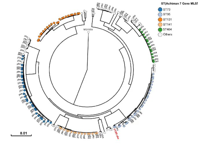
Phylogenetic distribution of pks island in UPEC strains
78
All 223 E. coli strains isolated during this sampling campaign were whole genome sequenced.
79
The phylogenetic distribution of these E. coli isolates was typical of strains which cause UTIs
80
2. A majority of strains belonged to the phylogroups B2 (69%) and D (15%) (Fig. 2 & Table
81
S1). Forty three percent of strains harboured pks island. All the pks+ strains belonged to the
82
phylogenetic group B2 and to the most common sequence types (STs) of extra-intestinal
83
pathogenic E. coli such as ST73, ST95, ST141 and ST404 (Fig. 2). As expected for
extra-84
intestinal pathogenic E. coli, multiple known or suspected virulence genes were also present in
85
the genome of these pks+ strains. pks island are thus widely present in the typical UPEC strains
86
that cause community-acquired UTIs.
87 88
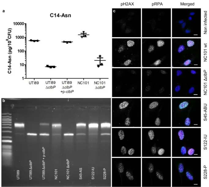
UPEC strains carrying pks island produce the genotoxin colibactin
89
We next sought to confirm that pks+ UPEC strains expressed colibactin. We first evaluated the
90
ability of UPEC strain UTI89, commonly used in rodent models of UTI, to produce colibactin
91
and to be genotoxic. We detected the colibactin C14-Asn cleavage product in bacterial cultures
92
(Fig. 3a) and confirmed the strain's genotoxicity on exogenous double-stranded DNA (Fig. 3b).
93
These effects were comparable to those observed with strain NC101 (Fig. 3a-b) which has been
94
shown to be pro-carcinogenic in different colorectal cancer mouse models. The same genotoxic
95
effects were also observed with three clinical pks+ UPEC isolates from asymptomatic
4
bacteriuria, cystitis or pyelonephritis cases (Fig. 3b). As was shown for NC101, infecting
97
human epithelial cells with pks+ UPEC strains induced the formation of nuclear pRPA and
98
pH2AX foci, indicating that the DNA of exposed cell is damaged (Fig. 1b, Fig. 3c). All
99
together, these results demonstrate that UPEC strains pks island are functional, mediate the
100
synthesis of colibactin and are genotoxic.
101 102
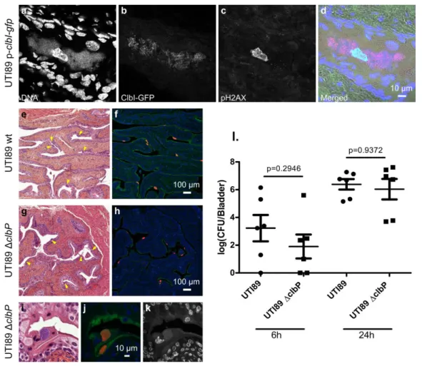
Colibactin is produced during UTIs and induces bladder urothelium DNA damage
103
In order to monitor the expression of the pks synthesis machinery during a UTI, we transformed
104
UPEC strain UTI89 with a plasmid expressing a GFP tagged pks island-encoded polyketide
105
ClbI synthase from its endogenous promoter. In bladder tissue collected 6 hours after infection,
106
we observed ClbI-GFP expressing bacteria in intracellular bacterial communities (IBCs) inside
107
superficial umbrella cells which line the lumen of the bladder (Fig. 4a-d & S1). Production of
108
colibactin was further confirmed by the detection of C14-Asn in pooled urines from 8 mice 24
109
hours after infection (C=0.66 pg/µL). We next assessed whether the metabolically active pks
110
machinery was associated with DNA damage in bladder cells. Phosphorylation of H2AX was
111
readily detected in nuclei of umbrella cells containing IBCs, 6 hours after infection with
wild-112
type E. coli UTI89 with or without the p-clbI-gfp plasmid (Fig. 4c-d & S2). A clbP mutant
113
unable to produce colibactin was very weakly genotoxic or not genotoxic at all (Fig. S2),
114
although equally capable of colonising the bladder (Fig. 4l) and inducing the formation of IBCs
115
(Fig. 4e-k). These results show that colibactin is expressed in vivo, in the bladder from the very
116
early stages of UTI and can induce DNA damage.
117 118
Colibactin induces urothelial cell DNA damage in the regenerative compartment
119
Bladders from mice infected with wild-type E. coli UTI89 exhibited pH2AX positive nuclei in
120
superficial umbrella cells, but also in urothelial basal cells (Fig. 5a-c). Bladders from mice
121
infected with the clbP mutant did not exhibit any pH2AX positive cells. pH2AX positive cells
122
nevertheless reappeared after complementation with the wild-type clbP allele (Fig. 5d-f & S3).
123
The basal urothelium compartment harbours keratin-14 positive (Krt14+) progenitor cells,
124
which are important for urothelium renewal following injury. There was an overrepresentation
125
of Krt14+ cells in urothelial tissue infected with wild-type or with clbP mutant UTI89 (Fig.
126
S4). Importantly, bladders infected with the genotoxic wild-type UTI89 strain exhibited basal
127
urothelial cells that were positive for both the regenerative cell marker Krt14 and the DNA
128
damage marker pH2AX (Fig. 5g-l). Thus, during UTI with a pks+ UPEC, colibactin induces
129
DNA damage in superficial and basal regenerative urothelial cells.
5 131
Discussion
132
It is now increasingly clear that pks+ E. coli strains found in the intestinal microbiota may play
133
a role in the aetiology and pathogenesis of colorectal cancer. These E. coli strains of
134
phylogenetic group B2 are often the same ones responsible for UTIs, but to date, no study has
135
been conducted on colibactin and UTIs. To the best of our knowledge the current study is the
136
first to report the presence of a C14-Asn signature, a metabolite of colibactin production, in the
137
urine of patients infected with pks+ UPEC. Thus, colibactin is produced during UTIs, one of
138
the most common human bacterial infections worldwide. We used a mouse model of human
139
UTI, to demonstrate that colibactin is produced and that it induces DNA damage in urothelial
140
cells, specifically in bladder Krt14+ progenitor cells.
141
Our study confirms the frequent occurrence of pks+ UPEC observed in Europe and the US,
142
irrespective of infection severity 10–13. This high prevalence in human subjects is in contrast to
143
the apparent lack of selective advantages associated with the production of colibactin in UTIs
144
mouse models. Our results suggest that colibactin is not essential for the ability of UPEC to
145
colonise the bladder or to form IBCs, whereas colibactin has been shown to play a role in the
146
pathogenicity of E. coli in extraintestinal infections such as septicemia or meningitis 27,28.
147
However, it should be noted that the mouse model used omits a key step in the pathogenesis of
148
UTIs: the domination and emergence from the intestinal reservoir 29. E. coli strains carrying pks
149
island are commonly observed among strains with a greater ability to persist in the intestinal
150
microbiota 3. In addition to the potential role of colibactin in modulating the intestinal
151
microbiota and promoting gut colonisation, enzymes encoded by pks island are essential for the
152
synthesis of siderophores and siderophore-microcins 30,31. Siderophores are major determinants
153
in the domination of the intestinal niche 32. They confer upon strains that produce them the
154
ability to outcompete other bacteria for iron, a rare essential nutrient. Moreover,
siderophore-155
microcins, antimicrobial peptides which target phylogenetically-linked enterobacteria help
156
UPEC to implant and proliferate within their intestinal reservoir, a step which precedes UTI 10.
157
The role of pks island in UPEC cannot merely be reduced to a determinant of intestinal
158
colonisation. Indeed, our current study shows that the pks machinery is also active in the urinary
159
tract and even within IBCs. Bacterial multiplication in these structures is intense, with a
160
doubling time of nearly 30 min, which requires an efficient and optimised bacterial metabolism
161
33. Although colibactin is a small molecule, the metabolic cost of its production, i.e. expressing
162
and operating the PK-NRP biosynthesis machinery is very high: nearly 1000 times higher than
6
that of peptide synthesis 34. If UPEC trigger such energetically inexpedient assembly lines
164
during UTIs, they must derive an adaptive benefit from it. One may speculate that other
165
products of the machinery may confer this advantage. In vitro, there is indeed a wide diversity
166
of metabolites produced by the pks machinery, ranging from other putative forms of
167
“colibactins” (macrocyclic metabolites for example) to other smaller metabolites, which
168
potentially vary between strains 15,35. The biological function of these metabolites remains to
169
be elucidated, but some of these metabolites may be relevant to the pathogenesis of infections.
170
We have, for instance, recently described the synthesis of C12-Asn-GABA, by the pks
171
machinery of the probiotic Nissle 1917 strain and shown its digestive pain-relieving activity 36.
172
The production of such an analgesic metabolite coupled with the increased production of
173
siderophores by pks+ E. coli strains may provide a selective advantage to colonising the urinary
174
tract, in addition to the digestive tract.
175
Irrespective of the role of pks island in the virulence of UPECs, the fact remains that these
176
strains are genotoxic to bladder cells, particularly to bladder progenitor cells.
Colibactin-177
induced DNA damage is incompletely repaired, resulting in the accumulation of gene mutations
178
and cell transformation 21,26,37. We have previously shown that post-exposure chromosomal
179
instability to colibactin can persist in daughter cells 21. DNA damage could also be propagated
180
to contiguous cells by the induction of a senescence-associated secretory phenotype in the
181
affected cells and the production of ROS, that could also explain the pattern of pH2AX positive
182
patches of cells 24 hours after infection 37,38. In the current study, we observed that some murine
183
bladder progenitor cells (Krt14+) display pH2AX positive nuclei. In humans, Krt14 expression
184
characterises undifferentiated bladder cancers with particularly poor prognoses 39,40. Rodent
185
bladder cancer models indicate that a significant proportion of cancerous tissue derives from
186
Krt14 positive cells 41. Thus, colibactin damage to Krt14+ cells may initiate the formation and
187
perpetuate the propagation of DNA lesions. The colibactin mutational signature has recently
188
been identified in colorectal cancers but also in urinary tract cancers 26,42. Currently the main
189
risk factors for bladder cancers are tobacco and occupational exposure to solvents, which are
190
more frequently investigated than UTIs 43. However, a large worldwide bladder cancer case
191
control study recently showed that regular UTIs were epidemiologically associated with an
192
increased risk of urinary bladder cancer 44. Our findings suggest that pks+ UPEC UTIs may be
193
an additional risk factor, particularly in cases of chronic and regular infections, irrespective of
194
whether symptoms are present or not. We detected C14-Asn in the urine of patients with
195
asymptomatic bacteriuria, which are usually not treated with antibiotics and can thus persist for
7
periods of up to many years 45. A better understanding of the consequences of colibactin
197
production could prompt a systematic search for pks island in UPEC isolates or C14-Asn in the
198
urine of patients at risk.
199 200
Materials and methods
201
Bacterial strains
202
The archetypal E. coli strains used in this study were the UPEC strain UTI89 46 and the
203
colitogenic E. coli strain NC101 22. The UTI89 and NC101 ΔclbP mutants were constructed
204
using the lambda Red recombinase method 47 with primers IHAPJPN29 and IHAPJPN30, as
205
previously described 14. The pMB702 construct (referred to as p-clbP) was used for
206
complementation of the UTI89 ΔclbP mutant 48. For the in vivo complementation assay, the
207
UTI89 ΔclbP mutant was transformed with the pCM17-clbP plasmid. Briefly, the clbP gene
208
was PCR-amplified from pBRSK-clbP 48, with primers clbP-F-Bam_pm
(5’-209
atGGATCCatgacaataatggaacacgttagc-3’) and pBRSK-F-Bam_pm (5’-atGGATCCcaagctcgga
210
attaaccctc-3’) and cloned into the pCM17 vector 49 BamHI site. The ClbI C-terminal GFP
211
fusion was constructed using the Gibson Assembly kit (New England Biolabs, MA, USA).
212
Briefly, the clbI and gfp genes were amplified by PCR using the following primers:
pK184-213 clbI-gb1(5’-GATTACGAATTCGAGCTCGGTACCCATGGCAGAGAATGATTTTGG-3’); 214 clbI-gfp-gb2 (5’-215 CTTCTCCTTTTCCGCCTCCTCCGCCCTCATTAATCATGTCGTTAACTAG-3’); clbI-216 gfp-gb3 (5’-217 GATTAATGAGGGCGGAGGAGGCGGAAAAGGAGAAGAACTTTTCACTGG-3’) and 218 gfp-pK184-gb4 (5’-219 TGCAGGTCGACCTCGAGGGATCCCCTTATTTGTATAGTTCATCCATGCC-3’). A 220
glycine linker (5’-GGCGGAGGAGGCGGA-3’) was introduced at the clbI and gfp junction for
221
flexibility. PCR amplified fragments and SmaI-digested pK184 vector were assembled
222
according to the manufacturer's recommendations.
223 224
Collection of clinical strains and urines
225
The collection of 225 E. coli strains from urine samples at the Adult Emergency Department of
226
Toulouse University Hospital, France, between July and October 2017, was previously
227
described 10. According to the French regulations relating to observational database analyses,
228
the study did not require specific informed consent. Urine was collected from 223 patients
229
(women and men, under 75 years) with either pyelonephritis (104), symptomatic infections
8
excluding pyelonephritis (cystitis) (83), or asymptomatic bacteriuria (36) without urological
231
comorbidities or catheterisation. All strains were identified as E. coli by matrix-assisted laser
232
desorption/ionisation time-of-flight mass spectrometry (Microflex LT MALDI-TOF MS,
233
Bruker Daltonik GmbH, Germany). Samples of the corresponding urine collected in boric acid
234
tubes were stored at -80 °C until lipid analysis.
235 236
C14-Asn quantification
237
Extraction and quantification of C14-Asn was performed as previously described 36. Briefly, 5
238
µL internal standard mixture (Deuterium-labelled compound at 400 ng/mL) was added to the
239
bacterial pellets of 24 h DMEM cultures or to 500 µL of urine samples before crushing,
240
followed by addition of cold methanol (MeOH) (15% final volume) and homogenisation. After
241
centrifugation, supernatants were solid phase extracted on HLB plates (OASIS® HLB 2 mg,
242
96-well plate, Waters, Ireland). Following washing with H2O/ MeOH (90:10, v/v) and elution
243
with MeOH, samples were evaporated twice under N2 and finally resuspended in 10 µL MeOH.
244
Separation and quantification of C14-Asn was performed on a high-performance liquid
245
chromatography coupled to tandem mass spectrometry system (G6460 Agilent) 36. Limit of
246
detection was 2.5 pg and limit of quantification was 5 pg.
247 248
Sequencing data, sequence alignments and phylogenetic analyses
249
Whole genome sequencing was performed using the Illumina NextSeq500 Mid Output platform
250
(Integragen, Evry, France) to generate 2 x 150 bp paired-end reads, at approximately
251
80x average coverage. Genome de novo assembly and analysis were performed with the
252
BioNumerics 7.6 software (Applied Maths) and Enterobase (http://enterobase.warwick.ac.uk/).
253
For SNP-based phylogenetic trees, core genome alignments were generated after mapping raw
254
reads to the E. coli MG1655 genome. The core genome phylogenetic tree was inferred with the
255
Maximum-likelihood algorithm using Enterobase for B2 phylogroup strains.
256 257
Exogenous DNA cross-linking assay
258
As previously described 18, the pUC19 plasmid, linearised with BamHI, was exposed to bacteria
259
pre-grown (3 x 10⁶ CFU) in DMEM 25 mM Hepes for 40 min at 37 °C. The DNA was purified
260
and 100 ng submitted to denaturing gel DNA electrophoresis (40 mM NaOH and 1 mM EDTA,
261
pH ~12.0). After a neutralisation step, the gel was stained with GelRed and visualised under
262
UV using the ChemiDoc Imaging System (BioRad).
263 264
9
pH2AX and pRPA immunofluorescence analysis of post infected HeLa cells
265
HeLa cells (ATCC CCL2) were cultured and infected in 8-well chamber slides (Labtek), as
266
previously described 18. 24 hours after passaging, Hela cells were infected with bacteria
267
precultured in DMEM 25 mM HEPES at a multiplicity of infection (MOI) of 50, for 4 h. Cells
268
were then washed and incubated overnight in cell culture medium with 100 µg/ml gentamicin.
269
Immunofluorescence analysis was then performed as previously described 18. Briefly, cells
270
were pre-extracted in PBS 0.1%, Triton X-100 and fixed in PBS 4% formaldehyde,
271
permeabilised, blocked with MAXblock medium (Active Motif) and stained with antibodies
272
against pH2AX (1:500, JBW301, Millipore) and S33p-RPA32 (1:500, A300-264A, Bethyl),
273
diluted in MAXblock 0.05%, Triton X-100. Cells were washed 3 times for 5 min in PBS 0.05%,
274
Triton X-100 and incubated with secondary AlexaFluor 488 or 568 (Invitrogen) diluted 1:500
275
in MAXblock medium with 1 µg/ml DAPI (Sigma). Slides were mounted with Fluoroshield
276
(Sigma) and examined on a Leica SP8 laser scanning confocal microscope in sequential mode
277
with LasX software (Leica), while keeping the same laser and detector settings between
278
different wells. Final images were processed and edited with the ImageJ software.
279 280
Mouse UTI model
281
Animal infections were performed in accordance with the European directives for the protection
282
of animals used for scientific purposes (2010/63/EU). An ethics committee approved the
283
protocol (number CEEA-122 2014-53). Female, 6-8 weeks old, C3H/HeN mice (Janvier Labs)
284
were infected transurethrally as previously described 50. Briefly, UPEC were cultivated
285
statically in LB 50 and resuspended to an inoculum of 108 CFU in 50 µl PBS. Mice under 4%
286
isoflurane anaesthesia were inoculated twice at one-hour intervals with a pump that delivered
287
10 µL/s. At 6 or 24 hours, mice were euthanised, bladders were harvested, homogenised for
288
bacterial enumeration on agar plates, stored in OCT compound at -80 °C or fixed in 4%
289
formaldehyde after filling the bladder prior to paraffin embedding. Each experiment was
290
conducted in duplicate with 5 to 8 mice per group.
291 292
Histological and immunofluorescence analyses of mouse bladder tissue
293
Histological bladder slices were prepared using standard protocols and processed for
294
haematoxylin-eosin staining then scanned using a Pannoramic 250 scanner (3DHistech). Final
295
images were captured using CaseViewer software (3DHistech).
296
For GFP and pH2AX detection on OCT compound-embedded bladders, 8 µm thick sections
297
were dried on Superfrost plus glass slides, fixed for 15 min in 4% formaldehyde then rinsed
10
with PBS, blocked and permeabilised with MaxBlock, 0.3% Triton X100. Primary anti-GFP
299
(Rabbit, Abcam 6556) and anti-pH2AX (Mouse, Millipore #O5-636) antibodies were diluted
300
at 1:200 in MaxBlock, 0.3% triton X100. After washing in PBS, 0.05% Triton X100, slides
301
were incubated with secondary antibodies at 1:200 (Alexa 633 Goat anti-Rabbit antibody
302
(Invitrogen A21071) and Alexa 488 Goat anti-Mouse antibody (Life technologies A11029))
303
and DAPI. For pH2AX and Krt14 staining on paraffin-embedded bladders, 5 µm thick sections
304
were de-paraffinised and re-hydrated in xylene, ethanol and tap water. Unmasking was
305
performed with a 6-min trypsin digestion (Trypsin-EDTA solution, Sigma, T392) followed by
306
30 min incubation in citrate buffer pH 6 at 80–95°C. Sections were then blocked and
307
permeabilised in MaxBlock, 0.3% Triton X100. Slices were stained with anti-pH2AX (Rabbit,
308
Cell Signalling S9718) at a dilution of 1:200 and anti-Krt14 antibodies (Chicken, Ozyme
309
BLE906001) at a dilution of 1:250 in MaxBlock, 0.3% Triton X100. Sections were washed in
310
PBS, 0.05% Triton X100 and incubated with secondary antibodies (Alexa 633 Goat anti-Rabbit
311
antibody, Invitrogen A21071, at a dilution of 1:200; Alexa 555 Goat anti-Chicken antibody,
312
Invitrogen A32932, at a dilution of 1:500). Images were acquired as with infected cultured cells.
313 314
Fluorescence in situ hybridisation (FISH) of mouse bladder tissue
315
Five micron paraffin-embedded bladder sections were de-paraffinised in
316
xylene/ethanol. Sections were incubated in lysozyme solution (10 mg/ml, Sigma, France) for
317
15 minutes at 37 °C and exposed to 100 µl ofuniversal bacterial 16 S fluorescent rRNA probe
318
(Eub338, GCTGCCTCCCGTAGGAGT-Cy5’, Eurofins, France) at a concentration of 5 ng/µl,
319
in hybridisation buffer (20 mM Tris-HCl, pH7.4, 0.9 M NaCl, 0.01% SDS) at 46°C for 3 hours
320
51. Sections were then incubated in a 48 °C prewarmed saline-sodium citrate wash buffer (30
321
mM sodium citrate, 300 mM sodium chloride, pH7.4, Invitrogen, France) for 20 minutes. To
322
stain polysaccharide-rich content, sections were counterstained with FITC-conjugated wheat
323
germ agglutinin (Invitrogen, France) at a 1:1000 dilution in PBS buffer for 30 min. Slides were
324
mounted with Fluoroshield containing DAPI. Images were acquired using a confocal laser
325
scanning microscope (Zeiss LSM 710) and final images were processed and edited with the
326 ImageJ software. 327 328 Statistical analyses 329
Graphical representation and statistical analyses were carried out using GraphPad Prism 8.3. P
330
values were calculated using two-tailed Mann-Whitney U test.
331 332
11 333
References
334
1. Flores-Mireles, A. L., Walker, J. N., Caparon, M. & Hultgren, S. J. Urinary tract
335
infections: epidemiology, mechanisms of infection and treatment options. Nat. Rev.
336
Microbiol. 13, 269–284 (2015).
337
2. Klein, R. D. & Hultgren, S. J. Urinary tract infections: microbial pathogenesis,
host-338
pathogen interactions and new treatment strategies. Nat. Rev. Microbiol. (2020)
339
doi:10.1038/s41579-020-0324-0.
340
3. Nowrouzian, F. L. & Oswald, E. Escherichia coli strains with the capacity for long-term
341
persistence in the bowel microbiota carry the potentially genotoxic pks island. Microb.
342
Pathog. 53, 180–182 (2012).
343
4. Tenaillon, O., Skurnik, D., Picard, B. & Denamur, E. The population genetics of
344
commensal Escherichia coli. Nat. Rev. Microbiol. 8, 207–217 (2010).
345
5. Mobley, H. L. T., Donnenberg, M. S. & Hagan, E. C. Uropathogenic Escherichia coli.
346
EcoSal Plus 3, (2009).
347
6. Ulett, G. C. et al. Uropathogenic Escherichia coli virulence and innate immune responses
348
during urinary tract infection. Curr. Opin. Microbiol. 16, 100–107 (2013).
349
7. Johnson, J. R. & Russo, T. A. Molecular epidemiology of extraintestinal pathogenic
350
Escherichia coli. EcoSal Plus 8, (2018).
351
8. Mobley, H. L. T. Measuring Escherichia coli gene expression during human urinary tract
352
infections. Pathog. Basel Switz. 5, (2016).
353
9. Mobley, H. L. et al. Pyelonephritogenic Escherichia coli and killing of cultured human
354
renal proximal tubular epithelial cells: role of hemolysin in some strains. Infect. Immun.
355
58, 1281–1289 (1990).
356
10. Massip, C., Chagneau, C. V., Boury, M. & Oswald, E. The synergistic triad between
357
microcin, colibactin, and salmochelin gene clusters in uropathogenic Escherichia coli.
358
Microbes Infect. (2020) doi:10.1016/j.micinf.2020.01.001.
359
11. Krieger, J. N., Dobrindt, U., Riley, D. E. & Oswald, E. Acute Escherichia coli prostatitis
360
in previously health young men: Bacterial virulence factors, antimicrobial resistance, and
361
clinical outcomes. Urology 77, 1420–1425 (2011).
362
12. Salvador, E. et al. Comparison of asymptomatic bacteriuria Escherichia coli isolates from
363
healthy individuals versus those from hospital patients shows that long-term bladder
364
colonization selects for attenuated virulence phenotypes. Infect. Immun. 80, 668–678
365
(2012).
12
13. Dubois, D. et al. Cyclomodulins in urosepsis strains of Escherichia coli. J. Clin.
367
Microbiol. 48, 2122–2129 (2010).
368
14. Nougayrède, J.-P. et al. Escherichia coli induces DNA double-strand breaks in eukaryotic
369
cells. Science 313, 848–851 (2006).
370
15. Vizcaino, M. I., Engel, P., Trautman, E. & Crawford, J. M. Comparative metabolomics
371
and structural characterizations illuminate colibactin pathway-dependent small molecules.
372
J. Am. Chem. Soc. 136, 9244–9247 (2014).
373
16. Jiang, Y. et al. Reactivity of an unusual amidase may explain colibactin’s DNA
cross-374
linking activity. J. Am. Chem. Soc. (2019) doi:10.1021/jacs.9b02453.
375
17. Brotherton, C. A. & Balskus, E. P. A prodrug resistance mechanism is involved in
376
colibactin biosynthesis and cytotoxicity. J. Am. Chem. Soc. 135, 3359–3362 (2013).
377
18. Bossuet-Greif, N. et al. The colibactin genotoxin generates DNA interstrand cross-links in
378
infected cells. mBio 9, e02393-17 (2018).
379
19. Wilson, M. R. et al. The human gut bacterial genotoxin colibactin alkylates DNA. Science
380
363, eaar7785 (2019).
381
20. Xue, M. et al. Structure elucidation of colibactin and its DNA cross-links. Science
382
eaax2685 (2019) doi:10.1126/science.aax2685.
383
21. Cuevas-Ramos, G. et al. Escherichia coli induces DNA damage in vivo and triggers
384
genomic instability in mammalian cells. Proc. Natl. Acad. Sci. U. S. A. 107, 11537–11542
385
(2010).
386
22. Arthur, J. C. et al. Intestinal inflammation targets cancer-inducing activity of the
387
microbiota. Science 338, 120–123 (2012).
388
23. Dejea, C. M. et al. Patients with familial adenomatous polyposis harbor colonic biofilms
389
containing tumorigenic bacteria. Science 359, 592–597 (2018).
390
24. Dalmasso, G., Cougnoux, A., Delmas, J., Darfeuille-Michaud, A. & Bonnet, R. The
391
bacterial genotoxin colibactin promotes colon tumor growth by modifying the tumor
392
microenvironment. Gut Microbes 5, 675–680 (2014).
393
25. Buc, E. et al. High prevalence of mucosa-associated E. coli producing cyclomodulin and
394
genotoxin in colon cancer. PloS One 8, e56964 (2013).
395
26. Pleguezuelos-Manzano, C. et al. Mutational signature in colorectal cancer caused by
396
genotoxic pks + E. coli. Nature 580, 269–273 (2020).
397
27. McCarthy, A. J. et al. The genotoxin colibactin is a determinant of virulence in
398
Escherichia coli K1 experimental neonatal systemic infection. Infect. Immun. 83, 3704–
399
3711 (2015).
13
28. Marcq, I. et al. The genotoxin colibactin exacerbates lymphopenia and decreases survival
401
rate in mice infected with septicemic Escherichia coli. J. Infect. Dis. 210, 285–294 (2014).
402
29. Moreno, E. et al. Relationship between Escherichia coli strains causing acute cystitis in
403
women and the fecal E. coli population of the host. J. Clin. Microbiol. 46, 2529–2534
404
(2008).
405
30. Massip, C. et al. Deciphering the interplay between the genotoxic and probiotic activities
406
of Escherichia coli Nissle 1917. PLOS Pathog. 15, e1008029 (2019).
407
31. Martin, P. et al. Interplay between siderophores and colibactin genotoxin biosynthetic
408
pathways in Escherichia coli. PLoS Pathog. 9, e1003437 (2013).
409
32. Pi, H. et al. Role of catecholate siderophores in gram-negative bacterial colonization of
410
the mouse gut. PloS One 7, e50020 (2012).
411
33. Justice, S. S. et al. Differentiation and developmental pathways of uropathogenic
412
Escherichia coli in urinary tract pathogenesis. Proc. Natl. Acad. Sci. U. S. A. 101, 1333–
413
1338 (2004).
414
34. Amoutzias, G. D., Chaliotis, A. & Mossialos, D. Discovery strategies of bioactive
415
compounds synthesized by nonribosomal peptide synthetases and type-I polyketide
416
synthases derived from marine microbiomes. Mar. Drugs 14, (2016).
417
35. Li, Z.-R. et al. Macrocyclic colibactin induces DNA double-strand breaks via
copper-418
mediated oxidative cleavage. Nat. Chem. (2019) doi:10.1038/s41557-019-0317-7.
419
36. Pérez-Berezo, T. et al. Identification of an analgesic lipopeptide produced by the probiotic
420
Escherichia coli strain Nissle 1917. Nat. Commun. 8, 1314 (2017).
421
37. Cougnoux, A. et al. Bacterial genotoxin colibactin promotes colon tumour growth by
422
inducing a senescence-associated secretory phenotype. Gut 63, 1932–1942 (2014).
423
38. Secher, T., Samba-Louaka, A., Oswald, E. & Nougayrède, J.-P. Escherichia coli
424
producing colibactin triggers premature and transmissible senescence in mammalian cells.
425
PloS One 8, e77157 (2013).
426
39. Lerner, S. P. et al. Bladder cancer molecular taxonomy: Summary from a consensus
427
meeting. Bladder Cancer Amst. Neth. 2, 37–47.
428
40. Volkmer, J.-P. et al. Three differentiation states risk-stratify bladder cancer into distinct
429
subtypes. Proc. Natl. Acad. Sci. U. S. A. 109, 2078–2083 (2012).
430
41. Papafotiou, G. et al. KRT14 marks a subpopulation of bladder basal cells with pivotal role
431
in regeneration and tumorigenesis. Nat. Commun. 7, (2016).
14
42. Dziubańska-Kusibab, P. J. et al. Colibactin DNA damage signature indicates causative
433
role in colorectal cancer. bioRxiv 819854 (2019) doi:10.1101/819854. Preprint at:
434
https://www.biorxiv.org/content/10.1101/819854v1
435
43. Saginala, K. et al. Epidemiology of bladder cancer. Med. Sci. Basel Switz. 8, (2020).
436
44. Vermeulen, S. H. et al. Recurrent urinary tract infection and risk of bladder cancer in the
437
Nijmegen bladder cancer study. Br. J. Cancer 112, 594–600 (2015).
438
45. Lindberg, U. et al. Asymptomatic bacteriuria in schoolgirls. II. Differences in escherichia
439
coli causing asymptomatic bacteriuria. Acta Paediatr. Scand. 64, 432–436 (1975).
440
46. Mulvey, M. A., Schilling, J. D. & Hultgren, S. J. Establishment of a persistent Escherichia
441
coli reservoir during the acute phase of a bladder infection. Infect. Immun. 69, 4572–4579
442
(2001).
443
47. Datsenko, K. A. & Wanner, B. L. One-step inactivation of chromosomal genes in
444
Escherichia coli K-12 using PCR products. Proc. Natl. Acad. Sci. 97, 6640–6645 (2000).
445
48. Dubois, D. et al. ClbP Is a prototype of a peptidase subgroup involved in biosynthesis of
446
nonribosomal peptides. J. Biol. Chem. 286, 35562–35570 (2011).
447
49. Morin, C. E. & Kaper, J. B. Use of stabilized luciferase-expressing plasmids to examine
448
in vivo-induced promoters in the Vibrio cholerae vaccine strain CVD103-HgR. FEMS
449
Immunol. Med. Microbiol. 57, 69–79 (2009).
450
50. Hung, C.-S., Dodson, K. W. & Hultgren, S. J. A murine model of urinary tract infection.
451
Nat. Protoc. 4, 1230–1243 (2009).
452
51. Motta, J.-P. et al. Active thrombin produced by the intestinal epithelium controls mucosal
453
biofilms. Nat. Commun. 10, (2019).
454 455
Acknowledgements
456
The authors thank Alexandre Perrat, Frédéric Auvray and Laurent Cavalié for their help with
457
genomic DNA extraction and sequence analysis and Pauline Dragot for her excellent technical
458
help. We thank the technical microbiology laboratory staff from the Toulouse University
459
Hospital for helping to culture and isolate E. coli strains. The authors also wish to thank the
460
staff of the Tri GenoToul imaging facility, Toulouse (particularly Sophie Allart and Danièle
461
Daviaud), the experimental histology facility (CREFRE, Toulouse) and the lipidomic facility
462
of MetaToul, Toulouse. The authors thank the team members for their helpful discussions and
463
critical reading of the manuscript and Sébastien Déjean for his help with data analysis.
464 465
15
This work was supported by grants from the French National Agency for Research (ANR)
466
(UTI-TOUL ANR-17-CE35-0010 and LiBacPain ANR-18-CE14-0039). C.V.C was supported
467
by a two-year grant from the French National Institute for Health and Medical Research (poste
468
d’accueil INSERM 2018). The funding bodies did not contribute to the study design, collection
469
of data, interpretation of results or to the decision to submit the work for publication.
470 471
Authors contribution
472
C.M., C.V.C. collected human clinical samples (urines and strains). C.M., C.V.C, E.O. analysed
473
the strains. C.V.C., P.L.F., N.C. performed lipid extractions and analyses. A.S., N.B.G., P.M.,
474
C.V.C. designed and constructed the molecular engineered strains. C.V.C., N.B.G., J-P.N.
475
performed in vitro genotoxicity assays. C.F., M.F., P.M. designed and carried out in vivo
476
experiments. P.M., C.B. and C.V.C. analysed in vivo samples. C.V.C., J-P.N., J-P.M. performed
477
histological and microscopic analyses. C.V.C., C.M., J-P.N., J-P.M., P.M., N.C., M-P.R., H.C.,
478
M.F., E.O. contributed to the analysis and interpretation of the data. C.V.C, J-P.N., C.M., E.O.
479
drafted and wrote the paper. E.O. obtained dedicated funding to support this work.
480 481
Competing interests statement
482
The authors declare no competing interests.
16
Figures
484 485
486
Fig. 1: Synthesis of the genotoxin, colibactin, releases the C14-Asn metabolite detected in
487
the urine of patients with UTI. a. Colibactin is first synthesised as a prodrug, precolibactin
488
and then cleaved by ClbP peptidase thereby releasing the mature genotoxic dimeric colibactin,
489
and C14-Asn. b. Colibactin alkylates both strands of the DNA helix, generating an interstrand
490
cross-link. In response to this DNA damage, the host cell machinery recruits and phosphorylates
491
the H2AX and RPA proteins. c. Concentration of C14-Asn in human urines according to the
492
presence of a pks island in the genome of the corresponding UPEC isolate as determined by
493
LC-MS/MS. All individual data points are shown on a violin plot, with a two segments linear
494
Y range. C14-Asn was below the LC-MS/MS detection limit in the urine of patients infected
495
with UPEC isolates that did not contain a pks island.
17 497
Fig. 2: Human pks+ UPEC belong to major lineages of extraintestinal pathogenic E. coli
498
from phylogroup B2. A phylogenetic tree based on whole genome analysis was constructed
499
and rooted on E. coli MG1655. Each circle represents an individual UPEC strain. Main
500
Sequence Types are grouped by colours. The presence of a pks island is denoted adjacent to
501
each circle. The archetypal UPEC strain UTI89, which is pks+, was also included in this tree.
18 503
Fig. 3: The archetypal cystitis E. coli strain UTI89 and clinical pks+ UPEC isolates
504
produce colibactin and induce DNA damage. a. LC-MS/MS quantification of the C14-Asn
505
cleavage product released following colibactin maturation by ClbP peptidase in bacterial pellets
506
of E. coli strains UTI89 and NC101 and their respective ΔclbP mutants. Quantifications were
507
performed in triplicate and represented as a mean ± standard error of the mean (SEM). b. DNA
508
interstrand cross-links formed after exposure of linear double stranded DNA to UTI89, NC101
509
and their respective ΔclbP mutants as well as three representative clinical human UPEC
510
isolates, visualised by electrophoresis under denaturing conditions. DNA with interstrand
cross-511
links migrates at an apparent molecular weight of twice that of DNA without cross-links. In the
512
absence of any crosslinking DNA migrates as a single denatured strand. S45, S122, S228:
513
Human UPEC strains from AS=asymptomatic bacteriuria; IU=cystitis; P=pyelonephritis
514
respectively. c. S33p-RPA32 and pH2AX immunofluorescence staining of HeLa cells, 16 hours
515
after infection with the reference NC101 strain, its ΔclbP mutant or human UPEC strains (S45,
19
S122, S228). pH2AX and S33p-RPA32=pRPA: grayscale. Merged: green=pH2AX;
517
magenta=S33p-RPA32; blue=DAPI; scale bar=10 µm.
20 519
Fig. 4: Colibactin is produced during UTIs and induces DNA damage. a-d. Confocal
520
microscopic detection of ClbI-GFP expression and pH2AX in frozen bladder sections
521
containing an IBC, 6 hours post infection with UTI89, hosting the p-clbI-gfp fusion plasmid.
522
The individual channel images are shown in grayscale. In the merged (d) image: blue=DNA,
523
magenta=ClbI-GFP, green=pH2AX, grey=phase contrast. e-k. Bladder sections 6 hours post
524
inoculation with wild-type UTI89 (e, f) or the isogenic ΔclbP mutant (g-k) stained with
525
haematoxylin-eosin (e, g, i), FISH (f, h, j) or DAPI (k, grayscale) all of which detect IBCs
526
(arrows). In the FISH images: blue=DAPI stain DNA; green= FITC-conjugated wheat germ
527
agglutinin (WGA) stained polysaccharides; red=bacterial cells, labelled with the universal 16S
528
FISH probe. l. Bladder tissue E. coli counts, 6 h and 24 h after infection with wild-type E. coli
529
UTI89 (circles) or ΔclbP (squares). Each data point corresponds to one mouse, with mean ±
530
standard error of the mean (SEM) shown for each group. n=6. Mann-Whitney U test.
21 532
22
Fig. 5: Colibactin induces DNA damage in urothelial cells of the regenerative
533
compartment. a-f. DAPI (DNA: a, d), pH2AX (b, e) and haematoxylin-eosin (H&E: c, f)
534
staining of paraffin-embedded bladder sections 24 hours after UTI89 wild-type (a-c) or
535
UTI89ΔclbP (d-f) infection. The individual fluorescence channel images are shown in
536
grayscale. See also Fig. S3. g-l. Paraffin-embedded bladders sections, were immuno-stained for
537
pH2AX and Krt14, 24 hours post infection with wild-type UTI89. g-i: merged images:
538
blue=DNA, red=Krt14, green=pH2AX. j-l: individual channel images shown at higher
539
magnification in grayscale. Pink arrows identify cells positive for both Krt14 and pH2AX. Scale
540
bar = 10 µm.