Analysis of sequence-selective guanine oxidation by
biological agents
by
Yelena Margolin
B.S. in Chemistry, Massachusetts Institute of Technology, 1999 B.S. in Biology, Massachusetts Institute of Technology, 1999
Submitted to the Department of Biological Engineering in Partial Fulfillment of the requirements for the degree of
Ph.D. in Toxicology at the
MASSACHUSETTS INSTITUTE OF TECHNOLOGY
@ Massachusetts
October 007
Institute of Technology. All rights reserved.
ARHIMES MAS~SACHUtU' INSITUT
OF TEOHNOLOGY
JAN
2
8
2008
LIBRARIES
Signature of Author:
Department of Biological Engineering October, 2007
Certified by:
Accepted by
Peter C. Dedon Professor of Toxicp ogy and Biological Engineering
S
/
Thesis supervisor
: ,, " % sor of Chairr n, Committee Alan Grodzinsky Biological Engineering for Graduate StudentsThis doctoral thesis has been examined by a committee of the Department of Biological Engineering as follows:
Associate Professor Bevin P. Engelward
Chairperson
Professor Peter C. Dedon
Supervisor
eforP ssor John M. Essigmann
Professor Steven R. Tannenbaum
1I~~V
-Professor John M. Essiemann
"-Analysis of sequence-selective guanine oxidation
by biological agents
by
Yelena Margolin
Submitted to the Department of Biological Engineering on October 5, 2007 in partial fulfillment of the requirements for the degree of Doctor of Philosophy in
Toxicology
ABSTRACT
Oxidatively damaged DNA has been strongly associated with cancer, chronic degenerative diseases and aging. Guanine is the most frequently
oxidized base in the DNA, and generation of a guanine radical cation (G'") as an
intermediate in the oxidation reaction leads to migration of a resulting cationic hole through the DNA n-stack until it is trapped at the lowest-energy sites. These sites reside at runs of guanines, such as 5'-GG-3' sequences, and are
characterized by the lowest sequence-specific ionization potentials (IPs). The charge transfer mechanism suggests that hotspots of oxidative DNA damage induced by electron transfer reagents can be predicted based on the primary
DNA sequence. However, preliminary data indicated that
nitrosoperoxycarbonate (ONOOCO2"), a mediator of chronic inflammation and a one-electron oxidant, displayed unusual guanine oxidation properties that were the focus of present work.
As a first step in our study, we determined relative levels of guanine oxidation, induced by ONOOCO2 in all possible three-base sequence contexts (XGY) within double-stranded oligonucleotides. These levels were compared to
the relative oxidation induced within the same guanines by photoactivated riboflavin, a one-electron reagent. We found that, in agreement with previous studies, photoactivated riboflavin was selective for guanines of lowest IPs located within 5'-GG-3' sequences. In contrast, ONOOCO2" preferentially reacted with guanines located within 5'-GC-3' sequences characterized by the highest IPs. This demonstrated that that sequence-specific IP was not a determinant of guanine reactivity with ONOOCO2". Sequence selectivities for both reagents
were double-strand specific. Selectivity of ONOOCO2 for 5'-GC-3' sites was also
observed in human genomic DNA after ligation-mediated PCR analysis. Relative yields of different guanine lesions produced by both ONOOCO2" and riboflavin varied 4- to 5-fold across all sequence contexts.
To assess the role of solvent exposure in mediating guanine oxidation by ONOOCO2", relative reactivities of mismatched guanines with ONOOCO2" were
measured. The majority of the mismatches displayed an increased reactivity with ONOOCO2 as compared to the fully matched G-C base-pairs. The extent of reactivity enhancement was sequence context-dependent, and the greatest levels of enhancement were observed for the conformationally flexible guanine-guanine (G-G) mismatches and for guanine-guanines located across from a synthetic abasic site.
To test the hypothesis that the negative charge of an oxidant influences its reactivity with guanines in DNA, sequence-selective guanine oxidation by a negatively charged reagent, Fe+2-EDTA, was assessed and compared to guanine oxidation produced by a neutral oxidant, y-radiation. Because both of these agents cause high levels of deoxyribose oxidation, a general method to quantify sequence-specific nucleobase oxidation in the presence of direct strand
breaks was developed. This method exploited activity of exonuclease III (Exo III), a 3' to 5' exonuclease, and utilized phosphorothioate-modified synthetic oligonucleotides that were resistant to Exo III activity. This method was employed to determine sequence-selective guanine oxidation by Fe+2-EDTA complex and y-radiation and to show that both agents produced identical guanine
oxidation pattems and were equally reactive with all guanines, irrespective of their sequence-specific IPs or sequence context. This showed that negative
charge was not a determinant of Fe+2-EDTA-mediated guanine oxidation.
Finally, the role of oxidant binding on nucleobase damage was assessed
by studying sequence-selective oxidation produced by DNA-bound Fe+2 ions in the presence of H202. We found that the major oxidation targets were thymines
located within 5'-TGG-3' motifs, demonstrating that while guanines were a required element for coordination of Fe+2 to DNA, they were not oxidized.
Our results suggest that factors other than sequence-specific IPs can act as major determinants of sequence-selective guanine oxidation, and that current models of guanine oxidation and charge transfer in DNA cannot be used to adequately predict the location and identity of mutagenic lesions in the genome.
Acknowledgements
I am very grateful to my thesis advisor, Prof. Peter C. Dedon, for all of his
support, generosity and encouragement, especially through the difficult times when things did not work out as expected. I have learned a great deal from him and have enjoyed interacting with him both on a professional and personal level.
I feel that I am very lucky to have had an opportunity to work in his lab.
I am indebted to all the past and present members of the Dedon lab both
for their friendship and for their willingness to share their scientific knowledge and expertise with me. I would particularly like to acknowledge the help of Dr.
Michael Demott, Dr. Min Dong, Dr. Christianne Collins, and Dr. Marita Barth. Debra Dederich, Jose McFaline, Bahar Edrissi, and Dr. Erin Prestwich proofread parts of this thesis. Olga Parkin, Christine Marzilli, Dawn Erickson and Jackie Goodluck-Griffith have made my everyday life in the lab easy and organized, and
I will be forever thankful for their help.
I am very grateful to the members of my thesis committee Prof. Bevin P.
Engelward, Prof. John M. Essigmann and Prof. Steven R. Tannenbaum for their continuing encouragement and advice, as well as their enthusiasm for science and sense of humor. I very much appreciate the time and the knowledge they shared with me.
I would like to acknowledge our collaborators in the Geacintov laboratory at the NYU Department of Chemistry Prof. Nicholas E. Geacintov, Prof. Vladimir
Shafirovich, Dr. Young-Ae Lee and Dr. Fabian A. Rodriguez, who have
contributed much to our understanding of the mechanisms of guanine oxidation
by the carbonate radical anion.
I would like to thank the members my family - my father Yulis, my mother
Raisa, my sisters Irene and Masha, my cousin Iva, and my husband Mark for their love and support.
During my undergraduate years at MIT, I was extremely fortunate to have had an opportunity to work in the laboratory of Prof. Vemon M. Ingram, who was not only an outstanding scientist, but also a very kind and gracious human being.
Table of Contents
Abstract... 3 Acknowledgements ... ... 5 Abbreviations... 9 List of Figures ... 10 List of Tables ... ... ... 13 Chapter 1. Introduction Abstract ... ... 14Biological importance of guanine oxidation ... 15
Chemistry of guanine oxidation... ... 17
Determinants of guanine reactivity in double-stranded DNA... 21
Unusual guanine oxidation by nitrosoperoxycarbonate... 25
References ... ... 29
Chapter 2. Paradoxical hotspots for guanine oxidation by a chemical mediator of inflammation Abstract... 35
Introduction... ... 36
Materials and Methods... 42
Results... 49
Discussion... 57
References ... 63
Chapter 3. Solvent exposure as a determinant of sequence-selective guanine oxidation by nitrosoperoxycarbonate Abstract... 68
Introduction... 69
Materials and Methods... 72
Results... 78
Discussion ... 83
Chapter 4. Development of a general method to quantify sequence-specific nucleobase damage in the presence of strand breaks
Abstract ... 91
Introduction... ... ... ... ... ... 92
Materials and Methods... ... 95
Results ... 98
Discussion ... 103
References... ... 105
Chapter 5. Role of electrostatic interactions in sequence-selective oxidation of guanines in DNA Abstract... 107
Introduction... 108
Materials and Methods ... 113
Results... 117
Discussion ... 126
References ... 132
Chapter 6. Guanine is not the primary target of nucleobase oxidation mediated by unchelated Fe+2 ions. Abstract... 136
Introduction ... 137
Materials and Methods ... ... 139
Results ... ... 141
Discussion... 144
References ... 146
Chapter 7. Conclusions and future directions Conclusions ... ... 148
References ... 153
Abbreviations
102 8-oxoG 'OH ddH20 DGh DNA E. coli EDTA Exo III FapyG Fpg G' G + Gh y-32P-ATP H202 hr Im LMPCR min NHE NMR NO' N02" 02" Oa ONOO ONOOCOj PNK RNS ROS Sp Tris singlet oxygen 8-oxo-7,8-dihydroguanine hydroxyl radicalcarbonate radical anion distilled and deionized water dehydroguanidinohydantoin deoxyribonucleic acid Escherichia coli ethylenediaminetetraacetic acid exonuclease III 2,6-diamino-4-hydroxy-5-formamidopydmidine formamidopyrimidine-DNA glycosylase
neutral guanine radical guanine radical cation
guanidinohydantoin
adenosine 5'-triphosphate, [1,32P]
hydrogen peroxide hour
imidazolone
ligation-mediated polymerase chain reaction minute
normal hydrogen electrode nuclear magnetic resonance nitric oxide
nitrogen dioxide radical superoxide
oxaluric acid peroxynitrite
nitrosoperoxycarbonate polynucleotide kinase reactive nitrogen species reactive oxygen species spiroiminohydantoin
List of Figures
Figure 1-1. ROS and RNS produced by activated macrophages
during chronic inflammation... 16
Figure 1-2. Oxidation of guanine by electron transfer ... 20 Figure 1-3. Products of 8-oxoguanine oxidation by electron transfer... 21 Figure 1-4. Oxidative electron transfer through DNA helix.. ... 22 Figure 1-5. Relative number of hot piperidine-sensitive guanine
lesions produced by photoactivated riboflavin in XGY sequence
contexts versus their calculated sequence-specific IPs... 25
Figure 1-6. Normalized frequency of Fpg-sensitive DNA damage
and mutation frequency after exposure to ONOOCO2 ... ... ... 27
Figure 2-1. Decomposition of peroxynitrite in the absence and
presence of CO2.... ... ... . ... . . .. 39 Figure 2-2. Guanine lesions observed at high and low ONOO'
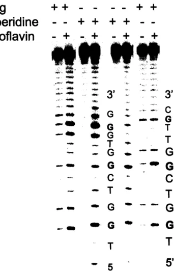
fluxes in phosphate buffer at physiological pH... 41 Figure 2-3A. A typical picture of a sequencing gel used for
quantification of relative guanine damage induced by
photoactivated riboflavin... 50
Figure 2-3B. A typical picture of a sequencing gel used for
quantification of relative guanine damage induced by ONOOCO2"... 51
Figure 2-4. Sequence-selective guanine oxidation in duplex DNA
by ONOOCO2"and riboflavin-mediated photooxidation... 53
FIgure 2-5. Sequence-selective guanine oxidation in
single-stranded DNA by ONOOCOj and riboflavin-mediated
photooxidation... ... 54 Figure 2-6. Sequence-selective guanine oxidation by
Figure 2-7. Sequence selectivity of photooxidized
riboflavin-and ONOOCO2-induced guanine oxidation chemistry... 57
Figure 3-1. Representative gel picture used for quantification of relative damage induced within mismatched guanines by
O NO O C O 2... . . . .. . . 81
Figure 3-2. Relative reactivities of matched and mismatched
guanines with ONOOCO2 ... 82
Figure 3-3. Predominant structures of G-C, G-T, G-A and
G-G base-pairs in solution at pH 7.4... ... 85
Figure 4-1. Structure of a phosphorothioate linkage ... 93
Figure 4-2. General approach for removing background of
direct strand breaks for sequencing gel analysis of base lesions... 94 Figure 4-3. Use of Exonuclease III to unmask guanine oxidation ... 101
Figure 4-4. Relative reactivities of guanines with ONOOCO2
and photoactivated riboflavin measured in the absence or
presence of Exo III ... 102 Figure 4-5. Addition of EDTA combined with filtration through
Micropure-EZ filter is sufficient to completely remove Exolll activity ... 103
Figure 5-1. Representative lanes of the sequencing gels used for quantitative analysis of guanine damage induced by
Fe+2-EDTAIH 202, Fe+2-EDTA, and y-radiation... 119
Figure 5-2. Relative reactivities of guanines within all possible three-base sequence contexts (XGY) with Fe+2-EDTA/N H202 and
y-radiation, hot piperidine treatment... 120 Figure 5-3. Relative reactivities of guanines within all possible
three-base sequence contexts (XGY) with Fe+2-EDTA, hot
piperidine treatment... 121 Figure 5-4. Relative reactivities of guanines within all possible
three-base sequence contexts (XGY) with Fe+2-EDTA/N H202
and y-radiation, Fpg treatment... 123
Figure 5-5. Relative reactivities of guanines within all possible
Figure 5-6. Ratios of relative amounts of Fpg to piperidine-sensitive guanine lesions produced by Fe*2-EDTAH 202, Fe+2-EDTA,
and y-radiation within different sequence contexts... 125 Figure 5-7. Hydroxyl radical-mediated guanine oxidation ... 130 Figure 6-5. Oxidative nucleobase damage induced in
List of Tables
Table 2-1. Sequences of oligonucleotides used for the
analysis of relative guanine damage induced by
photoactivated riboflavin and ONOOCO2 ... ... 45
Table 3-1. Sequences of oligonucleotides used for mismatch
damage analysis... ... 74 Table 3-2. Sequences of oligonucleotides used for Tm measurements 75
Table 3-3. Melting temperatures of oligonucleotides containing matched and mismatched guanines within
four representative sequence contexts... 79
Table 5-1. Sequences of oligonucleotides used for analysis of guanine damage induced by Fe+2-EDTA, Fe+2-EDTA/H 202
and y-radiation... ... 113
Table 6-1. Sequences of oligonucleotides used Fe+2-mediated
Chapter
1
Introduction
ABSTRACT
DNA damage resulting from oxidative stress has been strongly associated
with cancer, chronic degenerative diseases and aging. Both the nucleobase and deoxyribose moieties of the DNA are targets for oxidative damage. Guanine is the most frequently oxidized base because of its low ionization potential, and it
can be converted into a variety of primary and secondary oxidation products. Generation of a guanine radical cation (G"÷) as an intermediate in the oxidation
reaction leads to migration of the resulting cationic hole through the DNA I-stack until it is trapped at runs of guanines which are characterized by the lowest sequence-specific ionization potentials. This charge transfer mechanism suggests that hotspots of oxidative DNA damage induced by electron transfer reagents can be predicted based on the primary DNA sequence. However, preliminary data indicates that guanine oxidation mediated by
nitrosoperoxycarbonate (ONOOCO2"), a mediator of chronic inflammation, may
BIOLOGICAL IMPORTANCE OF GUANINE OXIDATION
Cellular DNA is being constantly subjected to attack by a variety of oxidizing agents, including environmental pollutants, ionizing radiation, and
endogenous reactive intermediates. The latter are responsible for the majority of oxidative damage sustained by nuclear and mitochondrial DNA in aerobic cells, and are overproduced during conditions of chronic inflammation and oxidative stress (1-3). These reactive oxygen and nitrogen species (ROS and RNS, respectively) include hydrogen peroxide (H202), superoxide (02"), nitric oxide
(NO'), peroxynitrite (ONOO), nitrosoperoxycarbonate (ONOOCO2") and the hydroxyl radical ('OH) (4).
A variety of deleterious conditions, including chronic neurodegenerative
diseases, artherosclerosis, and carcinogenesis are associated with oxidative stress and chronic inflammation (4-8). For example, recent estimations link approximately 20% of all cancers to chronic inflammatory conditions, such as pancreatitis, inflammatory bowel disease, hepatitis, and inflammation of the prostate that lead to the increased risk of malignancies in the affected organs, while chronic infection of gastric mucosa with H. pylori causes an increased risk of gastric cancer (9-11). Oxidative DNA damage is a strong component of the
pathologies associated with such diseases, and has been suggested to contribute to disease progression (5). For example, the mechanism of carcinogenesis associated with chronic inflammation is thought to involve chromosomal instability arising from DNA damage by ROS and RNS produced
by activated macrophages and neutrophils in inflamed tissues (5, 12) (Figure 1-1). yloperoxds
H
202 NO2 02 N20 %r Nious NO Anhydrde Macrophage+Cl-
HOCIHOBr NO2 l"rnuNO
2 0 Oxidation Halogenation Oxidation Niration DNA RNA Nitrosatlon Lipids Deaminaton Proteins Oxidation Nitration NItrosoperoxycarbonateFigure 1-1. ROS and RNS (shown in red) produced by the activated macrophages and neutrophils during chronic inflammation.
While both deoxyribose and nucleobase oxidation constitute the spectrum of DNA damage chemistries, guanine stands out as the major oxidation target due to its low ionization potential and high reactivity with a variety of oxidizing agents (13, 14). A study by Polyak et al. found that the majority of mutational
hotspots identified within mitochondrial DNA from human colorectal cancer cell lines resided at guanine residues, thus providing direct evidence for the
dominating nature and biological importance of oxidative guanine damage mediated by ROS (15). In fact, 8-oxoguanine, the most abundant oxidative
associated with oxidative stress (16, 17). The following sections will briefly discuss the chemistry of guanine oxidation and the determinants of hotspots of oxidative guanine damage in double-stranded DNA.
CHEMISTRY OF GUANINE OXIDATION
Guanine is characterized by the lowest ionization potential of all canonical
DNA bases (Eo = 1.29 V vs normal hydrogen electrode, NHE) and is, therefore,
the major target of oxidative damage in DNA (18). The mechanism of guanine oxidation is highly dependent on the nature of the oxidizing species and may involve [4+2] cycloaddition of singlet oxygen (102), direct transfer of an electron from the guanine base, or addition of a hydroxyl radicals ('OH) onto the double bonds of a guanine heterocycle (19). Nevertheless, subsequent transformations of the reactive intermediates produced in each case result in their eventual conversion to the same final lesions (19). Mechanisms of generation of primary and secondary guanine oxidation products by a variety of damaging agents have recently been reviewed (19-21), and a brief summary will be presented here with the focus on guanine oxidation mediated by electron transfer reagents as the most relevant to the present discussion.
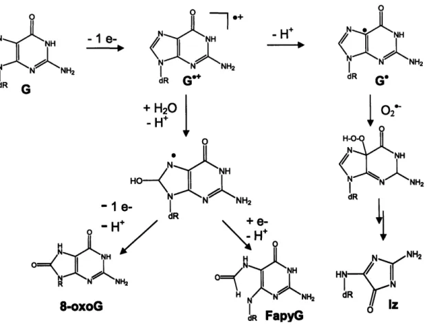
The initial step in oxidation of a guanine base by electron transfer involves abstraction of an electron from guanine and generation of a guanine radical cation (G'4) (Figure 1-2). The deprotonated form of this intermediate, a relatively
time-resolved spectroscopic methods (22-24). Deprotonation of Go* in the context of
double-stranded DNA has been demonstrated to proceed in a two-phase process
with the rate constants of 1.3 x 10⁷ s'1 and 3 x 10⁶ S-1 for the fast and slow
processes, respectively (25). These phases correspond to deprotonation of the
G°* moiety in the G:C base-pair to generate G':(C+H)*, followed by the loss of a proton by cytosine to produce G':C (25).
The rate of G' recombination with other radical species is close to being diffusion-controlled, and was measured to be 4.7 x 10⁸ s"' and 4.3 x 10⁸ s-1 for superoxide (02") and nitrogen dioxide radical (NO2I), respectively (26, 27). A
hydroperoxide intermediate results from recombination of G' with 02" and is a commonly accepted precursor of imidazolone (Figure 1-1), a major guanine oxidation product that forms in high yields in DNA oxidation reactions mediated
by type I photosensitizers, such as benzophenone or riboflavin (19, 28-30).
Trapping of a G`* by a water molecule is a commonly accepted route to
formation of 8-oxo-7,8-dihydroguanine or 8-oxoguanine (8-oxoG), another major guanine oxidation product (19, 21). Addition of a water molecule to the C8 of guanine, followed by deprotonation, generates a reducing G8*OH species (Figure
1-1) (19). Loss of a second electron by this intermediate leads to its eventual
transformation to 8-oxoG, while presence of reductants in the reaction mixture results in formation of 2,6-diamino-4-hydroxy-5-formamidopyrimidine (FapyG), the reduced and ring-opened form of 8-oxoG (31). 8-oxoG is generally
considered to be the most abundant product that forms in high yields upon oxidation of double-stranded DNA by a variety of oxidizing agents (29, 32-34).
The modified base 8-oxoG is highly prone to oxidation as its ionization potential (Eo = 0.74 V vs NHE) is even lower than that of guanine (35).
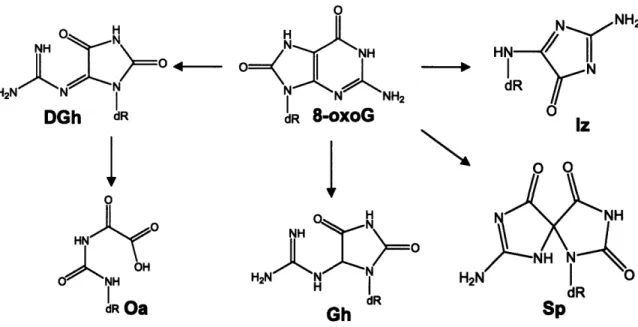
Therefore, once it forms, 8-oxoG becomes a target for oxidation and a precursor for a number of secondary oxidation lesions (19). The steps of 8-oxoG oxidation by electron transfer are analogous to those for guanine and involve electron transfer from 8-oxoG to an oxidant with subsequent generation of an 8-oxoG radical cation (8-oxoG"÷) that can undergo deprotonation or hydration (19-21, 36).
Final lesions arising from 8-oxoG oxidation are listed in Figure 1-3 and include spiroiminohydantoin (Sp), guanidinohydantoin (Gh), imidazolone (Im), and
dehydroguanidinohydantoin (DGh), a precursor to oxaluric acid (Oa) and a major product of 8-oxoG oxidation by type I photosensitizers in double-stranded
A --1 - I NH H N N NH2I dR Ge +
+
H
20
i
-H' o NNH H N N40 NH2-1
e-
I
dR NH -HH 0 NH H NH N N NH2 8-oxoG i dR FapyG 0 N NH dRG
02 H-0 NH N NH2 dRFigure 1-2. Oxidation of guanine by electron transfer. Abbreviations: G,
guanine; G"÷, guanine radical cation; G', neutral guanine radical; 8-oxoG,
8-oxo-7,8-dihydroguanine; Iz, imidazolone; Fapy, 2,6-diamino-4-hydroxy-5-formamidopyrimidine.
C, H2 .. N O . N rR' L GVqf-1
0 0
S O H2 N N H2N 0RSdRdR
dR Oa Gh dR SpFigure 1-3. Products of 8-oxoguanine oxidation by electron transfer. Abbreviations: 8-oxoG, 8-oxo-7,8-dihydroguanine; Iz, imidazolone; Sp,
spiroiminohydantoin, Gh, guanidinohydantoin; DGh, dehydroguanidinohydantoin; Oa, oxaluric acid.
DETERMINANTS OF GUANINE REACTIVITY IN DOUBLE-STRANDED DNA
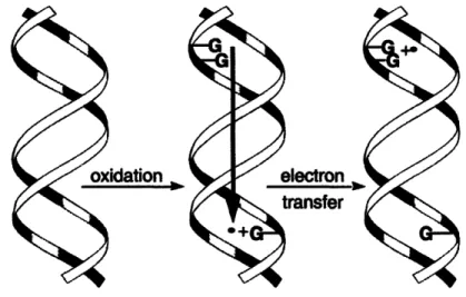
Oxidative charge transfer. An initial transfer of an electron from guanine to the oxidizing agent produces a cationic hole within DNA I-stack that can
migrate long distances though the helix until being irreversibly trapped (Figure 1-4) (37). Trapping involves deprotonation or hydration of G"+ and results in
formation of oxidative guanine lesions (38). Long-range oxidative charge transfer has been directly observed within synthetic assemblies of double-stranded
oligonucleotides covalently linked at their 5' ends to a photooxidizing agent, such
DGh
as a [Rh(phi)2bpy']3+ (phi = phenanthrenequinone diimine; bpy' =
4'-methylbipyridine-4-butyric acid) complex studied by the Barton group (37), or an anthraquinone studied by the Schuster group (39). A cationic hole initially
injected into the n-stack upon photoactivation of the tethered oxidant has been observed to cause oxidative damage at the sites located up to 200 A away from the 5' end of such synthetic DNA assemblies (39, 40).
Figure 1-4. Oxidative electron transfer through DNA helix. Adapted from (38).
The mechanism of long-range electron hole migration through DNA has been a subject of intensive investigations and is currently believed to involve short-range fast superexchange combined with a longer-range hopping mechanism. In the fast superexchange process, a cationic hole tunnels over short distances through a DNA bridge between the electron donor and electron acceptor, whereas migration of charge over longer distances consists of distinct
"hops" between guanine bases (41). The Schuster group proposed that the latter involves thermally-assisted hopping of a "polaron", a structure consisting of a cationic hole delocalized in a sequence-dependent manner over several
base-pairs in a DNA helix (42). Recently, Lewis et al. have directly observed a cross-over from superexchange to the hopping mechanism that occurred at the
donor-acceptor distance of about 20 A, or about 5 base-pairs (43). Short-range
electron hole migration occurs on a very short time-scale of around 109-1012 S-1
(41), while the rate for a single hopping step from an isolated guanine to a
guanine doublet has been measured to be khop = 106 - 10 s6 " (44). The rates for
the long-range electron hole migration are comparable to the rates of G°*
trapping through deprotonation or hydration (25).
Guanine runs as the most efficient electron hole traps. It has been
consistently noted that guanines located at the 5' positions within runs of guanines, such as 5'-GG-3' and 5'-GGG-3' motifs, acted as the most efficient electron hole traps during oxidative charge transfer. Indeed, the charge transfer
mechanism has been evoked to explain preferential damage of guanine runs that has been observed for a variety of structurally diverse one-electron oxidants, including photoactivatable Rh(lll) complexes (37), anthraquinones (39), riboflavin (45), pterins (46) and benzophenone (47); Ru(llI) metallointercalators (48) and
Ni(ll) complexes in the presence of sulfite (49); as well as direct laser irradiation
(50). Conversely, preferential damage at 5' positions of guanine runs has been a
widely accepted hallmark of oxidative charge migration induced by a DNA damaging agent acting through electron transfer.
Saito and co-workers have examined the properties of GG-3' and
5'-GGG-3' sequences that conferred to the 5' guanines the ability to act as the most
efficient electron hole traps. They showed that stacking interactions of bases in these sequence contexts resulted in lowering guanine's sequence-specific
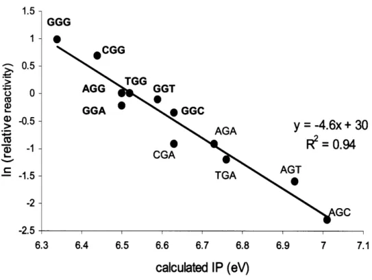
ionization potentials (IPs) that, in turn, determined guanine's reactivity toward one-electron oxidation (51, 52). In a comprehensive study, they used short oligonucleotides containing guanines in all possible three-base sequence contexts (XGY) in a one-electron oxidation reaction mediated by photoactivated riboflavin, and quantified relative amounts of piperidine-sensitive lesions induced within each guanine using sequencing gel (45). These relative reactivity values for guanines were then compared to guanines' sequence-specific IPs, calculated
by ab initio methods. A strong inverse correlation was observed between IPs
and relative reactivities, and guanines located at the 5' position within 5'-GG-3' sequence contexts were characterized by the lowest sequence-specific IPs and highest reactivities toward one-electron oxidation (Figure 1-5). This experiment demonstrated that sequence-specific IPs were the major determinants of guanine's reactivity with electron transfer reagents in double-stranded DNA.
1-..
0.5
S0.5 -- 1.52 --2.5 GGG 0 30 4 6.3 6.4 6.5 6.6 6.7 6.8 6.9 7 7.1 calculated IP (eV)Figure 1-5. Relative numbers of hot piperidine-sensitive guanine lesions produced by photoactivated riboflavin in XGY sequence contexts versus their calculated sequence-specific ionization potentials (IPs). Data points
corresponding to sequences containing runs of guanines are marked in bold. Adapted from (45).
UNUSUAL GUANINE OXIDATION BY NITROSOPEROXYCARBONATE
Chronic inflammatory conditions are characterized by overproduction of ROS and RNS that are secreted by activated macrophages and neutrophils to cause elimination of the infections agents (53). The same species can also cause substantial damage to the surrounding biomolecules, including lipid
25
1
rperoxidation and oxidation and nitration of lipids and DNA (5, 54) (Figure 1-1).
Peroxynitrite (ONOO-), a powerful oxidant and DNA damaging agent, is produced
by recombination of macrophage-derived nitric oxide (NO') and superoxide (02")
that occurs with a diffusion-controlled rate of 6.6-19 x 109 M-1 s' (55, 56). In physiological tissues, where carbon dioxide (CO2) is present at an approximate concentration of 1 mM, decomposition of ONOO proceeds via bimolecular
recombination with CO2 at the rate of 2.9 x 10⁴ M1 s-1, resulting in the formation
of nitrosoperoxycarbonate (ONOOCO2") (57). Scission of the 0-0 bond in this very unstable intermediate produces carbonate anion (C030) and nitrogen
dioxide (N02') radicals that can escape the solvent cage in 30-35% of cases to react with surrounding biomolecules, causing damage (58, 59). Both ONOOCO2"
and independently generated C0 3-have been previously shown to cause
selective oxidation of guanines in double-stranded DNA (60, 61). In the case of
CO03 , guanine oxidation has been demonstrated to proceed through electron transfer with the generation of a G°*, a charge transfer intermediate, the
deprotonated form of which has been detected in pulse radiolysis experiments involving guanine-containing oligonucleotides (22).
Total DNA damage uaton requency I~~aI
DIII
ID
)
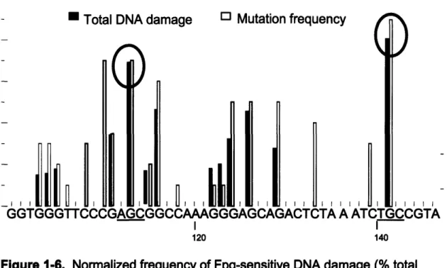
GGTGGGTTCCCGAGCGGCCAAAGGGAGCAGACTCTA A ATCTGCCGTA I I 120 140Figure 1-6. Normalized frequency of Fpg-sensitive DNA damage (% total damage) and mutation frequency (total number of mutations) after exposure to ONOOCO2j. Circled are the observed hotspots of total DNA damage, with their sequence contexts underlined. Adapted from (61).
Our interest in sequence-selective guanine oxidation by ONOOCO2"
derives from an initial observation of unusual damage patterns produced within a short DNA fragment upon treatment with ONOOCO2". Specifically, an
experiment was carried out by the Tannenbaum, Dedon and Wogan groups with the purpose of correlating the sites of DNA damage and mutational hotspots
produced by ONOOCO2" within a supF gene in E. coli cells (61). While a strong
correlation between sites of guanine damage and mutations has indeed been observed, examination of oxidative damage hotspots induced within a 135 bp
restriction fragment revealed that guanine runs, characterized by the lowest
27 SM
sequence-specific ionization potentials, sustained only relatively low levels of oxidative damage (Figure 1-5). Moreover, it was found that AGC-3' and
5'-TGC-3' sites, characterized by the highest sequence-specific ionization
potentials, were the hotspots of ONOOCO2--induced damage (Figure 1-5).
These preliminary observations contradicted the predictions of based on the oxidative charge transfer model and suggested that sequence-specific ionization potentials were not the predictors of oxidative damage hotspots produced by
ONOOCO2 .
Building upon these observations, this work explores the basis of the unusual sequence selectivity of guanine oxidation by ONOOCO2. It has been previously observed that guanines located within single-stranded
oligonucleotides displayed a higher reactivity toward ONOO" and ONOOCO2" than guanines located within duplex DNA (60, 62). This has led us to
hypothesize than solvent exposure may play a more dominant role in selection of oxidation targets by ONOOCO2- than sequence-specific ionization potentials. Solvent exposure is indeed a determining factor of the reactivity of DNA bases with chemical probes of DNA structure such as Ni(ll) complexes (63),
bromoacetaldehyde (64), diethylpyrocarbonate, and hydroxylamine (65). Additionally, we have also hypothesized that unfavorable electrostatic
interactions between the negatively charged ONOOCO2" and the DNA backbone precluded access of ONOOCO2 to the bases stably stacked inside the DNA helix, limiting its reactivity to the sites with more solvent exposed guanines.
As an initial step in exploring the unusual guanine oxidation by
ONOOCO2-, a comprehensive and a systematic study was undertaken to define
relative reactivities of guanines in all possible three-base sequence contexts (XGY) with ONOOCO2. The role of solvent exposure in determining guanine reactivity with ONOOCO2 was explored by measuring oxidation of mismatched guanines in several sequence contexts. To test the hypothesis that the negative charge of ONOOCO2 favors its selectivity for the more solvent-exposed bases, sequence-selective guanine oxidation by another negatively charged oxidant, the
Fe+2-EDTA compex, was studied and compared to guanine oxidation produced by y-radiation, a neutral oxidant. Because both Fe+2-EDTA and y-radiation produce high level direct strand breaks that can interfere with nucleobase oxidation analysis (66), a method to remove the high strand break background has been developed. Finally, the role of oxidant binding in influencing sequence-selective nucleobase oxidation has been examined by analyzing damage
produced by DNA-bound Fe+2 ions. Overall, our studies explored the factors that determine the location and identity of mutagenic oxidation lesions in the genome.
REFERENCES
1. Ames, B. N., Shigenaga, M. K., and Hagen, T. M. (1993) Oxidants,
antioxidants, and the degenerative diseases of aging. Proc Natl Acad Sci
USA 90, 7915-7922
2. Jaruga, P., and Dizdaroglu, M. (1996) Repair of products of oxidative DNA
base damage in human cells. Nucleic Acids Res 24, 1389-1394
3. Richter, C. (1995) Oxidative damage to mitochondrial DNA and its relationship to ageing. Int J Biochem Cell Biol 27, 647-653
4. Valko, M., Leibfritz, D., Moncol, J., Cronin, M. T., Mazur, M., and Telser, J. (2007) Free radicals and antioxidants in normal physiological functions
and human disease. Int J Biochem Cell Biol 39, 44-84
5. Bartsch, H., and Nair, J. (2006) Chronic inflammation and oxidative stress in the genesis and perpetuation of cancer: role of lipid peroxidation, DNA damage, and repair. Langenbecks Arch Surg 391, 499-510
6. Hussain, S. P., Hofseth, L. J., and Harris, C. C. (2003) Radical causes of cancer. Nat Rev Cancer 3, 276-285
7. Mercer, J., Mahmoudi, M., and Bennett, M. (2007) DNA damage, p53, apoptosis and vascular disease. Mutat Res 621, 75-86
8. Reynolds, A., Laurie, C., Lee Mosley, R., and Gendelman, H. E. (2007) Oxidative stress and the pathogenesis of neurodegenerative disorders. Int
Rev Neurobiol 82, 297-325
9. Coussens, L. M., and Werb, Z. (2002) Inflammation and cancer. Nature 420, 860-867
10. De Marzo, A. M., Platz, E. A., Sutcliffe, S., Xu, J., Gronberg, H., Drake, C.
G., Nakai, Y., Isaacs, W. B., and Nelson, W. G. (2007) Inflammation in prostate carcinogenesis. Nat Rev Cancer 7, 256-269
11. Lochhead, P., and EI-Omar, E. M. (2007) Helicobacter pylori infection and gastric cancer. Best Pract Res Clin Gastroenterol 21, 281-297
12. Ohshima, H., Tatemichi, M., and Sawa, T. (2003) Chemical basis of inflammation-induced carcinogenesis. Arch Biochem Biophys 417, 3-11
13. Steenken, S., Jovanovic, S.V. (1997) How easily oxidizable is DNA? One-electron reduction potentials of adenosine and guanosine radicals in aqueous solution. J Am Chem Soc 119, 617-618
14. Burrows, C. J., and Muller, J. G. (1998) Oxidative Nucleobase Modifications Leading to Strand Scission. Chem Rev 98, 1109-1152
15. Polyak, K., Li, Y., Zhu, H., Lengauer, C., Willson, J. K., Markowitz, S. D., Trush, M. A., Kinzler, K. W., and Vogelstein, B. (1998) Somatic mutations of the mitochondrial genome in human colorectal tumours. Nat Genet 20,
291-293
16. Loft, S., and Poulsen, H. E. (1996) Cancer risk and oxidative DNA damage in man. J Mol Med 74, 297-312
17. Loft, S., Fischer-Nielsen, A., Jeding, I. B., Vistisen, K., and Poulsen, H. E.
(1993) 8-Hydroxydeoxyguanosine as a urinary biomarker of oxidative DNA
damage. J Toxicol Environ Health 40, 391-404
18. Steenken, S., Jovanovic, S.V. (1997) How easily oxidized is DNA? One-electron reduction potentials of adenosine and guanosine radicals in aqueous solution. J Am Chem Soc 119, 617-618
19. Pratviel, G., and Meunier, B. (2006) Guanine oxidation: one- and two-electron reactions. Chemistry 12, 6018-6030
20. Neeley, W. L., and Essigmann, J. M. (2006) Mechanisms of formation, genotoxicity, and mutation of guanine oxidation products. Chem Res
21. Niles, J. C., Wishnok, J. S., and Tannenbaum, S. R. (2006) Peroxynitrite-induced oxidation and nitration products of guanine and 8-oxoguanine: structures and mechanisms of product formation. Nitric Oxide 14, 109-121 22. Shafirovich, V., Dourandin, A., Huang, W., and Geacintov, N. E. (2001)
The carbonate radical is a site-selective oxidizing agent of guanine in double-stranded oligonucleotides. J Biol Chem 276, 24621-24626
23. Shafirovich, V., Dourandin, A., Huang, W. D., Luneva, N. P., and Geacintov, N. E. (1999) Oxidation of guanine at a distance in
oligonucleotides induced by two-photon photoionization of 2-aminopurine.
Journal of Physical Chemistry B 103, 10924-10933
24. Stemp, E. D. A., Arkin, M. R., Barton, J. K. (1997) Oxidation of guanine in
DNA by Ru(phen)2(dppz)3+ using the flash-quench technique. J Am Chem
Soc 119, 2921-2925
25. Kobayashi, K., Tagawa, S. (2003) Direct observation of guanine radical cation deprotonation in duplex DNA using pulse radiolysis. J Am Chem
Soc 125, 10213-10218
26. Misiaszek, R., Crean, C., Joffe, A., Geacintov, N. E., and Shafirovich, V. (2004) Oxidative DNA damage associated with combination of guanine and superoxide radicals and repair mechanisms via radical trapping. J Biol
Chem 279, 32106-32115
27. Misiaszek, R., Crean, C., Geacintov, N. E., and Shafirovich, V. (2005) Combination of nitrogen dioxide radicals with 8-oxo-7,8-dihydroguanine and guanine radicals in DNA: oxidation and nitration end-products. J Am
Chem Soc 127, 2191-2200
28. Kupan, A., Sauliere, A., Broussy, S., Seguy, C., Pratviel, G., and Meunier, B. (2006) Guanine oxidation by electron transfer: one- versus two-electron oxidation mechanism. Chembiochem 7, 125-133
29. Douki, T., and Cadet, J. (1999) Modification of DNA bases by photosensitized one-electron oxidation. Int J Radiat Biol 75, 571-581
30. Kino, K., Saito, I. (1998) Product analysis of GG-specific photooxidation of DNA via electron transfer: 2-aminoimidazolone as a major guanine
oxidation product. J Am Chem Soc 120, 7373-7374
31. Candeias, L. P., and Steenken, S. (2000) Reaction of HO* with guanine derivatives in aqueous solution: formation of two different redox-active
OH-adduct radicals and their unimolecular transformation reactions. Properties of G(-H)*. Chemistry 6, 475-484
32. Yu, H., Venkatarangan, L., Wishnok, J. S., and Tannenbaum, S. R. (2005) Quantitation of four guanine oxidation products from reaction of DNA with varying doses of peroxynitrite. Chem Res Toxicol 18, 1849-1857
33. Cadet, J., Delatour, T., Douki, T., Gasparutto, D., Pouget, J. P., Ravanat,
J. L., and Sauvaigo, S. (1999) Hydroxyl radicals and DNA base damage.
Mutat Res 424, 9-21
34. Ravanat, J. L., and Cadet, J. (1995) Reaction of singlet oxygen with 2'-deoxyguanosine and DNA. Isolation and characterization of the main oxidation products. Chem Res Toxicol 8, 379-388
35. Steenken, S., Jovanovic, S.V., Bietti, M., Bernhard, K. (2000) The trap depth (in DNA) of 8-oxo-7,8-dihydro-2'deoxyguanosine as derived from
electron-transfer equilibria in aqueous solution. J Am Chem Soc 122,
2373-2374
36. Shafirovich, V., Cadet, J., Gasparutto, D., Dourandin, A., Huang, W. D., and Geacintov, N. E. (2001) Direct spectroscopic observation of
8-oxo-7,8-dihydro-2 '-deoxyguanosine radicals in double-stranded DNA generated by one-electron oxidation at a distance by 2-aminopurine
radicals. Journal of Physical Chemistry B 105, 586-592
37. Hall, D. B., Holmlin, R. E., and Barton, J. K. (1996) Oxidative DNA damage through long-range electron transfer. Nature 382, 731-735
38. Giese, B. (2002) Long-distance electron transfer through DNA. Annu Rev Biochem 71, 51-70
39. Ly, D., Sanii, L., Schuster, G. B. (1999) Mechanism of charge transport in DNA: internally-linked anthraquinone conjugates support phonon-assisted
polaron hopping. J Am Chem Soc 121, 9400-9410
40. Nunez, M. E., Hall, D. B., and Barton, J. K. (1999) Long-range oxidative damage to DNA: effects of distance and sequence. Chem Biol 6, 85-97 41. Wagenknecht, H. A. (2006) Electron transfer processes in DNA:
mechanisms, biological relevance and applications in DNA analytics. Nat Prod Rep 23, 973-1006
42. Henderson, P. T., Jones, D., Hampikian, G., Kan, Y., and Schuster, G. B.
(1999) Long-distance charge transport in duplex DNA: the
phonon-assisted polaron-like hopping mechanism. Proc Nat Acad Sci U S A 96,
8353-8358
43. Lewis, F. D., Zhu, H., Daublain, P., Fiebig, T., Raytchev, M., Wang, Q.,
and Shafirovich, V. (2006) Crossover from superexchange to hopping as the mechanism for photoinduced charge transfer in DNA hairpin
conjugates. J Am Chem Soc 128, 791-800
44. Lewis, F. D., Liu, X., Liu, J., Miller, S. E., Hayes, R. T., and Wasielewski, M. R. (2000) Direct measurement of hole transport dynamics in DNA. Nature 406, 51-53
45. Saito, I., Nakamura T., Nakatani K., Yoshioka Y., Yamaguchi K., Sugiyama H. (1998) Mapping of the hot spots for DNA damage by one-electron oxidation: Efficacy of GG doublets and GGG triplets as a trap in long-range hole migration. J Am Chem Soc 120, 12686-12687
46. Ito, K., and Kawanishi, S. (1997) Photoinduced hydroxylation of
deoxyguanosine in DNA by pterins: sequence specificity and mechanism. Biochemistry 36, 1774-1781
47. Nakatani, K., Dohno, C., Nakamura, T., Saito, I. (1998) p-Cyano
substituted benzophenone as an excellent photophore for one-electron oxidation of DNA. Tetrahedron Lett 39, 2779-2782
48. Arkin, M. R., Stemp, E. D., Pulver, S. C., and Barton, J. K. (1997) Long-range oxidation of guanine by Ru(lll) in duplex DNA. Chem Biol 4, 389-400
49. Muller, J. G., Hickerson, R. P., Perez, R. J., Burrows, C. J. (1997) DNA damage from sulfite autoxidation catalyzed by a nickel(ll) peptide. J Am
Chem Soc 119, 1501-1506
50. Spassky, A., and Angelov, D. (1997) Influence of the local helical
conformation on the guanine modifications generated from one-electron DNA oxidation. Biochemistry 36, 6571-6576
51. Saito, I., Takayama, M., Sugiyama, H., Nakatani, K., Tsuchida, A.,
Yamamoto, M. (1995) Photoinduced DNA cleavage via electron transfer: demonstration that guanine residues located 5' guanine are the most electron-donating sites. J Am Chem Soc 117, 6406-6407
52. Sugiyama, H., Saito, I. (1996) Theoretical studies of GG-specific photocleavage of DNA via electron transfer: significant lowering of
ionization potential and 5'-localization of HOMO of stacked GG bases in B-form DNA. J Am Chem Soc 118, 7063-7068
53. Nathan, C., and Shiloh, M. U. (2000) Reactive oxygen and nitrogen
intermediates in the relationship between mammalian hosts and microbial pathogens. Proc Natl Acad Sci U S A 97, 8841-8848
54. Dedon, P. C., and Tannenbaum, S. R. (2004) Reactive nitrogen species in the chemical biology of inflammation. Arch Biochem Biophys 423, 12-22 55. Huie, R. E., and Padmaja, S. (1993) The reaction of no with superoxide.
Free Radic Res Commun 18, 195-199
56. Kissner, R., Nauser, T., Bugnon, P., Lye, P. G., and Koppenol, W. H. (1997) Formation and properties of peroxynitrite as studied by laser flash photolysis, high-pressure stopped-flow technique, and pulse radiolysis.
Chem Res Toxicol 10, 1285-1292
57. Lymar, S. V., Hurst, J.K. (1995) Rapid reaction between peroxynitrite ion and carbon dioxide: implications for biological activity. J Am Chem Soc
117, 8867-8868
58. Squadrito, G. L., Pryor, W. A. (2002) Mapping the reaction of peroxynitrite with C02: energetics, reactive species and biological implications. Chem
Res Toxicol 15, 885-895
59. Lymar, S. V., Jiang, Q., and Hurst, J. K. (1996) Mechanism of carbon dioxide-catalyzed oxidation of tyrosine by peroxynitrite. Biochemistry 35, 7855-7861
60. Burney, S., Niles, J. C., Dedon, P. C., and Tannenbaum, S. R. (1999) DNA damage in deoxynucleosides and oligonucleotides treated with peroxynitrite. Chem Res Toxicol 12, 513-520
61. Tretyakova, N. Y., Burney, S., Pamir, B., Wishnok, J. S., Dedon, P. C., Wogan, G. N., and Tannenbaum, S. R. (2000) Peroxynitrite-induced DNA damage in the supF gene: correlation with the mutational spectrum. Mutat
Res 447, 287-303
62. Gu, F., Stillwell, W. G., Wishnok, J. S., Shallop, A. J., Jones, R. A., and Tannenbaum, S. R. (2002) Peroxynitrite-induced reactions of synthetic oligo 2'-deoxynucleotides and DNA containing guanine: formation and stability of a 5-guanidino-4-nitroimidazole lesion. Biochemistry 41, 7508-7518
63. Chen, X., Burrows, C. J., and Rokita, S. E. (1992) Conformation-specific detection of guanine in DNA: ends, mismatches, bulges, and loops. J Am
Chem Soc 114, 322-325
64. Kohwi-Shigematsu, T., Gelinas, R., and Weintraub, H. (1983) Detection of an altered DNA conformation at specific sites in chromatin and
supercoiled DNA. Proc Nat/ Acad Sci U S A 80, 4389-4393
65. Ross, M. K., Said, B., Shank, R. C. (2000) DNA-damaging effects of genotoxins in mixture: modulation of covalent binding to DNA. Toxicol Sci 53, 224-236
66. Kennedy, L. J., Moore, K., Jr., Caulfield, J. L., Tannenbaum, S. R., and Dedon, P. C. (1997) Quantitation of 8-oxoguanine and strand breaks produced by four oxidizing agents. Chem Res Toxicol 10, 386-392
Chapter 2
Paradoxical hotspots for guanine oxidation by a
chemical mediator of inflammation
ABSTRACT
Peroxynitrite (ONOO') is produced during chronic inflammation and may contribute to carcinogenesis through oxidative DNA damage. Its decomposition in the presence of carbon dioxide leads to formation of nitrosoperoxycarbonate
(ONOOCO2), an unstable intermediate that produces carbonate anion (CO3-)
and nitrogen dioxide (NO2') radicals through disproportionation. When added to the buffer containing bicarbonate, ONOO- leads to selective oxidation of
guanines in the DNA, as does independently generated CO3", through a
mechanism involving electron transfer from guanine and generation of a guanine radical cation (G"+) as an intermediate. Previous studies of ONOOCO
2 -mediated
DNA damage have indicated that, unlike other classical, one-electron oxidants
that are selective for guanines of lowest ionization potentials (IPs) located within guanine runs, ONOOCO2" causes preferential damage at guanines located within 5'-AGC-3' and 5'-TGC-3' sequences characterized by the highest IPs. In a
have determined relative reactivities of guanines within all possible three-base sequence contexts in double-stranded oligonucleotides, and have compared them to the relative reactivities of the same guanines toward photooxidized riboflavin, a classical, one-electron oxidant. In agreement with previous studies, we have determined that photooxidized riboflavin was selective for guanines of lowest IPs, located within guanine runs. In contrast, ONOOCO2 preferentially
reacted with guanines located within 5'-GC-3' motifs, characterized by the highest IPs. Sequence selectivities for both reagents were double-strand specific. ONOOCO2-mediated oxidation of human genomic DNA exhibited a similar selectivity for 5'-GC-3' motifs, as was determined by ligation-mediated
PCR analysis. We have also demonstrated sequence-dependent variation in the guanine oxidation chemistry, as relative yields of different guanine oxidation
products, produced by both ONOOCO2 and photooxidized riboflavin, varied as a function of sequence. Our results demonstrate that factors other than
sequence-specific ionization potentials have a role in the selection of targets by
ONOOCOj, and this complicates efforts to predict the location and chemistry of
mutagenic DNA oxidation in the genome.
INTRODUCTION
The association between chronic inflammation and an elevated cancer risk is now well established. A recent estimation links approximately 20% of all cancers to chronic inflammatory conditions, such as pancreatitis, inflammatory
bowel disease, hepatitis, and inflammation of the prostate that lead to the increased risk of malignancies in the affected organs, while chronic infection of gastric mucosa with H. pylori causes an increased risk of gastric cancer (1-3). The mechanism of carcinogenesis associated with chronic inflammation is thought to involve chromosomal instability arising from DNA damage by reactive
oxygen and nitrogen species (ROS and RNS) produced by the activated macrophages and neutrophils in the inflamed tissues (4, 5) (Figure 1-1).
Peroxynitrite (ONOO'), a powerful oxidant and a DNA damaging agent, is a major example of such a species, produced by a reaction of macrophage-derived nitric oxide (NO') with superoxide (02'-) that occurs with a diffusion-controlled rate of 6.6-19 x 109 M-1 s- (6, 7). The rate of 020" dismutation by
superoxide dismutase (SOD) is about 3-8 times lower (k = 2.5 x 10⁹ M-1 s•),
allowing for initial accumulation of ONOO" in tissues (8). Indeed, microelectrode-based measurements have established that a single activated RAW264.7
macrophage produces -9 fmol of ONOO" in approximately 50 seconds, from estimated -30 fmol of NO' and 24 fmol of 02" generated by the same cell per oxidative burst (9). It has been further suggested that because ONOO- is
released into a microenvironment of only a few femtoliters, local ONOO'
concentrations may reach into tens of millimolar (9).
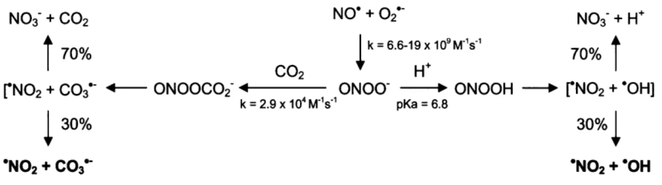
Decomposition of peroxynitrite proceeds very rapidly in an aqueous
solution at physiological pH, and is dependent on the presence of carbon dioxide
(CO2) in the reaction buffer. In the absence of CO2, ONOO' decomposes with a half-life of about 1 second and involves partial protonation to form peroxynitrous
acid (10, 11). This species can undergo homolytic bond scission to produce nitrogen dioxide radical (NO2') and a hydroxyl radical (OH') in a solvent cage, approximately 70% of which degrades into N03 and H', while the remaining 30%
escapes the solvent cage and can diffuse across biological membranes to react with surrounding biomolecules (Scheme 2-1) (10, 12). Indeed, peroxynitrite has been shown to oxidize proteins, cause lipid peroxydation and induce both deoxyribose and nucleobase oxidation and nitration in the DNA (13). In
physiological tissues, where carbon dioxide (CO2) is present at an approximate concentration of 1 mM, decomposition of peroxynitrite proceeds via bimolecular
recombination of ONOO" and CO2 at the rate of 2.9 x 10⁴ M-1 s-1, resulting in the formation of nitrosoperoxycarbonate (ONOOCO2") (14). Analogous to
decomposition of peroxynitrous acid, scission of the 0-0 bond in this very unstable intermediate produces carbonate anion (CO3") and nitrogen dioxide
(NO2*) radicals that can escape solvent cage in 30-35% of the cases to react with surrounding biomolecules, causing damage (Scheme 2-1) (11, 15). Consistent with formation of OH* in the absence, and of CO3" in the presence of CO2 during
ONOO' decomposition, a change in ONOO'-mediated DNA damage chemistry
has been observed, with the spectrum of lesions shifting from deoxyribose
oxidation leading to strand breaks toward predominant nucleobase oxidation (16,
17). Guanine (G) is the most frequently oxidized nucleobase in the DNA
because of its low ionization potential (1.29 V vs. NHE), and its selective damage has indeed been observed within oligonucleotides reacted with ONOO' in the presence of 25 mM bicarbonate (17, 18).
NO3- + 002 NO' + 02* NO3 + H÷
70%
k = 6.6-19 x 109 M's70%
CO2 H'
rNO2 + C0 3 •- ONOOCO2 C ONOO" -H ONOOH - ['N0 2 + "OH]
k = 2.9 x 10⁴ M'S1"1 pKa = 6.8
30% 30%
"N02 + CO3" "NO2 + "OH
Figure 2-1. Decomposition of peroxynitrite in the absence and presence of CO2.
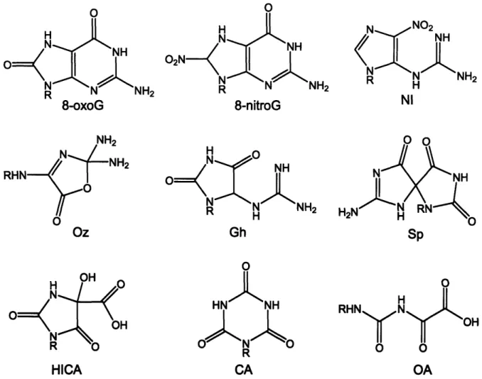
The chemistry of guanine oxidation by ONOO- has been extensively investigated and has recently been reviewed (19). The spectrum of lesions arising from ONOO--induced guanine damage at physiological pH of 7.4 in the phosphate buffer includes both primary and secondary oxidation, as well as nitration products (Figure 2-2). Secondary oxidation products arise from oxidation of 8-oxoguanine (8-oxoG), the most studied guanine lesion that is inherently more prone to oxidation than G due to its low ionization potential (E0 = 0.74 V vs. Eo = 1.29 V vs. NHE, respectively) (20). The nitration products of guanine characterized to date include a stable nitroimidazole (NI) lesion and 8-nitroguanine (8-nitroG); the latter depurinates within several hours to produce abasic sites (17). Primary oxidation products include 8-oxoG and imidazolone (lz) with its hydrolysis product, oxazolone (Oz) (Figure 2-1) (17, 19, 21-23). Once formed, 8-oxoG is further oxidized to yield spiroiminodihydantoin (Sp), guanidinohydantoin (Gh), HICA, oxaluric acid (OA), and cyanuric acid (CA) as final products that can be detected by liquid chromatography and mass
spectrometry (LC/MS) (19, 24-26). In addition, Iz and Oz can also be formed from oxidation of 8-oxoG (19, 27). The concentration of ONOO- and its mode of addition to the reaction mixture greatly influence relative efficiencies of formation of various secondary products. If a very high local concentration of ONOO- is instantaneously achieved by bolus addition, the secondary oxidative lesion spectrum will be dominated by cyanuric acid, oxaluric acid, and oxazolone (19, 23, 26). When ONOO' is slowly infused into a reaction mixture over time, HICA, Sp and Gh will be primarily observed (19, 25).
Guanine oxidation by independently generated products of ONOOCO2-decomposition, C03" and NO2', has also been investigated. Specifically, it was
demonstrated that CO3", but not NO2%, is capable of selectively oxidizing guanine
through electron transfer to produce the guanine radical cation (G°*) (28, 29).
The deprotonated form of this intermediate, the neutral guanine radical (G'), has been detected in pulse radiolysis experiments involving guanine-containing oligonucleotides (28). The reaction of G' with 02* leads to formation of Im as a major product, while its reaction with NO2* leads to production of NI (30-32). Recent work has also shown that final products of guanine oxidation by CO'3 in
the absence of N02' are Sp and Gh, produced through the intermediacy of
8-oxoG (25, 33, 34). Unlike CO3', NO2" does not oxidize guanine, but its oxidation of 8-oxoG leads to the same final products (29, 32).
O0 H N H R N NH2 8-oxoG RHN" Oz O H R N NH 8-nitroG NH 0 NH Gh 0 HNCANH R CA 0 S o OH OA
Figure 2.2. Guanine lesions observed at high and low ONOO' fluxes in phosphate buffer at physiological pH.
As has been reviewed in Chapter 1, oxidizing agents that act by electron transfer and generate G"+ cause selective oxidation of runs of guanines within
double-stranded DNA, a result of migration of the electron hole through the
it-stack to the sites of lowest ionization potentials (35, 36). Surprisingly, guanine runs did not emerge as oxidative damage hotspots in the studies that probed sequence-specific guanine damage induced within a restriction fragment by
Kilf Sp H N N R HICA
ONOOCO2- (17). Instead, guanines at positions 113 and 141 in the supF gene, located within 5'-AGC-3' and 5'-TGC-3' sequences contexts, respectively, sustained the highest levels of damage (17). These contexts are characterized
by the highest sequence-specific guanine ionization potentials, as has been
previously calculated using ab initio methods by Saito and co-workers (36). We have pursued this curious observation by systematically defining sequence-specific reactivity of ONOOCO2". This has been accomplished by determining relative levels of oxidative damage, sustained by guanines within all possible sixteen three-base sequence contexts located within short, single-stranded or duplex oligonucleotides. In addition, we have also utilized ligation-mediated PCR
(LMPCR) to analyze sequence-specific guanine damage induced by ONOOCO2 within genomic DNA isolated from human TK6 cells. We have compared these
relative damage levels to the ones induced by photoactivated riboflavin, a classical one-electron oxidant that is selective for guanine runs. We have also indirectly probed the influence of the sequence context on the efficiencies of formation of various oxidative lesions in the DNA.
MATERIALS AND METHODS
Materials. Synthetic oligonucleotides were purchased from Integrated
DNA Technologies (Coralville, IA). ONOO' was purchased from Cayman Chemical (Ann Arbor, MI). Riboflavin, piperidine and TEMED (N,N,N',N'-Tetramethylethylenediamine) were purchased from Sigma-Aldrich. Urea, boric
acid, tris base, ammonium persulfate and 40% solution of acrylamide:bis (19:1) were obtained from American Bioanalytical (Natick, MA). K2HPO4, KH2PO4,
NaHCO3 and EDTA (ethylenediaminetetraacetic acid, sodium salt) were
purchased from Mallinckrodt Baker (Phillipsburg, NJ). All chemicals were used without further purification. Chelex-100 was purchased from Bio-Rad (Hercules, CA), and Sephadex G-25 mini-spin columns were purchased from Roche
Diagnostics. E. Coli Fpg (formamidopyrimidine [fapy]-DNA glycosylase) was purchased from Trevigen (Gaithersburg, MD), and T4 PNK (polynucleotide kinase) was obtained from New England Biolabs (Ipswich, MA). [y-32P]-ATP with
activity of 6000 Ci/mmol was purchased from Perkin Elmer (Waltham, MA). Distilled and deionized water (ddH20) was purified using a Milli-Q system from Millipore (Bedford, MA) and was used for all experiments.
Preparation of buffers. A buffer containing 150 mM potassium
phosphate and 25 mM sodium bicarbonate at pH 7.4 was prepared and treated with Chelex-100 resin overnight at 4 OC to remove adventitious metals. Following treatment, the buffer was passed through a 0.2 micron filter and stored at 4 OC.
Design of oligonucleotides. The oligonucleotide sequences used for analyses were adapted from previous studies and were of the type 5'
-CGTACTCTTTGGTXiGYITX 2GY2TTCTTCTAT - 3' (36). This sequence
contains consensus portions on both 5' and 3' ends, an invariant TGG sequence at the same position in all oligonucleotides for normalizing damage (underlined),
and two variable guanine sequence contexts X1GY, and X2GX2. For each
oligonucleotide, counterparts with the relative positions of X1GY, and X2GX2
reversed were also analyzed to account for longer-range sequence effects. Table 2-1 shows sequences of all oligonucleotides used in experiments.
Purification of synthetic oligonucleotides. To remove the background
of oxidized bases, all oligonucleotides were treated with 1 M piperidine at 900 C
for 30 minutes and lyophilized prior to gel purification. The oligonucleotides were re-dissolved in TE buffer, pH 8.0 with 20% glycerol, and loaded on a 20%
acrylamide:bis, 8M urea preparative gel electrophoresis. The gel was run in lx
TBE at a constant power of 70 watts for 6 hours. Oligonucleotides were
visualized by UV shadowing, cut out from the gel, eluted by overnight shaking in
0.5 M ammounium acetate and 10 mM magnesium acetate, filtered and ethanol
precipitated. All oligonucleotides were desalted by Sephadex G-25 spin columns and transferred to 150 mM potassium phosphate, 25 mM sodium bicarbonate buffer before use.
Oligonucleotide name Oligonucleotide sequence (5' to 3') S1 CGTACTCTT TGGTTGATGGGTTCTTTCTAT 1S CGTACTCTT TGGTGGGTTGATTCTTTCTAT S2 CGTACTCTT TGGTCGGTTGCTTCTTTCTAT 2S CGTACTCTT TGGTTGCTCGGTTCTTTCTAT S3 CGTACTCTT TGGTAGTTGGATTCTTTCTAT 3S CGTACTCTT TGGTGGATAGTTTCTTTCTAT S4 CGTACTCTT TGGTAGGTTGTTTCTTTCTAT 4S CGTACTCTTTGGTTGTTAGGTTCTTTCTAT S5 CGTACTCTTTGGTCGCTCGATTCTTTCTAT 5S CGTACTCTTTGGTCGATCGCTTCTTTCTAT S6 CGTACTCTTTGGTAGCTAGATTCTTTCTAT 6S CGTACTCTT TGGTAGATAGCTTCTTTCTAT S8 CGTACTCTTTGGTGGCTCGTTTCTTTCTAT 8S CGTACTCTTTGGTCGTTGGCTTCTTTCTAT S9 CGTACTCTT TGGTAGCTGGTTTCTTTCTAT 9S CGTACTCTT TGGTGGTTAGCTTCTTTCTAT
Table 2-1. Sequences of oligonucleotides used for the analysis of relative guanine damage induced by photoactivated riboflavin and ONOOCO2". Relative reactivities of guanines shown in bold within underlined sequence contexts have been quantified, and the invariant TGG sequence used for normalizing damage is italicized.
Labelling and annealing of olilgonucleotides. Oligonucleotides were 5'-end labeled with 32P in a reaction containing approximately 100 pmol of 5' ends,