Regulation of major histocompatibility complex class I expression by
NF-kB-related proteins in breast cancer cells
Emmanuel Dejardin
1,4, ValeÂrie Deregowski
1,4, Roland Greimers
2, Zhenzi Cai
3, Salem Chouaib
3,
Marie-Paule Merville
1and Vincent Bours
11Laboratory of Medical Chemistry/Medical Oncology, and2Laboratory of Pathology, University of LieÁge, Belgium;3INSERM
CJF 9411 `Cytokines et Immunite Antitumorale', Institut Gustave Roussy, Villejuif, France Downregulation of MHC Class I antigens has been
observed in many cancers and usually results from a decreased gene transcription. A reporter CAT gene dependent on the MHC Class I kB site or on a longer promoter is transactivated by NF-kB complexes contain-ing p65 or RelB. p100 as well as IkB-a are potent inhibitors of this transcription and p100 sequesters RelB and p65 complexes in the cytoplasm of breast cancer cells. However, although p100 is highly expressed in a number of breast cancer cell lines, MHC Class I antigen expression was observed on all the cell lines we analysed and could be further induced by stimulation with the cytokines IFN-g or TNF-a. Stable transfection of a unresponsive mutated IkB-a Ser 32-36 expression vector showed that TNF-a induced MHC Cl I expression in an NF-kB-dependent way while IFN-g did it independently of any NF-kB activation.
Keywords: MHC class I; NF-kB, breast cancer
Introduction
The function of the Major Histocompatibility Complex (MHC) is critical for antigen presentation and immune response (Accolla et al., 1995). MHC Class I (MHC Cl I) antigens are required for cell recognition and subsequent destruction by cytotoxic T lymphocytes. Down-regulation or loss of MHC Cl I antigens have been described in many solid tumors and probably allow them to escape T cell-mediated immune surveillance (Blanchet et al., 1992; Garrido et al., 1995). Moreover, MHC Cl I expression is lower in metastases than in primary tumors or in metastatic compared to non metastatic cell lines (Cordon-Cardo et al., 1991; Plaskin et al., 1993).
Several mechanisms can account for a down-regulation of MHC Cl I antigens function in cancers: loss of MHC Cl I genes, mutation in the b2-microglobulin associated protein or defects in the assembling and cellular transport of MHC Cl I / b2-microglobulin/peptide antigen complexes (Garrido et al., 1995). However, the low expression of MHC Cl I antigens is very often associated with a decrease of gene transcription.
The promoter of the MHC Cl I gene contains three major regulatory elements: the enhancer A or
Class I regulatory element (CRE), the sequence ICS (Interferon Consensus Sequence) and the enhancer B (Garrido et al., 1995). The enhancer A can be divided in two regions, I and II. Region I binds the transcription factors NF-kB, KBF1 and H2TF1 (Baldwin and Sharp, 1987, 1998; IsraeÈl et al., 1987).
NF-kB is an ubiquitous transcription factor regulat-ing the expression of a large variety of genes and viruses (Siebenlist et al., 1994). It is made itself from a family of proteins which are characterized by a conserved Rel homology domain (RelHD) responsible for dimerization, nuclear translocation and speci®c DNA-binding. Among these proteins, p65 (RelA), RelB and c-Rel contain one or two transactivating domains (Siebenlist et al., 1994). Two other proteins belonging to the same family, p50 and p52 do not harbor any transactivating motif and can thus inhibit NF-kB-dependent transcription by binding to DNA as homodimers (Bours et al., 1990, 1992; Kieran et al., 1990; Neri et al., 1991; Franzoso et al., 1992).
NF-kB complexes are sequestered in the cytoplasm of most resting cells by inhibitory proteins belonging to the IkB family (Beg and Baldwin, 1993; Baeuerle and Henkel, 1994; Siebenlist et al., 1994; Miyamoto and Verma, 1995). The members of the human IkB family are IkB-a, IkB-b, IkB-e, p100 and p105 (Haskill et al., 1991; Rice et al., 1992; Beg and Baldwin, 1993; Mercurio et al., 1993; Whiteside et al., 1997). Bcl-3 is also a member of this family but, at least in some cellular types, is located in the nucleus and participates in NF-kB transactivating activity (Ohno et al., 1990; Bours et al., 1993; Nolan et al., 1993; Watanabe et al., 1997).
NF-kB has been shown to participate in the control of the MHC Cl I genes basal expression as well as in their transcriptional upregulation following treatment by retinoids and TNF-a (IsraeÈl et al., 1987, 1989; Logeat et al., 1991; Segars et al., 1993; van't Veer et al., 1993). Moreover, inhibition of NF-kB activity has been associated with the downregulation of MHC Cl I expression in tumor cell lines and in adenovirus 12-transformed cells (Blanchet et al., 1992; van't Veer et al., 1993; Schouten et al., 1995; Liu et al., 1996). KBF1 has been puri®ed and is identical to transactively inactive p50 homodimers (Yano et al., 1987; Kieran et al., 1990). It indeed acts as a repressor of MHC Cl I expression in metastatic cancer cells (Plaskin et al., 1993).
The proteins p100 and p105, precursors of p52 and p50 respectively, function as IkB-like molecules (Rice et al., 1992; Mercurio et al., 1993; Dejardin et al., 1995) and thus sequester NF-kB complexes in the cytoplasm.
Correspondence: V Bours
4Equally contributed and both should be considered as ®rst authors
Received 14 July 1997; revise 28 January 1998; accepted 29 January 1998
A downregulation of p105 processing into p50 has been associated with decreased MHC Cl I expression in adenovirus 12-transformed cells (Schouten et al., 1995). The H2TF1 transcription factor was shown to contain the p100 protein which thus could bind in vitro the MHC Cl I kB site (Potter et al., 1993; Scheinman et al., 1993). However, several reports con®rmed that p100 is mostly localized in the cytoplasm where it sequesters other NF-kB proteins (Mercurio et al., 1993; Potter et al., 1993; Scheinman et al., 1993; Dejardin et al., 1995). In a previous report, we demonstrated high expression of p100 in breast cancer cell lines and in primary tumors (Dejardin et al., 1995). In these cell lines, p100 is the major NF-kB inhibitor and sequesters most p65 protein in the cytoplasm.
The putative role of p100 in human cancer cells remains to be elucidated. The H2TF1 data indicate that p100 probably plays a role in the regulation of MHC Cl I expression in normal and maybe in cancer cells. It is not clear, however, whether p100 sequesters other NF-kB proteins in the cytoplasm and thus downregulates MHC Cl I expression or whether trace amounts of H2TF1 or p100 can translocate to the nucleus and participate in the basal expression of MHC Cl I proteins.
This study investigates the regulation of MHC Cl I expression in breast cancer cells by NF-kB and IkB proteins. It demonstrates that, in transient transfection assays, NF-kB complexes containing p65 or RelB can activate transcription of a reporter CAT-gene driven by the kB site from the MHC Cl I promoter or by a more complete promoter. We also show a novel trimeric cytoplasmic complex formed of p100, p50 and RelB in MDA-MB-231 breast adenocarcinoma cells. While we could not ®nd any correlation between p100 expression and basal or induced MHC Cl I expression on the surface of breast cancer cell lines, stable expression of a
unresponsive IkB-a mutant inhibited Tumor Necrosis Factor-a- but not Interferon-g-induced MHC Cl I expression.
Results
NF-kB transactivates through the kB site from the MHC class I promoter
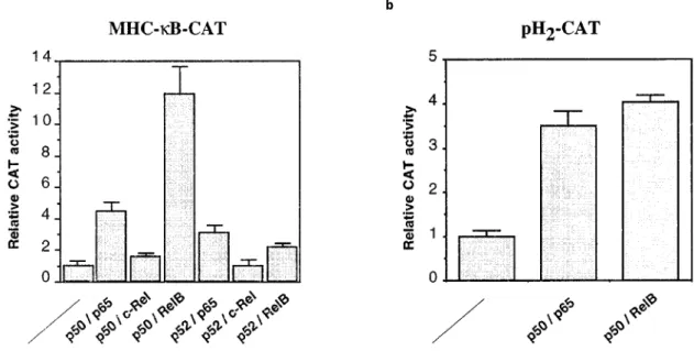
The NF-kB site of the MHC Cl I was inserted in position 756 of the minimal c-fos promoter of a CAT reporter plasmid in order to study the NF-kB transactivation eciency through this particular site. This reporter plasmid, named MHC-kB-CAT, was transfected into the breast cancer derived cell line MCF7 A/Z together with expression vectors directing the production of various NF-kB-related proteins. The transactivating activities of p50/p65, p52/p65, p50/c-Rel, p52/c-p50/c-Rel, p50/RelB and p52/RelB heterodimers were determined by measuring CAT activities in the transfected MCF7 A/Z cells (Figure 1a). The p65 and RelB-containing complexes induced signi®cant CAT activity. The most important eect was observed with p50/p65 or p50/RelB (about a 10-fold induction over control CAT activity) whereas c-Rel-containing com-plexes did not transactivate the reporter plasmid. The same experiments were repeated in MDA-MB-435 cells and showed similarly that p50/p65 and RelB contain-ing complexes were the most active for the transactiva-tion of the MHC-kB-CAT plasmid while c-Rel complexes were only weak transactivators (data not shown).
This transactivating eect was dose-dependent since transfections with increasing amounts of p50/p65 or p50/RelB expression vectors led to progressively increased CAT activities (data not shown).
a b
Figure 1 Various NF-kB complexes transactivate the MHC Class I promoter. MCF7 A/Z cells were transfected with expression vectors for various NF-kB-related proteins together with a CAT reporter plasmid containing a single kB site from the MHC Cl I gene promoter (MHC-kB-CAT) (a) or a longer MHC Cl I promoter (pH2-CAT) (b). 0.5 mg of each expression vector were
transfected as indicated in the ®gure together with 3 mg of the reporter plasmid. The ®gure shows the relative CAT activity over the activity observed with the CAT vector alone after normalization to the protein concentration of the extracts. Each column represents the mean of three independent experiments (+ sd). The total amount of transfected DNA was kept constant throughout the experiment by adding appropriate amounts of the expression vector without insert
Experiments performed with a longer MHC Cl I
promoter regulating CAT expression (pH2-CAT)
showed that the p50/p65 and p50/RelB complexes can also stimulate transcription through this promoter (Figure 1b).
Inhibition of NF-kB-dependent transactivation by p100 and IkB-a
As we had previously observed high p100 expression in human breast cancers (Dejardin et al., 1995), p100 and IkB-a-mediated inhibition of NF-kB-induced transacti-vation of the MHC-kB-CAT reporter plasmid were compared. MCF7 A/Z cells were transfected with ®xed amounts of the p50 and p65 or p50 and RelB expression vectors together with increasing amounts of p100 or IkB-a expression vectors (Figure 2). In these conditions, a strong and dose-dependent inhibition of the CAT expression was observed with both inhibitory proteins. This inhibitory eect seemed to be more dramatic with IkB-a than with p100 as the transfection of 0.5 mg of the IkB-a expression vector already completely abolished the induced transcription. Inter-estingly, in the same experimental conditions, the IkB-like protein p105 did not produce any inhibition of the transactivation (data not shown).
p100 sequesters RelB in the cytoplasm of breast cancer cells
The expression of various NF-kB and IkB-related proteins in breast cancer cell lines was investigated. p100 expression can easily be detected in a number of breast adenocarcinoma cell lines (MDA-MB-231, MDA-MB-435, T47D and MCF7 A/Z) (Figure 3) as well as in primary breast cancers (Dejardin et al., 1995). Among these cell lines, the highest level of p100 expression was observed in MDA-MB-435 cells (Figure 3). Similarly, immunoblots demonstrated RelB expres-sion in these four cell lines with the strongest signal
observed in MDA-MB-231 cells and the weakest in MCF7 A/Z cells (Figure 3). The level of p65 expression was similar in the four cell lines (Figure 3).
We had previously shown that in MDA-MD-435 cells, p100 is the major NF-kB inhibitor and sequesters most p50/p65 complexes in the cytoplasm (Dejardin et al., 1995). To investigate how p100 and IkB-a form cytoplasmic complexes with p65 or RelB, cytoplasmic extracts from the same four breast cancer cell lines were immunoprecipitated with antibodies directed against p65 or RelB. The precipitated materials were analysed by immunoblots with speci®c antibodies recognizing IkB-a or p100 (Figure 4). p65 was sequestered in the cytoplasm by both IkB-a and p100 in all the cell lines with the exception of MDA-MB-435 while RelB coimmunoprecipitates only with p100 and not with IkB-a in all four cell lines. In MDA-MB-435 cells, highly expressed p100 sequesters all detected p65 and RelB in the cytoplasm as we could not observe any coimmunoprecitation of these two proteins with IkB-a. The speci®city of the immunoprecipitations was veri®ed by the addition of the peptides used to generate the antibodies (Figure 4). These experiments con®rmed that p100 was the major inhibitor of RelB-containing NF-kB complexes as already demonstrated by others (Dobrzanski et al., 1995).
p100 can form trimeric complexes with p50 and p65 in Jurkat and in MDA-MB-435 cells (Kanno et al., 1994; Dejardin et al., 1995). Double immunoprecipita-tions were performed to determine whether p100/p50/ RelB complexes were formed in breast cancer cells. In these experiments, cytoplasmic extracts were ®rst immunoprecipitated with RelB antibodies and the supernatant was discarded. The immune complexes were then dissociated with an excess of RelB peptides and the supernatant was immunoprecipitated with anti-p50 antibodies. The material which had been immunoprecipitated successively by RelB and p50 antibodies was ®nally analysed on immunoblots with antibodies recognizing speci®cally p100 (Figure 5). In
a b
Figure 2 p100 and IkB-a inhibit NF-kB-dependent transactivation of the MHC-kB-CAT reporter plasmid. MCF7 A/Z cells were transfected with expression vectors for p50, p65 and RelB (0.5 mg each) together with the MHC-kB-CAT reporter plasmid (3 mg). Increasing amounts of expression vectors for IkB-a (a) or p100 (b) were cotransfected as indicated in the ®gure
these experimental conditions, p100/p50/RelB com-plexes were only detected in the MDA-MB-231 cells. In the other cell lines, p100 forms a complex with RelB as shown in Figure 4 but it is apparently not engaged in multimeric complexes with p50 and RelB (Figure 5).
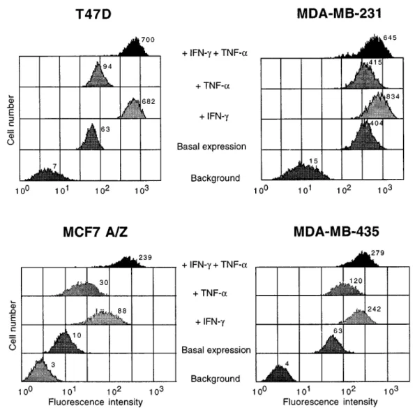
Induction of MHC Cl I expression by IFN-g and TNF-a in breast cancer cells
The expression of MHC Cl I proteins on the surface of breast cancer cells was studied by ¯ow cytometry in basal conditions and after stimulation with Interferon-g (IFN-Interferon-g) and TNF-a (FiInterferon-gure 6). In basal conditions, the four cell lines investigated showed some expression of MHC Cl I proteins. The lowest expression was observed in MCF7 A/Z cells and the highest in MDA-MB-231 cells. There is no correlation between the basal level of MHC Cl I proteins expression and that of p100 (compare Figures 6 and 3).
Cells were then stimulated with the cytokines IFN-g, TNF-a or a combination of them. IFN-g (100 U/ml) induced MHC Cl I in the four cell lines and most signi®cantly in cells demonstrating low basal MHC Cl I expression (Figure 6). Cell stimulation with TNF-a also induced MHC Cl I expression in three out of the four cell but the eect observed was not as strong as with IFN-g (Figure 6). A combination of both cytokines generated in the MCF7 A/Z cells an increase of MHC Cl I expression corresponding at least to the addition of the eect obtained which each of them alone. In the three other cell lines, the stimulation observed with IFN-g was not boosted by the addition of TNF-a.
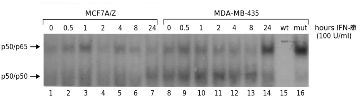
To study whether this MHC Cl I induction could be related to cytokine-induced NF-kB activation, we performed Electrophoretic Mobility Shift assays with nuclear extracts from MDA-MB-435 and MCF7 A/Z cells. As previously demonstrated in a number of cell types, TNF-a rapidly induced nuclear NF-kB DNA-binding activity in both cell lines (data not shown). Conversely, IFN-g stimulation did not induce any detectable NF-kB activity in MCF7 A/Z cells and generated only a very weak and delayed (24 h) NF-kB activity in MDA-MB-435 cells (Figure 7, lane 14).
MDA-MB-435 MDA-MB-231 T47D MCF7 A/Z
RelB
p65
p100
p52
Figure 3 Expression of p100, p65 and RelB in breast cancer cell lines. Equal amounts of total cell extracts (10 mg) from four dierent breast cancer cell lines (MDA-MB-435, MDA-MB-231, T47D and MCF7 A/Z) were analysed by immunoblots for expression of RelB, p65 and p100. Speci®c bands are indicated in the ®gure. p52 refers to the processed form of p100
p65 + Peptide RelB + Peptide p65 RelB p65 + Peptide RelB + Peptide p65 RelB
T47D MDA-MB-231 MCF7 A/Z MDA-MB-435 IP IB p100 IB I κB-α
Figure 4 p100 and RelB are coimmunoprecipitated from cytoplasmic extracts. Cytoplasmic extracts from four breast cancer cell lines were immunoprecipitated (IP) with anti-p65 or anti-RelB antibodies. The immunoprecipitated material was then analysed on immunoblots (IB) for the presence of IkB-a (left panel) or p100 (right panel). As controls, the immunoprecipita-tions were also performed in the presence of the p65 and RelB peptides used to generate the antibodies
C MDA-MB-435 MCF7 A/Z MDA-MB-231 T47D
p100
*
Figure 5 p100 forms a trimeric complex with p50 and RelB in MDA-MB-231 cells. Cytoplasmic extracts from the breast adenocarcinoma cell lines were ®rst immunoprecipitated with anti-RelB antibodies. The immunoprecipitated complexes were then dissociated with an excess of the RelB peptide and the supernatants were re-immunoprecipitated with anti-p50 antibo-dies. The immunoprecipitated material was ®nally analysed on immunoblots for the presence of p100. The speci®c band is indicated. The broad band indicated by an asterisk corresponds to the reaction of the secondary antibody used for the immunoblot with the immunoprecipitating antibodies. The lane C shows protein extracts directly analysed by immunoblot without previous immunoprecipitation
Regulation of MHC Class I expression by NF-kB proteins
Mutations of the serines 32 and 36 of IkB-a phosphorylation sites abolish IkB-a degradation following a number of external stimuli and thus prevent NF-kB activation (Brown et al., 1995; Traeckner et al., 1995; Whiteside et al., 1995). Basal and induced MHC Cl I expression was thus compared in MCF7 cells stably transfected with the pcDNA3 expression vector containing or not the mutant IkB-a gene. It has been previously shown that induction of NF-kB DNA-binding activity was abolish in these stably transfected MCF7 MAD cells (Cai et al., 1997). The basal expression of MHC Cl I proteins, as measured by FACS analysis, was signi®cantly lower in MCF7 MAD cells than in control cells (Figure 8, basal expression). In the MCF7 MAD cells, the ¯uorescence intensity was reduced to the background level suggesting a complete inhition of MHC Cl I expression. However, a signi®cant induction of MHC Cl I expression could still be observed in these
cells after IFN-g stimulation while TNF-a treatment was without eect (Figure 8). The same experiment was then reproduced with MCF7 A/Z cells. Again, stable transfection of the mutated IkB-a vector completely abolished NF-kB activation as demonstrated by EMSAs (data not shown). In the MCF7 A/Z MAD cells, as compared with cells transfected with an empty expression vector, the basal MHC Cl I expression was not signi®cantly decreased and remained higher that the background level (Figure 8). Again, cells that expressed the mutated IkB-a protein did not show any induction of MHC Cl I expression following TNF-a stimulation (Figure 8). Similar observations were also made with stably transfected HCT116 colon carcinoma cells and OVCAR-3 ovarian carcinoma cells expressing the mutated IkB-a protein (data not shown).
Discussion
Investigating the mechanisms regulating MHC Cl I expression is most important for our understanding
Figure 6 Expression of MHC Class I proteins in breast cancer cell lines. Four breast cancer cell lines were analysed by ¯ow cytometry for basal and stimulated expression of MHC Cl I proteins. Background lanes correspond to the signal obtained in the presence of an irrelevant ®rst antibody (puri®ed mouse IgG1). Control lanes refer to the basal expression of MHC Class I proteins in unstimulated cells. The cell lines T47D, MDA-MB-231, MCF7 A/Z and MDA-MB-435 were stimulated for 48 h with IFN-g (100 U/ml) alone, with TNF-a (100 U/ml) alone or with both cytokines at the same time. The relative ¯uoresence intensity is indicated next to each peak. This experiment was performed independently twice
of immune response and carcinogenesis. The NF-kB transcription factor certainly plays a key role in this regulation but its precise eect on the MHC Cl I promoters has yet to be determined. In this report, we con®rmed that, as already shown by others in various cell types (Drew et al., 1993, 1995; Scheinman et al., 1993; Segars et al., 1993), NF-kB complexes activated transcription of the MHC Class I antigens in breast cancer cells.
Stable transfections of the uninducible IkB-a mutant abolished basal MHC Cl I expression in MCF7 breast adenocarcinoma cells but not in the other cells we analysed. It is not surprising that such a mutant does not in¯uence basal MHC Cl I expression as serines 32 and 36 had been shown to be phosphorylated in response to stimuli such as proin¯ammatory cytokines (Brown et al., 1995; Traeckner et al., 1995; Whiteside et al., 1995). In other words, basal NF-kB activity is not in¯uenced by phosphorylation of these IkB-a two serine residues but rather by the IkB-a or IkB-b PEST sequence or by the IkB-a ankyrin repeat domain (Krappmann et al., 1996; Van Antwerp et al., 1996; McKinsey et al., 1996; Good and Sun, 1996).
The mutated IkB-a inhibitor completely blocked MHC Cl I induction by TNF-a in MCF7 and MCF7 A/Z cells as well as in the colon carcinoma HCT116 and ovarian carcinoma OVCAR-3 cells. We could thus conclude that TNF-a-induced MHC Cl I expression is regulated by NF-kB. However, complete inhibition of NF-kB activation does not prevent MHC Cl I induc-tion by IFN-g. Indeed, we observed that IFN-g rapidly activated the transcription factor IRF-1 in breast cancer cells while it induced NF-kB DNA-binding only very faintly (Figure 7 and data not shown). IFN-g-induced MHC Cl I expression in these breast cancer cell lines is thus NF-kB-independent.
RelB-containing NF-kB complexes transactivate MHC Class I promoters in breast cancer cells. Moreover, ¯ow cytometry analysis indicated high basal expression of MHC Cl I in the MDA-MB-231 cells and a much lower expression in MCF7 A/Z cells. In these cells, MHC Cl I expression might be correlated with the level of RelB expression although such a correlation should be con®rmed on a larger number of cell lines. These observations con®rm that the RelB protein might be a major regulator of antigen presentation. It was indeed reported that RelB is
expressed in dendritic cells and is required for the dierentiation of these antigen presenting cells (Burkly et al., 1995; Weih et al., 1995). Moreover, RelB is implicated in the constitutive expression of kB-dependent genes (Dobrzanski et al., 1994; Weih et al., 1996).
p100 acts as an IkB molecule and sequesters NF-kB complexes in the cytoplasm of various cell types. It was indeed demonstrated that p100 can form cytoplasmic complexes with p65, p50 and RelB (Mercurio et al., 1993; Scheinman et al., 1993; Kanno et al., 1994; Dejardin et al., 1995). Previous reports indicated that RelB complexes poorly interact with IkB-a, p105 or Bcl-3 and are preferentially inhibited by p100 in B lymphocytes (Lernbecher et al., 1994; Dobrzanski et al., 1994, 1995). Our data con®rm that, in breast cancer cells, RelB-containing complexes are not inhibited by IkB-a but by p100, indicating a preferential interaction between RelB and the IkB-like molecule p100. In transient transfections however, as there is no possible competition with p100, overexpressed IkB-a interact with p50/RelB complexes and block transcription of the MHC-kB-CAT (Figure 2).
It was also shown that p100 can form trimeric complexes with p50 and p65 in the cytoplasm of Jurkat and MDA-MB-435 cells, presumably through an interaction of the p100 ankyrin repeats with p50 or p65 nuclear translocation signal (Kanno et al., 1994; Dejardin et al., 1995). In this report, we immunopre-cipitated from MDA-MB-231 cells a novel form of multimeric complex which associates RelB, p50 and p100. This observation thus con®rms that triple complexes are formed with p100. However, p100 is much more stable than IkB-a and p105 and does not respond to the same stimuli as IkB-a (Dejardin et al., unpublished data). Although a previous study showed p100 processing into p52 after PMA stimulation of Hela cells (Mercurio et al., 1993), our data indicate that, in breast cancer cells, these p100-containing multimeric complexes constitutes a separate pool of NF-kB factors which are released after speci®c, yet to be identi®ed, stimuli.
Despite our experiments and previous data on p100 and RelB interaction, we did not observe any
correlation between basal or TNF-a-induced
MHC C1 I expression and the level of p100 expression in the studied cell lines. Indeed, the basal MHC Cl I 0 0.5 1 2 4 8 24 0 0.5 1 2 4 8 24 wt mut p50/p65 p50/p50 hours IFN-γ (100 U/ml) MCF7A/Z MDA-MB-435 1 2 3 4 5 6 7 8 9 10 11 12 13 14 15 16
Figure 7 Analysis of NF-kB DNA-binding after interferon-g stimulation of breast cancer cell lines MCF7 A/Z and MDA-MB-435. Nuclear extracts were prepared following IFN-g stimulation for various times as indicated. 5 mg of nuclear proteins from MCF7 A/Z (lanes 1 ± 7) and MDA-MB-435 (lanes 8 ± 14) were mixed with a labelled probe corresponding to the kB site from the human MHC Cl I promoter. For competition assays, a 206molar excess of unlabelled wild type (wt) or mutated (mut) MHC Cl I probe was added to the binding reactions containing nuclear extract from MDA-MB-435 stimulated for 24 h. Supershifting experiments con®rmed that the faster migrating complex was the p50/p50 homodimer while the slower one was the p50/p65 heterodimer (data not shown). Same results were obtained with the palindromic kB probe
expression is higher in MDA-MB-435 cells than in MCF7 A/Z cells which express p100 at a much lower level.
This observation indicates that, although highly expressed p100 forms stable cytoplasmic complexes with all detectable p65 and RelB proteins in MDA-MB-435 cells, it does not completely block NF-kB activity as the IkB-a mutant does. Two hypotheses could explain such an observation. Either, p100 allows small amounts of NF-kB to translocate to the nucleus and to regulate basal and TNF-a-induced transcription of the MHC Cl I gene; this hypothesis is supported by the observation that p100 is not as ecient as IkB-a in inhibiting NF-kB-dependent transcription (Figure 2) and that stable expression of p100 in MCF7 A/Z cells does not completely block NF-kB nuclear translocation and does not modify basal or induced MHC Cl I expression (data not show). Alternatively, p100 itself can bind the MHC Cl I promoter as a part of the H2TF1 complex and this complex would thus be responsible for uninduced MHC Cl I expression
(Potter et al., 1993; Scheinman et al., 1993). We do not favor this last hypothesis as we never observed any p100 expression in the nucleus of breast cancer cells.
In summary, the present report demonstrates that NF-kB proteins regulate basal and TNF-a-induced but not IFN-g-induced MHC Cl I expression in breast cancer cells and that highly expressed p100 probably does not inhibit this expression.
Materials and methods
Cell culture and biological reagents
The human breast cancer cell lines MCF7, MCF7 A/Z, MDA-MB-435, MDA-MB-231 and T47D were grown in RPMI 1640 medium (Gibco BRL) supplemented with 1% antibiotics, 1% glutamine and 10% fetal bovine serum (FBS). The cancer cell line MCF7 A/Z is a generous gift from Professor Mareel (University of Ghent, Belgium).
In the experiment described in Figures 6 and 9, the cells were treated with 100 U/ml of TNF-a (Boehringer
Figure 8 MHC Cl I expression in MCF7 and MCF7 A/Z cells stably transfected with an IkB-a mutant expression vector. The expression of MHC Cl I was measured by FACS analysis in MCF7 and MCF7 A/Z cells transfected with a pcDNA3 empty expression vector (left panels) or with an expression vector coding for an IkB-a protein mutated at serines 32 and 36 (MCF7 MAD and MCF7 A/Z MAD). Background lanes correspond to the signal obtained in the presence of an irrelevant ®rst antibody (puri®ed mouse IgG1-FITC). Basal expression refers to the expression of MHC Class I proteins in unstimulated cells. The cells were stimulated either with IFN-g or TNF-a for 48 h as indicated in the ®gure. The relative ¯uorescence intensity is indicated next to each peak. This experiment was performed independently twice
Mannheim, Germany) or 100 U/ml of IFN-g (Boehringer Mannheim) for 48 h.
Transient transfections and CAT assays
MDA-MB-435 and MCF7 A/Z cells were transfected using liposomes (DOTAP system, Boehringer Mannheim, Ger-many) following the manufacturer's instructions. CAT assays were performed as described previously (Neumann et a., 1987; Bours et al., 1992).
The MHC-kB-CAT reporter plasmid was constructed by inserting the kB site from the H-2Kb promoter (5'-GATCTCAACGGCAGGCGGGGATTCCCTC-3') at posi-tion 756 of a minimal c-fos promoter (Yano et al., 1987; Bours et al., 1992). A longer construct (pH2-CAT: a gift from Dr A Israel, Institut Pasteur, Paris, France) containing a 2027 bp HindIII ± NruI fragment from the mouse H-2Kb promoter was also used (Daniel-Vedele et al., 1985). The expression vectors contained the cDNAs for p50, p65 (RelA), c-Rel, RelB, p52, IkB-a or p100 cloned into the PMT2T expression vector at the EcoRI site (Bours et al., 1992). Immunoblots, immunoprecipitations and protein quanti®cation Cellular extracts, immunoblots, immunoprecipitations and protein quanti®cation were performed as described (Dejardin et al., 1995; Bonizzi et al., 1996). Immunoblots were analysed by chemiluminescence (ECL, Amersham). The antibodies used were: an anti-RelB anti-peptide antibody (Santa Cruz Biotechnology, Santa Cruz, CA), an anti-p100 monoclonal antibody (a gift from Dr Siebenlist, NIH, Bethesda, MD), and an p65 anti-peptide antibody (Santa Cruz Biotechnology, Santa Cruz, CA).
Double immunoprecipitations
Cytoplasmic extracts were ®rst immunoprecipitated with a speci®c anti-RelB-peptide antibody and protein A-sephar-ose beads. The supernatant was discarded and the beads were incubated in TNT buer (Tris 20 mM pH 7.5, NaCl 200 mM, Triton-X-100 1%) in the presence of an excess (5 mg of peptide for 5 ml of antibody) of the speci®c RelB peptide for 16 h at 48C. The resulting supernatant containing the released multimeric RelB complexes was then immunopreciptated with anti-p50 antibodies and protein A-sepharose beads to isolate complexes containing both p50 and RelB. The ®nal immune complexes were separated by SDS ± PAGE and examined by immunoblot analysis with anti-p100/p52 monoclonal antibody.
Electrophoretic mobility shift assays
Nuclear extracts were prepared as previously described (Dejardin et al., 1995). The oligonucleotide probe used was the palindromic kB site (Bonizzi et al., 1996) or the
MHC Cl I kB site with the sequence 5'-TTGGAAGGA-GAGGGGATTCCCCCTGCCGTTG-3'. A 20-fold excess of unlabelled wild type or mutated probe (5'-TTGGAAG-GAGATCTATTCCCCCTGCCGTTG-3') was used for competition experiments.
Flow cytometry
Adherent cells were ®rst treated with dispase II (Boehringer Mannheim, Germany) and washed twice in PBS. 56105 cells were then incubated for 30 min at 48C with a monoclonal antibody directed against the proteins HLA A, B, C (Pharmingen, San Diego, CA) coupled with ¯uorescein isothiocyanate (FITC). After three washes in PBS, the cells were analysed using a ¯ow cell sorter (FACStar Plus, Becton Dickinson, San Jose, CA) with a 100 mW air-cooled argon laser (Spinnaker 1161; Spectra Physics, Mountain View, CA) and the CellQuest software (Macintosh, Facstation; Becton Dickinson, San Jose, CA). For each sample, 3000 cells were analysed and ¯uorescence histograms (1024 channels, log scale) were constructed. Stable transfections
The MCF7 cells stably transfected with the pcDNA3 empty vector (pcN-183) or with the mutated IkB-a expression vector (MCF7 MAD) were previously described (Cai et al., 1997).
The MCF7 A/Z cells were transfected with the linearized pcDNA3 empty vector or with the mutated IkB-a expression vector (MCF7 A/Z MAD) using the DOTAP system (Boehringer Mannheim). Forty-eight hours after transfec-tion, transfected cells were selected in a medium containing G418 at 0.5 mg/ml. After 15 days of selection, 10 resistant colonies were isolated and grown separetly. Positive clones were selected for their ability to prevent nuclear NF-kB translocation following TNF-a stimulation.
Acknowledgements
We thank Dr U Siebenlist (NIH, Bethesda, MD) for the NF-kB antibodies, Dr A Israel (Institut Pasteur, Paris, France) for the pH2-CAT construct and for the IkB-a mutant plasmid and Professor Mareel (University of Ghent, Belgium) for the MCF7 A/Z cells. We are most thankful to Professor J Gielen for his support and critical comments on the manuscript. We thank G Bonizzi and A-C Hellin fot the MA-CF7 A/Z, OVA-CAR-3 and HA-CT 116 stable transfectants. M-PM and VB are Research Associ-ates at the National Fund for Scienti®c Research (Belgium). ED and VD are supported by FRIA fellow-ships. This work has been supported by grants from the National Fund for Scienti®c Research (Belgium), TeÂleÂvie (Belgium) and the Centre Anti CanceÂreux (University of LieÁge, Belgium).
References
Accolla RS, Adorini L, Sartoris S, Sinigaglia F and Guardiola J. (1995). Immunol. Today, 16, 8 ± 11.
Baeuerle PA and Henkel T. (1994). Ann. Rev. Immunol., 12, 141 ± 179.
Baldwin ASJ and Sharp PA. (1987). Mol. Cell. Biol., 7, 305 ± 313.
Baldwin ASJ and Sharp PA. (1988). Proc. Natl. Acad. Sci. USA, 85, 723 ± 727.
Beg AA and Baldwin AS. (1993). Genes Dev., 7, 2064 ± 2070. Blanchet O, Bourge JF, Zinszner H, Israel A, Kourilsky P, Dausset J, Degos JL and Paul P. (1992). Proc. Natl. Acad. Sci. USA, 89, 3488 ± 3492.
Bonizzi G, Dejardin E, Piret B, Piette J, Merville M-P and Bours V. (1996). Eur. J. Biochem., 242, 544 ± 549. Bours V, Villalobos J, Burd PR, Kelly K and Siebenlist U.
(1990). Nature, 348, 76 ± 80.
Bours V, Burd PR, Brown K, Villalobos J, Park S, Ryseck R-P, Bravo R, Kelly K and Siebenlist U. (1992). Mol. Cell. Biol., 12, 685 ± 695.
Bours V, Franzoso G, Azarenko V, Park S, Kanno T, Brown K and Siebenlist U. (1993). Cell, 72, 729 ± 739.
Brown K, Gerstberger S, Carlson L, Franzoso G and Siebenlist U. (1995). Science, 267, 1485 ± 1488.
Burkly L, Hession C, Ogata L, Reilly C, Marconi LA, Olson D, Tizard R, Cate R and Lo D. (1995). Nature, 373, 531 ± 536.
Cai Z, KoÈrner M, Tarantino N and Chouaib S. (1997). J. Biol. Chem., 272, 96 ± 101.
Cordon-Cardo C, Fuks Z, Drobnjak M, Moreno L, Eisenbach L and Feldman M. (1991). Cancer Res., 51, 6372.
Daniel-Vedele F, Israel A, Benicourt C and Kourilsky P. (1985). Immunogenetics, 21, 601 ± 611.
Dejardin E, Bonizzi G, BellahceÁne A, Castronovo V, Merville M-P and Bours V. (1995). Oncogene, 11, 1835 ± 1841.
Dobrzanski P, Ryseck R-P and Bravo R. (1994). EMBO J., 13, 4608 ± 4616.
Dobrzanski P, Ryseck R-P and Bravo R. (1995). Oncogene, 10, 1003 ± 1007.
Drew PD, Lonergan M, Goldstein ME, Lampson LA, Ozato K and McFarlin DE. (1993). J. Immunol., 150, 3300 ± 3310.
Drew PD, Franzoso G, Becker KG, Bours V, Carlson LM, Siebenlist U and Ozato K. (1995). J. Interferon Cytokine Res., 15, 1037 ± 1045.
Franzoso G, Bours V, Park S, Tomita-Yamaguchi M, Kelly K and Siebenlist U. (1992). Nature, 359, 339 ± 342. Garrido F, Cabrera T, Lopez-Nevot MA and Ruiz-Cabello
F. (1995). Adv. Cancer Res., 67, 155 ± 195.
Good L and Sun S-C. (1996). J. Virol., 70, 2730 ± 2735. Haskill S, Beg AA, Tompkins SM, Morris JS, Yurochko AD,
Sampson-Johannes A, Mondal K, Ralph P and Baldwin ASJ. (1991). Cell, 65, 1281 ± 1289.
IsraeÈl A, Kimura A, Kieran M, Yano O, Kanellopoulos J, Le Bail O and Kourilsky P. (1987). Proc. Natl Acad. Sci. USA, 84, 2653 ± 2657.
IsraeÈl A, Le Bail O, Hatat D, Piette J, Kieran M, Logeat F, Wallach D, Fellous M, and Kourilsky P. (1989). EMBO J., 8, 3793 ± 3800.
Kanno T, Franzoso G and Siebenlist U. (1994). Proc. Natl. Acad. Sci. USA, 91, 12634 ± 12638.
Kieran M, Blank V, Logeat F, Vandekerckhove J, Lottspeich F, Bail OL, Urban MB, Kourilsky P, Baeuerle PA and IsraeÈl A. (1990). Cell, 62, 1007 ± 1018.
Krappmann D, Wulczyn FG and Scheidereit C. (1996). EMBO J., 15, 6716 ± 6726.
Lernbecher T, Kistler B and Wirth T. (1994). EMBO J., 13, 4060 ± 4069.
Liu X, Ge R and Ricciardi RP. (1996). Mol. Cell. Biol., 16, 398 ± 404.
Logeat F, IsraeÈl N, Ten R, Blank V, Le Bail O, Kourilsky P and IsraeÈl A. (1991). EMBO J., 10, 1827 ± 1832.
McKinsey TA, Brockman JA, Scherer DC, Al-Murrani SW, Green PL and Ballard DW. (1996). Mol. Cell. Biol., 16, 2083 ± 2090.
Mercurio F, DiDonato JA, Rosette C and Karin M. (1993). Genes Dev., 7, 705 ± 718.
Miyamoto S and Verma IM. (1995). Adv. Cancer Res., 66, 255 ± 292.
Neri A, Chang C-C, Lombardi L, Salina M, Corradini P, Maiolo AT, Chaganti RSK and Dalla-Favera R. (1991). Cell, 67, 1075 ± 1087.
Neumann JF, Morency C and Russian K. (1987). Biotechniques, 5, 444 ± 447.
Nolan GP, Fujita T, Bhatia K, Huppi C, Liou H-C, Scott ML and Baltimore D. (1993). Mol. Cell. Biol., 13, 3557 ± 3566.
Ohno H, Takimoto G and McKeithan TW. (1990). Cell, 60, 991 ± 997.
Plaskin D, Baeuerle PA and Eisenbach L. (1993). J. Exp. Med., 177, 1651 ± 1662.
Potter DA, Larson CJ, Eckes P, Schmid RM, Nabel GM, Verdine GL and Sharp PA.(1993). J. Biol. Chem., 268, 18882 ± 18890.
Rice NR, MacKichan ML and IsraeÈl A. (1992). Cell, 71, 243 ± 253.
Scheinman RI, Beg AA and Baldwin AS. (1993). Mol. Cell. Biol., 13, 6089 ± 6101.
Schouten JG, Van der Eb AJ and Zantema A. (1995). EMBO J., 14, 1498 ± 1507.
Segars JH, Nagata T, Bours V, Medin JA, Franzoso G, Blanco JCG, Drew PD, Becker KG, An J, Tang T, Stephany DA, Neel B, Siebenlist U and Ozato K. (1993). Mol. Cell. Biol., 13, 6157 ± 6169.
Siebenlist U, Franzoso G and Brown K. (1994). Annu. Rev. Cell. Biol., 10, 405 ± 455.
Traenckner EB, Pahl HL, Henkel T, Schmidt KN, Wilk S and Baeuerle PA. (1995). EMBO J., 14, 2876 ± 2883. Van Antwerp DJ and Verma IM. (1996). Mol. Cell. Biol., 16,
6037 ± 6045.
van't Veer LJ, Beijersbergen RL and Bernards R. (1993). EMBO J., 12, 195 ± 200.
Watanabe N, Iwamura T, Shinoda T and Fujita T. (1997). EMBO J., 16, 3609 ± 3620.
Weih F, Carrasco D, Durham SK, Barton DS, Rizzo CA, Ryseck R-P, Lira SA and Bravo R. (1995). Cell, 80, 331 ± 40.
Weih F, Lira SA and Bravo R. (1996). Oncogene, 12, 445 ± 449.
Whiteside ST, Ernst MK, Le Bail O, Laurent-Winter C, Rice N and IsraeÈl A. (1995). Mol. Cell. Biol., 15, 5339 ± 5345. Whiteside ST, Epinat JC, Rice NR and IsraeÈl A. (1997).
EMBO J., 16, 1413 ± 1426.
Yano O, Kanellopoulos J, Kieran M, Le Bail O, IsraeÈl A and Kourilsky P. (1987). EMBO J., 6, 3317 ± 3324.