HAL Id: inserm-00380194
https://www.hal.inserm.fr/inserm-00380194
Submitted on 7 May 2009HAL is a multi-disciplinary open access
archive for the deposit and dissemination of sci-entific research documents, whether they are pub-lished or not. The documents may come from teaching and research institutions in France or abroad, or from public or private research centers.
L’archive ouverte pluridisciplinaire HAL, est destinée au dépôt et à la diffusion de documents scientifiques de niveau recherche, publiés ou non, émanant des établissements d’enseignement et de recherche français ou étrangers, des laboratoires publics ou privés.
Functional organization of a Schwann cell enhancer.
Eric Denarier, Reza Forghani, Hooman Farhadi, Samar Dib, Nancy Dionne,
Hana Friedman, Pierre Lepage, Thomas Hudson, Régen Drouin, Alan Peterson
To cite this version:
Eric Denarier, Reza Forghani, Hooman Farhadi, Samar Dib, Nancy Dionne, et al.. Functional organi-zation of a Schwann cell enhancer.. Journal of Neuroscience, Society for Neuroscience, 2005, 25 (48), pp.11210-7. �10.1523/JNEUROSCI.2596-05.2005�. �inserm-00380194�
Section: Development/Plasticity/Repair
Senior Editor : Dr. Pat Levitt
Functional organization of a Schwann cell enhancer.
Eric Denarier1 , Reza Forghani1 , Hooman F. Farhadi1 , Samar Dib1 , Nancy Dionne1 , Hana Friedman1 , Pierre Lepage2 , Thomas J. Hudson2 , Régen Drouin3
and Alan C. Peterson1
#
1
Laboratory of Developmental Biology, Molecular Oncology Group H-5, McGill University Health Centre, 687 Pine Ave. West, Montreal, Quebec, Canada, H3A 1A1.
2
McGill University and Genome Quebec Innovation Centre. 740, Dr Penfield Ave. Montreal, Qc, Canada H3A 1A4.
3
Service de Génétique Médicale, Dept of Pediatrics, Centre Hospitalier Universitaire de Sherbrooke, Hôpital Fleurimont. 3001, 12th Avenue North, Sherbrooke, Québec
CANADA J1H 5N4
#To whom correspondence should be addressed. Corresponding author : Alan C. Peterson
Laboratory of Developmental Biology, Molecular Oncology Group H-5, McGill University Health Centre, 687 Pine Ave. West, Montreal, Quebec, Canada, H3A 1A1. Phone: (514) 934-1934 ext. 35846/35835
Fax: (514) 843-1478
E-mail: alan@devbiol2.molonc.mcgill.ca
7 figures, 1 table, 24 pages
Keywords : Myelin Basic Protein, Schwann cell, Enhancer, phylogenetic footprint,
transgenesis,
Acknowledgments
This work was supported by grants from the Canadian Institutes of Health Research, The Canadian MS Foundation, the Canadian Genetic Diseases Network, Genome Canada and Genome Quebec. E.D. was supported by Commissariat à l’Energie Atomique, France. Doctoral fellowships were provided by CIHR (R.F. and H.F.), McGill University Health Centre (S.D.) and MS Society (N.D.). T.J.H. is recipient of an investigator Award from CIHR and a Clinician-scientist Award in Translational Research from the Burroughs Wellcome Fund. R.D. holds the “Canada Research Chair in Genetics, Mutagenesis and Cancer”. We thank P. Charnay for providing the pET-Krox plasmid. The authors are grateful to M. Beaudoin, F. Bourdeau, N. Dallaire, C. Dy, T. Fernandez, H. Furtenbacher, I. Paradis, I. Tretjakoff and P. Valera for excellent technical support.
Abstract
Myelin Basic Protein (MBP) gene expression is conferred in oligodendrocytes and Schwann cells by different upstream enhancers. In Schwann cells, expression is controlled by a 422 bp enhancer lying –9kb from the gene. We show here that it contains 22 mammalian conserved motifs > 6 bps. To investigate their functional significance different combinations of wild type or mutated motifs were introduced into reporter constructs that were inserted, in single copy, at a common HPRT docking site in ES cells. Lines of transgenic mice were derived and the subsequent qualitative and quantitative expression phenotypes were compared at different stages of maturation. In the enhancer core, seven contiguous motifs cooperate to confer Schwann cell specificity while different combinations of flanking motifs engage, at different stages of Schwann cell maturation, to modulate expression level. Mutation of a Krox-20 binding site reduces the level of reporter expression while mutation of a potential Sox element silences reporter expression. This potential Sox motif was also found conserved in other Schwann cell enhancers suggesting that it contributes widely to regulatory function. These results demonstrate a close relationship between phylogenetic footprints and regulatory function and suggest a general model of enhancer organization. Finally, this investigation demonstrates that in vivo functional analysis supported by controlled transgenesis can be a robust complement to both molecular and bioinformatics approaches to understand regulatory mechanisms.
Introduction
Myelin is elaborated in the peripheral nervous system (PNS) and in the central nervous system (CNS) by Schwann cells and oligodendrocytes respectively. MBP belongs to a family of co-regulated genes that participates in the formation of myelin in both cell types and several upstream regulatory modules variously contribute to its regulation in the two lineages (Farhadi et al., 2003) (Fig.1A). Mod1 and Mod2, located in the proximal 5’ flanking sequence, drive expression in myelinating oligodendrocytes while Mod 3, located further upstream confers expression in mature oligodendrocytes. Expression in Schwann cells is governed by Mod4 located, in the mouse, at - 9kb (Forghani et al., 2001).
To locate the regulatory sequences controlling genes expressed in Schwann cells, previous investigations exploited reporter constructs containing specific promoter or enhancer sequences. Regulatory sequences from P0 (Feltri et al., 1999), PMP-22 (Maier et al., 2003) and the transcription factors Oct-6 (Mandemakers et al., 2000) and Krox-20 (Ghislain et al., 2002) have been evaluated extensively in transgenic mice. However, the precise location, identity and functional relevance of individual transcription factor binding elements are yet to be defined for any such locus.
Several transcription factors are known components of the mechanism engaged to regulate Schwann cell differentiation. In the absence of the zinc finger transcription factor Krox-20, or the POU-domain transcription factor Oct-6, Schwann cells arrest at the promyelinating stage (Topilko et al., 1994; Bermingham et al., 1996). In Oct-6 mutants myelination resumes following a two-week pause while in Krox-20 mutants this block is insurmountable. Sox-10, known to cooperate with Oct6 (Kuhlbrodt et al., 1998) and
Krox-20 (Bondurand et al., 2001), is also required for myelin formation (Britsch et al., 2001). Thus, differentiation of the Schwann cell lineage is controlled through a regulatory network of transcription factors acting at multiple stages of Schwann cell maturation. Evidence from in vitro studies suggests that these same factors may regulate individual myelin genes at later stages of maturation (Peirano et al., 2000; Musso et al., 2001).
Here we attempted to refine the localization of functional elements in the MBP Mod4 Schwann cell enhancer by extending comparative analysis to additional species. We show, using a controlled strategy of transgenesis (Bronson et al., 1996), that phylogenetic footprints within this enhancer are functional regulatory elements. Further, functional analysis led to a model of enhancer structure in which targeting elements, clustered in the core, combine with enhancing activity contributed from multiple flanking motifs to confer fine control over cell specificity and expression levels. Notable differences in the enhancing elements engaged during myelin elaboration and maintenance demonstrate that the MBP expression program is controlled by different transcription factor repertoires at different stages of development. By specific mutation, we show that Krox-20, and a motif with potential Sox10 binding activity, play major roles in enhancer function. Further, the Sox motif is conserved in other enhancer sequences active in myelinating Schwann cells suggesting that it contributes widely to myelin gene expression in Schwann cells.
Materials and methods
Generation of constructs for HPRT docked transgenesis: The MBP –9.5kb and
adding a 80 bp PCR product to the SacII site 5’ of clone –9.0 kb. To clone Mod4 sub-fragments we first generated an hsp-LacZ Entry vector in which hsp-LacZ is cloned into an Eco RV site of the pENTRIA vector (Invitrogen). Mod4 sub-domains were ligated upstream of the hsp promoter in reverse orientation relative to endogenous MBP. Entry vectors were used for in vitro recombination into the HPRT Destination vector that includes homology arms for the HPRT locus using the LR clonase reaction kit (Invitrogen). The final Destination vector was amplified, sequenced across the insert and linearized by Age I. 40mg of targeting construct were used to transfect ES cells. Individual motif mutations were generated by a two step PCR introducing a TTGTT ‡CGAGC substitution in 135-M14mut, TGA‡ATT in 135-M16mut, ACAA‡CCGC in SA-M18mut, ACA‡GTT in SA-M20mut.
Derivation of transgenic mice: Transgenic animals bearing constructs in the HPRT
docking site were generated by transfection of Destination constructs bearing the HPRT targeting cassette into the BK4 (for SA) or BPES5 cells, a laboratory derived heterozygous cell line. Homologous recombination simultaneously restores the deleted HPRT locus in cells and inserts a single copy of the reporter construct into the HPRT 5’ flanking region. Restoration of HPRT expression confers resistance to HAT selection thus permitting positive selection for clones derived from the desired homologous recombination event. Following selection and PCR screening, ES clones were used to inject blastocysts. DNAs prepared from chimeras or germline females were analyzed by Southern blot to check for single integration.
Remyelination experiments. To induce peripheral nerve regeneration, 2-month old male
mice were anesthetized and unilateral sciatic nerve crushes were performed as described (Farhadi et al., 2003). The injury site was marked with India ink. Following 1 or 2 weeks of recovery, injured and contra-lateral control nerves were recovered and dissected into similar proximal and distal segments. b-galactosidase activity was measured in distal segments and compared to expression levels in uninjured contra-lateral samples.
Histochemical detection of b-galactosidase activity. Histochemical staining was performed as described (Forghani et al., 2001).
Quantitation of b-galactosidase activity. Sciatic nerves were dissected from back-cross 2 to 3 C57BL/6 male mice and snap frozen in liquid nitrogen. 48 Samples were homogenized using a mixer mill (Qiagen) with 2 minute burst in 250ul of lysis buffer. Total protein concentrations were measured for all extracts in triplicate by the Bradford procedure (Bio-Rad) using a BSA standard curve. b-galactosidase activity was detected using Galacto-Star chemiluminescent assay system (Applied Biosystems). Standard curves were generated with serial dilutions of b-galactosidase (Roche) in duplicate.
Quantification of MBP RNA. Sciatic nerves were dissected from anesthetized mice and
snap frozen in liquid nitrogen. RNA was prepared from pools of 4 nerves (or 8 nerves from P2 and P4 mice) with the Qiagen RNAeasy Lipid mini kit according to the manufacturer’s protocol. The RNA was reverse transcribed using Superscript II (Invitrogen) and random hexamers according to the manufacturer’s protocol. Quantitative PCR was carried out using the Roche LightCycler FastStart DNA Master plus SYBR Green I kit. The cDNA was diluted 1/10 and 1/20 and then measured using
2ml in a 20ml reaction. For each time point, three pools of nerves were measured and averaged. MBP cDNA was amplified with the following primers:
CGAGAACTACCCATTATGGCTCCC and TGGAGGTGGTGTTCGAGGTGTC. GAPDH was amplified
with the following primers: ACCACAGTCCATGCCATCAC and TCCACCACCCTGTTGCTGTA.
Results are expressed as MBP mRNA moles/GAPDH mRNA moles.
In vivo footprinting: Peripheral nerves from wild type or Trembler mice, both on a
C57Bl/6 background, were analyzed. Trembler mice were evaluated here because the chronic demyelination/remyelination they experience leads to an enrichment of myelinating Schwann cells in their peripheral nerves. Sciatic nerves were obtained from P4–7 normal or adult trembler mice, treated with DMS and their DNA prepared for Maxam & Gilbert sequencing. Mice were killed and their sciatic nerves exposed. Each nerve was immersed in situ in Ringer’s solution containing 0.5% DMS for 6 minutes and then washed in cold Ca2+
and Mg2+
free PBS for 1-5 min, then transferred to buffer B+C (approximately 20 nerves per ml). DNA was prepared and treated as described (Drouin et al., 2001). 800bp of the sequence surrounding Mod4 were analyzed on both strands using different primer sets. As sequence marker, genomic DNA from liver was prepared for sequencing. Methylated guanines from DMS exposures and chemically damaged DNA were converted into strand breaks by hot piperidine treatment. Strand break frequencies were estimated on an alkaline agarose gel (Drouin et al., 1996). Only consistent band intensity differences between in vivo and in vitro samples in two different experiments were scored as footprints.
Sequence analysis: The rat Mod4 sequence was obtained through the rat genome
sequence available at http://genome.ucsc.edu/. An MBP containing BAC from the genomic Chicken BAC library (BACPAC resource, Oakland Research Institute) was identified using a PCR probe for chicken MBP exon 1. Positive clones were identified and amplified. DNA was sheared and ligated to pBluescript vectors (Nancy Dionne, unpublished results). Sequences were aligned by ClustalW using MacVector 7.0 software. VISTA plot of M4 sequence was created using VISTA server (http://gsd.lbl.gov/vista/)
Electrophoretic mobility shift assays: Oligonucleotide probes were produced by
annealing of the complementary strand and filling the two side overhangs with aP32
d C T P a n d K l e n o w . M 1 1 C T A G C C G G C A G C C A C A T G C C T T T C, M11mut
C T A G C C G G C A G A A T T C T G C C T T T C, M12-5’ C T A G T G C C T T T C A T A G A T G C A G A A, M12-5’mut
CTAGTGCCTTTGAATTCTGCAGAA, M14 CTAGGGGCCTTTTTGTTTCCTGTG, M14mut
CTAGGGGCCTTTCGAGCTCCTGTG, M 1 6 C T A G T C C C A G G T G A C C C C A A G C C C, M 1 6 m u t
CTAGTCCCAGGATTC C C C A A G C C C , M20 C T A G T A G C C G G G C C C A C A C G C C C A , K r o x
CTAGGTTGTACGCGGGGGCGGTTAGT. Preparation of sciatic nerve extracts: 150 sciatic nerves
from P10 mice were dissected, extracts were prepared and EMSA reactions (30ml) performed as described (Forghani et al., 1999). Bacterialy expressed Krox was produced from pET-Krox plasmid as described (Nardelli et al., 1992). For supershift 2.5mg of antibody (anti-Krox-20 Covance #PRB-236P, Sp1 Upstate #07-124) was added to the reaction mix before addition of the probe and preincubated 15 min with the extracts.
Results
Inter-species sequence comparisons reveal conserved motifs
within Mod4
The MBP 5’ flanking sequence contains four non-coding sequences (Mod1 to Mod4) eachwith human/mouse conservation extending over 100 bps at 75% or greater identity (Farhadi et al., 2003). Mod4 lies 9kb upstream of the MBP start site and corresponds to a 422 bp region of conservation with Schwann cell enhancer activity. To reveal motifs that may correspond to functional transcription factor binding sites we extended the Mod 4 sequence comparison to rat and chicken. Mouse/rat, mouse/human and mouse/chicken comparison yield respective sequence identities of 91%, 76% and 56%. Twenty-two motifs of at least 6 bps (M1 to M22) are conserved in many mammalian species and of these, 7 are invariant in the more distantly related chicken species (Fig. 1).
In vivo footprinting reveals protein/DNA interactions in the core
Mod4 sequence
To locate the regions of DNA-transcription factor interaction in Mod4 region, we screened an 800 bp Mod4 containing sequence using the LMPCR based in vivo footprinting approach. DNA was derived from sciatic nerve samples from normal mice. To potentially enrich for the population of Schwann cells actively elaborating myelin, additional samples were prepared from Trembler mice. Sciatic nerves were pre-treated with dimethylsulfate, DNA was purified and subsequently analyzed for footprints. Within the 800 bp region screened in this analysis, three footprints were detected, all located in the core of the conservedMod4 sequence. One is a protected guanine residue in motif 9, and two are hypersensitive sites in motif 15 (Fig. 1 and 2). These observations suggest
that a protein complex binds to the most conserved Mod4 region while the sequences surrounding Mod4 lack similar interactions with transcription factors. Consequently, DNA-protein interaction analysis and functional characterization focused on Mod4, guided by the location of phylogenetic footprints.
MBP developmental programming is exposed by
species-specific Mod4 expression.
To determine if human and chicken Mod4 sequences are capable of productive interaction with mouse transcription factors, each was ligated to an hsp68-promoted LacZ reporter gene and investigated for expression in transgenic mice. To eliminate the variation associated with random integration at different insertion sites, constructs were docked, in single copy and predetermined orientation, in the 5’ flanking sequence of the HPRT locus (Bronson et al., 1996). Mod4 sequences from both species drove reporter gene expression in Schwann cells in mature mice (Fig. 3 A and B). The human sequence conferred a typical post-natal expression phenotype initiating high-level expression at the commencement of myelin formation. In contrast, the chicken Mod4 reporter remained silent during pre-weaning development, when both the endogenous MBP locus and the human Mod4 reporter are highly expressed. Rather, expression initiated in the peri-weaning period when myelin formation in the PNS is approaching a mature state. Thus, Mod4 elements essential for expression during mammalian myelin formation are either non-existent or too divergent in the chicken sequence to be recognized by mouse factors. Further, the location of such essential elements must be limited to the polymorphisms existing between the chicken and human sequence (Fig. 1). The subsequent expression of the chicken Mod4 reporter in mature mice demonstrates that the MBP relevant
transcription factor repertoire undergoes significant changes as Schwann cells progress from myelin elaboration to myelin maintenance.
Mapping of the targeting sequence within Mod4
To map the location of targeting elements within mouse Mod4, multiple Mod4 sub-domains were analyzed individually (Fig. 3, A and C). The 5’ 210 bps lying between Bst XI and Sac II (BS), containing conserved motifs M1 through M9, failed to drive Schwann cell expression. In contrast, robust expression was conferred by the 3’ 213 bp lying between Sac II to Avr II (SA) that contains motifs 10 to 22. Expression of this construct is specific to the PNS as no staining was observed in spinal cord white matter where oligodendrocytes elaborate myelin (Fig. 3, B and C). Of note, two non-overlapping fragments of SA; 85 bps containing M10-M13 and 107bps containing M14-M20 individually showed no activity. Thus, one or more elements within each fragment are needed to confer targeting activity. As a 135 bp sequence containing M10-M16 drives Schwann cell expression, we conclude that at least one of the essential elements is located within the 50bp containing M14-M16.
Subfragments of Mod4 confer responsiveness to axonal
signaling
In rat, expression of many myelin-related genes follows the program of myelin acquisition, with peak expression realized in the third week ex utero (Stahl et al., 1990). We show here that MBP mRNA follows a similar program in mouse. To determine if the core enhancer sequences contained in the SA and 135 bp fragments are sufficient to confer this developmental expression program, the quantitative phenotypes realized from
the respective reporter genes were evaluated throughout development.Both constructs yield post-natal expression phenotypes that track the major features of the endogenous MBP program; for SA, a 6 fold increase occurs from P2 to P14 , followed by a decrease to one-fourth the peak level by 3 months of age (Fig. 4A). However, peak of expression occurs earlier for these constructs, which could be due to different stabilities of lacZ and MBP mRNA or transcriptional differences related to the constructs or their insertion environment. One notable exception, observed for both the SA and 135 sequences, is expression in fetal nerves commencing at E13.5 dpc(data not shown). Such precocious expression indicates that they lack one or more elements essential for normal silencing of MBP transcription in Schwann cell precursors.
High-level expression of genes encoding the major myelin proteins requires that Schwann cells achieve and maintain axonal contact. Following axon transection, denervated Schwann cells in the distal nerve segment abruptly down-regulate numerous myelin related genes such as MBP (LeBlanc and Poduslo, 1990). These are re-expressed only when contact with in-growing axons is reestablished. To determine if the elements within the SA construct are sufficient to confer this injury/remyelination response, ß-galactosidase activity was evaluated in sciatic nerves regenerating in response to crush injury (Fig. 4B). Consistent with maintained axonal responsiveness, one week post-crush, ß-galactosidase activity in the distal segment was reduced to 1/3 thatexpressed by uninjured contralateral nerves. When in-growing axons reestablish contact with Schwann cells in the distal segment (approximately two weeks post-crush), ß-galactosidase levels were indistinguishable from control nerves and this restored activity was maintained 4
weeks following injury, when axon regeneration and remyelination are largely complete. The same axon responsive expression program was revealed for the 135bp construct (Histochemical analysis. Data not shown)Thus, the expression of both the 213 bp SA and internal 135bp sequence parallel MBP expression in response to axonal signals.
Mod4 contains distinct targeting and enhancing sub-domains
As observed in both developing and remyelinating preparations, Schwann cell targeting and responsiveness to axon signals are conferred by elements located within the core 135 bp sequence. To evaluate the potential role played by motifs located outside this domain, constructs bearing additional motifs were analyzed for both qualitative and quantitative expression phenotypes. When compared to the 135 bp sequence, SA extends for 78 bps to include M17 to M22 and it drives expression at 3-fold greater levels (Fig. 5). In the 5’ half of Mod4, where M1 to M9 are located, three constructs regulated by contiguous MBP 5’ flanking sequence, terminating at different sites within or 5’ of conserved Mod4 sequence, were analyzed. The sequence terminating at SacII includes motifs 10 to 22. The sequence terminating at –9.08kb adds M5 through M9 and demonstrates a marked increase in expression level during the pre-weaning period of myelin formation. Extension to –9.5kb adds M1 through M4 and leads to an additional increase in expression levels in both juvenile and adult mice. Thus, one or all of the M5–M9 motifs confer regulatory information and recognize factor/s that distinguish pre-weaning myelin elaboration from myelin maintenance in mature mice.
To assign function to specific elements we performed both band shift assays using motif specific oligonucleotides and expression analysis with constructs bearing single motif mutations. Electrophoretic mobility shift assays (EMSAs) were performed with probes representing 8 conserved motifs in the SA sequence (Fig. 1B). While such putative elements could function in diverse cell types, we searched for those that could engage transcription factors expressed in Schwann cells by incubating the oligonucleotides with nuclear extracts prepared from P10 sciatic nerves. Consistent shifts were detected with 5 of the 8 probes (M11, M12-5’, M14, M16 and M20) (Fig. 6A and B). None were competed with oligonucleotides bearing 3-5bpsubstitutions in the motif cores. Thus, some motifs appear to engage factors expressed in Schwann cells while those that were not shifted may require cooperative binding not achievable on short oligonucleotides or engage factors expressed in different cell types.
Motifs M14 and M16 are located in the 50 bp shown to be essential for Schwann cell targeting and M18 and M20 are the most conserved motifs present in the enhancing region of SA. Therefore, we focused our search for candidate transcription factor binding elements on these sites. Using rVISTA software (Loots et al., 2002) several candidates with known relevance to Schwann cell biology and myelination were revealed. M14 and M18 contain Sox family binding sites, although we were unable to confirm this function by supershift. M16 contains sites for AP1 and nuclear receptors RORA1 and ER (Desarnaud et al., 2000; Miskimins and Miskimins, 2001) while M20 is a consensus binding site for Krox-20.
To determine if M20 can bind Krox-20, we used a Krox-20 antibody in a supershift analysis (Chavrier et al., 1988). With sciatic nerve extract, the M20 oligonucleotide
yields a complex of 3 retarded bands of which 2 are specifically competed by an excess of cold oligonucleotide. When the Krox-20 antibody is present in the reaction it displaces the upper specific complex. As a control, the incubation was performed with Sp1 antibody and only the non-specific complex was displaced. As a further test of specificity, we expressed Krox-20 in bacteria and used bacterial extract for M20 band shift; a single complex formed and it was specifically competed with a Krox control oligonucleotide. In addition, the sequence of M8, in the 5’ half of Mod4, is compatible with Krox-20 binding activity and consistent with this function, it also binds Krox-20 from sciatic nerve extracts or bacterial extracts (data not shown). We conclude that motifs M8 and M20 are able to bind Krox-20 and therefore, Krox-20 is a factor contributing to Mod4 enhancer activity in Schwann cells.
To test the in vivo function conferred through individual motifs, constructs were derived in which substitution mutationswere introduced into the core of M14, M16, M18 and M20 motifs (Fig. 1) and their consequences on reporter gene expression phenotypes were analyzed in transgenic mice. When introduced in the context of the core 135 bp sequence, M14 and M16 mutations each affected expression; the M14 mutation entirely silenced expression throughout post-natal development while the M16 mutation reduced expression to 20% of control levels in young animals and to 40% in adults (Fig. 6C). The large reduction of expression observed with both mutants suggests that a cooperative mechanism, involving both motifs, confers functionality to the 135bp sub-domain.
M18 and M20 were similarly mutated but in the context of the longer SA sequence. Both mutations caused expression levels to decline to approximately 50% of control
values at most developmental stages. An exception is the M18 mutation at P4 where activity reached only 30% of the control value.
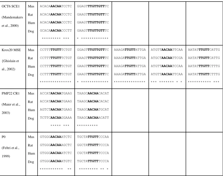
As M14 and M18 share a common AACAA sequence (close to the Sox protein consensus AACAA(T/A)RG for Sox9 in Transfac), and both motifs play an essential role in enhancer function, they may bind a particularly critical factor. To determine if this factor could play a similar role in the regulation of other Schwann cell expressed genes, we searched for this motif in those sequences shown previously to confer Schwann cell targeting in transgenic mice. As shown in Table 1 (Supplementary data) the motif was present in multiple copies and conserved in the Oct6 SCE, Krox-20 MSE, PMP22 CR1, and a conserved region lying in 6kb of sequence upstream of the P0 gene. This observation suggests that the factor bound to M14 motif is a widely used component of the mechanism regulating myelin gene expression in Schwann cells.
Functional organization of Mod4
The functional organization of Mod4 shares numerous features with the regulatory mechanism controlling the well-characterized endo-16 locus of the sea urchin (Yuh et al., 1998). Specifically, enhancer function is conferred through a small number of targeting elements operating in concert with multiple additional elements modulating activity in response to physiological and developmental changes. When the different constructs investigated in the present study are aligned with the Mod 4 sequence conservation plot (Fig.7) the elements necessary for Schwann cell specificity are found in two peaks of conservation at the 5’ and 3’ ends of the core 135 bp targeting sequence. In combination, they are essential for the basic Schwann cell targeting function. In contrast, the two peaks
of conservation flanking the 135 bp targeting sequence contribute enhancing activities, some of which are restricted to defined stages in Schwann cell maturation.
Discussion
In this investigation we show that Mod4 is composed of 22 conserved motifs. We provide evidence that most, if not all, contribute to Schwann cell enhancer activity with motif M14 playing an essential role in enhancer function while others modulate quantitative output.
M14 and M18 are putative Sox family binding elements. As Sox proteins contain a high mobility group domain known to bend DNA, Sox binding is expected to facilitate the cooperative binding of additional transcriptional activators leading to functional complexes (Bustin, 1999; Ellwood et al., 2000). Consistent with this role, Sox10 is known to modulate expression of myelin genes such as P0 in the PNS (Peirano et al., 2000) and MBP in the CNS (Wei et al., 2004). Through interaction with M14, it may also be the critical factor required for the formation of the MBP Mod4 Schwann cell enhancer complex. Remarkably, the core M14 and M18 sequence was found, in multiple copies, in all known sequences capable of driving Schwann cell expression in transgenic mice. Thus, Sox proteins may play a fundamental role in the regulation of multiple genes expressed by differentiating Schwann cells. A similar analysis with the other Mod4 motifs should indicate how widely the factors and elements engaged in the enhancer activity are used in coordinating the overall myelination program.
In addition to the two putative Sox protein binding sites, Mod4 also contains two motifs that are able to bind Krox-20. Schwann cells arrest in a premyelinating state in
has been shown (Musso et al., 2001). Here we show that Krox-20 plays a direct role in MBP regulation. However in our 135bp construct no obvious binding site for Krox-20 was found suggesting that Krox-20 is not crucial but rather, as seen for the periaxin gene (Parkinson et al., 2003), is used to amplify the activity of the enhancer. These combined results suggest that Krox-20 plays an important role in late myelin gene expression.
Binding site redundancy has been observed in multiple enhancers leading to a general model in which enhancers are thought to activate through multiple combinations of bound factors (Berman et al., 2002). We show here that such a redundancy exists within Mod4. By focusing our functional analysis on short, but still functional sub-sequences, we demonstrated large consequences of motif mutation and deletion. Whether consequences of the same magnitude would be elicited by the motif mutations introduced in the context of the entire module remains to be demonstrated.
Using in vivo functional analysis, we demonstrate that Mod4 enhancer activity requires simultaneous contributions from elements located in both targeting and enhancing sub-domains. A more proximal MBP oligodendrocyte enhancer (Mod3) demonstrates a similar structure (N. Dionne, personnal communication) suggesting a general model of enhancer structure/function in which targeting, once established, allows for fine-tuning of expression phenotypes through the lateral recruitment of enhancing elements. Evolutionary diversification of such lateral elements could accommodate species-specific regulatory requirements and consistent with this hypothesis, divergence between mammalian and chicken Mod4 is more pronounced outside the targeting core. The extent to which this model is generally applicable will become evident as the identity
and location of functional elements in additional tissue specific enhancers become known.
A recent study (Taveggia et al., 2004) also used reporter constructs to characterize the MBP sequences important for expression in primary cultures of Schwann cells and oligodendrocytes. In such preparations, the Mod4 sequence, in the context of the –9.0kb MBP construct, was found to enhance activity in oligodendrocytes. In contrast, amongst the transgenic lines we have evaluated to date, no construct regulated by Mod4, or its derivatives, expresses in oligodendrocytes. However we cannot exclude the possibility that Mod4 modulates the quantitative oligodendrocyte expression controlled by other MBP modules. Alternatively, Taveggia et al. point out that glial cells cultured in the absence of neurons may not provide a normal regulatory environment. Consistent with this limitation, we show here, in-vivo, that Mod4 enhancer sub-domains are highly responsive to axon signals.
The consequences of the experimental mutations introduced in this study suggest that naturally occurring variation within regulatory sequences could lead to significant gene deregulation (Knight, 2005). Mutations in conserved motifs could variously silence, or significantly down regulate, transcription at multiple developmental stages. The variable age of onset in numerous diseases, including myelinopathies, could, in part, be caused by variation affecting the stage specific regulatory motifs of key genes.
Finally, this investigation shows the important role in vivo functional analysis can play in the investigation of mammalian regulatory mechanisms. Few techniques are capable of accurately revealing the expression phenotypes conferred by specific regulatory elements and these are most widely applied to non-vertebrate models or in
vitro preparations. The results of this investigation further demonstrate that the controlled construct docking strategy introduced by Bronson et al. (1996) can be applied as an effective strategy to reveal high-resolution qualitative and quantitative in vivo expression phenotypes. By supporting comprehensive access to temporal, spatial and quantitative regulatory phenotypes, this robust in vivo approach emerges as an effective complement to both bioinformatics and molecular investigations on the structure and function of mammalian regulatory sequence.
References
Berman BP, Nibu Y, Pfeiffer BD, Tomancak P, Celniker SE, Levine M, Rubin GM, Eisen MB (2002) Exploiting transcription factor binding site clustering to identify cis-regulatory modules involved in pattern formation in the Drosophila genome. Proc Natl Acad Sci U S A 99:757-762.
Bermingham JR, Jr., Scherer SS, O'Connell S, Arroyo E, Kalla KA, Powell FL, Rosenfeld MG (1996) Tst-1/Oct-6/SCIP regulates a unique step in peripheral myelination and is required for normal respiration. Genes Dev 10:1751-1762. Bondurand N, Girard M, Pingault V, Lemort N, Dubourg O, Goossens M (2001) Human
Connexin 32, a gap junction protein altered in the X-linked form of Charcot-Marie-Tooth disease, is directly regulated by the transcription factor SOX10. Hum Mol Genet 10:2783-2795.
Britsch S, Goerich DE, Riethmacher D, Peirano RI, Rossner M, Nave KA, Birchmeier C, Wegner M (2001) The transcription factor Sox10 is a key regulator of peripheral glial development. Genes Dev 15:66-78.
Bronson SK, Plaehn EG, Kluckman KD, Hagaman JR, Maeda N, Smithies O (1996) Single-copy transgenic mice with chosen-site integration. Proc Natl Acad Sci U S A 93:9067-9072.
Bustin M (1999) Regulation of DNA-dependent activities by the functional motifs of the high-mobility-group chromosomal proteins. Mol Cell Biol 19:5237-5246.
Chavrier P, Zerial M, Lemaire P, Almendral J, Bravo R, Charnay P (1988) A gene encoding a protein with zinc fingers is activated during G0/G1 transition in cultured cells. Embo J 7:29-35.
Drouin R, Gao S, Holmquist G (1996) Agarose gel electrophoresis for DNA damage analysis. New-York: Plenum Press.
Drouin R, Therrrien J-P, Angers M, Ouellet S (2001) In vivo DNA analysis. Totowa NJ: Humana Press.
Ellwood KB, Yen YM, Johnson RC, Carey M (2000) Mechanism for specificity by HMG-1 in enhanceosome assembly. Mol Cell Biol 20:4359-4370.
Desarnaud F, Bidichandani S, Patel PI, Baulieu EE, Schumacher M (2000)
Glucocorticosteroids stimulate the activity of the promoters of peripheral myelin protein-22 and protein zero genes in Schwann cells. Brain Res 865:12-16.
Farhadi HF, Lepage P, Forghani R, Friedman HC, Orfali W, Jasmin L, Miller W, Hudson TJ, Peterson AC (2003) A combinatorial network of evolutionarily conserved myelin basic protein regulatory sequences confers distinct glial-specific phenotypes. J Neurosci 23:10214-10223.
Feltri ML, D'Antonio M, Quattrini A, Numerato R, Arona M, Previtali S, Chiu SY, Messing A, Wrabetz L (1999) A novel P0 glycoprotein transgene activates expression of lacZ in myelin-forming Schwann cells. Eur J Neurosci 11:1577-1586.
Forghani R, Nesbitt J, Snipes J, Shooter EM, Peterson A (1999) Preparation of nuclear extracts from myelinating Schwann cells. J Neurosci Methods 89:129-132. Forghani R, Garofalo L, Foran DR, Farhadi HF, Lepage P, Hudson TJ, Tretjakoff I,
Valera P, Peterson A (2001) A distal upstream enhancer from the myelin basic protein gene regulates expression in myelin-forming schwann cells. J Neurosci 21:3780-3787.
Ghislain J, Desmarquet-Trin-Dinh C, Jaegle M, Meijer D, Charnay P, Frain M (2002) Characterisation of cis-acting sequences reveals a biphasic, axon-dependent regulation of Krox20 during Schwann cell development. Development 129:155-166.
Knight JC (2005) Regulatory polymorphisms underlying complex disease traits. J Mol Med 83:97-109.
Kuhlbrodt K, Herbarth B, Sock E, Hermans-Borgmeyer I, Wegner M (1998) Sox10, a novel transcriptional modulator in glial cells. J Neurosci 18:237-250.
LeBlanc AC, Poduslo JF (1990) Axonal modulation of myelin gene expression in the peripheral nerve. J Neurosci Res 26:317-326.
Loots GG, Ovcharenko I, Pachter L, Dubchak I, Rubin EM (2002) rVista for comparative sequence-based discovery of functional transcription factor binding sites. Genome Res 12:832-839.
Maier M, Castagner F, Berger P, Suter U (2003) Distinct elements of the peripheral myelin protein 22 (PMP22) promoter regulate expression in Schwann cells and sensory neurons. Mol Cell Neurosci 24:803-817.
Mandemakers W, Zwart R, Jaegle M, Walbeehm E, Visser P, Grosveld F, Meijer D (2000) A distal Schwann cell-specific enhancer mediates axonal regulation of the Oct-6 transcription factor during peripheral nerve development and regeneration. Embo J 19:2992-3003.
Miskimins R, Miskimins WK (2001) A role for an AP-1-like site in the expression of the myelin basic protein gene during differentiation. Int J Dev Neurosci 19:85-91. Musso M, Balestra P, Bellone E, Cassandrini D, Di Maria E, Doria LL, Grandis M,
Mancardi GL, Schenone A, Levi G, Ajmar F, Mandich P (2001) The D355V mutation decreases EGR2 binding to an element within the Cx32 promoter. Neurobiol Dis 8:700-706.
Nardelli J, Gibson T, Charnay P (1992) Zinc finger-DNA recognition: analysis of base specificity by site-directed mutagenesis. Nucleic Acids Res 20:4137-4144.
Parkinson DB, Dickinson S, Bhaskaran A, Kinsella MT, Brophy PJ, Sherman DL,
Sharghi-Namini S, Duran Alonso MB, Mirsky R, Jessen KR (2003) Regulation of the myelin gene periaxin provides evidence for Krox-20-independent myelin-related signalling in Schwann cells. Mol Cell Neurosci 23:13-27.
Peirano RI, Goerich DE, Riethmacher D, Wegner M (2000) Protein zero gene expression is regulated by the glial transcription factor Sox10. Mol Cell Biol 20:3198-3209. Stahl N, Harry J, Popko B (1990) Quantitative analysis of myelin protein gene expression
during development in the rat sciatic nerve. Brain Res Mol Brain Res 8:209-212. Taveggia C, Pizzagalli A, Fagiani E, Messing A, Feltri ML, Wrabetz L (2004)
Characterization of a Schwann cell enhancer in the myelin basic protein gene. J Neurochem 91:813-824.
Topilko P, Schneider-Maunoury S, Levi G, Baron-Van Evercooren A, Chennoufi AB, Seitanidou T, Babinet C, Charnay P (1994) Krox-20 controls myelination in the peripheral nervous system. Nature 371:796-799.
Wei Q, Miskimins WK, Miskimins R (2004) Sox10 acts as a tissue-specific transcription factor enhancing activation of the myelin basic protein gene promoter by p27Kip1 and Sp1. J Neurosci Res 78:796-802.
Yuh CH, Bolouri H, Davidson EH (1998) Genomic cis-regulatory logic: experimental and computational analysis of a sea urchin gene. Science 279:1896-1902.
Figure legends
Figure 1. A: Schematic representation of the mouse MBP gene 5’ flanking region
showing the 4 conserved non-coding modules. B: Sequence alignments of Mod4 from four different species. 22 motifs of at least 6bp are conserved in mammals. Motifs are highlighted and designated M1 to M22. In vivo footprint analysis revealed a protected guanine (open circle over the sequence) and hypersensitive sites (filled circles). The Mod4 sequences evaluated for enhancer activity in reporter constructs are delineated by arrowheads over the sequence. Oligonucleotides used in EMSA (M11 to M21) are indicated by lines under the sequence and the nucleotides substituted in motif mutations are delineated by rectangles.
Figure 2. Protein-DNA interactions are revealed in Mod4 by an in vivo footprinting
assay. P10 normal mice (WT) and adult trembler mice (TR) were pretreated with dimethylsulfate. DNA prepared from their sciatic nerves was compared to similarly treated purified DNA (sequencing reaction lane C, T+C, A and G). Protection is detected on base 182 while bases 309 and 310 are hypersensitive.
Figure 3. A: Distribution of conserved Mod4 motifs in sequences analyzed for in vivo
function. Sequences driving Schwann cell expression are indicated in blue. B: ß-galactosidase histochemistry was performed on whole mount preparations of spinal cords
human Mod4 construct expresses in spinal roots at P11 and in the adult while the chicken Mod4 construct expresses in the spinal roots only in adults. C: The SA and 135bp constructs are expressed in spinal roots, but not in spinal cord oligodendrocytes. Note: the ventral spinal cord shown here demonstrates obvious labeling of the central artery but as this is typical of diverse reporter constructs docked at HPRT, it does not represent Mod4 specific enhancer activity.
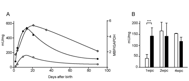
Figure 4. Reporter constructs are responsive to axon signals. A: b-galactosidase activity
in sciatic nerves of mice bearing the SA (black circles) or 135bp construct (white circles) peak during primary myelin formation in post-natal development and follow the MBP RNA accumulation (white diamonds). B: Following nerve crush, b-galactosidase activity in distal nerve segment (white) of mice bearing SA construct, was compared to that in the uninjured contralateral nerve (black) at 1, 2 and 4 weeks post crush. Means +/- SD. *** equals t test result of P<0.001. n=7, 6 and 2 at 1, 2 and 4 weeks post-crush respectively.
Figure 5 Developmental expression programs realized from constructs containing
progressive deletions of Mod4 motifs. A: SA and 135bp sequences ligated to the minimal hsp promoter B: Constructs with contiguous MBP 5’ flanking sequences. The sequence terminating at -9.0kb at the SacII shares the 5’ terminus of SA. Extension to -9.08kb adds motifs M9 to M5 and further extension to -9.5 includes all 22 Mod4 motifs. Means +/-SD. *** equals t-test result of P<0.001. NS: not significant. n > 5 except –9.08kb P21 where n=3.
Figure 6 A: Interaction of motifs M11, M12-5’, M14 and M16 detected by EMSA.
Labeled oligonucleotides were incubated with sciatic nerve extracts from P10 mice. Competition was performed with the oligonucleotides indicated (top of each lane). Note that two specific complexes are formed with oligonucleotide M16. B: M20 binds Krox-20 from sciatic nerve extracts (left panel) or bacterially expressed Krox-Krox-20 (right panel). Competition was achieved with the indicated oligos. Characterization of Krox-20 in sciatic nerve extracts was done by supershift with Krox-20 antibody and Sp1 antibody as a control. C: b-galactosidase activity in sciatic nerve samples from mice bearing control constructs (135 and SA) or mutated constructs (135M16mut and SAM18mut and SAM20mut). The 135M14mut has no activity and is not represented. Means +/- SD, t–test results are indicated as P<0.05: *; P<0.001: ***, n> 5.
Figure 7: VISTA plot of Mod4 sequence comparisons using a 20 bp window. Mouse and
human (open) and mouse and chicken (filled) identities are displayed. The sequences with different attributed functions are shaded in grey. The related Mod4 sequences introduced into constructs are indicated below the VISTA plot.
Table 1
OCT6 SCE1 (Mandemakers et al., 2000) Mus Rat Hum Dog ACACAAACAATCCTC ACACAAACAATCCTC ACACAAACAACCCTC GCACAAACAACCCTT ********* *** GGAGTTTGTTGTTTC GAAGTTTGTTGTTTC GAAGTTTGTTGTTTC GAAGTTTGTTGTTTC * ************* Krox20 MSE {Ghislain et al., 2002) Mus Rat Hum Dog CCTTTTTGTTTCTGT CCTTTTTGTTTCTGT CCTTTTTGTTTCTGT CCTTTTTGTTTCTGT *************** GGAGTTTGTTGTTTC GAAGTTTGTTGTTTC GAAGTTTGTTGTTTC GAAGTTTGTTGTTTC * ************* AAAGATTGTTATTGA AAAGATTGTTATTGA AAAGATTGTTATTGA AAAGATTGTTATTGA *************** ATGTTAACAATTCAA ATGCTAACAATTCGA ATGTTAACAATCCAA ATGTTAACAATTCAA *** ******* * * AATATTTGTTCATTG AATATTTGTTCATTG AATATTTGTTCTTTG AATATTTGTTCTTTG *********** *** PMP22 CR1 (Maier et al., 2003) Mus Rat Hum Dog ACCATAACAATGAAG ACCATAACAATGAAG AGTCTAACAATGAAG TCTTCAACAAGGAAA ***** *** TAAGCAACAACACAT TAAGCAACAACACAC TAAGCAACAATGCAT TAAGCAACAAACATT ********** P0 (Feltri et al., 1999) Mus Rat Hum Dog GTGGGAACAAATCTC GTGGGAACAAAGCTC GTGGGAACAAATCTC GTGGGAACAAATGTC *********** ** TGCTATTGTTCCCAA GGCTATTGTTTCCCA CGCTATTGTTTCCCA TGCTATTGTTTCCCA ********* ** *Table 1 legend: MBP Mod4 core of motif 14 is found conserved in all the sequences
governing Schwann cell expression in transgenic mice. The homology with M14 (bold) is shown with conservation of the surrounding sequence in 4 mammals.
Figure 4 Denarier et al.