HAL Id: inserm-00370150
https://www.hal.inserm.fr/inserm-00370150
Submitted on 24 Mar 2009
HAL is a multi-disciplinary open access
archive for the deposit and dissemination of
sci-entific research documents, whether they are
pub-lished or not. The documents may come from
teaching and research institutions in France or
abroad, or from public or private research centers.
L’archive ouverte pluridisciplinaire HAL, est
destinée au dépôt et à la diffusion de documents
scientifiques de niveau recherche, publiés ou non,
émanant des établissements d’enseignement et de
recherche français ou étrangers, des laboratoires
publics ou privés.
Transient ciliogenesis involving Bardet-Biedl syndrome
proteins is a fundamental characteristic of adipogenic
differentiation.
Vincent Marion, Corinne Stoetzel, Dominique Schlicht, Nadia Messaddeq,
Michael Koch, Elisabeth Flori, Jean Marc Danse, Jean-Louis Mandel, Hélène
Dollfus
To cite this version:
Vincent Marion, Corinne Stoetzel, Dominique Schlicht, Nadia Messaddeq, Michael Koch, et al..
Transient ciliogenesis involving Bardet-Biedl syndrome proteins is a fundamental characteristic of
adipogenic differentiation.. Proceedings of the National Academy of Sciences of the United States
of America , National Academy of Sciences, 2009, 106 (6), pp.1820-5. �10.1073/pnas.0812518106�.
�inserm-00370150�
Transient ciliogenesis involving Bardet-Biedl
syndrome proteins is a fundamental characteristic
of adipogenic differentiation
Vincent Mariona,1, Corinne Stoetzela, Dominique Schlichta, Nadia Messaddeqb, Michael Kochb, Elisabeth Floric,
Jean Marc Dansea, Jean-Louis Mandelb,d, and He´le`ne Dollfusa
aLaboratoire Physiopathologie des Syndromes Rares He´re´ditaires, AVENIR-Inserm, EA3949, Faculte´ de Me´decine de Strasbourg, Universite´ Louis Pasteur, 11
rue Humann, 67085 Strasbourg, France;bInstitut de Ge´ne´tique et de Biologie Mole´culaire et Cellulaire, Inserm U596, CNRS, UMR7104; Universite´ Louis
Pasteur, Strasbourg, Illkirch, F-67400 France;cService de Cytoge´ne´tique, Hoˆpitaux Universitaires de Strasbourg, Avenue Molie`re, Strasbourg, France;
anddChaire de Ge´ne´tique Humaine, Colle`ge de France, Illkirch, F-67400 France
Communicated by Pierre Chambon, Institut de Ge´ne´tique et de Biologie Mole´culaire et Cellulaire, Illkirch-Cedex, France, December 10, 2008 (received for review September 4, 2008)
Bardet-Biedl syndrome (BBS) is an inherited ciliopathy generally associated with severe obesity, but the underlying mechanism remains hypothetical and is generally proposed to be of neuroen-docrine origin. In this study, we show that while the proliferating preadipocytes or mature adipocytes are nonciliated in culture, a typical primary cilium is present in differentiating preadipocytes. This transient cilium carries receptors for Wnt and Hedgehog pathways, linking this organelle to previously described regulatory pathways of adipogenesis. We also show that the BBS10 and BBS12 proteins are located within the basal body of this primary cilium and inhibition of their expression impairs ciliogenesis, activates the glycogen synthase kinase 3 pathway, and induces peroxisome proliferator-activated receptor nuclear accumulation, hence favor-ing adipogenesis. Moreover, adipocytes derived from BBS-patients’ dermal fibroblasts in culture exhibit higher propensity for fat accumulation when compared to controls. This strongly sug-gests that a peripheral primary dysfunction of adipogenesis par-ticipates to the pathogenesis of obesity in BBS.
adipogenesis兩 primary cilium 兩 ciliopathy 兩 obesity
O
rigins of human obesity are complex and the study of inherited obesity syndromes is of great interest in identifying specific pathways that may be also implicated in more common forms. Bardet-Biedl syndrome (BBS), an autosomal recessive disorder with extensive nonallelic heterogeneity, is mainly defined by obesity, renal dysfunction, retinal degeneration, cognitive impairment, and polydactyly (1–3), and has been linked to a defect at the level of the primary cilium biology. The primary cilium is a microtubule-based organelle that protrudes from the surface of almost all human cells, acting as an antenna involved in extracellular signal transduction, implicating major biological pathways such as Wingless (Wnt) and Hedgehog (4, 5). Its importance has been recently highlighted by the growing number of inherited disorders related to ciliary defects (6, 7), illustrating the widespread tissular functions of this organelle. BBS is an emblematic ciliopathy with 12 genes identified to date (BBS1–BBS12), of which BBS1, BBS10, and BBS12 are cumula-tively found mutated in more than 50% of the patients (1). Obesity is a cardinal feature of the disease, for which the ciliary pathogenesis remains to be clarified (8). The hypothesis of defects in the ciliated central nervous system neurons (9) that regulate fat storage has been explored and has gained recent support from studies of animal models (10–12). Moreover, as the adipocyte has been described to be a nonciliated cell (13) and is not referenced in the list of ciliated cells (http://www.bowserlab.org/primarycilia/cilialist.html), a direct implication of this cell in the pathogenesis of BBS-associated obesity has, so far, not been investigated.Adipocytes are derived from mesenchymal precursor cells that, when they become committed to preadipocyte lineage, can either stay dormant or undergo terminal differentiation in mature
adi-pocytes in a process described as adipogenesis (14). At this cross-road, several pathways antagonize each other: the antiadipogenic Wnt and Hh pathways are potent inhibitors of adipogenesis, whose activities need to be repressed before the cells can undergo final differentiation, whereas the peroxisome proliferator-activated re-ceptor-␥ (PPAR␥) and CCAAT-enhancer-binding proteins (c/ EBP␣, -) are potent pro-adipogenic factors (15–17). Indeed, it is now clear that PPAR␥ is the master regulator of adipogenesis because it is able to stimulate normal levels of fat cell differentiation in cells lacking C/EBP␣, whereas C/EBP␣ has no ability to induce adipogenesis in absence of PPAR␥ (18). Wnt signaling maintains the preadipocytes in an undifferentiated state, and its inhibition is sufficient to cause spontaneous adipogenesis (19). Hh signaling also inhibits adipocyte differentiation, but unlike Wnt total repression, Hh signaling has been described to be only reduced during adipo-genic differentiation with detectable levels present in mature adipocytes (20, 21). This down-regulation is not sufficient to trigger adipocyte differentiation, which makes Wnt signaling a more potent regulatory pathway of adipogenesis compared to Hh signaling.
Glycogen synthase kinase 3 (GSK3) is also a key regulator of adipogenesis (22) and is repressed by Wnt (19, 23). Indeed, when the Wnt pathway is active, GSK3 is inactivated through phosphor-ylation and is unable to phosphorylate-catenin. This leads to the nuclear translocation of-catenin, which represses differentiation. In contrast, in the absence of Wnt signaling, the unphosphorylated form of GSK3 is increased, which phosphorylates-catenin tar-geting it for proteolytic degradation (24). This decrease in nuclear -catenin is associated with the nuclear accumulation of PPAR␥. Although it is well established that Wnt and Hh pathways are playing key regulatory roles in adipogenesis, the exact cellular localization of their corresponding receptors in preadipocytes has, to our knowledge, not been determined.
We approached the pathogenesis of obesity in BBS by investi-gating the role of cilia-related BBS proteins in the adipocyte biology. Indeed, an up-regulation of several BBS genes during the early phase of adipogenic differentiation in culture has been recently reported (25). In the present study we focused on 2 chaperonine-like BBS proteins that we recently identified: BBS10 (26) and BBS12 (27). This led us to discover the transient formation
Author contributions: V.M. and H.D. designed research; V.M., C.S., D.S., and N.M. per-formed research; V.M., C.S., M.K., E.F., and J.M.D. contributed new reagents/analytic tools; V.M., D.S., J.-L.M., and H.D. analyzed data; and V.M., J.-L.M., and H.D. wrote the paper. The authors declare no conflict of interest.
Freely available online through the PNAS open access option.
1To whom correspondence should be addressed. E-mail: vincent.marion@medecine.
u-strasbg.fr.
This article contains supporting information online atwww.pnas.org/cgi/content/full/ 0812518106/DCSupplemental.
of a primary cilium that carries Wnt and Hh receptors, during preadipocyte differentiation. Inhibition of BBS10 and BBS12 ex-pression impairs this ciliogenesis and activates proadipogenic path-ways implicating GSK3 and PPAR␥. Moreover, using adipocytes derived from BBS-patients’ dermal fibroblasts, we were able to show increased fat accumulation in the adipocytes and higher secreted leptin levels compared to control fibroblasts.
Results
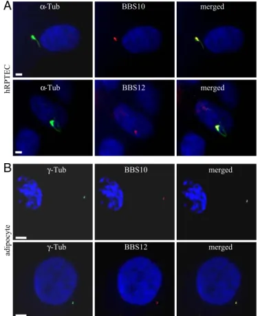
Cellular Localization of BBS10 and BBS12 Proteins.As BBS10 and BBS12 proteins have not yet been characterized, rabbit polyclonal antibodies were raised that detect in Western blots single bands of the expected size (data not shown). We used them to study the intracellular localization of the 2 endogenous proteins. The kidney tubular epithelial cells represent a well-defined and recognized model for ciliated cells (28, 29), and moreover, kidney dysfunction is another cardinal feature in BBS patients (30, 31). We tested the localization of BBS10 and BBS12 in human primary renal proximal tubular epithelial cells (hRPTEC). When these cells were grown to confluence, their cilium was readily immunolabeled using anti-acetylated␣-tubulin, with the basal body clearly seen at the base of the cilium (Fig. 1A). Immunodetection of BBS10 and BBS12 proteins showed that both proteins localized to the basal body of the primary cilium. Thus, these proteins appear to share the same localization as reported for BBS6 (32) that belongs to the same vertebrate-specific family of chaperonin-like proteins (27). This localization is thus more restricted than the localizations of the other BBS proteins belonging to the BBSome complex, which are
also found in the cilium (33, 34). In mature adipocytes derived from human white preadipocyte (HWP) [SI TextandFig. S1], BBS10 and BBS12 proteins were concentrated in 2 focal points colocalizing with the ␥-tubulin-labeled centrioles, as shown in the merged pictures (Fig. 1B).
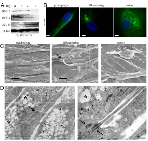
Ciliated Differentiating Preadipocytes. To analyze expression of BBS10 and BBS12 proteins during adipogenesis, we cultured the HWP to full confluence (D0) in preadipocyte growth medium, then switched to preadipocyte differentiation medium over 3 days, followed by a final change to adipocyte nutrition medium, leading to mature lipid accumulating adipocytes. Maximum expression levels of BBS10 and BBS12 proteins were detected in the first 2 days of adipogenic differentiation, followed by a net decrease as from day 4 to reach low basal expression levels (Fig. 2A). The glucose transporter4, GLUT4, was used as an adipogenic marker, expressed only in the mature adipocytes, to show that the BBS expression was progressively reduced with the proceeding of adipogenesis. This expression pattern appears thus similar to the previously described mRNA expression profile for the other BBS genes (25). The higher expression of ciliary proteins during adipogenesis prompted us to test for the presence of a cilium at 3 different stages: subconfluent dividing preadipocyte, confluent preadipocyte corresponding to maximum expression of BBS10 and BBS12, and mature adipocyte filled with lipid droplets (Fig. 2B). Interestingly, no cilium was detected in subconfluent preadipocytes and in mature adipocyte, but it was present in the confluent-differentiating preadipocyte. The same 3 cellular stages were examined by scanning electron microscopy, confirming the findings obtained by immunofluores-cence labeling (Fig. 2C). Transmission electron microscopy re-vealed the typical ultrastructural architecture of the primary cilium in the differentiating preadipocyte with the 2 centrioles, 1 at the base of cilium in the basal body (Fig. 2D), and the presence of lipid droplets. At higher magnification, the axoneme is seen emerging from the centriole in the basal body, which could be counted to 9⫹ 2 doublets on a transverse section of the cilium (SI Textand Fig. S2C).
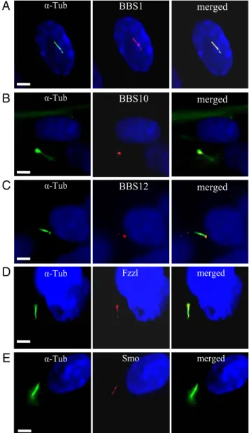
Characterization of the Primary Cilium During Adipogenesis.BBS1, one of the 2 most frequently mutated genes in patients, codes for a subunit of the recently described BBS protein complex, the BBSome, and localizes within the primary cilium (33). We inves-tigated whether BBS1 and the 2 chaperonin-like proteins studied herein share this same cellular localization in the differentiating preadipocyte. We costained the differentiating adipocyte for the cilium and for BBS1, BBS10, or BBS12. BBS1 was detected all along the adipocyte cilium (Fig. 3A) while BBS10 and BBS12 retained their cellular localization observed in unciliated fat cell, remaining associated to the centriole in the basal body of the primary cilium (Fig. 3 B and C). In the renal epithelium, the primary cilium harbors receptors for pathways like Wnt and Hh (35), whose signaling is dependent on the inherent intraflagellar transport (36). We there-fore tested if the differentiating preadipocyte primary cilium could be implicated in these pathways. We performed immunodetection of Wnt receptors using an antibody against a common epitope to the Fzzl receptors 1 to 10 (Fig. 3D). Specific Fzzl immunolabeling was found to localize in the cilium. In a similar way, we observed the presence on the cilium of Smoothened (Smo), a receptor involved in Hh signaling (Fig. 3E) as well as Patched1 (data not shown), suggesting the pivotal role of the cilium-associated signaling in the adipogenic program.
BBS10 and BBS12 Affect Ciliogenesis and Adipogenic Pathways.To investigate the implication of BBS10 and BBS12 proteins in the adipocyte transient ciliogenesis, we knocked down their expression using cellular Stealth RNAi delivery. Strong reduction of BBS10 and BBS12 protein contents were achieved in confluent D0 prea-dipocytes after treatment with the cognates RNAi (two different
B
A
BBS10 BBS12 α-Tub α-Tub merged merged hRPTEC γ-Tub γ-Tub BBS10 BBS12 adipoc yte merged mergedFig. 1. Localization of BBS10 and BBS12 proteins in renal epithelial cells and mature adipocytes. (A) Ciliated hRPTEC stained for cilium (acetylated ␣-tubu-lin,␣-Tub) and for BBS10 or BBS12. Primary cilium and basal body (Left). BBS10 and BBS12 proteins are detected adjacent to the nucleus (Middle) and local-ized at the basal body (Right, merged pictures). (B) Mature adipocytes stained for the centrioles with␥-tubulin (␥-Tub) and for either BBS10 or BBS12. BBS10 and BBS12 are centriolar proteins. (Scale bars, 5m.)
Marion et al. PNAS 兩 February 10, 2009 兩 vol. 106 兩 no. 6 兩 1821
CELL
RNAi for each gene gave similar results) (Fig. 4 A and B). Inhibition of BBS10 and BBS12 expression resulted in a significant reduction in the number of ciliated cells compared to control RNAi treated cells (Fig. 4 C and D). Because both BBS10 and BBS12 are chaperonine-like proteins and are both localized at the centriole, we wondered whether one would need the other for its centriolar localization. We performed immunofluorescence for BBS12 in BBS10-deprived cells and for BBS10 in BBS12-deprived cells (Fig. 4 E and F) and no difference in BBS10 localization was observed after BBS12 deprivation or vice versa.
We then tested whether BBS10 and BBS12 knock down affect key regulators of adipogenesis as GSK3,-catenin, and PPAR␥ based on an experimental procedure depicted in the scheme (SI Text and Fig. S3). Immunofluorescence detection of both the phosphorylated inactive and unphosphorylayed active forms of GSK3 isoform in the preadipocytes was carried out. Upon BBS10 and BBS12 inactivation, the unphosphorylated active form of GSK3 was increased compared to control transfected cells (Fig. 5A and B for ELISA quantification). Active GSK3 represses-catenin nuclear accumulation (24). Cytoplasmic and nuclear fractions were therefore isolated from confluent preadipocytes after BBS10 and BBS12 inactivation and loaded on gel for immunodetection. Higher nuclear-catenin protein content was detected in the control lane compared to the BBS10 and BBS12 knock-down lanes (Fig. 5C,
Right). Efficient separation between the nuclear and cytoplasmic
fractions was tested by Western blot using Histone H3 as a control marker (SI Text; see Fig. S1 E and F). Indeed, histone H3 was detected only in the nuclear fraction, demonstrating good separa-tion of the fracsepara-tions. Concomitant to the reducsepara-tion of nuclear -catenin, inhibition of BBS10 and BBS12 expression induced a higher nuclear accumulation of PPAR␥ in differentiating preadi-pocytes compared to control cells (Fig. 5D). This effect was confirmed by Western blot analysis (Fig. 5E). The increased level of PPAR␥ is accompanied by its redistribution in the nuclei,
appearing more homogenous than in cells that express normal levels of BBS10 and BBS12 proteins.
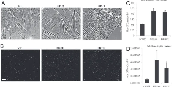
Adipocyte-Derived Fibroblasts from Patients Have a Higher Propensity to Triglyceride Accumulation.Dermal fibroblasts from BBS10 and BBS12 patients and healthy control were cultured in fibroblast growth medium. No difference in morphology was observed be-tween the BBS10- and BBS12-deficient cells compared to the wild-type control cells (Fig. 6A). At full confluence, the cells were differentiated into fat-accumulating cells as described in the SI
MethodsandFig. S4. After 14 days, the fat vacuoles were visualized
in the living cells (Fig. 6B). The intracellular triglyceride (37) levels were measured in control and BBS-deficient cells and the corre-sponding fluorescence levels were plotted (Fig. 6C). Significant increase in triglyceride content was observed in the BBS patients cells compared to the control cells. Concomitant to the increased level of triglycerides present in the cells with one BBS inactivated gene, there was a significantly higher leptin level secreted in the culture medium measured by ELISA (Fig. 6D).
Discussion
The present study demonstrates that a primary cilium is present on the confluent differentiating preadipocyte (SI Text, seeFig. S2 A and B). This ciliogenesis is spontaneous when full cellular conflu-ence is reached and doesn’t require serum withdrawal. In this respect, the HWP resembles the renal epithelial cells, which also exhibit spontaneous ciliogenesis. But at the difference of the renal monocilia, which is 9⫹ 0, the primary cilium carried by the human differentiating preadipocyte has a 9⫹ 2 (seeSI TextandFig. S2C) structure like the nonmotile kinocilium of hair cells in the cochlea (38, 39). Transient 9⫹ 2 cilia may act as developmental organelles at key points of cellular differentiation in the adipocyte and the kinocilium. The reported mouse models for BBS reproduce the human overweight condition (10). We verified that differen-tiating mouse preadipocytes (3T3-L1 cells) are also ciliated
B
C
D
β Tub D0*
*
*
preadipocyte differentiating mature
hum
an
10% SDS-PAGE
preadipocyte differentiating mature
BBS10
BBS12 0
A
Day 0 2 4 8GLUT4
Fig. 2. Up-regulation of BBS10 and BBS12 proteins and transient presence of primary cilium in early adi-pogenesis. (A) BBS10 and BBS12 protein levels during adipogenesis. HWP were cultured to D0 in preadipo-cyte growth medium. Differentiation was induced, and cellular extracts were immunoblotted at the indi-cated time points. BBS10 and BBS12 proteins are max-imally expressed in early differentiation. Glucose Transporter 4 (GLUT4, 45kDa) served as adipogenic marker and-tubulin served as loading control. (B) (Left to Right) Proliferating preadipocytes, confluent differentiating preadipocytes, mature adipocytes stained for cilium. Primary cilium is exclusively ob-served in differentiating preadipocyte. (Scale bars, 5 m.) (C) Representative scanning electron microscopic pictures of the same 3 stages. Cilium is clearly observ-able on the differentiating preadipocyte (Middle,
ar-row) and absent in preadipocyte and mature
adipo-cyte. (Scale bars, 10 m.) (D) Electron microscopic pictures showing cilium with the centriole at the base (arrow), axonemes inside the cilium. Asterix indicates the intracellular lipid droplets. Right panel shows the two centrioles (arrows). One centriole recruited at the basal body to support the axonemes. (Scale bar, 1m.)
(seeSI TextandFig. S2D), indicating that this is a common feature in adipogenesis between these 2 species.
The transient ciliated status is accompanied by an increased expression of the 2 recently identified BBS genes BBS10 and BBS12, consistent with the previous findings that other BBS genes are also up-regulated during early adipogenesis (25). The chaperonine-like BBS10 and BBS12 show centriolar/basal body localization, whereas the other BBS protein, which forms part of the BBSome, have also been detected all along the cilium as exemplified by the BBS1 detection (see Fig. 3). One can therefore think that BBS10 and BBS12 are required, based on their predicted function of chapero-nine, in assisting the formation of ciliary components and are not directly involved in intragellar transport. Both BBS10 and BBS12 appear essential for proper ciliogenesis during adipogenesis be-cause their absence inhibits ciliogenesis, as shown in Fig. 4. How-ever, they don’t require each other to localize to the centrioles, as no effect was detected on their respective localization following knockdown of the other one (Fig. 4 E and F). Alstro¨m syndrome,
another syndromic obesity condition recently classified as a cili-opathy, is caused by mutations in the ALMS1 gene coding for another ciliary protein. ALMS1 was recently shown to be regulated during adipogenesis (40), further illustrating the prominent link between ciliary proteins and adipogenesis.
The primary cilium carries Wnt and Hh receptors, which are ancient signaling pathways and important well-recognized regula-tors of adipogenesis. Upon inhibition of BBS10 and BBS12 protein expression, the differentiating preadipocyte loses the cilium that carries Wnt and Hh receptors. This is likely the cause of the observed increase of the active (unphosphorylated) form of GSK3, associated with a decrease in the nuclear content of -catenin. GSK3-mediated balance between -catenin and PPAR␥ has been previously documented and the inactivation of BBS12 BBS10 merged merged BBS1 merged Fzzl α-Tub merged Smo α-Tub merged α-Tub α-Tub α-Tub
A
B
C
D
E
Fig. 3. Adipocyte cilium-associated proteins. Differentiating HWP were immunostained for cilium and for BBS1, BBS10, BBS12, Fzzl1–10, and Smo. Left panels show the cilium, middle panels show the protein localization and right panels the merged pictures. (A) BBS1 is detected throughout the cilium. BBS10 and BBS12 are localized at the basal body (B) and (C), respectively. The Fzzl protein (D) and Smo (E) are also associated to the cilium. (Scale bars, 5m.)
A
1 2 3 4 1 2 3 4 BBS10 β Tub β Tub BBS12B
* * % of c ilia ted cells 1 2 3D
control BBS10 RNAi BBS12 RNAi
C
BBS10 RNAi BBS12 RNAi γ-Tub BBS12 merged γ-Tub BBS10 mergedE
F
100 50 0Fig. 4. Impaired cilium formation during adipogenesis upon BBS knock down. (A) Ninety percent confluent preadipocytes were transfected and cultured for 4 days in preadipocytes growth medium. Four days later, immunoblot analysis for BBS10 was done on control transfected RNAi duplexes (medium and low Nega-tive Universal Control Stealth RNAi) (lanes 1 and 2, respecNega-tively) and on BBS10 RNAi knockdown confluent preadipocyte cells (BBS10 duplexes 1 and 2) (SI
Methods, and lanes 3 and 4, respectively). Significant reduction in BBS10 protein
level was obtained. Anti--Tubulin was used as loading control. (B) BBS12 was inactivated in a similar way and cell lyzates analyzed by immunoblotting (lanes 1
and 2 were loaded with cellular extracts from medium and low Negative
Uni-versal Control Stealth RNAi transfected cells and lanes 3 and 4 corresponded to those from cells transfected with BBS12 duplexes 2 and 3). A significant reduction in BBS12 level was also observed. (C) BBS10 or BBS12 silencing to prevent cilio-genesis. Four days after RNAi-transfection, cilia presence was assessed. Represen-tative pictures of BBS10, BBS12, and control RNAi transfected cells are shown. Arrows indicate the primary cilium. (Scale bar, 40m.) (D) The percentage of ciliated cells: that is, (number of cilia/total number of nuclei)⫻ 100% was calculated and plotted. n⫽ 550 for each of 3 independent experiments. Signif-icant reduction in ciliated cells was obtained (control compared to BBS10: P⫽ 0.001 and control compared to BBS12: P⫽ 0.003). (E) Immunolocalization of BBS12 in BBS10-deprived preadipocytes showed that centriolar localization of BBS12 protein was unaffected in the absence of the BBS10. (Scale bar, 5m.) (F) In a similar way, BBS10 centriolar localization was also unaffected in BBS12-deprived preadipoctes. (Scale bar, 5m.)
Marion et al. PNAS 兩 February 10, 2009 兩 vol. 106 兩 no. 6 兩 1823
CELL
-catenin could favor the up-regulation of PPAR␥ (17) that we observed. Surprisingly, PPAR␥ was readily detected both by im-munofluorescence and by Western blot only 24 h after adipogenic induction mediated by medium change in these human differenti-ating preadipocytes, which seems to be in contradiction to previous reports showing the presence of PPAR␥ only 48 h after induction of adipogenesis (15, 41, 42). The expression of PPAR␥ has been extensively studied in murine models, especially the 3T3-L1 prea-dipocytes, which require mitotic clonal expansion before they can start expressing the genes producing the adipocyte phenotype including PPAR␥ (43). A major difference exists between the widely studied 3T3-L1 preadipocyte model and the primary human preadipocyte used in the present study, as human preadipocytes can proceed through terminal differentiation without postconfluence
mitosis (44), which may account for the earlier expression of PPAR␥ after adipogenic induction. To ascertain the proadi-pogenic effect of BBS10 and BBS12 deprivation, we knocked down BBS10 and BBS12 in conf luent preadipocytes and kept culturing them in the preadipocyte growth medium, a specially formulated medium to prevent adipogenesis, for 48 h before analyzing PPAR␥ expression (see SI Text and Fig. S2F). PPAR␥ was readily detected in the extract from the BBS10-and BBS12-deprived preadipocytes BBS10-and remained almost un-detectable in the control lysate. This proves that the absence of the cilium following BBS10 or BBS12 inactivation is suffi-cient to induce adipogenesis, probably by disrupting Wnt signaling, which could not be detected after the BBS knock down (data not shown).
E
C
B
A
GS K3-P GS K3 me rg edcontrol BBS10 RNAi BBS12 RNAi
D
1 2 3 β Tub PPARγ Cyto. nuclear 1 2 3 1 2 3 GS K3 β (ng/m L ) * *control BBS10 RNAi BBS12 RNAi
1 2 3
Fig. 5. Adipogenesis is favored in BBS-deprived adipocytes. (A) Representative immunofluorescence pictures for the de-tection of the inactive phosphorylated GSK3 (GSK3-P) and the active unphos-phorylated GSK3 in the differentiating preadipocytes following the indicated RNAi treatment. An increase in unphos-phorlated GSK3 was observed after inac-tivation of either BBS10 or BBS12. (Scale bars, 5m.) (B) Quantification of the active GSK3 isoform by ELISA. Increases of 20 and 35% in active GSK3 were obtained after BBS10 (lane 2) or BBS12 (lane 3) knock down compared to control RNAi (lane 1). (n⫽ 3; control compared to BBS10: P ⫽ 0.007 and control compared to BBS12: P⫽ 0.013). (C) Immunodetection of cytoplasmic and nuclear-catenin in confluent preadi-pocytes after 24 h transfection with con-trol, BBS10, and BBS12 RNAi.-catenin
pro-tein was detected at 94kDa. 1, control RNAi transfected; 2, BBS10 RNAi; 3, BBS12 RNAi. Homogenous propro-tein loading was verified (SI TextandFig. S1D) (D) Immunofluorescence for PPAR␥ in transfected preadipocyte after 24 h in differentiating medium. Increased PPAR␥ content was detected in BBS10- and BBS12 RNAi-treated cells compared to controls. (E) A band at 57 kDa was detected by Western blot. 1, control RNAi transfected; 2, BBS10 RNAi; 3, BBS12 RNAi. The Western blot results confirmed the increased PPAR␥ expression in the adipocytes following the indicated BBS knock down.
BBS10 BBS12 WT BBS10 BBS12 WT
A
B
C
D
Fig. 6. Increased fat content in adipocytes derived from BBS patients’ fibroblasts. (A) Representative pictures of human dermal fibroblasts with the indicated gene mutation cultured in FGM (SI Text,Methods) to full confluence before adipogenic induction. (Scale bar, 20m.) (B) Triglyceride accumulation after 14 days of culture in FDAM was stained with Adipored Assay Reagent. Increased fluorescent-labeled cells were observable in the BBS10- and BBS12-mutated cells (Middle and Right, respectively) compared to control wild-type cells (Left). (Scale bar, 60m.) (C) The lipid-derived fluorescence was measured at 572 nm for each condition (CONT for the wild-type healthy fibroblasts, BBS10-, and BBS12-mutated fibroblasts) and was expressed as total measured fluorescence per cell. (n⫽ 3) (control compared to BBS10:
P⫽ 0.001 and control compared to BBS12: P ⬍ 0.001). (D) Secreted leptin was measured in the culture medium by ELISA and was expressed as the total absorbance at 450 nm per cell. In parallel with the increased intracellular triglyceride content, higher quantities of leptin was detected in the medium from the BBS mutated cells compared to the healthy control (n⫽ 3) (control compared to BBS10: P ⫽ 0.05 and control compared to BBS12: P ⫽ 0.03).
Although stable gene knockdowns are useful, human fibroblasts from patients with characterized mutations often represent an invaluable tool to investigate the related diseases. Here, we were able to culture and reprogram dermal fibroblasts from two BBS patients to lipid-accumulating cells expressing PPAR␥ and GLUT4
(seeSI Text,Methods, andFig. S4). Interestingly, during the culture
of these cells, no difference was observed in cell growth or in structural cellular aspects (data not shown), although it has been described that BBS6, the other chaperonine-like protein of the BBS family, blocked cytokinesis yielding abnormal polynucleated cells (32). BBS6 protein may therefore possess a specific role in cell division, which both BBS10 and BBS12 don’t share. Based on the effect observed on PPAR␥ expression after specific BBS knock-down in the differentiating preadipocytes, intracellular triglyceride content and secreted leptin levels were measured, and significant increase in intracellular triglyceride contents was observed in the BBS mutated cells compared to the control cells (see Fig. 6 B–D). It is well recognized that leptin secretion is directly correlated to adiposity. An increase in leptin secretion was measured in the culture medium from the BBS mutated cells compared to the control cells, reproducing the human-observed phenotype of high-circulating leptin concentrations.
The pathophysiological pathways leading to obesity in the BBS patients have been suggested to be related to central nervous system control of body weight. BBS proteins are required for proper ciliary localization in neurons of the Mchr1 receptor, involved in regula-tion of feeding behavior (12). Hypothalamic ciliated neurons may be involved in the leptin resistance recently described in BBS patients and obese BBS knockout mouse models (10, 45). BBS gene inactivation in these neurons has been shown to reduce Pomc expression, a gene activated by leptin via Stat3 (10). Nonetheless,
this reduction cannot solely explain the extreme increase in body weight because the mice with specific inactivation of Stat3 and subsequent decreased Pomc expression exhibit a mild increase in total body weight and only a 2-fold increase in fat pad mass (46). The results presented in this article point out that the BBS-associated obesity may have a dual origin: the suggested central nervous system origin (10, 11) combined with a peripheral origin by increased adipogenesis via inhibition of Wnt signaling.
Lipodystrophy syndromes are to date the only syndromes related to primary abnormalities in adipogenesis. Obesity related to im-paired adipogenesis has, so far, not been reported, and it is believed that body mass increase is not directly related to the adipocyte differentiation. Very recently however, Cao et al. (47) presented evidence for a lipid-mediated endocrine network where the adipose tissue itself uses lipokines to communicate with other organs and regulate systemic metabolic homeostasis. We therefore suggest that adipogenesis itself may participate to the pathogenesis of obesity. This hypothesis warrants further investigations in vivo. To address this question and to dissect the associated molecular parameters, conditional knockout mice will be used to target specific BBS inactivation in the adipose tissue, which will allow us to understand the role of enhanced adipogenesis in the BBS-induced obesity.
Materials and Methods
For details, please seeSI Methods.
ACKNOWLEDGMENTS. We thank Rinus Verdonschot for his technical support
and Gilles Duval (Institut de Ge´ne´tique et de Biologie Mole´culaire et Cellulaire) for generating the rabbit polyclonal antibodies. This work was funded by the Agence Nationale pour la Recherche Call for rare disorders 2006, Retina FRANCE, and by the Institut National pour la Sante´ et le Recherche Me´dicale with the AVENIR program 2007.
1. Tobin JL, Beales PL (2007) Bardet-Biedl syndrome: beyond the cilium. Pediatr Nephrol 22:926 –936.
2. Blacque OE, Leroux MR (2006) Bardet-Biedl syndrome: an emerging pathomechanism of intracellular transport. Cell Mol Life Sci 63:2145–2161.
3. Benzinou M, et al. (2006) Bardet-Biedl syndrome gene variants are associated with both childhood and adult common obesity in French Caucasians. Diabetes 55:2876 –2882. 4. Saxena R, et al. (2007) Genome-wide association analysis identifies loci for type 2
diabetes and triglyceride levels. Science 316:1331–1336.
5. Marshall WF, Nonaka S (2006) Cilia: tuning in to the cell’s antenna. Curr Biol 16:R604 –R614. 6. Afzelius BA (2004) Cilia-related diseases. J Pathol 204:470 – 477.
7. Marshall WF (2008) The cell biological basis of ciliary disease. J Cell Biol 180:17–21. 8. Goldstone AP, Beales PL (2008) Genetic obesity syndromes. Front Horm Res 36:37– 60. 9. Katsanis N, Lupski JR, Beales PL (2001) Exploring the molecular basis of Bardet-Biedl
syndrome. Hum Mol Genet 10:2293–2299.
10. Rahmouni K, et al. (2008) Leptin resistance contributes to obesity and hypertension in mouse models of Bardet-Biedl syndrome. J Clin Invest 118:1458 –1467.
11. Mak HY, Nelson LS, Basson M, Johnson CD, Ruvkun G (2006) Polygenic control of
Caenorhabditis elegans fat storage. Nat Genet 38:363–368.
12. Berbari NF, Lewis JS, Bishop GA, Askwith CC, Mykytyn K (2008) Bardet-Biedl syndrome proteins are required for the localization of G protein-coupled receptors to primary cilia. Proc Natl Acad Sci USA 105:4242– 4246.
13. Alieva IB, Vorobjev IA (2004) Vertebrate primary cilia: a sensory part of centrosomal complex in tissue cells, but a ‘‘sleeping beauty’’ in cultured cells? Cell Biol Int 28:139 –150. 14. Rosen ED, Walkey CJ, Puigserver P, Spiegelman BM (2000) Transcriptional regulation of
adipogenesis. Genes Dev 14:1293–1307.
15. Bennett CN, et al. (2002) Regulation of Wnt signaling during adipogenesis. J Biol Chem 277:30998 –31004.
16. Cousin W, Fontaine C, Dani C, Peraldi P (2007) Hedgehog and adipogenesis: fat and fiction. Biochimie 89:1447–1453.
17. Liu J, Farmer SR (2004) Regulating the balance between peroxisome proliferator-activated receptor gamma and beta-catenin signaling during adipogenesis. A glyco-gen synthase kinase 3beta phosphorylation-defective mutant of beta-catenin inhibits expression of a subset of adipogenic genes. J Biol Chem 279:45020 – 45027. 18. Rosen ED, et al. (2002) C/EBPalpha induces adipogenesis through PPARgamma: a
unified pathway. Genes Dev 16:22–26.
19. Ross SE, et al. (2000) Inhibition of adipogenesis by Wnt signaling. Science 289:950 –953. 20. Cousin W, Dani C, Peraldi P (2006) Inhibition of the anti-adipogenic Hedgehog signal-ing pathway by cyclopamine does not trigger adipocyte differentiation. Biochem
Biophys Res Commun 349:799 – 803.
21. Suh JM, et al. (2006) Hedgehog signaling plays a conserved role in inhibiting fat formation. Cell Metab 3:25–34.
22. Zaragosi LE, et al. (2008) Effects of GSK3 inhibitors on in vitro expansion and differ-entiation of human adipose-derived stem cells into adipocytes. BMC Cell Biol 9:11. 23. Longo KA, et al. (2004) Wnt10b inhibits development of white and brown adipose
tissues. J Biol Chem 279:35503–35509.
24. Cohen P, Frame S (2001) The renaissance of GSK3. Nat Rev Mol Cell Biol 2:769 –776. 25. Forti E, Aksanov O, Birk RZ (2007) Temporal expression pattern of Bardet-Biedl
syn-drome genes in adipogenesis. Int J Biochem Cell Biol 39:1055–1062.
26. Stoetzel C, et al. (2006) BBS10 encodes a vertebrate-specific chaperonin-like protein and is a major BBS locus. Nat Genet 38:521–524.
27. Stoetzel C, et al. (2007) Identification of a novel BBS gene (BBS12) highlights the major role of a vertebrate-specific branch of chaperonin-related proteins in Bardet-Biedl syndrome. Am J Hum Genet 80:1–11.
28. Hildebrandt F, Zhou W (2007) Nephronophthisis-associated ciliopathies. J Am Soc
Nephrol 18:1855–1871.
29. Benzing T, Walz G (2006) Cilium-generated signaling: a cellular GPS? Curr Opin
Nephrol Hypertens 15:245–249.
30. O’Dea D, et al. (1996) The importance of renal impairment in the natural history of Bardet-Biedl syndrome. Am J Kidney Dis 27:776 –783.
31. Green JS, et al. (1989) The cardinal manifestations of Bardet-Biedl syndrome, a form of Laurence-Moon-Biedl syndrome. N Engl J Med 321:1002–1009.
32. Kim JC, et al. (2005) MKKS/BBS6, a divergent chaperonin-like protein linked to the obesity disorder Bardet-Biedl syndrome, is a novel centrosomal component required for cytokinesis. J Cell Sci 118:1007–1020.
33. Nachury MV, et al. (2007) A core complex of BBS proteins cooperates with the GTPase Rab8 to promote ciliary membrane biogenesis. Cell 129:1201–1213.
34. Blacque OE, et al. (2004) Loss of C. elegans BBS-7 and BBS-8 protein function results in cilia defects and compromised intraflagellar transport. Genes Dev 18:1630 –1642. 35. Satir P, Christensen ST (2007) Overview of structure and function of mammalian cilia.
Annu Rev Physiol 69:377– 400.
36. Huangfu D, et al. (2003) Hedgehog signalling in the mouse requires intraflagellar transport proteins. Nature 426:83– 87.
37. Schermer B, et al. (2005) Phosphorylation by casein kinase 2 induces PACS-1 binding of nephrocystin and targeting to cilia. EMBO J 24:4415– 4424.
38. Sobkowicz HM, Slapnick SM, August BK (1995) The kinocilium of auditory hair cells and evidence for its morphogenetic role during the regeneration of stereocilia and cutic-ular plates. J Neurocytol 24:633– 653.
39. Fliegauf M, Benzing T, Omran H (2007) When cilia go bad: cilia defects and ciliopathies.
Nat Rev Mol Cell Biol 8:880 – 893.
40. Romano S, et al. (2008) Regulation of Alstrom syndrome gene expression during adipo-genesis and its relationship with fat cell insulin sensitivity. Int J Mol Med 21:731–736. 41. Park BH, Qiang L, Farmer SR (2004) Phosphorylation of C/EBPbeta at a consensus
extracellular signal-regulated kinase/glycogen synthase kinase 3 site is required for the induction of adiponectin gene expression during the differentiation of mouse fibro-blasts into adipocytes. Mol Cell Biol 24:8671– 8680.
42. Darlington GJ, Ross SE, MacDougald OA (1998) The role of C/EBP genes in adipocyte differentiation. J Biol Chem 273:30057–30060.
43. Tang QQ, Otto TC, Lane MD (2003) Mitotic clonal expansion: a synchronous process required for adipogenesis. Proc Natl Acad Sci USA 100:44 – 49.
44. Rosen ED, MacDougald OA (2006) Adipocyte differentiation from the inside out. Nat
Rev Mol Cell Biol 7:885– 896.
45. Fath MA, et al. (2005) Mkks-null mice have a phenotype resembling Bardet-Biedl syndrome. Hum Mol Genet 14:1109 –1118.
46. Xu AW, Ste-Marie L, Kaelin CB, Barsh GS (2007) Inactivation of signal transducer and activator of transcription 3 in proopiomelanocortin (Pomc) neurons causes decreased pomc expression, mild obesity, and defects in compensatory refeeding. Endocrinology 148:72– 80.
47. Cao H, et al. (2008) Identification of a lipokine, a lipid hormone linking adipose tissue to systemic metabolism. Cell 134:933–944.
Marion et al. PNAS 兩 February 10, 2009 兩 vol. 106 兩 no. 6 兩 1825
CELL