HAL Id: hal-02437442
https://hal.archives-ouvertes.fr/hal-02437442
Submitted on 16 Jan 2020
HAL is a multi-disciplinary open access
archive for the deposit and dissemination of
sci-entific research documents, whether they are
pub-lished or not. The documents may come from
teaching and research institutions in France or
abroad, or from public or private research centers.
L’archive ouverte pluridisciplinaire HAL, est
destinée au dépôt et à la diffusion de documents
scientifiques de niveau recherche, publiés ou non,
émanant des établissements d’enseignement et de
recherche français ou étrangers, des laboratoires
publics ou privés.
Realization and characterization of diamond
micro-transducers for bio-chemical sensing
Alexandre Bongrain, Emmanuel Scorsone, Lionel Rousseau, Gaelle Lissorgues,
Samuel Saada, Philippe Bergonzo
To cite this version:
Alexandre Bongrain, Emmanuel Scorsone, Lionel Rousseau, Gaelle Lissorgues, Samuel Saada, et al..
Realization and characterization of diamond micro-transducers for bio-chemical sensing. Eurosensors
XXIII, Ecole Polytechnique Federale de Lausanne, Sep 2009, Lausanne, Switzerland. pp.754-757,
�10.1016/j.proche.2009.07.188�. �hal-02437442�
Procedia
Chemistry
www.elsevier.com/locate/procediaProceedings of the Eurosensors XXIII conference
Realization and characterization of diamond micro-transducers
for bio-chemical sensing
Alexandre bongrain
1, Emmanuel Scorsone
1, Lionel Rousseau
2, Gaëlle Lissorgues
2,
Samuel Saada
1, Philippe Bergonzo
1*
1CEA, LIST, Diamond Sensor Laboratory, CEA/Saclay, 91191 Gif sur Yvette, France. 2 University Paris Est, ESYCOM, ESIEE Cité Descartes, BP99, 93162 Noisy Le Grand, France
Abstract
We report on novel MEMS micro-transducers made of diamond and used for bio-sensing applications. To overcome the non straightforward micro-machining of diamond, we developed an original process involving the patterned growth of diamond using the CVD (chemical vapour deposition) technique, inside micro-machined silicon moulds.
Typical MEMS structures were successfully fabricated and include cantilevers and bridges. They were actuated using Laplace forces. The structures were characterized by measuring their first mode resonance (frequency and Q-factor) using laser interferometry. The measured data fitted the simulated data and comparison with equivalent silicon structures showed the superior resonant properties of diamond cantilevers. It implies that the later are potentially more sensitive transducers than their silicon counterpart.
Keyword: Poly-crystalline diamond, diamond micro-transducer, bio-chemical sensing
1. Introduction
In recent years, a great attention has been paid to the development of accurate mass-sensitive sensors for chemicals or biological compounds and the feasibility to detect specific gases, enzymes, proteins or DNA sequences using micro cantilevers has been demonstrated. [ref 1].
Over the last decade, silicon was used extensively for MEMS fabrication by taking advantage of the batch silicon-micromachining techniques developed for integrated circuits technology. For instance silicon was used for accelerometers, pressure sensors, resonators, or chemical/bio-chemical sensors [ref 2]. For emerging applications such as bio-sensing, diamond appears to be a promising alternative material for MEMS. Indeed, diamond features outstanding mechanical properties [ref 3] that include a high Young modulus. It implies that diamond MEMS will exhibit superior sensitivities where sensing is based on resonating structures. Diamond also features a high fractural strength which is linked to a high fabrication yield. Finally, the carbon terminated surface of diamond offers a wide range of possibilities for covalent grafting of specific bio-receptors on its surface [ref 4]. Hence the sensitive receptors can be strongly attached on diamond resonator sensor without deteriorating the mechanical properties as it
* Alexandre Bongrain. Tel.:+33 (0) 1 69 08 44 22; fax:.+33 (0) 1 69 08 76 79 E-mail address:Alexandre.bongrain@cea.fr.
1876-6196/09 ©2009Published by Elsevier B.V. doi:10.1016/j.proche.2009.07.188
Procedia Chemistry 1 (2009) 754–757
is often the case when an intermediate gold layer is deposited onto the transducer typically to allow grafting of thiol terminated sensitive receptors.
2. Experimental set up
2.1. Diamond MEMS fabrication
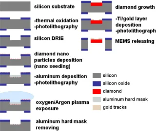
MEMS structures were fabricated using a process reported in previous work [ref 5]. This process involves the direct growth of diamond micro - structures inside silicon
moulds (Fig.1). Firstly a thermally oxidized 4-inch silicon wafer was selectively etched by DRIE in order to accurately pattern moulds in the substrate, using the silicon oxide layer as a hard mask. Then the substrate was seeded with diamond nanoparticles using a PVA/nanoparticle solution as described in [ref 6]. The technique enables the formation of a uniform density of diamond particles over the substrate in spite of the 3D aspect [ref 6]. Then we deposited an aluminium hard mask inside the silicon moulds to selectively etch the diamond particles layer outside the silicon moulds under a oxygen/argon plasma. After removal of the aluminium hard mask, diamond was grown inside the moulds using MPCVD process. Finally, gold tracks and strain gauges were deposited before releasing MEMS transducer by etching the substrate back side by DRIE.
Fig 1. Fabrication process of diamond MEMS 2.2. Diamond MEMS characterisation
The transducers were observed at the end of the fabrication process using a Field Emission Gun Scanning Electron Microscope. Then their resonant frequencies and Q-factor were measured using laser interferometry with a coherent laser source emitting in the range 620 to 690 nm and its associated interferometer (OFV511), a demodulator (OFV3001) and a spectrum analyser (Agilent 89410A). Structure actuation was achieved using Laplace forces. In this way, an alternative current, supplied by the spectrum analyser is injected through the gold tracks deposited along the structures edges while a permanent magnet is placed near the edge of the transducer in order to apply an alternative force normal to the structure plane. Diamond film thickness was accuratel measured by interferometry. In parallel, the structures’ resonant behaviour was simulated by finite elements method on Coventor simulation software using as input data the diamond thickness measured by interferometry and other physical parameters of our CVD poly-crystalline diamond, such as Young modulus, [ref 5].
3. Results and discussion
Examples of diamond micro structures are presented in Fig 2, where it is seen that the structures are well defined. It is therefore concluded that diamond growth was efficiently directed by the silicon moulds. Moreover, no structure bending or twisting is visible which suggests low strain in the diamond material. The measurements presented here were carried out on cantilevers having a thickness of 1.7 µ m as measured by interferometry, and length varying from 250 to 2000 µm.
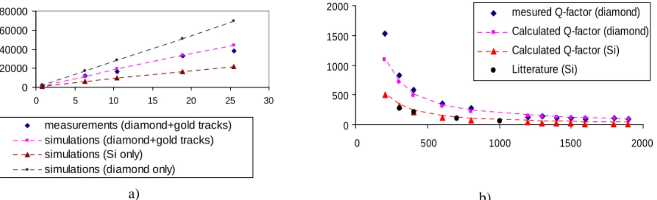
The first mode resonance frequencies of the cantilevers were measured and plotted against the ratio t/L², where t is the diamond film thickness and L the beam length (Fig 3.a). A linear relationship was obtained as expected as well as a good correlation between measured and simulated data when taking into account the integrated tracks. Hence,
755
a) b) c) d) Fig 2. SEM images of fabricated diamond structures
using the same model a similar plot was done for bare diamond and silicon cantilevers having the same dimensions as the cantilevers with embedded gold tracks. From the slopes of the lines, we extracted the value of the equivalent Young modulus of our diamond cantilevers which is equal to 756 GPa when taking into account the gold tracks. From this value we also worked out the corresponding Young modulus of the diamond used for fabricating the structures and found 986 GPa which corresponds to the typical values reported previously for poly-crystalline diamond [ref 7]. The presence of gold tracks on the structures significantly decreases the equivalent Young modulus of our resonators, and consequently their resonance frequency. Indeed the gold tracks thickness is not negligible when compared to the thickness of the poly-crystalline diamond (0.5 µm and 1.7 µ m, respectively) and both mass loading and Young modulus decrease contribute to lower the resonance frequency. Nevertheless the measured resonance frequency of the diamonds structures remains higher than simulated data for silicon.
Then the mass sensitivity of two of our diamond micro cantilevers was assessed using the measurement of their first mode resonant frequency and equation (1). The same calculation was done for equivalent modeled silicon cantilevers. The results are summarized in Table 1 and show that poly-crystalline diamond cantilevers exhibits sensitivity that is typically twice higher than the equivalent silicon structures.
oi i
f
t
L
W
f
m
S
6
4χ
ρ
−
=
∆
∆
=
(1)Finally, we measured the Q-factor of diamond cantilevers for the first mode resonance frequency in air and plotted the results against the length of the structures. In air, two components of the Q-factor can be considered: the effect of air damping and the effect of anchoring to the substrate. In the present case the beams are long enough to neglect the effect of the beams support on the Q-factor. Indeed Fig.3.b shows that the Q-factor decreases when the length of cantilevers increases. However for the shortest beams no decrease is visible as it is the case when anchoring affects the resonance properties of such structures. Hence we could model the Q-factor only by considering the air damping contribution using vibrating sphere model in viscous fluids. This model was used previously for silicon cantilevers to determine the influence of cantilevers dimension on the Q-factor [ref 8]. The expression of the Q-factor is given by (2)
+
=
δ
µ
π
ρ
χ
R
R
L
t
W
E
Q
i1
.
3
12
2 2 (2)This model fitted our measured data correctly. Then we also used this model for Q-factor values of silicon cantilevers having the same dimensions as our diamond cantilevers. Again this model fitted correctly some measured data found in the literature [ref 8, 9] (Fig 3.b.). This figure shows that the Q-factor of the silicon structures is approximately twice less than for their poly-crystalline diamond counterparts. Since diamond cantilevers exhibit a better Q-factor than the silicon ones diamond appears as a material of choice for decreasing the detection limit of such resonant transducers. We evaluated the mass resolution of two diamond cantilevers having a fixed width but different lengths using formula (3).
=
∆
2 max minZ
Q
f
TB
k
k
S
m
oi B eq (3)A. Bongrain et al. / Procedia Chemistry 1 (2009) 754–757
where B is the bandwidth of the complete system (i.e. transducer and electronics). For this calculation, we chose a typical value of B=1000 Hz and T=300 K. The results summarized in Table 1 show a potential lower limit of detection for diamond cantilevers. Moreover, both estimated sensitivity and limit of detection of our diamond cantilevers is adequate to detects 30-bases DNA molecules grafted on our transducers surface with a density per surface unit superior to 1011 cm-2 using method [ref 4].
0 20000 40000 60000 80000 0 5 10 15 20 25 30
measurements (diamond+gold tracks) simulations (diamond+gold tracks) simulations (Si only)
simulations (diamond only)
a) 0 500 1000 1500 2000 0 500 1000 1500 2000
mesured Q-factor (diamond) Calculated Q-factor (diamond) Calculated Q-factor (Si) Litterature (Si)
b)
Fig 3. Measured resonance frequency (3.a) and Q-factor (3.b) of fabricated diamond cantilevers and calculated first mode resonant frequency (3.a) and Q-factor (3.b) of both modeled diamond and silicon cantilevers
Table 1: Calculated sensitivity and mass-resolution from two characterized diamond and modeled silicon cantilevers
Poly-crystalline Diamond Silicon
Length (µm) Sensitivity (pg/Hz) Estimed mass resolution (pg) Sensitivity (pg/Hz) Estimed mass resolution (pg)
250 5.66 0.32 12.81 0.85
400 20.68 0.45 34.63 0.91
4. Conclusion
We introduced in this study poly-crystalline diamond micro-mechanical transducers prepared for bio-chemical sensing. Structures were fabricated using a process that involves the direct growth of nano-particles of diamond in silicon moulds. We characterized the fabricated structures by measuring their first mode resonant frequency and Q-factor and fit the data with simulations and analytical model, respectively. Comparing with equivalent silicon structures demonstrated the superiority of diamond for mass sensing applications. In future work those diamond based transducers will be used for developing biosensors. The diamond surfaces of the structures will be exploited for chemical or electrochemical covalent grafting of bioreceptors, using for instance diazonium salts as linker molecule to attach enzymes or DNA onto the transducers surfaces.
References
1. F. M. Battiston, J.-P. Ramseyer, H. P. Lang, M. K. Baller, Ch. Gerber,. Cantilever transducers as a platform for chemical and biological sensors. Review of scientific instrument 2004;Vol 75:Num 7.
2. W. Zhang, K. L. Turner. Application of parametric resonance amplification in a single-crystal silicon micro-oscillator based mass sensor, Sensors and actuators, 2005, 122, 23-30.
3. Y. Gurbuz, O. Esame, I. Tekin, W. P. Kang, J. L. Davidson, Diamond semiconductor technology for RF device applications, Solid-State Electronics, 49 (2005), 1055-1070.
4. C. E. Nebel, B. Rezek, D. Shin, H. Uetsuka, N. Yang, Diamond for bio-sensor applications, J. Phys. D: Appl. Phys 2007, 40, 6443-6466. 5. A. Bongrain, E. Scorsone, L. Rousseau, G. Lissorgues, C. Gesset, S. Saada, P. Bergonzo, Selective nucleation in silicon moulds for diamond MEMS fabrication, J. Micromech. Microeng 2009, 19.
6 E. Scorsone, S. Saada, J.C. Arnault, P. Bergonzo, J. Appl. Phys. in press DOI: 10.1063/1.3153118
7. N. Sepúlveda, J. Lu, D. M. Aslam, J. P. Sullivan: High-Performance Polycrystalline Diamond Micro- and Nanoresonators, J of microelectromechanical systems, 2008, 17, 473-482.
8. T. Ikehara, J. Lu, M. Konno, R. Maeda, T. Mihara, A high quality-factor silicon cantilever for a low detection-limit resonant mass sensor operating in air, J. Micromech. Microeng. 2007, 2491-2494.
9. K. Naeli, O. Brand, Dimensional considerations in achieving large quality factors for resonant silicon cantilevers in air, JAP 2009, 105, 014908.
757