HAL Id: hal-00330257
https://hal.archives-ouvertes.fr/hal-00330257
Submitted on 7 Aug 2007
HAL is a multi-disciplinary open access
archive for the deposit and dissemination of sci-entific research documents, whether they are pub-lished or not. The documents may come from teaching and research institutions in France or abroad, or from public or private research centers.
L’archive ouverte pluridisciplinaire HAL, est destinée au dépôt et à la diffusion de documents scientifiques de niveau recherche, publiés ou non, émanant des établissements d’enseignement et de recherche français ou étrangers, des laboratoires publics ou privés.
Picoplankton diversity in the South-East Pacific Ocean
from cultures
F. Le Gall, F. Rigaut-Jalabert, D. Marie, L. Garczarek, M. Viprey, A. Gobet,
D. Vaulot
To cite this version:
F. Le Gall, F. Rigaut-Jalabert, D. Marie, L. Garczarek, M. Viprey, et al.. Picoplankton diversity in the South-East Pacific Ocean from cultures. Biogeosciences Discussions, European Geosciences Union, 2007, 4 (4), pp.2699-2732. �hal-00330257�
BGD
4, 2699–2732, 2007 Picoplankton diversity in the South-East Pacific Ocean F. Le Gall et al. Title Page Abstract Introduction Conclusions References Tables Figures ◭ ◮ ◭ ◮ Back CloseFull Screen / Esc
Printer-friendly Version
Interactive Discussion
Biogeosciences Discuss., 4, 2699–2732, 2007 www.biogeosciences-discuss.net/4/2699/2007/ © Author(s) 2007. This work is licensed
under a Creative Commons License.
Biogeosciences Discussions
Biogeosciences Discussions is the access reviewed discussion forum of Biogeosciences
Picoplankton diversity in the South-East
Pacific Ocean from cultures
F. Le Gall1, F. Rigaut-Jalabert1, D. Marie1, L. Garczarek1, M. Viprey1, A. Gobet1,2, and D. Vaulot1
1
Station Biologique de Roscoff, UMR 7144, CNRS et Universit ´e Pierre et Marie Curie, Place G. Tessier, 29682, Roscoff, France
2
Present address: Max Planck Institute for Marine Microbiology, Celsiustrasse 1, 28209 Bremen, Germany
Received: 4 July 2007 – Accepted: 4 July 2007 – Published: 7 August 2007 Correspondence to: D. Vaulot (vaulot@sb-roscoff.fr)
BGD
4, 2699–2732, 2007 Picoplankton diversity in the South-East Pacific Ocean F. Le Gall et al. Title Page Abstract Introduction Conclusions References Tables Figures ◭ ◮ ◭ ◮ Back CloseFull Screen / Esc
Printer-friendly Version
Interactive Discussion Abstract
In late 2004, the BIOSOPE cruise sailed between the equatorial influenced waters off Marquesas islands and the nutrient enriched waters of the Chilean upwelling. Along the way, it explored the Southeast Pacific gyre centred around Easter Island, which is probably the most oligotrophic oceanic region on earth. During this cruise, we under-5
took a vigorous effort to isolate novel photosynthetic picoplanktonic eukaryotes. Two strategies were attempted on board: enrichment of samples with culture medium and sorting of specific populations by flow cytometry based on chlorophyll fluorescence. Over 1900 pre-cultures were started and then further purified by flow cytometry, serial dilution or pipette isolation to yield a total of 212 strains. These strains were char-10
acterized morphologically and for more than 50% of them, genetically, through partial sequencing of the 18 S rRNA gene.
Among the characterized strains, the largest number are stramenopiles (Heterokon-tophyta) with a record of 38 strains belonging to the species Pelagomonas calceolata (Pelagophyceae). Strains from the recently described genera Bolidomonas and Floren-15
ciella have been re-isolated for the first time since their description. Two other abundant groups are the Chlorophyta, especially Prasinophyceae, and the Haptophyta, espe-cially the genera Phaeocystis and Emiliania. A limited number of heterotrophic flag-ellates have also been isolated, all of them closely related to known species. Finally over a dozen of unicellular cyanobacteria strains have been obtained, some forming 20
unusual short chains.
Overall our strategy was quite successful since it allowed us to isolate a large num-ber of picoplankton strains but failed in two respects. First, apparently very few novel taxa have been obtained. One set of strains is related to Prasinoderma coloniale (Prasinococcales, Prasinophyceae) but their sequences are sufficiently different from 25
the latter to probably belong to a new genus or species. The sequences of two other strains are phylogenetically affiliated to stramenopile environmental sequences, prob-ably corresponding a new algal class. Second, very few strains have been obtained
BGD
4, 2699–2732, 2007 Picoplankton diversity in the South-East Pacific Ocean F. Le Gall et al. Title Page Abstract Introduction Conclusions References Tables Figures ◭ ◮ ◭ ◮ Back CloseFull Screen / Esc
Printer-friendly Version
Interactive Discussion
from the very oligotrophic central gyre itself. Future work should probably combine flow cytometry sorting with culture media and cultivation approaches specifically developed for oligotrophic water species.
1 Introduction
Thirty years ago, the existence of very small algal cells was discovered in marine waters 5
(Johnson and Sieburth, 1982; Waterbury et al., 1979). They were termed picoplank-ton, defined as smaller than 2–3 µm (Sieburth et al., 1978). It was soon realized that a significant fraction of photosynthetic biomass and primary production could be at-tributed to these tiny cells (Li et al., 1983; Platt et al., 1983). This small size frac-tion was found to be more important as chlorophyll concentrafrac-tion decreased, i.e. as 10
the degree of oligotrophy increased (Herbland et al., 1985). Within photosynthetic pi-coplankton, prokaryotes appeared early on as much less diversified than eukaryotes as they are dominated by two major cyanobacteria genera: Prochlorococcus and Syne-chococcus. This probably explains why we now know much more about photosynthetic picoplanktonic prokaryotes than eukaryotes. In particular, the genetic diversity of these 15
prokaryotes has been quite well characterized (Fuller et al., 2003; Rocap et al., 2002), representatives of key genotypes have been isolated in culture, and more recently quite a few genomes have been sequenced (Palenik et al., 2003; Rocap et al., 2003). It is now possible to map the distribution of key groups of cyanobacteria in oceanic waters and to assess the existing relationships between genotypes and ecotypes (Johnson et 20
al., 2006).
For photosynthetic picoeukaryotes, the situation is, in many respects, much less ad-vanced, one reason being their very wide phylogenetic diversity. They belong to at least four major lineages: Chlorophyta, Haptophyta, stramenopiles (or Heterokonto-phyta) and Alveolata. Moreover, extensive studies of their genetic diversity from en-25
vironmental samples only started less than 10 years ago (L ´opez-Garc´ıa et al., 2001; Moon-van der Staay et al., 2001). To date, about 20 species that are picoplanktonic
BGD
4, 2699–2732, 2007 Picoplankton diversity in the South-East Pacific Ocean F. Le Gall et al. Title Page Abstract Introduction Conclusions References Tables Figures ◭ ◮ ◭ ◮ Back CloseFull Screen / Esc
Printer-friendly Version
Interactive Discussion
sensu stricto (i.e. for which cells are always smaller than 3 µm) have been described
(Vaulot et al., 20071). Among these, knowledge about “flagship” species such as
Ostre-ococcus and Micromonas (both belonging to the order Mamiellales, Prasinophyceae) is progressing fast since the genome of several “ecotypes” has already been (or is cur-rently) sequenced (Derelle et al., 2006; Palenik et al., 2007). Their oceanic distribution 5
can be mapped using techniques such as fluorescent in situ hybridization (Not et al., 2005) or quantitative PCR (Marie et al., 2006). However, this only constitutes the tip of the iceberg as molecular approaches, in particular the analysis of 18S rDNA ge-netic libraries from the natural environment, have pointed out to a very wide diversity
at all taxonomic levels (Vaulot et al., 20071). For example, a new division of
photosyn-10
thetic eukaryotes, the picobiliphytes, has been recently discovered (Not et al., 2007). At the class, order or genus level many taxa are only known from their sequences. This is the case for example for Prasinophyceae clade VII B (Guillou et al., 2004) or for Chrysochromulina-related clades within the prymnesiophytes (Moon-van der Staay et al., 2000). For all these taxonomic groups, there is a critical need to obtain cultured 15
representatives. This concern is especially acute in open ocean oligotrophic regions due to the difficulty to isolate and maintain organisms adapted to low nutrient conditions that are often outgrown by fast dividing “weed” species.
The BIOSOPE cruise that sailed through the center of the South East Pacific gyre, probably the most oligotrophic place on earth, offered an opportunity to obtain cultures 20
from this unique environment. We performed sample enrichment with diluted culture medium following filtration to separate the smaller picoplankton cells from the rest of the plankton, a strategy that allowed us in the past to obtain novel taxa (Vaulot et al., 2004). We also targeted specifically photosynthetic picoeukaryotes by using flow cy-tometry sorting directly on board the ship. In the end, we obtained 212 cultures that 25
have been integrated to the Roscoff Culture Collection (RCC), more than half of which were characterized genetically by sequencing partially the 18S rRNA gene. These
cul-1
Vaulot, D., Eikrem, W., Viprey, M., and Moreau, H.: The diversity of eukaryotic marine picophytoplankton, FEMS Microbiol. Rev., submitted, 2007.
BGD
4, 2699–2732, 2007 Picoplankton diversity in the South-East Pacific Ocean F. Le Gall et al. Title Page Abstract Introduction Conclusions References Tables Figures ◭ ◮ ◭ ◮ Back CloseFull Screen / Esc
Printer-friendly Version
Interactive Discussion
tures encompass representatives of five major phylogenetic divisions: Cyanobacteria, Chlorophyta (mostly Prasinophyceae), stramenopiles, Haptophyta, Alveolata (dinoflag-ellates), Euglenozoa (bodonids).
2 Material and methods
2.1 Sampling
5
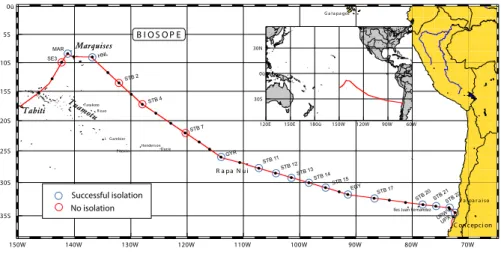
Samples were taken in general at two depths (surface layer and vicinity of the chloro-phyll maximum) at selected stations along the BIOSOPE cruise track (Fig. 1 and Ta-ble 1) using Niskin bottles mounted on a CTD frame. The oceanographic context of the
cruise is described in Claustre et al. (2007)2.
2.2 Primary cultures
10
We used two different strategies to obtain starter cultures. The first one was based on filtered seawater enriched with nutrients. The second one relied on single cell sorting by flow cytometry, targeting specific cell populations based on their size and pigment fluorescence. As cultures were examined several times during the cruise, many varia-tions were attempted in an effort to increase final culture yield.
15
2.2.1 Growth conditions used on board
All cultures were incubated on board in a thermostatic cabinet set at 20◦C. Two
light levels were obtained with 2 Sylvania 18 W tubes: white light around 140 µmol
photons.m−2s−1and blue light (Moon Light Blue paper, M.E.S, Nantes, France) around
8 µmol photons m−2s−1. We used three types of medium: K (Keller et al., 1987)
20
2
Claustre, H., Sciandra, A., Vaulot, D., and Raimbault, P.: Introduction to the special section: bio-optical and biogeochemical conditions in the South East Pacific in late 2004 – the BIOSOPE program, Biogeosciences Discuss., in preparation, 2007.
BGD
4, 2699–2732, 2007 Picoplankton diversity in the South-East Pacific Ocean F. Le Gall et al. Title Page Abstract Introduction Conclusions References Tables Figures ◭ ◮ ◭ ◮ Back CloseFull Screen / Esc
Printer-friendly Version
Interactive Discussion
for photosynthetic eukaryotes, Pro2 (Moore and Chisholm, 1999) for photosynthetic prokaryotes (Prochlorococcus and Synechococcus), and rice-based (Massana et al., 2004) for heterotrophic eukaryotes which were grown in the dark. Multi-well plates were wrapped with parafilm in order to avoid any evaporation during growth.
2.2.2 Enrichment cultures
5
About 500 mL of sample seawater was filtered by simple gravity through two super-posed (in an effort to provide more tight size fractionation) Nuclepore filters of 47 mm diameter, with either 0.6 µm or 3 µm porosity (Whatman International Ltd, Maidstone, UK). The filtrate was partitioned into 50 mL culture flasks (Sarstedt, Orsay, France) or, at one station (HNL3), into individual wells of 24-well plates to which we added either 10
1/10 or 1/100 of full strength K or Pro2 medium. In order to try to promote nitrogen fixing organisms, some cultures were started by simply amending sea water with iron
(as FeCl3) and phosphorus (as KH2PO4 at final concentrations of 3 nM and 0.4 µM,
respectively.
2.2.3 Cultures sorted by flow cytometry
15
Samples were run either un-concentrated or concentrated between 5 and 100-fold by tangential flow filtration using a 100 000 MWCO (Regenerated Cellulose – RC ref VF20C4) Vivaflow 200 cassette. Concentration was usually necessary to target the rarer cells. Between 1 to 500 000 cells were sorted using a FACSAria (Becton Dick-inson, San Jose CA) flow cytometer either into 24 or 48-well plates or directly into 20
10 mL polystyrene tubes pre-filled with medium diluted 100 times (Table 2). Different cell populations were discriminated based on side scatter as well as orange and red fluorescence following excitation at 488 nm (20 mW) and sorting was done either in purity or yield mode.
BGD
4, 2699–2732, 2007 Picoplankton diversity in the South-East Pacific Ocean F. Le Gall et al. Title Page Abstract Introduction Conclusions References Tables Figures ◭ ◮ ◭ ◮ Back CloseFull Screen / Esc
Printer-friendly Version
Interactive Discussion
2.3 Primary culture processing and establishment of strains
On board the ship, primary cultures (either enriched or flow sorted) were checked for growth once or twice (depending on how early in the cruise they were started) using flow cytometry and inverted microscopy. Cultures that displayed growth but appeared mixed were sorted a second time.
5
A first set of cultures were transferred back to Roscoff on the occasion of change of crew at Easter Island at mid-cruise. At the end of the cruise, cultures from the early part of the cruise (i.e. about two months old) that showed no evidence of containing photosynthetic cells based on flow cytometry analysis were discarded. All cultures grown in multi-well plates were transferred to 10 mL polystyrene tubes and brought 10
back to Roscoff in an ice box.
Once transferred back to Roscoff, cultures were monitored based on colour as well as with optical microscopy and flow cytometry. Cultures were purified either by serial dilution, solid medium plating, or individual cell pipetting under an inverted microscope. Strains that appeared to be pure were transferred to normal strength medium [PCR-15
S11 (Rippka et al., 2000), K, and rice for cyanobacteria, autotrophic and heterotrophic eukaryotes, respectively] and entered into the Roscoff Culture Collection (RCC) under new accession numbers (Table 2).
2.4 Strain characterization
Strains deposited to the RCC were characterized by optical microscopy. For each 20
strain, pictures were taken on live cultures with an Olympus BX51 microscope with a x100 objective using differential interference contrast (DIC) with a SPOT RT-slider digital camera (Diagnostics Instruments, Sterling Heights, MI). Average cell dimen-sion of each culture was determined from the pictures. Flagellated cells were also photographed after adding one drop of lugol to visualize flagellum shape, length and 25
number. Cyanobacteria were identified by their colour and shape. The morphology of a few strains was confirmed by whole-mount transmission electron microscopy. Cells
BGD
4, 2699–2732, 2007 Picoplankton diversity in the South-East Pacific Ocean F. Le Gall et al. Title Page Abstract Introduction Conclusions References Tables Figures ◭ ◮ ◭ ◮ Back CloseFull Screen / Esc
Printer-friendly Version
Interactive Discussion
were fixed for 15 min with 1% glutaraldehyde. A drop of fixed cells was deposited onto formvar-coated grids. When the drop had dried, grids were rinsed with distilled water. Cells on grids were stained with a saturated solution of uranyl acetate for 20 min and rinsed with distilled water. Photomicrographs were taken with a JEOL JEM-1200EX electron microscope.
5
A subset of strains was characterized by their partial 18 S ribosomal RNA gene se-quence. Cultures were grown in 50 mL flasks for 1–2 weeks depending on the growth of each strain and recovered by centrifugation at 11 000×g for 10 min. DNA was ex-tracted using 3% Cethyl Trimethyl Ammonium Bromide (CTAB, Doyle and Doyle, 1990).
DNA was then stored at –80◦C.
10
The 18S rRNA gene was amplified by polymerase chain reaction (PCR) using the primer set Euk328f and Euk329r (Moon-van der Staay et al., 2000) and the HotStarTaq Master Mix (Qiagen, Courtaboeuf, France). For PCR, a 15 min initial activation step of
the Polymerase at 95◦C, was followed by 40 cycles including 1 min of denaturation at
94◦C, 45 s of annealing at 57◦C and 75 s extension at 72◦C. The PCR program was
fin-15
ished by a final extension of 10 min at 72◦C followed by cooling at 4◦C. PCR products
were purified with the Qiaquick PCR purification kit (Qiagen) and controlled by elec-trophoresis on a 1% agarose gel. Partial 18 S rRNA gene sequences were determined from purified PCR products by using Big Dye Terminator V3.1 (Applied Biosystems, Foster city, CA, USA) and the internal primer Euk 528f (Elwood et al., 1985) run on an 20
ABI prism 3100 sequencer (Applied Biosystems, Courtaboeuf, France).
Sequences were compared to those available in public database with NCBI BLAST
web application. Sequences were also automatically aligned using the ARB
pro-gram (Ludwig et al., 2004) to a set of more than 20 000 high quality pre-aligned
eukaryotic sequences available from the Silva database (database SSURef: http:
25
//www.arb-silva.de). After manual refinement of the alignment, sequences were added to the reference tree provided with the SSURef database using the quick parsimony addition option. Sequences with high similarities were grouped together using Fast
Group II (http://biome.sdsu.edu/fastgroup/fg tools.htm) with the sequence match
BGD
4, 2699–2732, 2007 Picoplankton diversity in the South-East Pacific Ocean F. Le Gall et al. Title Page Abstract Introduction Conclusions References Tables Figures ◭ ◮ ◭ ◮ Back CloseFull Screen / Esc
Printer-friendly Version
Interactive Discussion
rameter set at 80 % and one or two representative sequences per group were cho-sen along with the closest publicly available sequence. Phylogeny analysis was
per-formed on aligned sequences with MEGA4 (http://www.megasoftware.net/, Tamura et
al., 2007). A neighbour-joining tree was computed from 394 common positions based on Kimura 2-parameter model distances using 1000 bootstrap replications. Sequences 5
have been submitted to GenBank under accession number xxx-xxx.
3 Results
3.1 Isolation success
All together more than 1900 starter cultures were established during the BIOSOPE cruise (Table 1) either as enrichment cultures following filtration through either 0.6 or 10
3 µm or by sorting specific populations into individual wells or tubes. From one to three purification steps were in general necessary to obtain pure cultures (Table 2). For example, enrichment cultures started at the beginning of the cruise were sorted at the end of the cruise and then purified by serial dilution back in the laboratory.
In the end, we obtained 188 autotrophic and 24 heterotrophic cultures which have 15
been deposited to the RCC (Table 2). Among these, 12 were subsequently lost and 25 remain not pure to this date. The latter are mostly autotrophic strains contaminated by heterotrophic eukaryotes. Cruise coverage was quite unequal with many strains obtained in mesotrophic regions and in the Chilean upwelling and much fewer from the central gyre (Table 2, Fig. 1). This reflects probably the difficulty to obtain cultures rep-20
resentative of extreme oligotrophic conditions, since nutrient additions even at relatively low concentrations are always much higher than those found in the environment. How-ever this unbalanced coverage is not only the consequence of the environment but also of practical considerations. Cultures started early during the cruise had a chance to be screened before the end of the cruise and therefore could be re-purified on-board the 25
ship. Conversely, cultures started late in the cruise were transported during their initial 2707
BGD
4, 2699–2732, 2007 Picoplankton diversity in the South-East Pacific Ocean F. Le Gall et al. Title Page Abstract Introduction Conclusions References Tables Figures ◭ ◮ ◭ ◮ Back CloseFull Screen / Esc
Printer-friendly Version
Interactive Discussion
growth phase, a period where they may be less fragile than once they have acclimated to more stable conditions. Refinements in culturing conditions that were implemented late in the cruise based upon results obtained in the first part of the cruise may also explain why our success rate was good by the end of the cruise. For example, starter cultures were sorted at the beginning of the cruise into 24 or 48-well plates. By mid-5
cruise, as we did not observe any growth under these conditions, we decided to switch to sorting into 10-mL tubes which seemed to result in a higher success rate.
Sorting was an important element since more than 65% of the final cultures had un-dergone a sorting step. The strategy that yielded most pure strains was first to establish an enrichment culture with either 0.6 or 3 µm filtered samples followed by sorting some-10
times later. In this case, it was often not necessary to perform further purification by serial dilution, saving this labour-intensive step. Sorting directly from natural samples was rarely sufficient to produce pure cultures and in most cases a second purification step had to be undertaken. It is difficult to determine whether sorting was successful in isolating the initially targeted population. We sorted sub-populations on the base 15
of side scatter and chlorophyll but each of these sub-populations does not appear to be uniform genetically and consists probably of a mixture of several taxa belonging to different algal classes (Shi, X. and Marie, D., unpublished).
3.2 Culture diversity
All purified cultures were examined by light microscopy, imaged digitally and their av-20
erage size was determined (Table 2 and Fig. 2). No attempts were made to record measurements for a large number of cells in each cultures and these data are there-fore only indicative. They confirm, however, that our efforts to target picoplankton were successful since the mode size for the culture set lies between 2.5 and 3 µm.
A large, randomly chosen, subset of cultures (115, Table 2, Table 3) was analysed 25
phylogenetically by sequencing either partially or, in a few cases, totally the 18 S rRNA gene. A few other cultures were identified based on their phenotypic characteristics (cyanobacteria, microplanktonic species).
BGD
4, 2699–2732, 2007 Picoplankton diversity in the South-East Pacific Ocean F. Le Gall et al. Title Page Abstract Introduction Conclusions References Tables Figures ◭ ◮ ◭ ◮ Back CloseFull Screen / Esc
Printer-friendly Version
Interactive Discussion
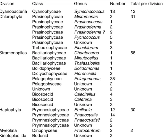
Representatives of cyanobacteria and of three major eukaryotic divisions containing photosynthetic organisms (stramenopiles, Chlorophyta, and Haptophyta) have been obtained in culture with the former most prevalent and the latter two in almost equal proportions (Table 4). Two dinoflagellate cultures have also been isolated.
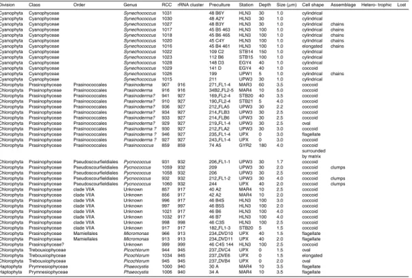
Thirteen strains of unicellular rod-shaped cyanobacteria have been obtained tenta-5
tively identified as Synechococcus. No Prochlorococcus was obtained despite the use of the Prochlorococcus specific Pro2 medium. Some of these cyanobacterial strains form short chains, exhibiting sometimes very elongated cells (Fig. 3, RCC 1027) con-trasting the usual Synechococcus morphology (Fig. 3, RCC 1022). Such strains mostly originated from the HNLC station near the Marquesas islands (Table 3) and could be 10
interesting since samples from this region displayed an unusually high fraction of chain-forming and colonial picocyanobacteria (Masquelier and Vaulot, 2007). Phylogenetic analyses of the 16 S rRNA gene will be necessary to determine the exact nature of these strains.
Chlorophyta, and more specifically Prasinophyceae, are important contributors to pi-15
coplankton and many strains have been isolated from marine waters in the past, some of them belonging to not yet described species (Guillou et al., 2004; Vaulot et al., 2004). Seventeen Chlorophyta strains have been isolated during BIOSOPE, mostly Prasino-phyceae. Among these, 11 are related to Prasinoderma coloniale (Prasinococcales), a picoplanktonic species that can form colonies surrounded by mucus. These strains 20
display the bilobed cup-shaped chloroplasts characteristics of P. coloniale (Hasegawa et al., 1996). However most of our strains do not seem to form colonies as P. coloniale does. Interestingly, one group of 9 sequences appear to form a separate clade (Fig. 4) with only 94.7% identity to P. coloniale (in contrast to the two other strains sharing 99.6% identity with P. coloniale) and possess large and highly similar insertions at least 25
330 bp long inside de 18 S rRNA gene starting at nucleotide position 862 of the P. colo-niale sequence. Phenotypically, strains from these group appear slightly smaller (Table 2) than those closely related to P. coloniale. They were isolated from near-surface wa-ters at a variety of stations, while the two strains more closely related to P. coloniale
BGD
4, 2699–2732, 2007 Picoplankton diversity in the South-East Pacific Ocean F. Le Gall et al. Title Page Abstract Introduction Conclusions References Tables Figures ◭ ◮ ◭ ◮ Back CloseFull Screen / Esc
Printer-friendly Version
Interactive Discussion
originated from the Marquesas area. A culture closely related to Prasinococcus cap-sulatus, a species that also belongs to the order Prasinococcales, has been recovered from the chlorophyll maximum at the GYR station. Cells display a polysaccharide cap-sule around the cell (Fig. 3, RCC 859), typical of this species (Miyashita et al., 1993). Five Prasinophyceae closely related to the picoplanktonic species Pycnococcus prova-5
solii (Pseudoscourfieldiales) have been isolated from two mid-depth samples in the Chilean upwelling. Eight strains belong to clade VII A of the Prasinophyceae (Guillou et al., 2004), a group which contains some cultured strains such as CCMP 1205 but for which no species has been described formally. All these strains consist of small (2 to 4 µm) coccoid cells lacking discriminating features (Fig. 3, RCC 857). Two sets 10
of strains originated from surface waters and one set from 100 m in the HNLC zone. Two Prasinophyceae strains from the Chilean upwelling belong to clade C of the very ubiquitous species Micromonas pusilla (Guillou et al., 2004). They possess an unusu-ally long flagellum (Fig. 3, RCC 913) that could be a diagnostic feature for that clade (Jouenne, F., personal communication). Finally, one culture (RCC 999) presents some 15
phylogenetic affinities to the Prasinophyceae but its partial 18 S rDNA sequence does not allow us to place it in any of the existing clades defined by Guillou et al. (2004). We also isolated from one sample of the Chilean upwelling, three cultures representative of another green algal class, the Trebouxiophyceae. These strains are phylogeneti-cally related to the recently established genus Picochlorum (Fig. 4) that regroups now 20
salt-tolerant Nanochlorum (Henley et al., 2004).
All Haptophyta cultures are part of the class Prymnesiophyceae. Sixteen strains be-long to the genus Phaeocystis, three from the upwelling region being more closely re-lated to the species P. jahnii which has been recently described from the Mediterranean Sea (Zingone et al., 1999) and forms loose colonies and 5 from the east of the gyre and 25
upwelling regions related to P. globosa that forms spherical colonies (Fig. 3, RCC 851). We also isolated 12 strains of Emiliania huxleyi, a few calcifying (Fig. 3, RCC 867) and most naked (Fig. 3, RCC 951), corresponding probably to diploid and haploid stages, respectively (Houdan et al., 2003). Two other unidentified coccolithophorids have also
BGD
4, 2699–2732, 2007 Picoplankton diversity in the South-East Pacific Ocean F. Le Gall et al. Title Page Abstract Introduction Conclusions References Tables Figures ◭ ◮ ◭ ◮ Back CloseFull Screen / Esc
Printer-friendly Version
Interactive Discussion
been obtained from the Marquesas and central gyre regions. Interestingly all Hapto-phyta strains were isolated from the top of euphotic zone (between 5 and 60 m).
Among stramenopiles, 38 cultures are closely related to the picoplanktonic species Pelagomonas calceolata (Pelagophyceae). More than half of them are flagellated (Fig. 3, RCC 879), fitting the original description of the species (Andersen et al., 1993), 5
and the rest, coccoid. However, the presence of a flagellum reflects probably more life cycle stages rather than taxonomical differences since both flagellated and non-flagellated strains have been isolated from the same sample (e.g. at 100 m at the HLN station). The presence of a thin theca characteristics of the species (Andersen et al., 1993) was confirmed by electron microscopy on strain RCC 879. P. calceolata 10
has been isolated at a variety of stations (Marquesas, HLNC, center of gyre, east of gyre and upwelling) both in surface and at 100 m, demonstrating that this species is truly ubiquitous in oceanic waters. Interestingly in the center of the South East gyre, Pelagomonas strains were isolated from very deep samples down to 160 m. Two Pelagophyceae strains (RCC 986 and 1024) with 18 S rDNA sequences displaying 15
slightly lower similarity to P. calceolata (Fig. 4) were recovered at 60 m depth from the Marquesas region. Both are picoplanktonic and coccoid, not displaying any specific morphological features. We isolated a novel strain with high similarity to Bolidomonas pacifica, a species that belongs to the recently described class of the Bolidophyceae (Guillou et al., 1999), closely related to the diatoms. Its morphology (presence of 2 un-20
equal flagella) was confirmed by electron microscopy. This is quite interesting since to our knowledge this is the first novel isolate from this class since its initial discovery. In the same manner, we isolated from Marquesas surface waters, two strains very closely related by their 18 S rDNA sequence to the recently described Dictyochophyceae pi-coplanktonic species Florenciella parvula (Eikrem et al., 2004). Similarity was also 25
confirmed by electron microscopy. Two photosynthetic stramenopile strains could not be assigned to any specific class as they shared homology with both Pinguiococcus (Pinguiophyceae) and Nannochloropsis (Eustigmatophyceae). Their 18 S sequences are almost identical to an environmental 18 S sequence (BL000921.5) recovered from
BGD
4, 2699–2732, 2007 Picoplankton diversity in the South-East Pacific Ocean F. Le Gall et al. Title Page Abstract Introduction Conclusions References Tables Figures ◭ ◮ ◭ ◮ Back CloseFull Screen / Esc
Printer-friendly Version
Interactive Discussion
Blanes Bay in the Mediterranean Sea (Fig. 4). They could belong to a new class, al-though the presence of refractive intracellular granules (Fig. 3, RCC 853) is quite rem-iniscent of what is observed in Nannochloropsis. Unfortunately, they have been lost in early 2007 following a breakdown in the air conditioning system of our culture facility. Their loss, which was almost the only one from a quite large collection, attests of their 5
sensitivity to change in environmental conditions and may explain why representatives of this group have not been isolated before.
Three diatoms, belonging to the genera Chaetoceros (Fig. 3, RCC 1025)Thalas-siosira, andMinutocellus were obtained from the upwelling region. The latter strain is quite interesting since its very small size (about 3 µm) connects it to picoplankton. Two 10
dinoflagellates belonging to the genus Prorocentrum, P. minimum (Fig. 3, RCC 922) and P. dentatum, were isolated from surface waters, east of the gyre.
Twelve heterotrophic strains from dark cultures growing on rice medium have been identified by their 18 S rRNA sequences. Nine belong to the bicosoecid lineage of the stramenopiles. Three cultures are quite closely related to the genus Caecitellus and 15
four more distantly related to Cafeteria. The two remaining strains were closely related to the bodonid (Euglenozoa) genera Rhynchomonas and Neobodo. All these genera are quite often recovered in cultures (Arndt et al., 2003).
4 Conclusions
Our large scale effort to isolate picoplanktonic strains from the Southeast Pacific Ocean 20
allowed us to obtain of 212 novel cultures, a large number of which are of picoplank-tonic size. The final number of cultures obtained is substantially higher than in previous efforts such as those linked to the PROSOPE and MINOS cruise in the Mediterranean Sea or the OLIPAC cruise in the Equatorial Pacific Ocean from which we obtained between 46 and 90 strains for each (Vaulot et al., 2004). Our initial intent was to 25
use mostly flow cytometry sorting to establish this strains. However as we experi-enced technical problems with flow cytometry in the first few days of the cruise and as
BGD
4, 2699–2732, 2007 Picoplankton diversity in the South-East Pacific Ocean F. Le Gall et al. Title Page Abstract Introduction Conclusions References Tables Figures ◭ ◮ ◭ ◮ Back CloseFull Screen / Esc
Printer-friendly Version
Interactive Discussion
we observed subsequently that the yield of the initially sorted samples was quite low, we decided to combine flow cytometry sorting with more classical enrichments. This proved to be quite a good recipe, especially since sorting based on photosynthetic pigment fluorescence appears as a good way to prevent contamination of cultures by heterotrophic eukaryotes, a problem plaguing some of our previous efforts. The appli-5
cation of sorting either before or after enrichment did not appear to affect dramatically the type of taxa isolated (Table 2).
The final diversity achieved is quite wide since we obtained representatives of most major photosynthetic divisions (Table 4). However it is clear that we globally failed to obtain representatives of environmental sequences for which no culture is available 10
yet. One interesting group of novel cultures was constituted by stramenopile strains RCC 853 and 862 from the central gyre which sequences were closely related to an en-vironmental sequence from the Mediterranean Sea (Fig. 4). Although these sequences had some affinities, based on BLAST, to Eustigmatophyceae and their morphology was somewhat similar to the latter, they probably belonged to a novel class. Despite the fact 15
that further studies are prevented since these strains have been lost, the strategy used (flow cytometry sorting followed by serial dilution) could be tried again to re-isolate them. Another interesting group is constituted by 9 cultures originating from the region east of the gyre and from the upwelling that are related to Prasinoderma but form a new clade clearly separated from the species P. coloniale (Fig. 4). They could belong 20
to a new species within the genus Prasinoderma or form a new genus. Interestingly, they are apparently not related to any published environmental sequence. All the other cultures obtained are related to described species or at least to established cultures. In particular, we have been successful at re-isolating two genera Bolidomonas and Florenciella that our group had previously isolated and described (Eikrem et al., 2004;
25
Guillou et al., 1999), but that had never been obtained again in culture since their ini-tial isolation. Interestingly, B. pacifica was iniini-tially isolated in exactly the same region
(between 2 and 16◦S) as the new strain (9◦S). In contrast, the only F. parvula strain
available previously originated from English Channel coastal waters, a very different 2713
BGD
4, 2699–2732, 2007 Picoplankton diversity in the South-East Pacific Ocean F. Le Gall et al. Title Page Abstract Introduction Conclusions References Tables Figures ◭ ◮ ◭ ◮ Back CloseFull Screen / Esc
Printer-friendly Version
Interactive Discussion
environment from that of the new strains. Moreover the 18S sequences of the latter differ slightly from that of F. parvula and they could belong to a novel species within this genus. Some of the cultures recovered correspond to ubiquitous species that were obtained from a wide range of environments. This is in particular the case for the two Haptophyta genera Emiliania, isolated from two of the four major regions investigated 5
(Marquesas, east of gyre) and Phaeocystis isolated from three regions (Marquesas, east of the gyre, Chilean upwelling) mostly in surface waters. For the latter genus, our strains may correspond to at least two different species, P. globosa and P. jah-nii. However, the largest number of strains obtained for a single taxon correspond to Pelagomonas isolated from a record of 13 different samples along the entire cruise
10
track ranging from oligotrophic (St B13) to eutrophic (UPX) and from surface (5 m) to very deep (160 m) samples. Although the similarity of their 18S rRNA gene sequence is very high (average p-distance=0.0018), it is likely that these strains present quite different growth responses to factors such as nitrogen supply or light levels and belong to different ecotypes, as observed previously for example for the genus Ostreococcus 15
(Rodr´ıguez et al., 2005).
From a biogeographic point of view, it is quite difficult to make any firm conclusion from this work. Many cultures belonging to a given taxonomic group were isolated from a variety of conditions and no specific pattern could be uncovered. Although there were some taxa unique to the central part of the gyre itself (stations 3 to 15) 20
such as Prasinococcus and the potentially novel class mentioned earlier, one should emphasize the low number of strains isolated from this region. This is probably linked to the fact that the media we used (K, Pro2), that are quite successful in general to isolate and maintain a wide variety of picophytoplankton strains, fail to mimic the drastic oligotrophic conditions met in the gyre. Moreover future isolation effort may need to 25
involve new culture approaches such as those successful to isolate fastidiously growing bacterial strains from the open ocean environment such as Peligibacter ubique (aka SAR11) that had escaped cultivation for quite a long time (Zengler et al., 2002).
Acknowledgements. We wish to thank the crew of the NO Atalante for their critical help in
BGD
4, 2699–2732, 2007 Picoplankton diversity in the South-East Pacific Ocean F. Le Gall et al. Title Page Abstract Introduction Conclusions References Tables Figures ◭ ◮ ◭ ◮ Back CloseFull Screen / Esc
Printer-friendly Version
Interactive Discussion repairing the FACSAria flow cytometer during the cruise as well as for their constant
avail-ability. We are grateful to all participants to the BIOSOPE cruise, especially H. Claustre and A. Sciandra, who coordinated the cruise and acted as chief scientists. Help for microscopy from F. Jouenne is kindly acknowledged. Financial support for this work was provided by the follow-ing programs and companies: ANR Biodiversit ´e (projet PICOFUNPAC), CNRS INSU PROOF,
5
Contrat de Plan Etat R ´egion (Souchoth `eque de Bretagne), Becton Dickinson.
References
Andersen, R. A., Saunders, G. W., Paskind, M. P., and Sexton, J.: Ultrastructure and 18 S rRNA gene sequence for Pelagomonas calceolata gen. and sp. nov. and the description of a new algal class, the Pelagophyceae classis nov. J. Phycol., 29, 701–715, 1993.
10
Arndt, H., Hausmann, K., and Wolf, M.: Deep-sea heterotrophic nanoflagellates of the Eastern Mediterranean Sea: qualitative and quantitative aspects of their pelagic and benthic occur-rence, Mar. Ecol. Prog. Ser., 256, 45–56, 2003.
Derelle, E., Ferraz, C., Rombauts, S., Rouze, P., Worden, A. Z., Robbens, S., Partensky, F., Degroeve, S., Echeynie, S., Cooke, R., Saeys, Y., Wuyts, J., Jabbari, K., Bowler, C., Panaud,
15
O., Piegu, B., Ball, S. G., Ral, J.-P., Bouget, F.-Y., Piganeau, G., De Baets, B., Picard, A., Delseny, M., Demaille, J., Van de Peer, Y., and Moreau, H.: Genome analysis of the smallest free-living eukaryote Ostreococcus tauri unveils many unique features, Proc. Natl. Acad. Sci. USA, 103, 11 647–11 652, 2006.
Doyle, J. J. and Doyle, J. L.: Isolation of plant DNA from fresh tissue, Focus, 12, 13–15, 1990.
20
Eikrem, W., Romari, K., Latasa, M., Le Gall, F., Throndsen, J., and Vaulot, D.: Florenciella
parvula gen. and sp. nov. (Dictyochophyceae, Heterokontophyta) a small flagellate isolated
from the English Channel, Phycologia, 43, 658–668, 2004.
Elwood, H. J., Olsen, G. J., and Sogin, M. L.: The small-subunit ribosomal RNA gene se-quences from the hypotrichous ciliates Oxytricha nova and Stylonychia pustulata, Mol. Biol.
25
Evol., 2, 399–410, 1985.
Fuller, N. J., Marie, D., Partensky, F., Vaulot, D., Post, A. F., and Scanlan, D. J.: Clade-specific 16 S ribosomal DNA oligonucleotides reveal the predominance of a single marine
Syne-chococcus clade throughout a stratified water column in the Red Sea, Appl. Environ.
Micro-biol., 69, 2430–2443, 2003.
30
BGD
4, 2699–2732, 2007 Picoplankton diversity in the South-East Pacific Ocean F. Le Gall et al. Title Page Abstract Introduction Conclusions References Tables Figures ◭ ◮ ◭ ◮ Back CloseFull Screen / Esc
Printer-friendly Version
Interactive Discussion Guillou, L., Chr ´etiennot-Dinet, M.-J., Medlin, L. K., Claustre, H., Loiseaux-de Go ¨er, S., and
Vaulot, D.: Bolidomonas: a new genus with two species belonging to a new algal class, the Bolidophyceae (Heterokonta), J. Phycol., 35, 368–381, 1999.
Guillou, L., Eikrem, W., Chr ´etiennot-Dinet, M. J., Le Gall, F., Massana, R., Romari, K., Pedr ´os-Ali ´o, C., and Vaulot, D.: Diversity of picoplanktonic prasinophytes assessed by direct nuclear
5
SSU rDNA sequencing of environmental samples and novel isolates retrieved from oceanic and coastal marine ecosystems, Protist, 155, 193–214, 2004.
Hasegawa, T., Miyashita, H., Kawachi, M., Ikemoto, H., Kurano, N., Miyachi, S., and Chihara, M.: Prasinoderma coloniale ge. nov. et sp. nov., a new pelagic coccoid prasinophyte from the western Pacific Ocean. Phycologia, 35, 170–176, 1996.
10
Henley, W. J., Hironaka, J. L., Guillou, L., Buchheim, M. A., Buchheim, J. A., Fawley, M. W., and Fawley, K. P.: Phylogenetic analysis of the “Nannochloris-like” algae and diagnoses of
Picochlorum oklahomensis gen. et sp nov (Trebouxiophyceae, Chlorophyta), Phycologia, 43,
641–652, 2004.
Herbland, A., Le Bouteiller, A., and Raimbault, P.: Size structure of phytoplankton biomass in
15
the equatorial Atlantic Ocean, Deep-Sea Res. I, 32, 819–836, 1985.
Houdan, A., Billard, C., Marie, D., Not, F., Saez, A. G., Young, J. R., and Probert, I.: Holococcolithophore-heterococcolithphore (Haptophyta) life cycles: flow cytometric analysis of relative ploidy levels, Systematics and Biodiversity, 4, 453–465, 2003.
Johnson, P. W. and Sieburth, J. M.: In-situ morphology and occurence of eucaryotic
pho-20
totrophs of bacterial size in the picoplankton of estuarine and oceanic waters, J. Phycol., 18, 318–327, 1982.
Johnson, Z. I., Zinser, E. R., Coe, A., McNulty, N. P., Woodward, E. M. S., and Chisholm, S. W.: Niche partitioning among Prochlorococcus ecotypes along ocean-scale environmental gradients, Science, 311, 1737–1740, 2006.
25
Keller, M. D., Selvin, R. C., Claus, W., and Guillard, R. R. L.: Media for the culture of oceanic ultraphytoplankton, J. Phycol., 23, 633–638, 1987.
Li, W. K. W., Subba Rao, D. V., Harrison, W. G., Smith, J. C., Cullen, J. J., Irwin, B., and Platt, T.: Autotrophic picoplankton in the tropical ocean, Science, 219, 292–295, 1983.
L ´opez-Garc´ıa, P., Rodriguez-Valera, F., Pedr ´os-Ali ´o, C., and Moreira, D.: Unexpected diversity
30
of small eukaryotes in deep-sea Antarctic plankton. Nature, 409, 603–607, 2001.
Ludwig, W., Strunk, O., Westram, R., Richter, L., Meier, H., Yadhukumar, Buchner, A., Lai, T., Steppi, S., Jobb, G., Forster, W., Brettske, I., Gerber, S., Ginhart, A. W., Gross, O., Grumann,
BGD
4, 2699–2732, 2007 Picoplankton diversity in the South-East Pacific Ocean F. Le Gall et al. Title Page Abstract Introduction Conclusions References Tables Figures ◭ ◮ ◭ ◮ Back CloseFull Screen / Esc
Printer-friendly Version
Interactive Discussion S., Hermann, S., Jost, R., Konig, A., Liss, T., Lussmann, R., May, M., Nonhoff, B., Reichel,
B., Strehlow, R., Stamatakis, A., Stuckmann, N., Vilbig, A., Lenke, M., Ludwig, T., Bode, A., and Schleifer, K. H.: ARB: a software environment for sequence data, Nucleic Acids Res., 32, 1363–1371, 2004.
Marie, D., Zhu, F., Balagu ´e, V., Ras, J., and Vaulot, D.: Eukaryotic picoplankton communities
5
of the Mediterranean Sea in summer assessed by molecular approaches (DGGE, TTGE, QPCR), FEMS Microbiol. Ecol., 55, 403–415, 2006.
Masquelier, S. and Vaulot, D.: Distribution of micro-organisms along a transect in the South-East Pacific Ocean (BIOSOPE cruise) from epifluorescence microscopy, Biogeosciences Discuss., accepted, 2007.
10
Massana, R., Balagu ´e, V., Guillou, L., and Pedr ´os-Ali ´o, C.: Picoeukaryotic diversity in an olig-otrophic coastal site studied by molecular and culturing approaches, FEMS Microbiol. Ecol., 50, 231–243, 2004.
Miyashita, H., Ikemoto, H., Kurano, N., Miyachi, S., and Chihara, M.: Prasinococcus capsulatus gen. et sp. nov., a new marine coccoid prasinophyte, J. Gen. Appl. Microbiol., 39, 571–582,
15
1993.
Moon-van der Staay, S. Y., De Wachter, R., and Vaulot, D.: Oceanic 18 S rDNA sequences from picoplankton reveal unsuspected eukaryotic diversity, Nature, 409, 607–610, 2001.
Moon-van der Staay, S. Y., van der Staay, G. W. M., Guillou, L., Vaulot, D., Claustre, H., and Medlin, L. K.: Abundance and diversity of prymnesiophytes in the picoplankton community
20
from the equatorial Pacific Ocean inferred from 18 S rDNA sequences, Limnol. Oceanogr., 45, 98–109, 2000.
Moore, L. R. and Chisholm, S. W.: Photophysiology of the marine cyanobacterium
Prochloro-coccus: Ecotypic differences among cultured isolates, Limnol. Oceanogr., 44, 628–638,
1999.
25
Not, F., Valentin, K., Romari, K., Lovejoy, C., Massana, R., T ¨obe, K., Vaulot, D., and Medlin, L.: Picobiliphytes, a new marine picoplanktonic algal group with unknown affinities to other eukaryotes, Science, 315, 252–254, 2007.
Not, F., Massana, R., Latasa, M., Marie, D., Colson, C., Eikrem, W., Pedr ´os-Ali ´o, C., Vaulot, D., and Simon, N.: Late summer community composition and abundance of photosynthetic
30
picoeukaryotes in Norwegian and Barents seas, Limnol. Oceanogr., 50, 1677–1686, 2005. Palenik, B., Brahamsha, B., Larimer, F. W., Land, M., Hauser, L., Chain, P., Lamerdin, J.,
Regala, W., Allen, E. E., McCarren, J., Paulsen, I., Dufresne, A., Partensky, F., Webb, E. A.,
BGD
4, 2699–2732, 2007 Picoplankton diversity in the South-East Pacific Ocean F. Le Gall et al. Title Page Abstract Introduction Conclusions References Tables Figures ◭ ◮ ◭ ◮ Back CloseFull Screen / Esc
Printer-friendly Version
Interactive Discussion and Waterbury, J.: The genome of a motile marine Synechococcus, Nature, 424, 1037–1042,
2003.
Palenik, B., Grimwood, J., Aerts, A., Rouze, P., Salamov, A., Putnam, N., Dupont, C., Jor-gensen, R., Derelle, E., Rombauts, S., Zhou, K., Otillar, R., Merchant, S. S., Podell, S., Gaasterland, T., Napoli, C., Gendler, K., Manuell, A., Tai, V., Vallon, O., Piganeau, G.,
5
Jancek, S., Heijde, M., Jabbari, K., Bowler, C., Lohr, M., Robbens, S., Werner, G., Dubchak, I., Pazour, G. J., Ren, Q., Paulsen, I., Delwiche, C., Schmutz, J., Rokhsar, D., Van de Peer, Y., Moreau, H., and Grigoriev, I. V.: The tiny eukaryote Ostreococcus provides genomic in-sights into the paradox of plankton speciation, Proc. Natl. Acad. Sci. USA, 104, 7705–7710, 2007.
10
Platt, T., Subba-Rao, D. V., and Irwin, B.: Photosynthesis of picoplankton in the oligotrophic ocean, Nature, 300, 701–704, 1983.
Rippka, R., Coursin, T., Hess, W., Lichtle, C., Scanlan, D. J., Palinska, K. A., Iteman, I., Parten-sky, F., Houmard, J., and Herdman, M.: Prochlorococcus marinus Chisholm et al. (1992) subsp. pastoris subsp. nov. strain PCC 9511, the first axenic chlorophyll a2/b2-containing
15
cyanobacterium (Oxyphotobacteria), Int. J. Syst. Evol. Microbiol., 50, 1833–1847, 2000. Rocap, G., Distel, D. L., Waterbury, J. B., and Chisholm, S. W.: Resolution of Prochlorococcus
and Synechococcus ecotypes by using 16 S-23S ribosomal DNA internal transcribed spacer sequences, Appl. Environ. Microbiol., 68, 1180–1191, 2002.
Rocap, G., Larimer, F. W., Lamerdin, J., Malfatti, S., Chain, P., Ahlgren, N. A., Arellano, A.,
20
Coleman, M., Hauser, L., Hess, W. R., Johnson, Z. I., Land, M., Lindell, D., Post, A. F., Regala, W., Shah, M., Shaw, S. L., Steglich, C., Sullivan, M. B., Ting, C. S., Tolonen, A., Webb, E. A., Zinser, E. R., and Chisholm, S. W.: Genome divergence in two Prochlorococcus ecotypes reflects oceanic niche differentiation, Nature, 424, 1042–1047, 2003.
Rodr´ıguez, F., Derelle, E., Guillou, L., Le Gall, F., Vaulot, D., and Moreau, H.: Ecotype
di-25
versity in the marine picoeukaryote Ostreococcus (Chlorophyta, Prasinophyceae), Environ. Microbiol., 7, 853–859, 2005.
Sieburth, J. M., Smetacek, V., and Lenz, J.: Pelagic ecosystem structure: heterotrophic com-partments of the plankton and their relationship to plankton size fractions, Limnol Oceanogr., 23, 1256–1263, 1978.
30
Tamura, K., Dudley, J., Nei, M., and Kumar, S.: MEGA4: Molecular Evolutionary Genetics Analysis (MEGA) Software Version 4.0. Mol. Biol. Evol., msm092, 2007.
Vaulot, D., Le Gall, F., Marie, D., Guillou, L., and Partensky, F.: The Roscoff Culture Collection
BGD
4, 2699–2732, 2007 Picoplankton diversity in the South-East Pacific Ocean F. Le Gall et al. Title Page Abstract Introduction Conclusions References Tables Figures ◭ ◮ ◭ ◮ Back CloseFull Screen / Esc
Printer-friendly Version
Interactive Discussion (RCC): a collection dedicated to marine picoplankton, Nova Hedwigia, 79, 49–70, 2004.
Waterbury, J. B., Watson, S. W., Guillard, R. R. L., and Brand, L. E.: Wide-spread occurence of a unicellular, marine planktonic, cyanobacterium. Nature, 277, 293–294, 1979.
Zengler, K., Toledo, G., Rappe, M., Elkins, J., Mathur, E. J., Short, J. M., and Keller, M.: Culti-vating the uncultured, Proc. Natl. Acad. Sci. USA, 99, 15 681–15 686, 2002.
5
Zingone, A., Chr ´etiennot-Dinet, M. J., Lange, M., and Medlin, L.: Morphological and genetic characterization of Phaeocystis cordata and P. jahnii (Prymnesiophyceae), two new species from the Mediterranean Sea, J. Phycol., 35, 1322–1337, 1999.
BGD
4, 2699–2732, 2007 Picoplankton diversity in the South-East Pacific Ocean F. Le Gall et al. Title Page Abstract Introduction Conclusions References Tables Figures ◭ ◮ ◭ ◮ Back CloseFull Screen / Esc
Printer-friendly Version
Interactive Discussion
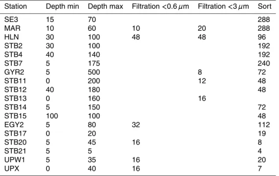
Table 1. Sampling stations and number of starter cultures for each station. Depths are
ex-pressed in meters.
Station Depth min Depth max Filtration <0.6 µm Filtration <3 µm Sort
SE3 15 70 288 MAR 10 60 10 20 288 HLN 30 100 48 48 96 STB2 30 100 192 STB4 40 140 192 STB7 5 175 240 GYR2 5 500 8 72 STB11 0 200 12 48 STB12 40 180 48 STB13 0 160 16 STB14 5 150 72 STB15 100 100 48 EGY2 5 80 32 112 STB17 0 20 19 STB20 5 45 16 8 STB21 5 5 4 UPW1 5 35 16 20 UPX 0 40 16 7 2720
BGD
4, 2699–2732, 2007 Picoplankton diversity in the South-East Pacific Ocean F. Le Gall et al. Title Page Abstract Introduction Conclusions References Tables Figures ◭ ◮ ◭ ◮ Back CloseFull Screen / Esc
Printer-friendly Version
Interactive Discussion
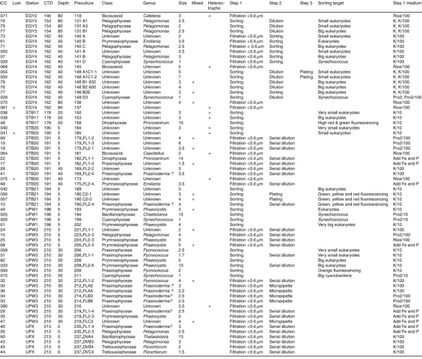
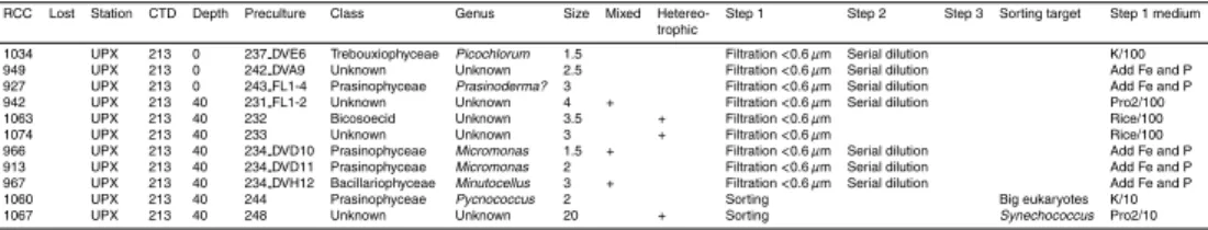
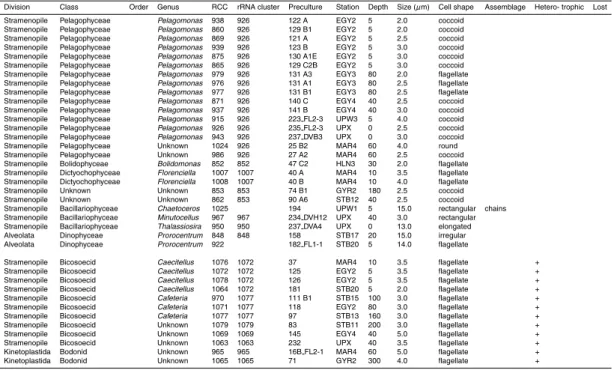
Table 2. List of strains deposited to the Roscoff Culture Collection (RCC) ordered according to
the sampled station. Steps 1, 2 and 3 refer to the different purification steps performed before the culture was entered to the RCC database.
RCC Lost Station CTD Depth Preculture Class Genus Size Mixed Hetereo- Step 1 Step 2 Step 3 Sorting target Step 1 medium trophic
923 MAR3 22 10 273 FL1-2 Unknown Unknown 2.5 + TFF enriched Serial dilution None 959 MAR3 22 10 273 FL2-2 Unknown Unknown 2.5 TFF enriched Serial dilution None 960 MAR3 22 10 273 FL2-3 Unknown Unknown 3 TFF enriched Serial dilution None 961 MAR3 22 10 273 FL2-7 Unknown Unknown 3 TFF enriched Serial dilution None 907 MAR3 22 60 271 FL1-4 Prasinophyceae Prasinoderma 3.5 TFF enriched Serial dilution None 1000 MAR4 28 10 30 A Prymnesiophyceae Phaeocystis 3.5 Filtration <3 µm Sorting Big eukaryotes K/100 872 MAR4 28 10 30 A2 Unknown Unknown 3 + Filtration <3 µm Dilution K/100 1001 MAR4 28 10 30 B Prymnesiophyceae Emiliania 3.5 Filtration <3 µm Sorting Small eukaryotes K/100 1002 MAR4 28 10 31A Prymnesiophyceae Phaeocystis? 4.7 + Filtration <3 µm Sorting Big eukaryotes K/100 1003 MAR4 28 10 31 B Unknown Unknown 3 Filtration <3 µm Sorting Small eukaryotes K/100 1004 MAR4 28 10 32 B Prymnesiophyceae Unknown 5 Filtration <3 µm Sorting Small eukaryotes K/100 1048 MAR4 28 10 32 B2 Unknown Unknown 3 Filtration <3 µm Sorting Eukaryotes K/100 911 MAR4 28 10 32B FL1-2 Prymnesiophyceae Emiliania 3.5 Filtration <3 µm Sorting Serial dilution Small eukaryotes K/100 962 MAR4 28 10 32B FL1-3 Prymnesiophyceae Emiliania 3.5 Filtration <3 µm Sorting Serial dilution Small eukaryotes K/100 963 MAR4 28 10 32B FL2-2 Unknown Unknown 5 Filtration <3 µm Sorting Serial dilution Small eukaryotes K/100 955 MAR4 28 10 32B HO22 Unknown Unknown 3 Filtration <3 µm Sorting Micropipette Small eukaryotes K/100 920 MAR4 28 10 32B HO3 Prymnesiophyceae Emiliania 3.5 Filtration <3 µm Sorting Micropipette Small eukaryotes K/100 921 MAR4 28 10 32B HO8 Prymnesiophyceae Emiliania 3 Filtration <3 µm Sorting Micropipette Small eukaryotes K/100 1005 + MAR4 28 10 33 A Unknown Unknown Filtration <3 µm Sorting Eukaryotes K/100 1049 MAR4 28 10 33 A2 Unknown Unknown 2.5 Filtration <3 µm Sorting Eukaryotes K/100 1006 MAR4 28 10 34 A Prymnesiophyceae Phaeocystis 3.5 Filtration <3 µm Sorting Big eukaryotes K/100 854 MAR4 28 10 34 B2 Unknown Unknown 2.5 + Filtration <3 µm Sorting Big eukaryotes K/100 956 MAR4 28 10 34B HO16 Unknown Unknown 3.5 Filtration <3 µm Sorting Micropipette Eukaryotes K/100 912 MAR4 28 10 34B HO17 Prymnesiophyceae Emiliania 4 Filtration <3 µm Sorting Micropipette Eukaryotes K/100 957 MAR4 28 10 34B HO23 Unknown Unknown 3.5 Filtration <3 µm Sorting Micropipette Eukaryotes K/100 914 MAR4 28 10 34B HO5 Prymnesiophyceae Emiliania 3 Filtration <3 µm Sorting Micropipette Eukaryotes K/100 958 MAR4 28 10 34B HO6 Prymnesiophyceae Emiliania 3.5 Filtration <3 µm Sorting Micropipette Eukaryotes K/100 916 MAR4 28 10 34B2 FL2-5 Prasinophyceae Prasinoderma 5 Filtration <3 µm Sorting Serial dilution Big eukaryotes 1076 MAR4 28 10 37 Bicosoecid Caecitellus 3.5 + Filtration <3 µm Rice/100 1007 MAR4 28 10 40 A Dictyochophyceae Florenciella 3.5 Filtration <0.6 µm Sorting Small eukaryotes Pro2/100 857 MAR4 28 10 40 A2 Prasinophyceae Unknown 2.5 Filtration <0.6 µm Sorting Big eukaryotes Pro2/100 1008 MAR4 28 10 40 B Dictyochophyceae Florenciella 4 Filtration <0.6 µm Sorting Big eukaryotes Pro2/100 855 MAR4 28 10 40 B2 Pelagophyceae Pelagomonas 3.5 Filtration <0.6 µm Sorting Very big eukaryotes Pro2/100 952 MAR4 28 10 41 A2 Unknown Unknown 2.5 Filtration <0.6 µm Dilution Pro2/100 1009 MAR4 28 10 41 S Unknown Unknown 4 Filtration <0.6 µm Sorting Small eukaryotes Pro2/100 856 MAR4 28 10 42 A2 Prasinophyceae Unknown 2 Filtration <0.6 µm Sorting Small eukaryotes Pro2/100 954 MAR4 28 10 43 A2 Unknown Unknown 2.5 Filtration <0.6 µm Dilution Pro2/100 1010 MAR4 28 10 43 PK Pelagophyceae Pelagomonas 3 Filtration <0.6 µm Sorting Prochlorococcus Pro2/100 953 MAR4 28 10 44 A2 Pelagophyceae Pelagomonas 3 Filtration <0.6 µm Dilution Pro2/100 989 MAR4 28 60 15 A Unknown Unknown 3 Filtration <3 µm Sorting Small eukaryotes K/100 982 MAR4 28 60 15 A2 Unknown Unknown 2.5 + Filtration <3 µm Dilution K/100 965 MAR4 28 60 16B FL2-1 Bodonid Unknown 5 + Filtration <3 µm Sorting Serial dilution Prochlorococcus Pro2/100 983 MAR4 28 60 17 A2 Unknown Unknown 2 Filtration <3 µm Dilution K/100 990 MAR4 28 60 17 B Unknown Unknown 2.5 Filtration <3 µm Sorting Small eukaryotes K/100 991 MAR4 28 60 18 A Unknown Unknown 2.5 Filtration <3 µm Sorting Small eukaryotes K/100 992 MAR4 28 60 18 B Prymnesiophyceae Phaeocystis 3.5 Filtration <3 µm Sorting Big eukaryotes K/100 993 MAR4 28 60 19 B Prymnesiophyceae Phaeocystis 3 Filtration <3 µm Sorting Big eukaryotes K/100 994 MAR4 28 60 19 C Prymnesiophyceae Phaeocystis? 5 Filtration <3 µm Sorting Prochlorococcus K/100 1073 + MAR4 28 60 22 Unknown Unknown + Filtration <3 µm Rice/100 984 MAR4 28 60 25 A2 Unknown Unknown 2 Filtration <0.6 µm Dilution Pro2/100 1024 MAR4 28 60 25B2 Pelagophyceae Unknown 4 Filtration <0.6 µm Dilution Pro2/100 985 MAR4 28 60 26 A2 Pelagophyceae Pelagomonas 2 Filtration <0.6 µm Dilution Pro2/100 986 MAR4 28 60 27A2 Pelagophyceae Pelagomonas 2.5 Filtration <0.6 µm Dilution Pro2/100 1043 HLN3 51 30 47 B1 Unknown Unknown 3.5 Filtration <3 µm Dilution K/10 1011 HLN3 51 30 47C1S Unknown Unknown 2.5 Filtration <3 µm Sorting Small eukaryotes K/100 852 HLN3 51 30 47 C2 Bolidophyceae Bolidomonas 2 Filtration <3 µm Dilution K/100 1044 + HLN3 51 30 47 C3 Unknown Unknown Filtration <3 µm Dilution K/100 1045 + HLN3 51 30 47 D1 Unknown Unknown Filtration <3 µm Dilution K/100 1030 HLN3 51 30 48 A2Y Cyanophyceae Synechococcus 1 Filtration <0.6 µm Sorting Synechococcus Pro2/10 850 HLN3 51 30 48 A5 Unknown Unknown 2 Filtration <0.6 µm Dilution Pro2/10 1046 HLN3 51 30 48 A6 Unknown Unknown 3 Filtration <0.6 µm Dilution Pro2/10 1027 HLN3 51 30 48 B3Y Cyanophyceae Synechococcus 1 Filtration <0.6 µm Sorting Synechococcus Pro2/10 1012 HLN3 51 30 48 B6V Unknown Unknown 10 Filtration <0.6 µm Sorting Very big eukaryotes Pro2/10 1031 HLN3 51 30 48 B6Y Cyanophyceae Synechococcus 1 Filtration <0.6 µm Sorting Synechococcus Pro2/10
BGD
4, 2699–2732, 2007 Picoplankton diversity in the South-East Pacific Ocean F. Le Gall et al. Title Page Abstract Introduction Conclusions References Tables Figures ◭ ◮ ◭ ◮ Back CloseFull Screen / Esc
Printer-friendly Version
Interactive Discussion
Table 2. Continued.
RCC Lost Station CTD Depth Preculture Class Genus Size Mixed Hetereo- Step 1 Step 2 Step 3 Sorting target Step 1 medium trophic
1013 HLN3 51 30 48 C1S Pelagophyceae Pelagomonas 2 Filtration <0.6 µm Sorting Small eukaryotes Pro2/100 858 HLN3 51 30 48 C3 Unknown Unknown 2.5 Filtration <0.6 µm Dilution Pro2/100 1014 HLN3 51 30 48 D5V Unknown Unknown 2.5 Filtration <0.6 µm Sorting Very big eukaryotes Pro2/100 880 HLN3 51 100 45 A2 475 Pelagophyceae Pelagomonas 1.5 Filtration <3 µm Dilution K/10 1019 HLN3 51 100 45 A2 478 Unknown Unknown 2.5 Filtration <3 µm Dilution K/10 995 HLN3 51 100 45 A2S Pelagophyceae Pelagomonas 2.5 Filtration <3 µm Sorting Small eukaryotes K/10 881 HLN3 51 100 45 A3E Pelagophyceae Pelagomonas 2 Filtration <3 µm Dilution K/10 883 HLN3 51 100 45 A5 Pelagophyceae Pelagomonas 2 Filtration <3 µm Dilution K/10 879 HLN3 51 100 45 B2E Pelagophyceae Pelagomonas 2.5 Filtration <3 µm Sorting Eukaryotes K/10 1016 HLN3 51 100 45 B4 461 Cyanophyceae Synechococcus 1 Filtration <3 µm Dilution K/10 1061 HLN3 51 100 45 B4 462 Pelagophyceae Pelagomonas 2 Filtration <3 µm Dilution K/10 1017 HLN3 51 100 45 B5 463 Cyanophyceae Synechococcus 1 Filtration <3 µm Dilution K/10 1062 HLN3 51 100 45 B5 464 Pelagophyceae Pelagomonas 2.5 Filtration <3 µm Dilution K/10 1018 HLN3 51 100 45 B6 465 Cyanophyceae Synechococcus 1 Filtration <3 µm Dilution K/10 884 HLN3 51 100 45 B6 466 Pelagophyceae Pelagomonas 2.5 Filtration <3 µm Dilution K/10 1020 HLN3 51 100 45 C4Y Cyanophyceae Synechococcus 1 Filtration <3 µm Sorting Synechococcus K/100 996 HLN3 51 100 46 B4S Prasinophyceae Unknown 3 Filtration <0.6 µm Sorting Small eukaryotes Pro2/10 997 HLN3 51 100 46 B5S Prasinophyceae Unknown 2 Filtration <0.6 µm Sorting Small eukaryotes Pro2/10 1021 HLN3 51 100 46 B6 Prasinophyceae Unknown 4 Filtration <0.6 µm Dilution Pro2/10 1032 HLN3 51 100 46 B7 Prasinophyceae Unknown 4 Filtration <0.6 µm ? Pro2/10 998 HLN3 51 100 46 C3S Prasinophyceae Unknown 2.5 Filtration <0.6 µm Sorting Small eukaryotes Pro2/100 999 HLN3 51 100 46 C4S 144 Prasinophyceae ? Unknown 2.5 Filtration <0.6 µm Sorting Small eukaryotes Pro2/100 859 GYR2 87 180 74 A5 Prasinophyceae Prasinococcus 4 Sorting Dilution Small eukaryotes K/100 853 + GYR2 87 180 74 B1 Unknown stramenopile Unknown 2.5 Sorting Dilution Small eukaryotes K/100 1047 + GYR2 87 180 74 B4 Unknown Unknown 6 Sorting Dilution Small eukaryotes K/100 863 + GYR2 87 180 74 D5 Unknown Unknown 3 Sorting Dilution Small eukaryotes K/100 964 GYR2 87 300 70 FL1-1 Unknown Unknown 2.5 Filtration <3 µm Serial dilution K/100 1065 GYR2 87 300 71 Bodonid Unknown 4 + Filtration <3 µm Rice/100 1068 GYR2 87 300 72 Unknown Unknown 3 + Filtration <3 µm Rice/100 987 STB11 121 200 79 A2 Unknown Unknown 3 + Filtration <3 µm Dilution K/100 1079 STB11 121 200 83 Bicosoecid Unknown 3 + Filtration <3 µm Rice/100 862 + STB12 125 40 90 A6 Unknown stramenopile Unknown 2.5 Sorting Dilution Small eukaryotes K 1066 STB13 129 0 105 Unknown Unknown 3 + Filtration <3 µm Rice/100 968 STB13 129 160 92 B Pelagophyceae Pelagomonas 2.5 Filtration <3 µm Sorting Big eukaryotes K/100 969 STB13 129 160 93 B Pelagophyceae Pelagomonas 2 Filtration <3 µm Sorting Big eukaryotes K/100 1077 STB13 129 160 97 Bicosoecid Unknown 3 + Filtration <3 µm Rice/100 988 STB13 129 160 98 A Pelagophyceae Pelagomonas 2.5 Filtration <3 µm Dilution Add Fe and P 866 STA14 132 5 108 B1 Prymnesiophyceae Unknown 4 + Sorting Dilution Small eukaryotes K; K/100 971 STB14 133 150 109 A1 Pelagophyceae Pelagomonas 2 Sorting Dilution Small eukaryotes K; K/100 972 STB14 133 150 109 B1 Pelagophyceae Pelagomonas 2 Sorting Dilution Big eukaryotes K; K/100 973 STB14 133 150 109 B2 Pelagophyceae Pelagomonas 2.5 Sorting Dilution Big eukaryotes K; K/100 974 STB14 133 150 109 B3 Pelagophyceae Pelagomonas 2.5 Sorting Dilution Big eukaryotes K; K/100 1022 STB14 133 150 109 C2 Cyanophyceae Synechococcus 1 Sorting Dilution Synechococcus Pro2; Pro2/100 975 STB14 135 75 110 A1 Pelagophyceae Pelagomonas 2.5 Sorting Dilution Very small eukaryotes K; K/100 970 STB15 137 100 111 B1 Bicosoecid Cafeteria 3 + Sorting Dilution Small eukaryotes K; K/100 980 STB15 137 100 111 B2 Pelagophyceae Pelagomonas 2 Sorting Dilution Small eukaryotes K; K/100 981 STB15 137 100 111 C1E Pelagophyceae Pelagomonas 2 Sorting Dilution Big eukaryotes K; K/100 978 STB15 137 100 111 D1E Pelagophyceae Pelagomonas 2 + Sorting Dilution Big eukaryotes K; K/100 1023 STB15 137 100 112 B6 Cyanophyceae Synechococcus 1 Sorting Dilution Synechococcus Pro2; Pro2/100 869 EGY2 146 5 121 A Pelagophyceae Pelagomonas 2.5 Filtration <0.6 µm Sorting Small eukaryotes K/100 870 + EGY2 146 5 121 B Prymnesiophyceae Phaeocystis 5 Filtration <0.6 µm Sorting Big eukaryotes K/100 938 EGY2 146 5 122 A Pelagophyceae Pelagomonas 2 Filtration <0.6 µm Sorting Small eukaryotes K/100 940 EGY2 146 5 122 B Prymnesiophyceae Phaeocystis 5 Filtration <0.6 µm Sorting Big eukaryotes K/100 868 EGY2 146 5 122 C Prymnesiophyceae Emiliania 4 Filtration <0.6 µm Sorting Big eukaryotes K/100 1051 EGY2 146 5 123 A Unknown Unknown 3 Filtration <0.6 µm Sorting Small eukaryotes Pro2/100 939 EGY2 146 5 123 B Pelagophyceae Pelagomonas 3 Filtration <0.6 µm Sorting Big eukaryotes Pro2/100 1072 EGY2 146 5 125 Bicosoecid Caecitellus 3.5 + Filtration <0.6 µm Rice/100 1078 EGY2 146 5 126 Bicosoecid Caecitellus 3.5 + Filtration <0.6 µm Rice/100 1052 EGY2 146 5 129 A1 545 Unknown Unknown 2.5 Sorting Dilution Very small eukaryotes K; K/100 1053 EGY2 146 5 129 A2 Unknown Unknown 2.5 Sorting Dilution Very small eukaryotes K; K/100 864 EGY2 146 5 129 A3 Unknown Unknown 2 Sorting Dilution Very small eukaryotes K; K/100 860 EGY2 146 5 129 B1 Pelagophyceae Pelagomonas 2 Sorting Dilution Very small eukaryotes K; K/100 1035 EGY2 146 5 129 B2 Unknown Unknown 2.5 Sorting Dilution Very small eukaryotes K; K/100 1036 EGY2 146 5 129 B3 661 Unknown Unknown 3 + Sorting Dilution Very small eukaryotes K; K/100 861 EGY2 146 5 129 C1 552 Prymnesiophyceae Phaeocystis 3 Sorting Dilution Small eukaryotes K; K/100 1037 EGY2 146 5 129 C1 662 Unknown Unknown 2.5 Sorting Dilution Small eukaryotes K; K/100 865 EGY2 146 5 129 C2B Pelagophyceae Pelagomonas 3 Sorting Sorting Eukaryotes K; K/100 867 EGY2 146 5 130 A1 Prymnesiophyceae Emiliania 5 Sorting Dilution Eukaryotes K; K/100 874 EGY2 146 5 130 A1BC Unknown Unknown 4 Sorting Sorting Eukaryotes K; K/100 875 EGY2 146 5 130 A1E Pelagophyceae Pelagomonas 3 Sorting Sorting Eukaryotes K; K/100