HAL Id: inserm-00311223
https://www.hal.inserm.fr/inserm-00311223
Submitted on 26 Oct 2010
HAL is a multi-disciplinary open access
archive for the deposit and dissemination of
sci-entific research documents, whether they are
pub-lished or not. The documents may come from
teaching and research institutions in France or
abroad, or from public or private research centers.
L’archive ouverte pluridisciplinaire HAL, est
destinée au dépôt et à la diffusion de documents
scientifiques de niveau recherche, publiés ou non,
émanant des établissements d’enseignement et de
recherche français ou étrangers, des laboratoires
publics ou privés.
The PHD domain of Np95 (mUHRF1) is involved in
large-scale reorganization of pericentromeric
heterochromatin.
Roberto Papait, Christian Pistore, Ursula Grazini, Federica Babbio, Sara
Cogliati, Daniela Pecoraro, Laurent Brino, Anne-Laure Morand, Anne-Marie
Dechampesme, Fabio Spada, et al.
To cite this version:
Roberto Papait, Christian Pistore, Ursula Grazini, Federica Babbio, Sara Cogliati, et al.. The PHD
domain of Np95 (mUHRF1) is involved in large-scale reorganization of pericentromeric
heterochro-matin.. Molecular Biology of the Cell, American Society for Cell Biology, 2008, 19 (8), pp.3554-63.
�10.1091/mbc.E07-10-1059�. �inserm-00311223�
Molecular Biology of the Cell Vol. 19, 3554 –3563, August 2008
The PHD Domain of Np95 (mUHRF1) Is Involved in
Large-Scale Reorganization of Pericentromeric
Heterochromatin
Roberto Papait,*
†Christian Pistore,*
†Ursula Grazini,
‡Federica Babbio,*
Sara Cogliati,* Daniela Pecoraro,* Laurent Brino,
§㥋Anne-Laure Morand,
§㥋Anne-Marie Dechampesme,
§㥋Fabio Spada,
¶Heinrich Leonhardt,
¶Fraser McBlane,
‡Pierre Oudet,
§㥋and Ian Marc Bonapace*
§*Department of Structural and Functional Biology, University of Insubria, 21052 Busto Arsizio (VA), Italy;
‡
Department of Experimental Oncology, European Institute of Oncology, 20141 Milano, Italy;
¶BioCenter and
Center for Integrated Protein Science (CIPS), Ludwig-Maximilians-Universita¨t Mu¨nchen (LMU), D-82152
Planegg-Martinsried Munich, Germany;
§De´partement de Biologie du Cancer, Institut de Ge´ne´tique et de
Biologie Mole´culaire et Cellulaire, Institut National de la Sante´ et de la Recherche Me´dicale U596, Centre
National de la Recherche Scientifique Unite´ Mixte de Recherche 7104, Universite´ Louis Pasteur Strasbourg,
67400 Illkirch Cedex, Strasbourg, France;
㛳Tranfected Cell Array Platform, Cance´ropoˆle du Grand Est, 67400
Illkirch Cedex, Strasbourg, France
Submitted October 22, 2007; Revised May 12, 2008; Accepted May 16, 2008 Monitoring Editor: Yixian Zheng
Heterochromatic chromosomal regions undergo large-scale reorganization and progressively aggregate, forming chromo-centers. These are dynamic structures that rapidly adapt to various stimuli that influence gene expression patterns, cell cycle progression, and differentiation. Np95-ICBP90 (m- and h-UHRF1) is a histone-binding protein expressed only in proliferating cells. During pericentromeric heterochromatin (PH) replication, Np95 specifically relocalizes to chromo-centers where it highly concentrates in the replication factories that correspond to less compacted DNA. Np95 recruits HDAC and DNMT1 to PH and depletion of Np95 impairs PH replication. Here we show that Np95 causes large-scale modifications of chromocenters independently from the H3:K9 and H4:K20 trimethylation pathways, from the expression levels of HP1, from DNA methylation and from the cell cycle. The PHD domain is essential to induce this effect. The PHD domain is also required in vitro to increase access of a restriction enzyme to DNA packaged into nucleosomal arrays. We propose that the PHD domain of Np95-ICBP90 contributes to the opening and/or stabilization of dense chromocenter structures to support the recruitment of modifying enzymes, like HDAC and DNMT1, required for the replication and formation of PH.
INTRODUCTION
The chromosomal regions that carry constitutive heterochro-matin tend to undergo large-scale reorganization and to progressively aggregate to form clusters called chromo-centers (Hochstrasser and Sedat, 1987). The biological sig-nificance of chromocenters is not yet fully understood. These structures, produced by the higher order conformation of various heterochromatic areas, might represent regions of
permanently silenced chromatin (Polo and Almouzni, 2006). They are not static or inaccessible, but dynamic, and display the potential to rapidly adapt to various stimuli that influ-ence gene expression patterns, cell cycle progression, and cell differentiation (Cheutin et al., 2003; Festenstein and Ara-gon, 2003). Alterations in chromocenter number, dimension, and nuclear distribution have been observed during termi-nal differentiation and studies in various organisms have convincingly demonstrated a relationship between nuclear topology and transcriptional silencing (Kosak and Grou-dine, 2004).
These highly compacted chromatin structures are easily detectable in mouse cells; they form the characteristic nod-ules stained by 4,6-diamidino-2-phenylindole (DAPI) and are mainly constituted of pericentromeric heterochromatin (PH). In mammals, PH is characterized by repeated DNA sequences, by high levels of specifically methylated forms of histone H3 and H4, by deacetylated histone H4, and by methylated DNA. These epigenetic modifications represent binding substrates for chromatin modifiers, like HP1 and MeCP2, that are thought to contribute both to the highly silent environment and to the structural organization of this
This article was published online ahead of print in MBC in Press (http://www.molbiolcell.org/cgi/doi/10.1091/mbc.E07–10 –1059) on May 28, 2008.
†These authors have equally contributed to the work.
Address correspondence to: Ian Marc Bonapace (ian.bonapace@ uninsubria.it).
Abbreviations used: DAPI, 4,6-diamidino-2-phenylindole; DNMT1, DNA methyltransferase 1; H3:K9met3, tri-methylated lysine 9 of histone H3; H4:K20met3, tri-methylated lysine 20 of histone H4; pHDB, PH duplication body; RNAi, RNA interference; siRNA, small interfering RNA oligonucleotides.
chromatin compartment (Maison and Almouzni, 2004; Brero
et al., 2005). Apparently, the high content of repeated DNA
sequences in heterochromatin enhances the condensation and aggregation properties that characterize these hetero-chromatin regions (Barr and Ellison, 1972).
During middle S phase, these highly compacted structures must be opened to allow the replication machinery to pro-ceed along DNA and to reconstitute the epigenetic marks and the silent state of this chromatin area. It has been pro-posed that these events occur at the pericentromeric dupli-cation body (pHDB; Quivy et al., 2004), in which parental chromatin from the interior is pulled out to the periphery, becomes transiently disrupted during replication, reassem-bled to form new chromatin, and finally pushed back inside the domain. The molecular mechanisms that produce the dynamic conformational changes of chromocenters are poorly understood although a role for some proteins has been proposed (Brero et al., 2005).
Np95 is a cell cycle–regulated and histone-binding protein expressed only in proliferating cells and involved in PH replication and formation (Papait et al., 2007). At the time of PH replication, Np95 specifically relocalizes to chromo-centers where it gets highly concentrated in the areas of active replication known as the PH duplication body (pHDB; Quivy et al., 2004). These areas correspond to less compacted DNA where the parental DNA is pulled out from the central core of the chromocenter and replicated. Newly synthesized nucleosomes are then deposited and epigeneti-cally modified to allow the formation of new heterochroma-tin domains. Np95 is part of the pHDB, and its ablation in the cell strongly reduces both DNA duplication of this area and PH reformation. This involves modification of the acet-ylation status of lysines 8, 12, and 16 of histone H4 and the silencing of major satellite repeats. Very recent studies show that UHRF1 plays a role in maintaining DNA methylation in mammalian cells by recruiting DNMT1 to hemi-methylated DNA (Bostick et al., 2007; Sharif et al., 2007), adding more evidence for a key role of Np95 in PH replication and in the maintenance of the epigenetic modifications required for PH formation.
In this article, we investigated the possibility that Np95 might have a role in the control of large-scale reorganization of chromocenters, thereby possibly contributing to the dy-namic changes of these dense chromatin areas that occur during PH replication.
MATERIALS AND METHODS
Cells and Adenovirus
NIH-3T3 cells was grown in DMEM supplemented with 10% fetal bovine serum (FBS). The Np95 adenovirus Np95), and adenovirus Track (Ad-Track) have been described previously (Bonapace et al., 2002).
Mouse Suv39h1/2 double-null (dn; Peters et al., 2001), dnmt1, 3a, and 3b triple knockout (dnmt TKO) embryonic stem (ES) cells (Tsumura et al., 2006), and wild-type mouse fibroblasts (NIH-3T3) were cultured in DMEM supple-mented with 10% fetal bovine serum (Seromed, Berlin, Germany).
DNA Transfection and RNA Interference
NIH-3T3 cells were transfected with vectors expressing myc-tagged Np95 using FuGENE 6 (Roche, Indianapolis, IN) according to the manufacture’s instructions. For experiments in Figure 5, 2g of pcDNA3.1-myc-his tag (Invitrogen, Carlsbad, CA) recombinant plasmids containing the wild type (wt), and the deletion mutants indicated in Figure 5 were transfected into NIH3T3 and Suv39h1/2dn cells using (Lipofectamine, Invitrogen). Forty-eight hours later, cells were fixed in 4% paraformaldehyde in phosphate-buffered saline (PBS) at room temperature (RT) for 10 min or pretreated with 0.5% Triton X-100 in PBS for 10 min on ice before fixation. Fixed cells were then permeabilized with 1% Triton X-100 in PBS for 10 min, followed by incubation in blocking solution (2% bovine serum albumin in PBS) for 30 min.
dnmt TKO ES cells were transfected using FuGene HD (Roche) transfection reagent according to the manufacturer’s instructions.
For RNA interference, NIH3T3 cells were transfected with 20 nM small interfering RNA (siRNA) duplex using Oligofectamine (Invitrogen). Two rounds of transfection were done for all experiments. Cells were analyzed 24 h after the last transfection. siRNA oligos were from Ambion (Austin, TX), and the targeting sequences were as follows: RNA interference (RNAi) con-trol: AAAACGAGGCAGGAAAGGCGGTT; RNAi Np95: AACGCGGCTTCT-GGTATGATGTT.
Protein Extraction and Immunofluorescence
Proteins were extracted as described previously (Citterio et al., 2004). Triton X-100 extraction was performed by treating with 0.5% Triton X-100 in PBS for 10 min on ice, followed by three washes in PBS, before lysis or immunoflu-orescence.
FISH and Immunofluorescence
Immunofluorescence procedures were as described previously (Citterio et al., 2004). Antibodies used were as follows: rabbit polyclonals anti-Np95 (Bona-pace et al., 2002); anti-HP1␣, , and ␥ (Euromedex, Strasbourg, France); goat polyclonal lamin B (Santa Cruz Biotechnology, Santa Cruz, CA). anti-MeCP2 (Sigma-Aldrich, St. Louis, MO), anti-H:K9met3 and anti-H4:K20 (Ab-cam, Cambridge, United Kingdom), mouse monoclonal anti-5mC (Calbio-chem, Darmstadt, Germany). Secondary antibodies were from Jackson Laboratories (Bar Harbor, ME). Nuclear counterstaining was performed with DAPI. Samples for DNA-FISH were mounted in Vectashield antifade (Vector Laboratories, Burlingame, CA), whereas for immunofluorescence, with Mowiol (Calbiochem).
FISH with a mouse major satellite-specific probe was performed as de-scribed in Weierich et al. (2003). In brief, NIH-3T3 cells were fixed with 4% paraformaldehyde (PFA) in 1⫻ PBS 36 h after DNA transfection. Cells were permeabilized with 0.5% Triton X-100/1⫻ PBS followed by incubation in 20% glycerol and a repeated freezing-thawing step in liquid nitrogen. Finally, incubated in 0.1 N HCl for 5 min. Until hybridization, coverslips with fixed and pretreated cells were stored in 50% formamid/2⫻ SSC at 4°C.
The probe was generated by PCR using 5⬘-GACGACTTGAAAAATGAC-GAAATC-3⬘ (MajF1-for) and 5⬘-CATATTCCAGGTCCTTCAGTGTGC-3⬘ (MajR1-rev) as primers and pCR4-MajSat9-2 plasmid as template (kind gift from T. Jenuwein, Research Institute of Molecular Pathology and The Vienna Biocenter, Vienna, Austria) and labeled by nick translation using Biotin-16-dUTP (Roche).
Labeled DNA was coprecipitated with salmon sperm DNA and mouse Cot-1. Hybridization mixture in all cases consisted of 50% formamid/10% dextran sulfate/2⫻ SSC. Cells and probe DNA were denatured simulta-neously at 75°C for 2 min and hybridization was performed for 2 or 3 d at 37°C on a hot-block in humid conditions, and posthybridization washes were performed with 2⫻ SSC at 37°C and 0.1⫻ SSC at 60°C, respectively. Bio-16-dUTP in major satellite probe was detected by two layers of avidin-Alexa488 (Molecular Probes, Eugene, OR) and FITC-conjugated goat avidin anti-bodies (Vector Laboratories).
Cells were analyzed with a fluorescent microscope (BX51; Olympus, Melville, NY) equipped with 100⫻ plan. Pictures were acquired with a color camera (DP50; Olympus). Blots were digitalized with an Epson scan system (Expression 1600 Pro; Epson, Long Beach, CA).
Picture Management
All pictures were managed with Adobe Photoshop (Adobe Systems, San Jose, CA) and Canvas (Deneba Software, Miami, FL). Quantitative analysis of chromocenters of Figure 1 were done with ImageJ (http://rsb.info.nih.gov/ ij/; National Institutes of Health, Bethesda, MD).
In Vitro Restriction Enzyme Accessibility Assay
Nucleosome arrays were reconstituted on 2.8-kb plasmid pFM218-H5 by high-salt dialysis using a modification of published methods (Baumann et al., 2003). Briefly, plasmid DNA and histones extracted from CV1 cells were mixed in 2 M NaCl/TE buffer plus 0.05% Np40. The Histone:DNA weight ratio was usually 1.5:1. Reconstitution mixes were reduced by dialysis at 26°C from 2 M to 50 mM NaCl/TE/Np40 buffer. For each accessibility assay, arrays contained 25 ng of DNA. Assays were performed for 3 h at 30°C in a buffer containing 10 mM Tris-HCl, pH 7.6, 1 mM EDTA, 4 MgCl2, 4 mM ATP, pH 8, 2g of glutathione S-transferase (GST), 1 or 2 g GST-Np95, or 200 ng of Brg1, or 200 ng of Drosophila nuclear extracts and 4 U of SfcI restriction enzyme. Products were separated by agarose gel electrophoresis and visual-ized by hybridization with radiolabeled pFM218-H5 plasmid DNA.
RESULTS
Functional Ablation of Np95 Induces Clustering of PH
Np95 relocalization to chromocenters at the time of PH replication leads to a high concentration of the protein in
these areas. Specific colocalization of Np95 with replication factories, which always corresponds with a less compacted DNA, suggested to us a possible role of the protein in chromocenter dynamics. We reasoned that if Np95 has a role in this process, we would expect that quantitative variations of the protein in the cell would lead to major changes in chromocenter structure.
To this end, we treated NIH-3T3 cells with siRNA against Np95, which resulted in a reduction of Np95 protein. This caused clustering of PH, visualized by a reduction in the number (Figure 1A; cf. picture 3 with 4; and 1B: cf. picture 7 with 8) and an increase in the size (Figure 1B; cf. picture 7 with 8) of the intense DAPI spots corresponding to chromo-centers. We performed a quantitative analysis with ImageJ software (NIH; version 1.32j) to calculate the number and the size of chromocenters in control (ctrl) and RNAi-Np95–treated cells (Figure 1B, pictures 9 and 10). The anal-ysis of⬃200 cells per experiment per treatment is shown in Figure 1, C and D. In RNAi-Np95 experiments, 88% of the cells display 20 or less chromocenters, whereas in RNAi-ctrl experiments⬍42% of cells display this number of chromo-centers (Figure 1C), as shown previously for NIH-3T3 cells (Cerda et al., 1999). The differences become more obvious when counting the number of cells that display 15 or less chromocenters: more than 57% in the RNAi-Np95–treated
cells and around 13% in RNAi-ctrl treated cells. The average size of the chromocenters in RNAi-Np95–treated cells is more than double with respect to the RNAi-ctrl cells (Figure 1D).
Clustering of chromocenters (present results) and impair-ment of PH replication in the absence of Np95 (Papait et al., 2007), together with the specific relocalization of Np95 to PH in middle S phase and association to the areas of less dense chromatin (Papait et al., 2007), argue in favor of a role of the protein in chromocenter dynamics.
Clustering of PH Is Independent from Alterations of HP1, Histone H3:K9, and H4:K20 Methylation
We next checked if this clustering was accompanied by modifications of H3:K9 and H4:K20 trimethylation levels, two of the most relevant epigenetic modifications of this chromatin compartment. We also monitored the distribution and expression level of HP1, a key protein for PH organiza-tion.
As Figure 2 shows, no significant alterations of these three pericentromeric markers are observed in RNAi-Np95 exper-iments. The increased size of HP1, H3:K9met3, and H4: K20met3 dots observed in the absence of Np95, in fact, parallels the variations observed for the DAPI staining (cf. in Figure 2A, pictures b, e, h, k, n, and q, respectively, with c, f, i, l, o, and r). Western blots performed on protein extracts obtained from Np95-depleted or control cells indicate no major alterations of these markers, even when cells were pretreated with Triton X-100 to extract proteins weakly bound to chromatin (Figure 2B). A slight increase in HP1␣ and H4:K20met3 was observed.
A recent study by Wong and coworkers (Karagianni et al., 2008) has shown that ICBP90 and Np95 specifically bind to H3:K9met3 and that in Suv39h1/2dn cells Np95 is delocal-ized. We performed a detailed analysis of the cell cycle
Figure 1. Depletion of NP95 induces clustering of PH. (A) Silenc-ing efficiency of Np95 by siRNA treatment. NIH3T3 cells were grown on coverslips, transfected with an siRNA oligo against Np95 (Np95 RNAi) or with control siRNA oligo (control RNAi), and analyzed 48 h later by immunofluorescence (IF) using antibody against Np95 (pictures 2 and 4). Nuclear counterstaining was visu-alized with DAPI (pictures 1 and 2). Representative pictures are shown. (B) Quantitative analysis of chromocenter number and size. Single nuclei of NIH-3T3 cells were treated as in A. Pictures 9 and 10 show the same cell nuclei of picture 7 and 8 overlaid with the masks applied to the chromocenters as calculated with the ImageJ software (version 1.32j) for quantitative analysis. (C) Quantitative analysis of chromocenter number in RNAi-Np95–treated cells. Nu-clei of 200 cells per condition (RNAi-Np95 and RNAi ctrl treated) were analyzed with the ImageJ software as in pictures 9 and 10 of B. The number of chromocenters were then plotted with the following parameters: number of chromocenters (x) less or equal to 10; 11ⱕ xⱕ 15; 16 ⱕ x ⱕ 20; 21 ⱕ x ⱕ 25; and x ⱖ 26. (D) Quantitative analysis of chromocenter size in RNAi-Np95–treated cells. The same cells of panel C were analyzed for the size of the particles calculated on chromocenters by the ImageJ software. The area of each particle is expressed in ImageJ arbitrary units.
Figure 2. Clustering of PH is independent from the histone H3 methylation pathway. (A) NIH3T3 cells were grown and transfected as in Figure 1. Cells were then stained with anti-Np95 antibody, together with anti-Hp1␣, anti-H3:K9 trimethylated (H3:K9met3), and anti-H4-K20 (H4:K20met3) trimethylated, and analyzed by in-direct fluorescence. Nuclear counterstaining was visualized with DAPI. Representative pictures are shown. (B) Nuclear lysates from cells treated as in A were pretreated with or without Triton X-100 to extract weakly bound chromatin proteins and were analyzed by Western blot with the indicated antibodies.
R. Papait et al.
Molecular Biology of the Cell 3556
distribution of Np95 in Suv39h1/2dn cells (a kind gift of Dr. T. Jenuwein; Figure 3). We found that 1) Suv39h1/2dn cells grow much slower than NIH-3T3 cells and in the majority of the asynchronous growing cells Np95 appears as diffused as H3:K9met3 (Figure 3A; arrows indicate the cells in which Np95 colocalizes with chromocenters), a typical well-known pattern of Np95 in G1 cells (Uemura et al., 2000; Miura et al., 2001; Papait et al., 2007); 2) in synchronized Suv39h1/2dn cells, as expected, Np95 appears diffused at the G1/S bound-ary, but shows a higher concentration at the chromocenters at the onset of S phase and clearly relocalizes to the dense DAPI dots at the time of heterochromatin replication with a pattern indistinguishable from NIH-3T3 cells (Uemura et al., 2000; Papait et al., 2007; Figure 3B, lanes Middle S, II and IIIA) and it is part of the pHDB (Quivy et al., 2004; Figure 3C). We conclude that Np95s distribution is independent from the trimethylation status of lysine 9 of histone H3.
Altogether, these experiments indicate that in the absence of Np95 the clustering of PH we detect occurs indepen-dently from the nuclear distribution pattern and methyl-ation status of H3:K9 and H4:K20 and from the expression levels of HP1. These results suggest that chromocenter ag-gregation observed in the absence of Np95 is due to mech-anisms that are independent from the known epigenetic
markers that are critical for gene silencing and PH organi-zation. They also show that lower amounts of Np95 induce the clustering of PH, suggesting that this protein might have a role in the regulation of chromocenter number and size.
Overexpression of Np95 and of ICBP90 Mislocalizes HP1␣, -, and -␥ from PH
To verify this hypothesis, we overexpressed Np95-ICBP90 in NIH-3T3 cells. In Figure 4, we show that infection with a recombinant adenovirus expressing Np95 (AdNp95; Bona-pace et al., 2002), but not the control adenovirus (Ad-TRACK), causes an alteration of the immunofluorescence distribution of HP1 in a dose-dependent manner (see Figure 4C for the overexpression levels of Np95 after infection). HP1 is displaced from the heterochromatic DAPI spots and becomes diffused in the nucleoplasm (cf. pictures b with e, h, and m in Figure 4, A and B). The effect on HP1 was con-firmed by transfection experiments, which showed that overexpression of recombinant myc-tagged Np95-wt protein displaced all three forms of endogenous HP1 (HP1␣, -, and -␥) from PH (Figure 4D; cf. pictures b, h and p, respectively with e, m, and s). Confocal microscopy corroborated the results (Figure 4E; cf. pictures b with e). The same outcome was obtained with transfection of myc-tagged ICBP90-wt
Figure 3. Np95s distribution is unaffected in Suv39h1/2dn cells. (A) Colocalization be-tween Np95 and chromocenters in Suv 1/2 dn cells. Asynchronously growing Suv39h1/2dn cells were grown on coverslips and analyzed 48 h later by immunofluorescence (IF) using antibody against Np95 (red) and lysine 9 of histone H3 (green). Nuclear counterstaining was visualized with DAPI. Arrows indicate the cells that display colocalization. (B) Distri-bution of Np95 during S Phase. Suvh1/2 dn cells were synchronized in G0. At different times after release, cells were pulse-labeled with BrdU and then stained with anti-Np95 together with anti-BrdU. Nuclear counter-staining was visualized with DAPI. The distri-bution of nuclear DNA replication sites was classified into the five major types of patterns during S-phase that have been shown to be identical in primary, immortalized, and trans-formed mammalian cells: IA and IB, Early S and euchromatin replication; II and IIIA, Mid-dle S, pericentromeric heterochromatin repli-cation; IIIB, Late S, replication of centromeric heterochromatin and other structures (Ma et
al., 1998; Dimitrova and Berezney, 2002). (C)
Np95 is part of pHDB. Suv39h1/2dn cells in mid-S phase were pulse-labeled with BrdU and then stained with anti-Np95 together with anti-BrdU. Nuclear counterstaining was visu-alized with DAPI. Representative pictures are shown. The insets correspond to magnifica-tions of the areas indicated. On the right is a schematic representation of the pHDB of the inset.
protein, indicating that higher cellular levels of either the human and the murine proteins displace HP1 from PH (Figure 4F; cf. pictures b with e).
Overexpression of Np95 Produces Large-Scale Changes in Chromocenters Structure Independently from the H3:K9 Trimethylation Pathway, from DNA Methylation and from the Cell Cycle
Surprisingly, we also observed major changes in size, form, and distribution of the chromocenters that seemed to grad-ually dissolve after overexpression of both Np95 and the human orthologue ICBP90 (Figure 4, A and B; cf. picture c with f, i, and n; and 4D; cf. pictures c, i, and q with f, n, and t, respectively). At moderate levels of Np95 overexpression (MOI 60), DAPI nodules begun to change their characteristic “spot” pattern by assuming a more diffuse distribution in the nucleus. At higher levels of expression (multiplicity of infection [MOI 120]), DAPI spots became completely dis-persed, and the chromocenters were no longer distinguish-able in the nucleus.
Fluorescent in situ hybridization with a major satellite probe showed that dispersion of DAPI-bright
chromo-centers is accompanied by a similar decondensation of peri-centromeric satellite DNA (Lehnertz et al., 2003; Figure 4G, cf. picture b with e).
Immunodecoration of overexpressed and fixed cells with antibodies against trimethylated H3:K9 and H4:K20 shows that the distribution of these PH markers mirrors the altered DAPI staining (Figure 5A; cf. respectively, pictures 4 and 6 with 13 and 15; and 7 and 9 with 16 and 18). Strikingly, the decondensation effect of chromocenters is observed also in Suv39h1/2dn double null cells, which lack H3:K9met3, and in dnmt TKO ES cells, which lack DNA methylation. (Tsumura et al., 2006; Figure 5A; cf. respectively, pictures 19 and 21 with 22 and 24 for the Suv39h1/2dn cells; in this case the specific Ab utilized is anti-5-methyl-cytosine; and 26 with 28 for TKO cells). MeCP2 has been shown to play a relevant role in chromocenter dynamics and to induce chro-mocenter clustering during terminal differentiation and in overexpression independently from the H3:K9 trimethyla-tion pathway (Brero et al., 2005). Figure 5A (pictures 10, 11, and 12) shows that overexpression of Np95 delocalizes MeCP2, which also mirrors the altered DAPI staining. This
Figure 4. Large-scale alterations of chromo-centers induced by the overexpression of Np95. (A and B) Overexpression of GFP-adenovirus expressing recombinant Np95 (Ad-Np95; Bona-pace et al., 2002). NIH-3T3 cells were grown on coverslips and infected with either a Np95 re-combinant adenovirus (Ad-Np95; MOI 30, 60, and 120) or nonrecombinant E1A defective ad-enovirus (Ad-Track; MOI 120) as a control. For-ty-eight hours later the cells were fixed and analyzed by immunofluorescence (IF) using an-tibody against HP1␣, whereas nuclear counter-staining was visualized with DAPI. (A) Low-magnification images; (B) high-Low-magnification images of representative cells. (C) Western blot of the overexpressed cells. Forty-eight hours after in-fection with Ad-Np95 or Ad-TRACK as a control the NIH-3T3 cells were harvested and lysed, and Western blotting was performed with an antibody against Np95. Lanes are as follows: 1) No infection; 2) TRACK MOI 60; 3) TRACK MOI 120; 4) Npo95 MOI 30; 5) Ad-Npo95 MOI 60; 6) Ad-Ad-Npo95 MOI 120. (D and E) Overexpression of a recombinant pcDNA3.1-Np95- and pcDNA3.1-ICBP90-myc-His6-tagged
plasmid. NIH-3T3 cells were transfected with the pcDNA3.1-Np95-myc-His6-tagged plasmid
(D) or pcDNA3.1-ICBP90-myc-His6-tagged
plasmid (E). Forty-eight hours later the cells were then stained with anti-myc antibody to visualize the transfected cells, together with ei-ther anti-HP1␣, anti-HP1, and anti- HP1␥. Nu-clear counterstaining was visualized with DAPI. Representative pictures are shown. (F) Con-focal microscopy analysis of overexpression of Np95. NIH-3T3 cells were transfected as in D). Forty-eight hours later the cells were then stained with anti-myc antibody, to-gether with either anti-HP1␣, and analyzed by confocal microscopy. (G) Effects of Np95 overexpression on Major satellite DNA. NIH-3T3 cells were transfected as in D). For-ty-eight hours later the cells were then hy-bridized with a Biotin-16-dUTP labeled probe against the mouse Major satellite and detected with FITC-conjugated goat-anti-avidin antibodies.
R. Papait et al.
Molecular Biology of the Cell 3558
suggests that overexpression of Np95 is able to counteract the chromocenter clustering effect of MeCP2.
Western blot experiments conducted on protein extracts from cells overexpressing Np95 showed no significant vari-ations in the overall methylation state of H3:K9 or H4:K20 or in the level of expression of HP1 or MeCP2 (Figure 5B, left panel). At very high levels of overexpression and after pre-treatment with Triton X-100, however, a slight decrease of the tightly chromatin-bound levels of HP1 and MeCP2 are seen (Figure 5B, right panel). Nevertheless, at a multiplicity of infection 60 (MOI 60), which is sufficient to induce large-scale chromocenter modifications (see Figure 4A), the levels of H3:K9met3, H4:K20met3, MeCP2, and HP1 are compara-ble to controls (cf. in Figure 5B, lanes Ad-Np95 60, all anti-bodies, with Track 120).
The delocalization of HP1 and the dissolving effect on chromocenters was also independent of the cell cycle. Over-expression of Ad-Np95 in serum-starved NIH-3T3 cells, a cell cycle phase in which the protein is completely absent, had the same effect as that in asynchronously growing cells (Figure 5C).
The pattern of bromodeoxyuridine (BrdU) incorporation in growing cells after transfection of recombinant myc-tagged Np95-wt reflected the altered DNA organization. No specific replication foci were observable, and the BrdU was distributed homogenously throughout the nucleus (Figure 5D), although it resulted less intense, once more indicating that higher amounts of Np95 profoundly modified chroma-tin organization.
Altogether, these results indicate that large-scale chromo-center modifications induced by the overexpression of Np95 are independent of the H3:K9met3-H4:K20met3-HP1 path-ways, DNA methylation, and the clustering activity of MeCP2, and are independent from the cell cycle.
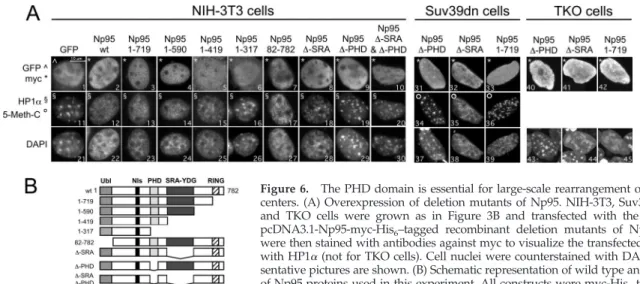
The PHD Domain Is Essential for the Large-Scale Changes in Chromocenter Structure
To investigate which domain of Np95-ICBP90 is involved in large-scale chromocenter modifications, we performed over-expression experiments in NIH3T3, Suv39h1/2dn, and TKO cells with various recombinant Np95 deletion mutants.
Np95 contains several protein domains. Progressing from the N to the C terminus, these are: a Ub-like domain, a putative nuclear localization signal, a PHD domain, an SRA-YDG domain and a RING finger domain (Figure 6B). In a previous study, we showed that Np95 is a RING-type E3 ubiquitin ligase (Citterio et al., 2004). The SRA-YDG domain has been implicated in the control of DNA methylation (Unoki et al., 2004; Bostick et al., 2007; Johnson et al., 2007; Sharif et al., 2007), the recruitment of HDAC (Unoki et al., 2004), and transcriptional silencing of major satellites (Papait
et al., 2007). We constructed deletion mutants of each of
these domains (Figure 6B) and evaluated the effects of their expression on the stability of chromocenters by immunoflu-orescence with antibodies against HP1␣ and by staining the PH areas with DAPI. Only those constructs retaining the PHD domain (Figure 6B, Np95-wt, -1-719, -1-590, -1-419, -82-782, and -⌬-SRA) were able to disaggregate chromo-centers and to delocalized HP1␣ from PH regions (Figure 6A; for HP1, cf. images 12, 13 ,14, 15, 17, and 18 with 16, 19, and 20; for DAPI, cf. images 22, 23, 24, 25, and 27 with 26, 29, and 30). Deletion of the PHD domain only (Figure 6A, ⌬-PHD mutant) is sufficient to impair the large-scale modi-fications observed with the wild-type protein in either NIH-3T3, Suv39h1/2dn or dnmt TKO cells. It has been shown that a RING point domain, but not ICBP90wt causes large-scale chromocenter changes (Karagianni et al., 2008). Our
experi-Figure 5. Large-scale alterations of chromocenters induced by the overexpression of Np95 are independent from DNA methylation, the methylation of histone H3:K9, H4:K20, of MeCP2 and of cell cycle. (A) NIH-3T3, Suv39h1/2dn and TKO cells were grown and transfected as in Figure 3B with the pcDNA3.1-Np95-myc-His6-tagged plasmid.
Forty-eight hours after transfection, the cells were immunostained with antibodies against MeCP2, H3:K9met3 and H4:K20met3 (NIH-3T3) or against 5-methyl-cytosine (5-Meth-C; Suv39h1/2dn). Nuclei were counterstained with DAPI. Representative pictures are shown. (B) Overexpression of Np95 in NIH-3T3 cells does not modify the meth-ylation pathway that leads to the formation of H3:K9met3 and H4: K20met3. NIH-3T3 cells were grown and infected as in Figure 3A. Forty-eight hours after infection, nuclear lysates were obtained from samples with or without Triton X-100 pretreatment. Western blotting was then performed using the indicated antibodies. (C) NIH-3T3 cells were grown on coverslips, serum-starved, and infected with Ad-Np95 MOI 120. Forty-eight hours later the cells were fixed and stained with antibody against HP1␣. Infected cells express GFP. Nuclei were coun-terstained with DAPI. The single-cell field is the enlargement of the boxed cell in the top panel. (D) NIH-3T3 cells, asynchronously growing on coverslips, were transfected with Np95-myc. Forty-eight hours later the cells were pulse-labeled for 10 min with BrdU, fixed, and stained with anti-myc and anti-BrdU antibodies.
ments in at least three type of mouse cells (NIH-3T3, Suv39h1/2dn, and TKO) indicate that overexpression of Np95wt or ICBP90wt or of the deletion mutants ⌬-SRA, ⌬-RING, ⌬-ubiquitin-like domain (82-782), but not ⌬-PHD always causes large-scale chromocenter modifications.
These data suggest that the known functions of Np95 (ubiquitin ligase activity determined by the RING domain, and HDAC recruitment and control of DNA methylation attributed to the SRA-YDG domain) are not involved in the ability of Np95 to disaggregate the chromocenter structures. They indicate, instead, a specific role for the PHD domain of Np95 in this process.
Np95 Increases the Access of Restriction Enzymes to DNA Packaged into Nucleosomal Arrays
During PH replication in middle S phase, the bulk of Np95 specifically relocalizes to and gets highly concentrated in the chromocenters where it occupies the areas of less compacted DNA that correspond to “replication factories.” The disag-gregating effects we observe on chromocenters in overex-pression experiments suggest that, in living cells, Np95 might contribute to the opening and/or stabilization of the dense chromatic structure to support the access and recruit-ment of modifying enzymes required for replication and formation of PH.
We therefore used in vitro experiments to investigate if Np95 is able to facilitate the access to modifying enzymes (restriction enzyme SfcI) to DNA (pFM218-H5 plasmid) packaged into nucleosome arrays (Figure 7A). This type of assay has been used by various authors to assess the chro-matin accessibility activity of various protein complexes in vitro (Almer et al., 1986; Varga-Weisz et al., 1997; Boyer et al., 2000; Shen et al., 2000; Alexiadis and Kadonaga, 2002). Re-combinant GST-Np95 was incubated with reconstituted chromatin in the presence or absence of the restriction en-zyme SfcI, separated by agarose gel electrophoresis, and visualized by hybridization with radiolabeled pFM218-H5 plasmid DNA. Figure 7B shows that 1 or 2g of GST-Np95 (Figure 7B, lanes 6 and 7), 2g of ICBP90 (Figure 7B, lane 14), and 200 ng of the positive control (Drosophila nuclear extracts; Figure 7B, lane 5), but not 2g of GST alone (Figure 7B, lane 4), increase access of a restriction enzyme to pack-aged DNA (Figure 7B, cf. lanes 4 and 5 with lane 6). Figure 7 further shows that deletion of the PHD domain, but not of the SRA domain strongly reduces this activity, although it
does not abolish it (Figure 7B, cf. lanes 6 –7 with 8 –9, 10 –11, and 12–13).
Altogether, these experiments indicate that Np95, by means of its PHD domain, is able to disaggregate
chromo-Figure 6. The PHD domain is essential for large-scale rearrangement of chromo-centers. (A) Overexpression of deletion mutants of Np95. NIH-3T3, Suv39h1/2dn, and TKO cells were grown as in Figure 3B and transfected with the indicated pcDNA3.1-Np95-myc-His6–tagged recombinant deletion mutants of Np95. Cells
were then stained with antibodies against myc to visualize the transfected cells and with HP1␣ (not for TKO cells). Cell nuclei were counterstained with DAPI. Repre-sentative pictures are shown. (B) Schematic representation of wild type and mutants of Np95 proteins used in this experiment. All constructs were myc-His6tagged.
Figure 7. Np95 induces chromatin template accessibility to a re-striction enzyme in an in vitro assay. (A) Schematic representation of the chromatin accessibility assay. Naked DNA was cut by the restriction endonuclease SfcI; if nucleosomes were reconstituted on the DNA, the enzyme was unable to cut; the presence of a remod-eller could mobilize histones and allow DNA cutting by SfcI. (B) Np95 increased template accessibility of the SfcI restriction enzyme to chromatinized DNA. Reconstituted nucleosomal arrays that were incubated in the presence either 2g of GST (lane 4), or 1 g (lanes 6, 8, 10 12) or 2 g (lane 7, 9, 11, 13) of wt-Np95, PHD, SRA, PHD-SRA deletion mutants (lanes 8 and9), or 2g of wt-ICBP90 (lane 14) were separated by agarose gel electrophoresis and visual-ized by hybridization with radiolabeled pFM218-H5 plasmid DNA. Nicked (N), linear (L), and supercoiled (S/C) forms of uncut pFM218-H5 plasmid DNA species (lane 1) are indicated to the left. The appearance of complete (C1, C2; left of pictures) or partial (p; right of pictures) SfcI digestion products are indicated.
R. Papait et al.
Molecular Biology of the Cell 3560
centers in vivo and to facilitate the access to DNA modifying enzymes in vitro. We suggest that Np95-ICBP90, when it specifically relocates to PH in middle S phase and becomes highly concentrated in the pHDB, could actively participate to render the chromatin of chromocenters more dynamic and potentially more accessible to modifying enzymes, such as HDAC and DNMT1.
DISCUSSION
In this article, we provide evidence that Np95 determines large-scale modifications of chromocenters and that the PHD domain is required for this process. Depletion of Np95 leads to a reduction in the number and a size increase of chromocenters, whereas overexpression of Np95-ICBP90 in-duces decondensation of chromocenters.
Remarkably, the dissolving effect on chromocenters is in-dependent from the H3:K9/K4:K20met3/HP1 pathway, from DNA methylation, and from the cell cycle. In Suv39h1/ 2dn and in dnmt TKO cells, indeed, overexpression of Np95 leads to the disappearance of the DAPI nodules. In NIH-3T3 cells, neither the histone trimethylation pattern in PH nor the expression levels of HP1 are significantly affected after Np95 depletion or overexpression. This, however, is not surprising because several studies show that the H3:K9 methylation pathway is not involved in chromocenter dy-namics. Accumulation of HP1 in PH regions does not cause large-scale chromocenter modifications (Mateos-Langerak et
al., 2007). The number and size of chromocenters in
Suv39h1/2dn cells are comparable to control cells (Peters et
al., 2001). Large-scale rearrangements of PH induced during
terminal differentiation of muscle cells and by overexpres-sion of MeCP2 are independent from H3:K9met3 and from HP1 levels (Brero et al., 2005). In plants, the methyltrans-ferase suvh4 mutant has normal chromocenters although H3:K9met2 is strongly reduced (Jasencakova et al., 2003; Naumann et al., 2005; Zemach et al., 2005).
It has also been suggested that extensive DNA ation is not necessary for PH clustering. The DNA methyl-binding protein MeCP2 was shown to assemble secondary chromatin structures independently from the methylated DNA-binding domain and from DNA methylation (Georgel
et al., 2003). However, during muscle terminal differentiation
the MBD domain of MeCP2 and of other MBD-containing proteins seems to be necessary and sufficient to increase percientromeric clustering (Brero et al., 2005). In plants, dis-ruption of chromocenter structures during dedifferentiation of specialized mesophyll cells into undifferentiated proto-plasts is not accompanied by changes in DNA or H3:K9 methylation and in transcriptional reactivation of silent genomic elements (Tessadori et al., 2007). The severe DNA hypomethylation mutants ddm1-5, ddm1-2, met1-1, and hog1 only show a limited decondensation of chromocenter het-erochromatin (Mittelsten Scheid et al., 2002; Probst et al., 2003). Deletion of Np95 results in a drastic reduction of DNA methylation levels in mouse ES cells and embryos (Bostick et al., 2007; Sharif et al., 2007). Our results indicate that loss of Np95 increases chromocenters size and that overexpression of Np95 in TKO cells disaggregates chromo-centers, once more suggesting that the chromocenter dy-namics we observe is independent from DNA methylation. Alterations in chromocenter structure by Np95 must, therefore, depend on different processes. We show that PHD is the domain required in vivo for the profound alterations of chromocenters structure after overexpression of Np95-ICBP90. It has been shown that a RING point domain, but not ICBP90wt, causes large-scale chromocenter changes
(Karagianni et al., 2008). Our experiments in at least three type of mouse cells (NIH-3T3, Suv39h1/2dn, and TKO) in-dicate that overexpression of Np95-ICBP90wt or of the de-letion mutants ⌬-SRA, ⌬-RING, ⌬-ubiquitin-like domain (82-782), but not⌬-PHD, always causes large-scale chromo-center modifications.
Significant sequence differences have been found in PHD domains, and accordingly, various activities have been as-signed to this domain (Bienz, 2006). The PHD domain is an important chromatin-binding module and is crucial for the function of proteins within chromatin-associated complexes that display chromatin-modifying activities, like Dnmt3L (Jia et al., 2007), BHC80 (Lan et al., 2007), ACF-1 (Eberharter
et al., 2004), and BPTF (Wysocka et al., 2006). The deletion or
functional impairment of the C-terminal PHD finger of ACF1 profoundly affected the nucleosome mobilization ca-pability of associated SNF2H in trans. Some of these com-plexes have been implicated in DNA replication (Corona and Tamkun, 2004) and the ACF1–SNF2H complex specifi-cally in PH replication (Collins et al., 2002).
Our in vitro experiments show that the PHD of Np95 is required to facilitate the access of a restriction enzyme (SfcI) to DNA packaged into nucleosome arrays, suggesting that in vivo this domain might enhance Np95s chromatin-bind-ing ability and favor the recruitment of chromatin modifiers. Indeed, the PHD domain of Np95 has a role in chromatin binding, although the SRA is critical (Citterio et al., 2004). Although a recent publication (Karagianni et al., 2008) shows that the PHD and SRA domains of Np95 are required for preferential binding to H3:K9met3 and that Np95 in Suv39h1/2dn cells does not bind heterochromatin, our ex-periments performed on those cells clearly show that the protein distribution is not affected by that genetic back-ground (Figure 3). In synchronized Suv39h1/2dn cells, Np95 exhibits foci of PH staining over a more diffuse pattern at the onset of S phase, relocalizes to chromocenters at the time of PH replication and is part of the pHDB, as it does in NIH-3T3 cells. A possible interpretation of this discrepancy is that Suv39h1/2dn cells grow slower and most cells are in G1, a cell cycle phase in which Np95 is well known to appear diffused in the nucleoplasm (Uemura et al., 2000; Miura et al., 2001; Papait et al., 2007).
The PHD-mediated large-scale PH reorganization might reflect changes that occur at a yet unknown level of chro-matin organization and disrupts the structure of chromo-centers, producing an open chromatin configuration. This view is reinforced by our FISH experiments, which show that major satellite DNA is disaggregated along with chro-mocenters. At the time of PH replication, the bulk of Np95 relocalizes to the chromocenters and always associates with the pHDB, which corresponds to the less dense chromatin areas. In these partially disaggregated chromocenter areas, parental DNA is pulled out and duplicated by the replica-tion machinery (Quivy et al., 2004). We propose the hypoth-esis that the PHD domain of Np95 is involved in PH repli-cation and formation by contributing to the “opening” of this chromatin compartment during replication. The SRA domain would recruit HDAC1 (Unoki et al., 2004; Papait et
al., 2007), contributing to the establishment of the repressive
environment that produces major satellite transcriptional repression (Papait et al., 2007). Our results showing chromo-center clustering and impairment of PH replication after Np95 depletion, are an indirect confirmation of this hypoth-esis.
The results described here, together with our previous studies, tend to suggest that higher amounts of this protein would contribute to a “proliferating” competence of the
cells, which is consistent with the observation of Np95-ICBP90 overexpression in many tumors (Mousli et al., 2003; Crnogorac-Jurcevic et al., 2005; Jenkins et al., 2005). Indeed, large-scale positional or structural modifications of hetero-chromatin have key roles in cellular transformation (Zink et
al., 2004) and may involve epigenetic regulation of gene
expression (van Driel et al., 2003).
In conclusion, we propose that Np95-ICBP90 has an im-portant role for chromocenter dynamics that is accom-plished independently from the H3:K9-H4:K20-HP1 path-way and from DNA methylation and that the PHD domain has an important role for chromocenter dynamics and might actively participate to the replication and reformation of PH domains in middle S phase.
ACKNOWLEDGMENTS
We thank Dr. Roberto Valli and Prof. Manuela Maserati for their help in FISH experiments; Prof. Peter Becker (Ludwig-Maximilians-Universität, Munich, Germany) for the gift of Drosophila nuclear extracts; Prof. Thomas Jenuwein for providing us with the Suv39h1/2dn cells; and Prof. Masaki Okano (Center for Developmental Biology, RIKEN, Kobe, Japan) for the TKO cells. We thank the core facilities of the IGBMC. This work was supported by grants from the Associazione Italiana Ricerca sul Cancro (AIRC), the Rett Syndrome Research Foundation, the Deutsche Forschungsgemeinschaft, and from Fondazione Cariplo. The transfected Cell Array platform thanks the Cance´ropoˆle du Grand Est, the Re´gion Alsace, the Conseil Ge´ne´ral du Bas Rhin, and the Communite´ Urbaine de Strasbourg. R.P. was supported by a fellowship from AIRC.
REFERENCES
Alexiadis, V., and Kadonaga, J. T. (2002). Strand pairing by Rad54 and Rad51 is enhanced by chromatin. Genes Dev. 16, 2767–2771.
Almer, A., Rudolph, H., Hinnen, A., and Horz, W. (1986). Removal of posi-tioned nucleosomes from the yeast PHO5 promoter upon PHO5 induction releases additional upstream activating DNA elements. EMBO J. 5, 2689 – 2696.
Barr, H. J., and Ellison, J. R. (1972). Ectopic pairing of chromosome regions containing chemically similar DNA. Chromosoma 39, 53– 61.
Baumann, M., Mamais, A., McBlane, F., Xiao, H., and Boyes, J. (2003). Regu-lation of V(D)J recombination by nucleosome positioning at recombination signal sequences. EMBO J. 22, 5197–5207.
Bienz, M. (2006). The PHD finger, a nuclear protein-interaction domain. Trends Biochem. Sci. 31, 35– 40.
Bonapace, I. M., Latella, L., Papait, R., Nicassio, F., Sacco, A., Muto, M., Crescenzi, M., and Di Fiore, P. P. (2002). Np95 is regulated by E1A during mitotic reactivation of terminally differentiated cells and is essential for S phase entry. J. Cell Biol. 157, 909 –914.
Bostick, M., Kim, J. K., Esteve, P. O., Clark, A., Pradhan, S., and Jacobsen, S. E. (2007). UHRF1 plays a role in maintaining DNA methylation in mammalian cells. Science 317, 1760 –1764.
Boyer, L. A., Logie, C., Bonte, E., Becker, P. B., Wade, P. A., Wolffe, A. P., Wu, C., Imbalzano, A. N., and Peterson, C. L. (2000). Functional delineation of three groups of the ATP-dependent family of chromatin remodeling enzymes. J. Biol. Chem. 275, 18864 –18870.
Brero, A., Easwaran, H. P., Nowak, D., Grunewald, I., Cremer, T., Leonhardt, H., and Cardoso, M. C. (2005). Methyl CpG-binding proteins induce large-scale chromatin reorganization during terminal differentiation. J. Cell Biol. 169, 733–743.
Cerda, M. C., Berrios, S., Fernandez-Donoso, R., Garagna, S., and Redi, C. (1999). Organisation of complex nuclear domains in somatic mouse cells. Biol. Cell 91, 55– 65.
Cheutin, T., McNairn, A. J., Jenuwein, T., Gilbert, D. M., Singh, P. B., and Misteli, T. (2003). Maintenance of stable heterochromatin domains by dy-namic HP1 binding. Science 299, 721–725.
Citterio, E., Papait, R., Nicassio, F., Vecchi, M., Gomiero, P., Mantovani, R., Di Fiore, P. P., and Bonapace, I. M. (2004). Np95 is a histone-binding protein endowed with ubiquitin ligase activity. Mol. Cell. Biol. 24, 2526 –2535. Collins, N., Poot, R. A., Kukimoto, I., Garcia-Jimenez, C., Dellaire, G., and Varga-Weisz, P. D. (2002). An ACF1-ISWI chromatin-remodeling complex is
required for DNA replication through heterochromatin. Nat. Genet. 32, 627– 632.
Corona, D. F., and Tamkun, J. W. (2004). Multiple roles for ISWI in transcrip-tion, chromosome organization and DNA replication. Biochim. Biophys. Acta 1677, 113–119.
Crnogorac-Jurcevic, T. et al. (2005). Proteomic analysis of chronic pancreatitis and pancreatic adenocarcinoma. Gastroenterology 129, 1454 –1463. Dimitrova, D. S., and Berezney, R. (2002). The spatio-temporal organization of DNA replication sites is identical in primary, immortalized and transformed mammalian cells. J. Cell Sci. 115, 4037– 4051.
Eberharter, A., Vetter, I., Ferreira, R., and Becker, P. B. (2004). ACF1 improves the effectiveness of nucleosome mobilization by ISWI through PHD-histone contacts. EMBO J. 23, 4029 – 4039.
Festenstein, R., and Aragon, L. (2003). Decoding the epigenetic effects of chromatin. Genome Biol. 4, 342.
Georgel, P. T., Horowitz-Scherer, R. A., Adkins, N., Woodcock, C. L., Wade, P. A., and Hansen, J. C. (2003). Chromatin compaction by human MeCP2. Assembly of novel secondary chromatin structures in the absence of DNA methylation. J. Biol. Chem. 278, 32181–32188.
Hochstrasser, M., and Sedat, J. W. (1987). Three-dimensional organization of Drosophila melanogaster interphase nuclei. I. Tissue-specific aspects of polytene nuclear architecture. J. Cell Biol. 104, 1455–1470.
Jasencakova, Z., Soppe, W. J., Meister, A., Gernand, D., Turner, B. M., and Schubert, I. (2003). Histone modifications in Arabidopsis— high methylation of H3 lysine 9 is dispensable for constitutive heterochromatin. Plant J. 33, 471– 480.
Jenkins, Y. et al. (2005). Critical role of the ubiquitin ligase activity of UHRF1, a nuclear RING finger protein, in tumor cell growth. Mol. Biol. Cell 16, 5621–5629.
Jia, D., Jurkowska, R. Z., Zhang, X., Jeltsch, A., and Cheng, X. (2007). Structure of Dnmt3a bound to Dnmt3L suggests a model for de novo DNA methylation. Nature 449, 248 –251.
Johnson, L. M., Bostick, M., Zhang, X., Kraft, E., Henderson, I., Callis, J., and Jacobsen, S. E. (2007). The SRA methyl-cytosine-binding domain links DNA and histone methylation. Curr. Biol. 17, 379 –384.
Karagianni, P., Amazit, L., Qin, J., and Wong, J. (2008). ICBP90, a novel methyl K9 H3 binding protein linking protein ubiquitination with heterochromatin formation. Mol. Cell. Biol. 28, 705–717.
Kosak, S. T., and Groudine, M. (2004). Form follows function: the genomic organization of cellular differentiation. Genes Dev. 18, 1371–1384.
Lan, F., Collins, R. E., De Cegli, R., Alpatov, R., Horton, J. R., Shi, X., Gozani, O., Cheng, X., and Shi, Y. (2007). Recognition of unmethylated histone H3 lysine 4 links BHC80 to LSD1-mediated gene repression. Nature 448, 718 –722. Lehnertz, B., Ueda, Y., Derijck, A. A., Braunschweig, U., Perez-Burgos, L., Kubicek, S., Chen, T., Li, E., Jenuwein, T., and Peters, A. H. (2003). Suv39h-mediated histone H3 lysine 9 methylation directs DNA methylation to major satellite repeats at pericentric heterochromatin. Curr. Biol. 13, 1192–1200. Ma, H., Samarabandu, J., Devdhar, R. S., Acharya, R., Cheng, P. C., Meng, C., and Berezney, R. (1998). Spatial and temporal dynamics of DNA replication sites in mammalian cells. J. Cell Biol. 143, 1415–1425.
Maison, C., and Almouzni, G. (2004). HP1 and the dynamics of heterochro-matin maintenance. Nat. Rev. Mol. Cell Biol. 5, 296 –304.
Mateos-Langerak, J., Brink, M. C., Luijsterburg, M. S., van der Kraan, I., van Driel, R., and Verschure, P. J. (2007). Pericentromeric heterochromatin do-mains are maintained without accumulation of HP1. Mol. Biol. Cell 18, 1464 –1471.
Mittelsten Scheid, O., Probst, A. V., Afsar, K., and Paszkowski, J. (2002). Two regulatory levels of transcriptional gene silencing in Arabidopsis. Proc. Natl. Acad. Sci. USA 99, 13659 –13662.
Miura, M., Watanabe, H., Sasaki, T., Tatsumi, K., and Muto, M. (2001). Dynamic changes in subnuclear NP95 location during the cell cycle and its spatial relationship with DNA replication foci. Exp. Cell Res. 263, 202–208. Mousli, M., Hopfner, R., Abbady, A. Q., Monte, D., Jeanblanc, M., Oudet, P., Louis, B., and Bronner, C. (2003). ICBP90 belongs to a new family of proteins with an expression that is deregulated in cancer cells. Br. J. Cancer 89, 120 –127.
Naumann, K., Fischer, A., Hofmann, I., Krauss, V., Phalke, S., Irmler, K., Hause, G., Aurich, A. C., Dorn, R., Jenuwein, T., and Reuter, G. (2005). Pivotal role of AtSUVH2 in heterochromatic histone methylation and gene silencing in Arabidopsis. EMBO J. 24, 1418 –1429.
R. Papait et al.
Molecular Biology of the Cell 3562
Papait, R., Pistore, C., Negri, D., Pecoraro, D., Cantarini, L., and Bonapace, I. M. (2007). Np95 is implicated in pericentromeric heterochromatin replica-tion and in major satellite silencing. Mol. Biol. Cell 18, 1098 –1106. Peters, A. H. et al. (2001). Loss of the Suv39h histone methyltransferases impairs mammalian heterochromatin and genome stability. Cell 107, 323–337. Polo, S. E., and Almouzni, G. (2006). Chromatin assembly: a basic recipe with various flavours. Curr. Opin. Genet Dev. 16, 104 –111.
Probst, A. V., Fransz, P. F., Paszkowski, J., and Mittelsten Scheid, O. (2003). Two means of transcriptional reactivation within heterochromatin. Plant J. 33, 743–749.
Quivy, J. P., Roche, D., Kirschner, D., Tagami, H., Nakatani, Y., and Al-mouzni, G. (2004). A CAF-1 dependent pool of HP1 during heterochromatin duplication. EMBO J. 23, 3516 –3526.
Sharif, J. et al. (2007). The SRA protein Np95 mediates epigenetic inheritance by recruiting Dnmt1 to methylated DNA. Nature 450, 908 –912.
Shen, X., Mizuguchi, G., Hamiche, A., and Wu, C. (2000). A chromatin remodelling complex involved in transcription and DNA processing. Nature 406, 541–544.
Tessadori, F., Chupeau, M. C., Chupeau, Y., Knip, M., Germann, S., van Driel, R., Fransz, P., and Gaudin, V. (2007). Large-scale dissociation and sequential reassembly of pericentric heterochromatin in dedifferentiated Arabidopsis cells. J. Cell Sci. 120, 1200 –1208.
Tsumura, A. et al. (2006). Maintenance of self-renewal ability of mouse em-bryonic stem cells in the absence of DNA methyltransferases Dnmt1, Dnmt3a and Dnmt3b. Genes Cells 11, 805– 814.
Uemura, T., Kubo, E., Kanari, Y., Ikemura, T., Tatsumi, K., and Muto, M. (2000). Temporal and spatial localization of novel nuclear protein NP95 in mitotic and meiotic cells. Cell Struct. Funct. 25, 149 –159.
Unoki, M., Nishidate, T., and Nakamura, Y. (2004). ICBP90, an E2F-1 target, recruits HDAC1 and binds to methyl-CpG through its SRA domain. Onco-gene 23, 7601–7610.
van Driel, R., Fransz, P. F., and Verschure, P. J. (2003). The eukaryotic genome: a system regulated at different hierarchical levels. J. Cell Sci. 116, 4067– 4075. Varga-Weisz, P. D., Wilm, M., Bonte, E., Dumas, K., Mann, M., and Becker, P. B. (1997). Chromatin-remodelling factor CHRAC contains the ATPases ISWI and topoisomerase II. Nature 388, 598 – 602.
Weierich, C., Brero, A., Stein, S., von Hase, J., Cremer, C., Cremer, T., and Solovei, I. (2003). Three-dimensional arrangements of centromeres and telo-meres in nuclei of human and murine lymphocytes. Chromosome Res. 11, 485–502.
Wysocka, J. et al. (2006). A PHD finger of NURF couples histone H3 lysine 4 trimethylation with chromatin remodelling. Nature 442, 86 –90.
Zemach, A., Li, Y., Wayburn, B., Ben-Meir, H., Kiss, V., Avivi, Y., Kalchenko, V., Jacobsen, S. E., and Grafi, G. (2005). DDM1 binds Arabidopsis methyl-CpG binding domain proteins and affects their subnuclear localization. Plant Cell 17, 1549 –1558.
Zink, D., Fischer, A. H., and Nickerson, J. A. (2004). Nuclear structure in cancer cells. Nat. Rev. Cancer 4, 677– 687.